Review

Inflammatory Mechanisms in a Neurovascular Disease: Cerebral Cavernous Malformation

1
Department of Neurosurgery, First Affiliated Hospital of Harbin Medical University, Harbin 150001, China
2
Neurovascular Surgery Program, Department of Neurological Surgery, The University of Chicago, Chicago, IL 60637, USA
3
Department of Neurological Surgery, University of Chicago Medicine, 5841 S Maryland, MC3026/Neurosurgery J341, Chicago, IL 60637, USA
*
Author to whom correspondence should be addressed.
These authors contributed equally to this work.
Brain Sci. 2023, 13(9), 1336; https://doi.org/10.3390/brainsci13091336
Submission received: 21 July 2023 / Revised: 6 September 2023 / Accepted: 11 September 2023 / Published: 17 September 2023
(This article belongs to the Special Issue Cerebrovascular Neurosurgery)

Abstract

:
Cerebral cavernous malformation (CCM) is a common cerebrovascular malformation causing intracranial hemorrhage, seizures, and focal neurologic deficits. A unique CCM lesional inflammatory microenvironment has been shown to influence the clinical course of the disease. This review addresses the inflammatory cell infiltrate in the CCM lesion and the role of a defined antigen-driven immune response in pathogenicity. We summarize immune mechanisms associated with the loss of the CCM gene and disease progression, including the potential role of immunothrombosis. We also review evidence of circulating inflammatory biomarkers associated with CCM disease and its clinical activity. We articulate future directions for this research, including the role of individual cell type contributions to the immune response in CCM, single cell transcriptomics of inflammatory cells, biomarker development, and therapeutic implications. The concepts are applicable for developing diagnostic and treatment strategies for CCM and for studying other neurovascular diseases.

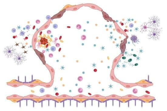
Graphical Abstract

1. Introduction

Cerebral cavernous malformation (CCM), also known as cerebral cavernous angiomas, is a common cerebrovascular malformation with a population prevalence estimated between 0.3 and 0.9% [1]. The lesion consists of dilated capillaries (caverns) with sluggish blood flow, containing thrombus at various stages of organization, in addition to perilesional blood products and a robust inflammatory cell infiltration. Symptomatic clinical presentations include seizures, focal neurologic deficits, and hemorrhagic stroke [2,3]. The CCM can be inherited in an autosomal dominant familial form (20–30% of cases) with multiple lesions throughout the brain or present in a sporadic form with a solitary lesion or lesions clustered around a developmental venous anomaly (70–80% of cases) [1,2]. The identification of genes implicated in CCM development has not fully explained the great variability in lesional clinical behavior [4]. Epidemiological studies have shown that patients can harbor an identical CCM genotype while experiencing different clinical courses of the disease during their lifetime [5,6,7,8,9,10].
Over the past 30 years, immune cell infiltration in CCMs, their interaction with the lesional milieu, and potential pathogenicity have been investigated [11,12,13,14,15,16]. Pro-inflammatory genotypes and gut microbiome factors have been implicated in lesional activity [6,17,18], and the CCM transcriptome studies and circulating biomarkers have endorsed the potential roles of immune response and inflammatory activity in this disease. We here review these concepts and how they influence potential biomarker development and therapy in CCM, as well as how they may be applied to other neurovascular diseases.

1.1. Inflammatory Cell Infiltrate in CCM, Predominance of B Cells and Their Clonal Expansion, Potential Pathogenicity

Familial and sporadic CCMs are histopathologically indistinguishable [4,13]. Regardless of genotype or recent clinical activity, resected lesions included a rich infiltration of B-cells and T-cells, human leukocyte antigen-DR-expressing cells, as well as macrophages [13]. As compared to arteriovenous malformations, a preponderance of B cells was documented in CCM and an oligoclonal IgG immune response in lesions [19], including oligoclonality of IgG mRNA [13]. De Souza et al. (2019) reported immune cell infiltration in association with CCM lesion aggressiveness [20]. Other authors have attributed the recurrence of CCM to local inflammatory conditions [21].
Later, Shi and co-workers demonstrated in situ B-cell clonal expansion and antigen-driven affinity maturation in CCMs [14]. These inflammatory cell niches are adjacent to abnormal vascular channels, suggesting an organ-intrinsic adaptive immune response in CCMs (Figure 1 and Figure 2).
A preclinical study showed that Ccm3 mice treated with anti-mouse BR3 to deplete B cells harbored smaller lesions, fewer mature CCMs [15], and decreased non-heme iron deposition, confirming the pathogenicity of the B-cell-mediated immune response in CCMs [15,22]. Additional studies further elucidated that these immune cells target cytoskeletal elements, including vimentin, myosin, and tubulin, that are commonly present in endothelial cells (ECs) and astrocytes [23]. This result also suggests that in situ inflammatory cells may react with other lesional cellular components such as neurons, pericytes, and other cell types [23].
Neutrophils, which have been identified in CCM, have been described as playing a role in the development of an inflammatory response and induction of a prothrombotic state in other diseases, such as intracerebral hemorrhage (ICH), cerebral arteriovenous malformations, and cancer [24,25,26,27]. The recruitment of neutrophils as well as the deposition of neutrophil extracellular traps (NETs) to neuroinflammatory sites have been shown in CCMs [28]. Nobiletti et al. (2022) demonstrated the role of the CCM1 gene (KRIT1) in the regulation of neutrophil adhesion and motility [29]. NET formation, also known as NETosis, is considered the innate immunity function of neutrophils [30], which can be triggered by proinflammatory cytokines as well as activated platelets and ECs [27,31]. Excessive NETosis can result in further endothelial damage due to its cytotoxic effect [28,32]. NETs could mediate adaptive immunity by inducing the secretion of proinflammatory chemokines/cytokines and/or present autoantigens to active B and T cells [33,34]. NET formation and release processes also depend on the generation of reactive oxygen species (ROS), a process shown to play an important role in CCM pathogenesis [35]. While the relationship of NOS to CCM pathogenicity requires further study, a Phase 2 trial targeting ROS to treat symptomatic CCM is ongoing (NCT05085561).
Macrophages and microglia identified in CCM have well-defined roles in the breakdown of red blood cells as well as heme iron and have been shown to react acutely following an ICH [13,36]. Activated microglia release pro-inflammatory mediators such as interleukin (IL)-1β, IL-6, tumor necrosis factor (TNF)-α, and ROS essential for the innate immune response, leading to neurotoxic sequelae [37,38]. Of interest, pro-inflammatory IL-1β has been suggested to be involved in the recruitment of leukocytes, while IL-6 may be a critical cytokine, controlling the transition from innate to adaptive immunity in the brain [39]. Moreover, TNF-α has been shown to play a role in T cell adhesion to brain ECs, which may occur during neuroinflammation [40]. Activation of microglia and the overproduction of ROS have been reported in the chronic inflammation microenvironment [41]. It contributes to secondary injury cascades through mitogen-activated protein kinase/nuclear factor-κB (MAPK/NF-κB) signaling and NOD-, LRR-, and pyrin domain-containing protein 3 inflammasome activation [41].
RhoA/Rho kinase (ROCK) signaling is negatively regulated by CCM proteins, and its dysregulation can increase endothelial permeability [42,43]. In addition, the activation of ROCK not only enhances leukocyte recruitment but also regulates B and T cell activation, proliferation, and cytokine production [44,45]. Lesional B and T cell infiltration was shown to be decreased in CCM preclinical models when blocking the Rho/ROCK signaling pathway [22]. Heterogeneities in the inflammatory cell populations within the lesional micro-environment have been shown to correlate with phenotype characteristics or clinical outcomes [13,46]. Sporadic CCMs harbor greater T-cell infiltration than familial CCMs [13]. Similarly, a higher Th17/Treg ratio has been observed in CCM with clinical symptoms suggestive of a primarily pro-inflammatory T cell phenotype [46].
Other researchers have linked immune responses in CCMs as well as other cerebrovascular anomalies to the lymphatic system and cerebrospinal fluid flow [47]. Low fluid shear stress conditions have been shown to contribute to the activation of cerebral cavernous malformation signaling pathways [48]. They have also been implicated in endothelial inflammation [49,50].

1.2. Transcriptomic Studies Highlight Inflammatory and Immune Mechanisms in CCM

The transcriptome of human and murine CCMs has clarified several features of this disease, such as lesion genesis, maturation, growth, and intralesional and extralesional hemorrhage. Some of the mechanistic findings observed in preclinical models of CCM disease were translated and validated in human CCM [16,51,52,53,54]. The transcriptomes of CCM lesional neurovascular units served as a reference to validate mechanistic hypotheses related to the pathogenesis of CCMs [16,55]. Interestingly, common pathways related to immune response, such as leukocyte migration and regulation of antigen receptor-mediated signaling are found across human CCMs, mouse BMECs, and Caenorhabditis elegans, as well as in two different genotypes (Ccm1 and Ccm3) [16]. In addition, several pathways related to macrophage cytokine production and activation (i.e., innate immunity) as well as activation and regulation of B and T cells (i.e., adaptive immunity) were identified in both human CCM and mouse BMECs [16]. The transcriptome of in vitro mouse brain microvascular ECs (BMECs) with induced allelic loss of Ccm3/Pdcd10 specifically identified several pathways associated with innate immunity pathways such as macrophage/granulocyte chemotaxis and migration [52].
Of interest, Chapman et al. (2019) reported that a mutation on kri-1(ok1251) in the C. elegans model affects several genes that are predicted to be involved in innate immunity [54]. These results suggest that a mutation in the CCM gene affects innate immunity pathways. Fusco et al. (2022) published transcriptome analyses revealing altered expression of genes involved in hypoxia, inflammation, and immune regulation in Ccm3 (Pdcd10)-depleted mouse endothelial cells [56].
Later, the analyses of the lesional transcriptome of acute and chronic Ccm3 mouse preclinical models showed that adaptive immune response pathways, such as chemotaxis and activation of neutrophils and leukocytes, chemokine production, and cytokine secretion, were only enriched in the chronic model with more mature lesions akin to symptomatic human CCMs [52]. The transcriptome of the acute Ccm3 model mostly identified enriched angiogenesis and cell proliferation pathways [52]. This result suggests that chronic bleeding in CCM accompanies an inflammatory stimulus, which may result in a local adaptive immune response. Finally, the transcriptome of surgically resected human CCMs with recent bleeding showed several enriched pathways associated with acute inflammation compared to non-recent bleeding [55]. The CCMs that recently bled likely carry unique biologic signatures, reflecting recent bleeding and subsequent vulnerability to hemorrhage [55].
The contribution of each cell type within the lesional milieu to CCM pathogenesis remains unclear. Pericytes, microglia, astrocytes, and neurons are necessary to maintain the integrity of the blood-brain barrier (BBB) and have also been associated with leukocyte infiltration and the local inflammatory response [12,57,58]. Pericytes normally regulate vascular integrity through close physical contact with ECs as well as paracrine signaling. Brain pericytes have also been shown to have similar functions to immune-regulating cells [59]. Recent preclinical studies on CCM lesional pericytes have identified leukocyte migration/proliferation, leukocyte cell-cell adhesion/activation, and the chemokine signaling pathway [57]. Moreover, pericytes express receptors for DAMPs and inflammatory mediators (IL-1β, IL-6, TNF-α, nitric oxide [NO], matrix metallopeptidase [MMP]-2, and MMP-9) that are associated with inflammatory responses in the brain [60,61]. In response to inflammatory stimuli, pericytes may also have macrophage-like functions such as phagocytosis and act as antigen-presenting cells by displaying antigens through major histocompatibility complex class II [61,62].

1.3. Immunothrombosis in CCM

Thrombosis is known to promote an inflammatory response involving a rich repertoire of immune cell infiltration [63,64,65]. The hallmarks of every CCM lesion are the organized thrombi in vascular caverns and leaky endothelium, with blood breakdown products in the surrounding brain parenchyma [11]. A complex interplay occurs between thrombotic and hemostatic regulatory pathways, mediated by cells of the neurovascular unit, and appears to lead to a proinflammatory milieu in brain tissue directly adjacent to the CCM [11,12,66]. A loss of hemostasis due to dysfunctional endothelium has been reported in CCM, resulting in upregulation of both pro- and anti-coagulant factors [51,67]. An upregulation of KLF4, a driver of CCM pathogenesis in mutant ECs, leads to the upregulation of the anticoagulant factors thrombomodulin (TM) and endothelial NO synthase (eNOS) [68]. A local increased level of TM and endothelial protein C receptors generates anticoagulant activated protein C, which could be associated with bleeding in CCMs [51].
Procoagulant domains that contribute to inflammation and the breakdown of endothelial integrity have been identified in CCM in conjunction with the inflammatory response induced by anticoagulant lesional domains [51,66]. This prothrombotic environment is a result of both endothelial and non-cell autonomous effects. ECs, as a proinflammatory effector, in the presence of Factor Xa can elevate expression of IL-8, IL-6, monocyte chemotactic protein-1, as well as intercellular adhesion molecule-1 (ICAM-1) and vascular cell adhesion molecule-1 (VCAM-1), which play roles in the promotion of leukocyte adhesion [69]. ECs are also responsible for the production of von Willebrand factor (vWF), which facilitates T-cell and neutrophil recruitment depending on the presence of platelets and the accessibility of their VWF-receptor glycoprotein (GP) Ib-IX-V complex [70]. Given a marked accumulation of vWF in the lumen of CCMs, recruitment of leukocytes can create positive feedback by increasing inflammatory cells around the CCM [11,67].
Perilesional astrocytes have been shown to upregulate coagulant tissue factor production in a Ccm3 murine model, likely due to increased shear stress caused by diminished actin rearrangement capability in CCM ECs [71,72]. Procoagulant domains and thrombi formation, in addition to low flow through caverns, induce a local hypoxia within the CCM, leading to increased production of endothelial NO and hypoxia-inducible factor (HIF)-1α [11,73]. Increased HIF-1α induces upregulation of VEGF-A in astrocytes, which has been linked to BBB breakdown by targeting endothelial tight junction proteins such as claudin-5 and occludin [73,74]. Astrocytes also play an active role as immune effector cells through the secretion of proinflammatory factors, including TNF-α and IL-1β, at high thrombin levels [75]. Increased levels of TNF-α have been shown to increase expression of ICAM-1 and VCAM-1, increasing trans-endothelial leukocyte migration and further contributing to the proinflammatory environment [76]. Similarly, microglia have shown reactive morphologies in a Ccm3 murine model within areas colocalized with thrombi and have been positively associated with the number of thrombi around lesions [12]. ICAM-1 stabilization and recruitment of neutrophils may form a feedback loop in which procoagulant-induced inflammation can lead to further endothelium injury and the release of cytokines such as IL-6, IL-8, and TNF-α [77,78,79,80]. These cytokines have been shown to promote the release of vWF from ECs [81,82], allowing for the recruitment of inflammatory cells into the lesional milieu.
These findings suggest a disrupted balance between pro- and anti-coagulant domains within the CCM to provide a nidus for the recruitment of pro-inflammatory cells and cytokines, further weakening the angio-architecture and leading to neurological sequelae (Figure 3).

1.4. Biomarkers and Clinical Features of CCM Disease

The U.S. FDA/NIH have outlined definitions for categorizing biomarkers in relation to their clinical contexts of use [83,84]. Molecular or imaging signatures have been proposed for the diagnosis, etiologic, and severity categorization of several neurological and neuro-vascular disorders [55,85,86,87,88]. The first association between the clinical course of CCM disease and circulating systematic compounds reported a hierarchical cluster of 5 plasma inflammatory cytokines, including IL-2, IFN-γ, TNF-α, IL-1β, and IL-6, defining CCM patients with a “high” inflammatory state, which was associated with seizures and more than one prior SH during a patient’s lifetime [89]. Several translational studies have recently reported that the complex inflammatory and angiogenic milieu observed in the transcriptome of CCMs is reflected in the plasma of CCM patients [16,55]. The lesional transcriptomes have since served as a reference to not only validate mechanistic hypotheses related to the pathogenesis of CCMs but also identify circulating mechanistically plausible candidate biomarkers [16,55]. While many of the biomarkers are not specific to CCM and have been invoked in other inflammatory conditions, they have been linked to the lesional CCM transcriptome and CCM-specific contexts of use. A novel paradigm that integrates multi-omic mechanistic discoveries and applies a likelihood-based computational model was developed to generate and improve biomarker candidates of disease activity [90]. This approach generated two weighted biomarkers based on the combination of plasma levels of inflammatory and angiogenic proteins, able to distinguish CCM patients recent (i.e., diagnostic) and impendent (i.e., prognostic) hemorrhagic activity with up to 80% sensitivity and specificity [55]. There is currently a multi-site initiative to develop highly accurate as well as generalizable diagnostic and prognostic biomarkers of symptomatic hemorrhage in CCMs (R01 NS114552), powered to examine the independent effects of confounders including sex, genotype, age, and lesion location.
The analyses of circulating genetic traits have also shown genetic polymorphisms within pro-inflammatory and immune response genes, such as TGFBR2, CD14, IL-6R, MSR1, IGH, and TLR4, that have been associated with total lesion count, number of large lesions, and intracerebral hemorrhage [6,8,17]. In addition, circulating microRNAs (miRNAs) have been suggested as possible mechanistic biomarkers of hemorrhagic activity for CCM disease. Lyne et al. (2019) identified 13 differently expressed (DE) miRNAs in the plasma of patients who experienced recent CCM-related brain bleeding [55]. Further computational analyses showed that miR-185-5p, one of these 13 DE miRNAs, had IL-10RA as a putative target, which was dysregulated in the lesional transcriptome of CCMs [55]. Of interest, lower plasma levels of IL-10 have been reported in CCM subjects who experienced recent bleeding [55].

1.5. Conclusions and Future Directions

A complex inflammatory cell environment and several potential immune mechanisms are inherent to CCM (Figure 3). More research on the role of the immune response in CCM is needed and may impact novel therapeutic and biomarker development. Future studies must parse out the individual roles of the inflammatory cells during lesion genesis and maturation and their involvement in clinical sequelae. Single-cell transcriptomic studies of inflammatory cells at various stages of lesion development could lead to more personalized treatments based on the lesion’s developmental stage [91,92,93,94]. These studies could also aid in the identification of molecules that can stratify patients based on lesion severity and prognosticate clinical events. Cytokines and chemokines may serve as biomarkers to predict disease characteristics due to the inflammatory microenvironment and increased vascular permeability. Of interest, IL-1β and IL-10 are currently being tested as biomarkers of hemorrhagic activity in CCM [90]. Other studies will motivate the repurposing of drugs that impact immune cells for therapeutic interventions [15]. In many ways, CCM remains a paradigmatic disease, as these concepts may be applicable to other neurovascular diseases where there is an interplay between vascular dysmorphism, immune response, and inflammation.

Author Contributions

Conceptualization, I.A.A., R.G., C.S., Y.L. and A.S.; Writing—Original Draft Preparation, Y.L., A.S., R.J.A.-F., S.H., A.B. and R.L.; Writing—Review and Editing, I.A.A., R.G., C.S. and R.S.; Supervision, I.A.A., R.G. and C.S.; Project Administration, I.A.A. and R.G.; Funding Acquisition, I.A.A. and C.S. All authors have read and agreed to the published version of the manuscript.

Funding

This work was supported by NINDS/NIH grant (P01 NS092521) for I.A.A., the National Natural Science Foundation of China (81771276 and 82071298) for C.S., the Key Project of Natural Science Foundation of Heilongjiang Province of China (ZD2016015) for C.S., and the Foundation for High-Level Returned Overseas Talents of Ministry of Human Resources and Social Security of China for C.S.

Conflicts of Interest

The authors declare no conflict of interest.

References

  1. Akers, A.; Al-Shahi Salman, R.; Awad, I.A.; Dahlem, K.; Flemming, K.; Hart, B.; Kim, H.; Jusue-Torres, I.; Kondziolka, D.; Lee, C.; et al. Synopsis of Guidelines for the Clinical Management of Cerebral Cavernous Malformations: Consensus Recommendations Based on Systematic Literature Review by the Angioma Alliance Scientific Advisory Board Clinical Experts Panel. Neurosurgery 2017, 80, 665–680. [Google Scholar] [CrossRef] [PubMed]
  2. Al-Shahi Salman, R.; Hall, J.M.; Horne, M.A.; Moultrie, F.; Josephson, C.B.; Bhattacharya, J.J.; Counsell, C.E.; Murray, G.D.; Papanastassiou, V.; Ritchie, V.; et al. Untreated clinical course of cerebral cavernous malformations: A prospective, population-based cohort study. Lancet Neurol. 2012, 11, 217–224. [Google Scholar] [CrossRef]
  3. Alalfi, M.O.; Lanzino, G.; Flemming, K.D. Clinical presentation, hemorrhage risk, and outcome in patients with familial cavernous malformations: A pragmatic prospective analysis of 75 patients. J. Neurosurg. 2023; Epub ahead of print 1–7. [Google Scholar] [CrossRef] [PubMed]
  4. Snellings, D.A.; Hong, C.C.; Ren, A.A.; Lopez-Ramirez, M.A.; Girard, R.; Srinath, A.; Marchuk, D.A.; Ginsberg, M.H.; Awad, I.A.; Kahn, M.L. Cerebral Cavernous Malformation: From Mechanism to Therapy. Circ. Res. 2021, 129, 195–215. [Google Scholar] [CrossRef]
  5. Denier, C.; Labauge, P.; Bergametti, F.; Marchelli, F.; Riant, F.; Arnoult, M.; Maciazek, J.; Vicaut, E.; Brunereau, L.; Tournier-Lasserve, E.; et al. Genotype-phenotype correlations in cerebral cavernous malformations patients. Ann. Neurol. 2006, 60, 550–556. [Google Scholar] [CrossRef]
  6. Choquet, H.; Pawlikowska, L.; Nelson, J.; McCulloch, C.E.; Akers, A.; Baca, B.; Khan, Y.; Hart, B.; Morrison, L.; Kim, H.; et al. Polymorphisms in inflammatory and immune response genes associated with cerebral cavernous malformation type 1 severity. Cerebrovasc. Dis. 2014, 38, 433–440. [Google Scholar] [CrossRef]
  7. Choquet, H.; Kim, H. Genome-wide Genotyping of Cerebral Cavernous Malformation Type 1 Individuals to Identify Genetic Modifiers of Disease Severity. Methods Mol. Biol. 2020, 2152, 77–84. [Google Scholar] [CrossRef] [PubMed]
  8. Choquet, H.; Trapani, E.; Goitre, L.; Trabalzini, L.; Akers, A.; Fontanella, M.; Hart, B.L.; Morrison, L.A.; Pawlikowska, L.; Kim, H.; et al. Cytochrome P450 and matrix metalloproteinase genetic modifiers of disease severity in Cerebral Cavernous Malformation type 1. Free Radic. Biol. Med. 2016, 92, 100–109. [Google Scholar] [CrossRef]
  9. Perrelli, A.; Retta, S.F. Polymorphisms in genes related to oxidative stress and inflammation: Emerging links with the pathogenesis and severity of Cerebral Cavernous Malformation disease. Free Radic. Biol. Med. 2021, 172, 403–417. [Google Scholar] [CrossRef]
  10. Trapani, E.; Retta, S.F. Cerebral cavernous malformation (CCM) disease: From monogenic forms to genetic susceptibility factors. J. Neurosurg. Sci. 2015, 59, 201–209. [Google Scholar]
  11. Globisch, M.A.; Onyeogaziri, F.C.; Jauhiainen, S.; Yau, A.C.Y.; Orsenigo, F.; Conze, L.L.; Arce, M.; Corada, M.; Smith, R.O.; Rorsman, C.; et al. Immunothrombosis and vascular heterogeneity in cerebral cavernous malformation. Blood 2022, 140, 2154–2169. [Google Scholar] [CrossRef]
  12. Lai, C.C.; Nelsen, B.; Frias-Anaya, E.; Gallego-Gutierrez, H.; Orecchioni, M.; Herrera, V.; Ortiz, E.; Sun, H.; Mesarwi, O.A.; Ley, K.; et al. Neuroinflammation Plays a Critical Role in Cerebral Cavernous Malformation Disease. Circ. Res. 2022, 131, 909–925. [Google Scholar] [CrossRef]
  13. Shi, C.; Shenkar, R.; Du, H.; Duckworth, E.; Raja, H.; Batjer, H.H.; Awad, I.A. Immune response in human cerebral cavernous malformations. Stroke 2009, 40, 1659–1665. [Google Scholar] [CrossRef] [PubMed]
  14. Shi, C.; Shenkar, R.; Kinloch, A.; Henderson, S.G.; Shaaya, M.; Chong, A.S.; Clark, M.R.; Awad, I.A. Immune complex formation and in situ B-cell clonal expansion in human cerebral cavernous malformations. J. Neuroimmunol. 2014, 272, 67–75. [Google Scholar] [CrossRef] [PubMed]
  15. Shi, C.; Shenkar, R.; Zeineddine, H.A.; Girard, R.; Fam, M.D.; Austin, C.; Moore, T.; Lightle, R.; Zhang, L.; Wu, M.; et al. B-Cell Depletion Reduces the Maturation of Cerebral Cavernous Malformations in Murine Models. J. Neuroimmune Pharmacol. 2016, 11, 369–377. [Google Scholar] [CrossRef]
  16. Koskimaki, J.; Girard, R.; Li, Y.; Saadat, L.; Zeineddine, H.A.; Lightle, R.; Moore, T.; Lyne, S.; Avner, K.; Shenkar, R.; et al. Comprehensive transcriptome analysis of cerebral cavernous malformation across multiple species and genotypes. JCI Insight 2019, 4, e126167. [Google Scholar] [CrossRef]
  17. Tang, A.T.; Choi, J.P.; Kotzin, J.J.; Yang, Y.; Hong, C.C.; Hobson, N.; Girard, R.; Zeineddine, H.A.; Lightle, R.; Moore, T.; et al. Endothelial TLR4 and the microbiome drive cerebral cavernous malformations. Nature 2017, 545, 305–310. [Google Scholar] [CrossRef]
  18. Polster, S.P.; Sharma, A.; Tanes, C.; Tang, A.T.; Mericko, P.; Cao, Y.; Carrion-Penagos, J.; Girard, R.; Koskimaki, J.; Zhang, D.; et al. Permissive microbiome characterizes human subjects with a neurovascular disease cavernous angioma. Nat. Commun. 2020, 11, 2659. [Google Scholar] [CrossRef]
  19. Shi, C.; Shenkar, R.; Batjer, H.H.; Check, I.J.; Awad, I.A. Oligoclonal immune response in cerebral cavernous malformations. Laboratory investigation. J. Neurosurg. 2007, 107, 1023–1026. [Google Scholar] [CrossRef] [PubMed]
  20. de Souza, J.M.; Aiex, C.A.; Silva Canedo, N.H.; Marques, S.A.; Blanco Martinez, A.M. Intraaxial and Extraaxial Cavernous Malformation with Venous Linkage: Immune Cellular Inflammation Associated with Aggressiveness. World Neurosurg. 2019, 131, 87–89. [Google Scholar] [CrossRef] [PubMed]
  21. Bianconi, A.; Salvati, L.F.; Perrelli, A.; Ferraris, C.; Massara, A.; Minardi, M.; Aruta, G.; Rosso, M.; Massa Micon, B.; Garbossa, D.; et al. Distant Recurrence of a Cerebral Cavernous Malformation in the Vicinity of a Developmental Venous Anomaly: Case Report of Local Oxy-Inflammatory Events. Int. J. Mol. Sci. 2022, 23, 14643. [Google Scholar] [CrossRef] [PubMed]
  22. Shenkar, R.; Peiper, A.; Pardo, H.; Moore, T.; Lightle, R.; Girard, R.; Hobson, N.; Polster, S.P.; Koskimaki, J.; Zhang, D.; et al. Rho Kinase Inhibition Blunts Lesion Development and Hemorrhage in Murine Models of Aggressive Pdcd10/Ccm3 Disease. Stroke 2019, 50, 738–744. [Google Scholar] [CrossRef]
  23. Zhang, D.; Kinloch, A.J.; Srinath, A.; Shenkar, R.; Girard, R.; Lightle, R.; Moore, T.; Koskimaki, J.; Mohsin, A.; Carrion-Penagos, J.; et al. Antibodies in cerebral cavernous malformations react with cytoskeleton autoantigens in the lesional milieu. J. Autoimmun. 2020, 113, 102469. [Google Scholar] [CrossRef] [PubMed]
  24. Carmona-Mora, P.; Ander, B.P.; Jickling, G.C.; Dykstra-Aiello, C.; Zhan, X.; Ferino, E.; Hamade, F.; Amini, H.; Hull, H.; Sharp, F.R.; et al. Distinct peripheral blood monocyte and neutrophil transcriptional programs following intracerebral hemorrhage and different etiologies of ischemic stroke. J. Cereb. Blood Flow. Metab. 2021, 41, 1398–1416. [Google Scholar] [CrossRef] [PubMed]
  25. Olsson, A.K.; Cedervall, J. NETosis in Cancer—Platelet-Neutrophil Crosstalk Promotes Tumor-Associated Pathology. Front. Immunol. 2016, 7, 373. [Google Scholar] [CrossRef]
  26. Shimada, K.; Yamaguchi, I.; Ishihara, M.; Miyamoto, T.; Sogabe, S.; Miyake, K.; Tada, Y.; Kitazato, K.T.; Kanematsu, Y.; Takagi, Y. Involvement of Neutrophil Extracellular Traps in Cerebral Arteriovenous Malformations. World Neurosurg. 2021, 155, e630–e636. [Google Scholar] [CrossRef]
  27. Li, C.; Xing, Y.; Zhang, Y.; Hua, Y.; Hu, J.; Bai, Y. Neutrophil Extracellular Traps Exacerbate Ischemic Brain Damage. Mol. Neurobiol. 2022, 59, 643–656. [Google Scholar] [CrossRef]
  28. Yau, A.C.Y.; Globisch, M.A.; Onyeogaziri, F.C.; Conze, L.L.; Smith, R.; Jauhiainen, S.; Corada, M.; Orsenigo, F.; Huang, H.; Herre, M.; et al. Inflammation and neutrophil extracellular traps in cerebral cavernous malformation. Cell Mol. Life Sci. 2022, 79, 206. [Google Scholar] [CrossRef]
  29. Nobiletti, N.; Liu, J.; Glading, A.J. KRIT1-mediated regulation of neutrophil adhesion and motility. FEBS J. 2023, 290, 1078–1095. [Google Scholar] [CrossRef]
  30. Yang, H.; Marion, T.N.; Liu, Y.; Zhang, L.; Cao, X.; Hu, H.; Zhao, Y.; Herrmann, M. Nanomaterial Exposure Induced Neutrophil Extracellular Traps: A New Target in Inflammation and Innate Immunity. J. Immunol. Res. 2019, 2019, 3560180. [Google Scholar] [CrossRef]
  31. Gupta, A.K.; Joshi, M.B.; Philippova, M.; Erne, P.; Hasler, P.; Hahn, S.; Resink, T.J. Activated endothelial cells induce neutrophil extracellular traps and are susceptible to NETosis-mediated cell death. FEBS Lett. 2010, 584, 3193–3197. [Google Scholar] [CrossRef]
  32. Villanueva, E.; Yalavarthi, S.; Berthier, C.C.; Hodgin, J.B.; Khandpur, R.; Lin, A.M.; Rubin, C.J.; Zhao, W.; Olsen, S.H.; Klinker, M.; et al. Netting neutrophils induce endothelial damage, infiltrate tissues, and expose immunostimulatory molecules in systemic lupus erythematosus. J. Immunol. 2011, 187, 538–552. [Google Scholar] [CrossRef] [PubMed]
  33. Carmona-Rivera, C.; Carlucci, P.M.; Goel, R.R.; James, E.; Brooks, S.R.; Rims, C.; Hoffmann, V.; Fox, D.A.; Buckner, J.H.; Kaplan, M.J. Neutrophil extracellular traps mediate articular cartilage damage and enhance cartilage component immunogenicity in rheumatoid arthritis. JCI Insight 2020, 5, e139388. [Google Scholar] [CrossRef]
  34. Domer, D.; Walther, T.; Moller, S.; Behnen, M.; Laskay, T. Neutrophil Extracellular Traps Activate Proinflammatory Functions of Human Neutrophils. Front. Immunol. 2021, 12, 636954. [Google Scholar] [CrossRef]
  35. Retta, S.F.; Glading, A.J. Oxidative stress and inflammation in cerebral cavernous malformation disease pathogenesis: Two sides of the same coin. Int. J. Biochem. Cell Biol. 2016, 81, 254–270. [Google Scholar] [CrossRef]
  36. Mracsko, E.; Veltkamp, R. Neuroinflammation after intracerebral hemorrhage. Front. Cell Neurosci. 2014, 8, 388. [Google Scholar] [CrossRef]
  37. Wang, W.Y.; Tan, M.S.; Yu, J.T.; Tan, L. Role of pro-inflammatory cytokines released from microglia in Alzheimer’s disease. Ann. Transl. Med. 2015, 3, 136. [Google Scholar] [CrossRef]
  38. Orihuela, R.; McPherson, C.A.; Harry, G.J. Microglial M1/M2 polarization and metabolic states. Br. J. Pharmacol. 2016, 173, 649–665. [Google Scholar] [CrossRef] [PubMed]
  39. Erta, M.; Quintana, A.; Hidalgo, J. Interleukin-6, a major cytokine in the central nervous system. Int. J. Biol. Sci. 2012, 8, 1254–1266. [Google Scholar] [CrossRef]
  40. Dasgupta, S.; Yanagisawa, M.; Krishnamurthy, K.; Liour, S.S.; Yu, R.K. Tumor necrosis factor-alpha up-regulates glucuronosyltransferase gene expression in human brain endothelial cells and promotes T-cell adhesion. J. Neurosci. Res. 2007, 85, 1086–1094. [Google Scholar] [CrossRef] [PubMed]
  41. Lo, J.; Liu, C.C.; Li, Y.S.; Lee, P.Y.; Liu, P.L.; Wu, P.C.; Lin, T.C.; Chen, C.S.; Chiu, C.C.; Lai, Y.H.; et al. Punicalagin Attenuates LPS-Induced Inflammation and ROS Production in Microglia by Inhibiting the MAPK/NF-kappaB Signaling Pathway and NLRP3 Inflammasome Activation. J. Inflamm. Res. 2022, 15, 5347–5359. [Google Scholar] [CrossRef] [PubMed]
  42. Stockton, R.A.; Shenkar, R.; Awad, I.A.; Ginsberg, M.H. Cerebral cavernous malformations proteins inhibit Rho kinase to stabilize vascular integrity. J. Exp. Med. 2010, 207, 881–896. [Google Scholar] [CrossRef] [PubMed]
  43. Whitehead, K.J.; Chan, A.C.; Navankasattusas, S.; Koh, W.; London, N.R.; Ling, J.; Mayo, A.H.; Drakos, S.G.; Jones, C.A.; Zhu, W.; et al. The cerebral cavernous malformation signaling pathway promotes vascular integrity via Rho GTPases. Nat. Med. 2009, 15, 177–184. [Google Scholar] [CrossRef] [PubMed]
  44. Noma, K.; Rikitake, Y.; Oyama, N.; Yan, G.; Alcaide, P.; Liu, P.Y.; Wang, H.; Ahl, D.; Sawada, N.; Okamoto, R.; et al. ROCK1 mediates leukocyte recruitment and neointima formation following vascular injury. J. Clin. Investig. 2008, 118, 1632–1644. [Google Scholar] [CrossRef]
  45. Ricker, E.; Chowdhury, L.; Yi, W.; Pernis, A.B. The RhoA-ROCK pathway in the regulation of T and B cell responses. F1000Res 2016, 5, F1000. [Google Scholar] [CrossRef]
  46. Castro, C.; Oyamada, H.A.A.; Cafasso, M.; Lopes, L.M.; Monteiro, C.; Sacramento, P.M.; Alves-Leon, S.V.; da Fontoura Galvao, G.; Hygino, J.; de Souza, J.; et al. Elevated proportion of TLR2- and TLR4-expressing Th17-like cells and activated memory B cells was associated with clinical activity of cerebral cavernous malformations. J. Neuroinflamm. 2022, 19, 28. [Google Scholar] [CrossRef]
  47. Rustenhoven, J.; Tanumihardja, C.; Kipnis, J. Cerebrovascular Anomalies: Perspectives From Immunology and Cerebrospinal Fluid Flow. Circ. Res. 2021, 129, 174–194. [Google Scholar] [CrossRef]
  48. Li, J.; Zhao, Y.; Coleman, P.; Chen, J.; Ting, K.K.; Choi, J.P.; Zheng, X.; Vadas, M.A.; Gamble, J.R. Low fluid shear stress conditions contribute to activation of cerebral cavernous malformation signalling pathways. Biochim. Biophys. Acta Mol. Basis Dis. 2019, 1865, 165519. [Google Scholar] [CrossRef]
  49. Qu, D.; Wang, L.; Huo, M.; Song, W.; Lau, C.W.; Xu, J.; Xu, A.; Yao, X.; Chiu, J.J.; Tian, X.Y.; et al. Focal TLR4 activation mediates disturbed flow-induced endothelial inflammation. Cardiovasc. Res. 2020, 116, 226–236. [Google Scholar] [CrossRef]
  50. Colombo, P.C.; Onat, D.; Harxhi, A.; Demmer, R.T.; Hayashi, Y.; Jelic, S.; LeJemtel, T.H.; Bucciarelli, L.; Kebschull, M.; Papapanou, P.; et al. Peripheral venous congestion causes inflammation, neurohormonal, and endothelial cell activation. Eur. Heart J. 2014, 35, 448–454. [Google Scholar] [CrossRef]
  51. Lopez-Ramirez, M.A.; Pham, A.; Girard, R.; Wyseure, T.; Hale, P.; Yamashita, A.; Koskimaki, J.; Polster, S.; Saadat, L.; Romero, I.A.; et al. Cerebral cavernous malformations form an anticoagulant vascular domain in humans and mice. Blood 2019, 133, 193–204. [Google Scholar] [CrossRef] [PubMed]
  52. Koskimaki, J.; Zhang, D.; Li, Y.; Saadat, L.; Moore, T.; Lightle, R.; Polster, S.P.; Carrion-Penagos, J.; Lyne, S.B.; Zeineddine, H.A.; et al. Transcriptome clarifies mechanisms of lesion genesis versus progression in models of Ccm3 cerebral cavernous malformations. Acta Neuropathol. Commun. 2019, 7, 132. [Google Scholar] [CrossRef] [PubMed]
  53. Lopez-Ramirez, M.A.; Fonseca, G.; Zeineddine, H.A.; Girard, R.; Moore, T.; Pham, A.; Cao, Y.; Shenkar, R.; de Kreuk, B.J.; Lagarrigue, F.; et al. Thrombospondin1 (TSP1) replacement prevents cerebral cavernous malformations. J. Exp. Med. 2017, 214, 3331–3346. [Google Scholar] [CrossRef] [PubMed]
  54. Chapman, E.M.; Lant, B.; Ohashi, Y.; Yu, B.; Schertzberg, M.; Go, C.; Dogra, D.; Koskimaki, J.; Girard, R.; Li, Y.; et al. A conserved CCM complex promotes apoptosis non-autonomously by regulating zinc homeostasis. Nat. Commun. 2019, 10, 1791. [Google Scholar] [CrossRef]
  55. Lyne, S.B.; Girard, R.; Koskimaki, J.; Zeineddine, H.A.; Zhang, D.; Cao, Y.; Li, Y.; Stadnik, A.; Moore, T.; Lightle, R.; et al. Biomarkers of cavernous angioma with symptomatic hemorrhage. JCI Insight 2019, 4, e128577. [Google Scholar] [CrossRef]
  56. Fusco, C.; Nardella, G.; Di Filippo, L.; Dejana, E.; Cacchiarelli, D.; Petracca, A.; Micale, L.; Malinverno, M.; Castori, M. Transcriptome Analysis Reveals Altered Expression of Genes Involved in Hypoxia, Inflammation and Immune Regulation in Pdcd10-Depleted Mouse Endothelial Cells. Genes 2022, 13, 961. [Google Scholar] [CrossRef]
  57. Dai, Z.; Li, J.; Li, Y.; Wang, R.; Yan, H.; Xiong, Z.; Wu, S.; Yang, X.; Lu, D.; Zhang, D.; et al. Role of pericytes in the development of cerebral cavernous malformations. iScience 2022, 25, 105642. [Google Scholar] [CrossRef]
  58. Bonkowski, D.; Katyshev, V.; Balabanov, R.D.; Borisov, A.; Dore-Duffy, P. The CNS microvascular pericyte: Pericyte-astrocyte crosstalk in the regulation of tissue survival. Fluids Barriers CNS 2011, 8, 8. [Google Scholar] [CrossRef]
  59. Rustenhoven, J.; Jansson, D.; Smyth, L.C.; Dragunow, M. Brain Pericytes As Mediators of Neuroinflammation. Trends Pharmacol. Sci. 2017, 38, 291–304. [Google Scholar] [CrossRef]
  60. ElAli, A.; Theriault, P.; Rivest, S. The role of pericytes in neurovascular unit remodeling in brain disorders. Int. J. Mol. Sci. 2014, 15, 6453–6474. [Google Scholar] [CrossRef]
  61. Kovac, A.; Erickson, M.A.; Banks, W.A. Brain microvascular pericytes are immunoactive in culture: Cytokine, chemokine, nitric oxide, and LRP-1 expression in response to lipopolysaccharide. J. Neuroinflamm. 2011, 8, 139. [Google Scholar] [CrossRef] [PubMed]
  62. Edelman, D.A.; Jiang, Y.; Tyburski, J.G.; Wilson, R.F.; Steffes, C.P. Lipopolysaccharide activation of pericyte’s Toll-like receptor-4 regulates co-culture permeability. Am. J. Surg. 2007, 193, 730–735. [Google Scholar] [CrossRef] [PubMed]
  63. Stark, K.; Massberg, S. Interplay between inflammation and thrombosis in cardiovascular pathology. Nat. Rev. Cardiol. 2021, 18, 666–682. [Google Scholar] [CrossRef]
  64. Engelmann, B.; Massberg, S. Thrombosis as an intravascular effector of innate immunity. Nat. Rev. Immunol. 2013, 13, 34–45. [Google Scholar] [CrossRef] [PubMed]
  65. Thakur, M.; Junho, C.V.C.; Bernhard, S.M.; Schindewolf, M.; Noels, H.; Doring, Y. NETs-Induced Thrombosis Impacts on Cardiovascular and Chronic Kidney Disease. Circ. Res. 2023, 132, 933–949. [Google Scholar] [CrossRef] [PubMed]
  66. Globisch, M.A.; Onyeogaziri, F.C.; Smith, R.O.; Arce, M.; Magnusson, P.U. Dysregulated Hemostasis and Immunothrombosis in Cerebral Cavernous Malformations. Int. J. Mol. Sci. 2022, 23, 12575. [Google Scholar] [CrossRef]
  67. Much, C.D.; Sendtner, B.S.; Schwefel, K.; Freund, E.; Bekeschus, S.; Otto, O.; Pagenstecher, A.; Felbor, U.; Rath, M.; Spiegler, S. Inactivation of Cerebral Cavernous Malformation Genes Results in Accumulation of von Willebrand Factor and Redistribution of Weibel-Palade Bodies in Endothelial Cells. Front. Mol. Biosci. 2021, 8, 622547. [Google Scholar] [CrossRef]
  68. Hamik, A.; Lin, Z.; Kumar, A.; Balcells, M.; Sinha, S.; Katz, J.; Feinberg, M.W.; Gerzsten, R.E.; Edelman, E.R.; Jain, M.K. Kruppel-like factor 4 regulates endothelial inflammation. J. Biol. Chem. 2007, 282, 13769–13779. [Google Scholar] [CrossRef]
  69. Senden, N.H.; Jeunhomme, T.M.; Heemskerk, J.W.; Wagenvoord, R.; van’t Veer, C.; Hemker, H.C.; Buurman, W.A. Factor Xa induces cytokine production and expression of adhesion molecules by human umbilical vein endothelial cells. J. Immunol. 1998, 161, 4318–4324. [Google Scholar] [CrossRef]
  70. Petri, B.; Broermann, A.; Li, H.; Khandoga, A.G.; Zarbock, A.; Krombach, F.; Goerge, T.; Schneider, S.W.; Jones, C.; Nieswandt, B.; et al. von Willebrand factor promotes leukocyte extravasation. Blood 2010, 116, 4712–4719. [Google Scholar] [CrossRef]
  71. Eddleston, M.; de la Torre, J.C.; Oldstone, M.B.; Loskutoff, D.J.; Edgington, T.S.; Mackman, N. Astrocytes are the primary source of tissue factor in the murine central nervous system. A role for astrocytes in cerebral hemostasis. J. Clin. Invest. 1993, 92, 349–358. [Google Scholar] [CrossRef] [PubMed]
  72. Macek Jilkova, Z.; Lisowska, J.; Manet, S.; Verdier, C.; Deplano, V.; Geindreau, C.; Faurobert, E.; Albiges-Rizo, C.; Duperray, A. CCM proteins control endothelial beta1 integrin dependent response to shear stress. Biol. Open 2014, 3, 1228–1235. [Google Scholar] [CrossRef]
  73. Lopez-Ramirez, M.A.; Lai, C.C.; Soliman, S.I.; Hale, P.; Pham, A.; Estrada, E.J.; McCurdy, S.; Girard, R.; Verma, R.; Moore, T.; et al. Astrocytes propel neurovascular dysfunction during cerebral cavernous malformation lesion formation. J. Clin. Investig. 2021, 131, e139570. [Google Scholar] [CrossRef] [PubMed]
  74. Argaw, A.T.; Gurfein, B.T.; Zhang, Y.; Zameer, A.; John, G.R. VEGF-mediated disruption of endothelial CLN-5 promotes blood-brain barrier breakdown. Proc. Natl. Acad. Sci. USA 2009, 106, 1977–1982. [Google Scholar] [CrossRef] [PubMed]
  75. Chen, X.; Zhang, H.; Hao, H.; Zhang, X.; Song, H.; He, B.; Wang, Y.; Zhou, Y.; Zhu, Z.; Hu, Y.; et al. Thrombin induces morphological and inflammatory astrocytic responses via activation of PAR1 receptor. Cell Death Discov. 2022, 8, 189. [Google Scholar] [CrossRef]
  76. Chandrasekharan, U.M.; Siemionow, M.; Unsal, M.; Yang, L.; Poptic, E.; Bohn, J.; Ozer, K.; Zhou, Z.; Howe, P.H.; Penn, M.; et al. Tumor necrosis factor alpha (TNF-alpha) receptor-II is required for TNF-alpha-induced leukocyte-endothelial interaction in vivo. Blood 2007, 109, 1938–1944. [Google Scholar] [CrossRef]
  77. Sugama, Y.; Tiruppathi, C.; Offakidevi, K.; Andersen, T.T.; Fenton, J.W., II; Malik, A.B. Thrombin-induced expression of endothelial P-selectin and intercellular adhesion molecule-1: A mechanism for stabilizing neutrophil adhesion. J. Cell Biol. 1992, 119, 935–944. [Google Scholar] [CrossRef]
  78. Bazzoni, F.; Cassatella, M.A.; Rossi, F.; Ceska, M.; Dewald, B.; Baggiolini, M. Phagocytosing neutrophils produce and release high amounts of the neutrophil-activating peptide 1/interleukin 8. J. Exp. Med. 1991, 173, 771–774. [Google Scholar] [CrossRef]
  79. McColl, S.R.; Paquin, R.; Menard, C.; Beaulieu, A.D. Human neutrophils produce high levels of the interleukin 1 receptor antagonist in response to granulocyte/macrophage colony-stimulating factor and tumor necrosis factor alpha. J. Exp. Med. 1992, 176, 593–598. [Google Scholar] [CrossRef]
  80. Zimmermann, M.; Arruda-Silva, F.; Bianchetto-Aguilera, F.; Finotti, G.; Calzetti, F.; Scapini, P.; Lunardi, C.; Cassatella, M.A.; Tamassia, N. IFNalpha enhances the production of IL-6 by human neutrophils activated via TLR8. Sci. Rep. 2016, 6, 19674. [Google Scholar] [CrossRef]
  81. Bernardo, A.; Ball, C.; Nolasco, L.; Moake, J.F.; Dong, J.F. Effects of inflammatory cytokines on the release and cleavage of the endothelial cell-derived ultralarge von Willebrand factor multimers under flow. Blood 2004, 104, 100–106. [Google Scholar] [CrossRef] [PubMed]
  82. Esmon, C.T. The interactions between inflammation and coagulation. Br. J. Haematol. 2005, 131, 417–430. [Google Scholar] [CrossRef] [PubMed]
  83. Amur, S.G.; Sanyal, S.; Chakravarty, A.G.; Noone, M.H.; Kaiser, J.; McCune, S.; Buckman-Garner, S.Y. Building a roadmap to biomarker qualification: Challenges and opportunities. Biomark. Med. 2015, 9, 1095–1105. [Google Scholar] [CrossRef] [PubMed]
  84. Amur, S.; LaVange, L.; Zineh, I.; Buckman-Garner, S.; Woodcock, J. Biomarker Qualification: Toward a Multiple Stakeholder Framework for Biomarker Development, Regulatory Acceptance, and Utilization. Clin. Pharmacol. Ther. 2015, 98, 34–46. [Google Scholar] [CrossRef]
  85. Kernagis, D.N.; Laskowitz, D.T. Evolving role of biomarkers in acute cerebrovascular disease. Ann. Neurol. 2012, 71, 289–303. [Google Scholar] [CrossRef] [PubMed]
  86. Lleo, A.; Cavedo, E.; Parnetti, L.; Vanderstichele, H.; Herukka, S.K.; Andreasen, N.; Ghidoni, R.; Lewczuk, P.; Jeromin, A.; Winblad, B.; et al. Cerebrospinal fluid biomarkers in trials for Alzheimer and Parkinson diseases. Nat. Rev. Neurol. 2015, 11, 41–55. [Google Scholar] [CrossRef]
  87. Backstrom, D.C.; Eriksson Domellof, M.; Linder, J.; Olsson, B.; Ohrfelt, A.; Trupp, M.; Zetterberg, H.; Blennow, K.; Forsgren, L. Cerebrospinal Fluid Patterns and the Risk of Future Dementia in Early, Incident Parkinson Disease. JAMA Neurol. 2015, 72, 1175–1182. [Google Scholar] [CrossRef]
  88. Moscoso, A.; Grothe, M.J.; Ashton, N.J.; Karikari, T.K.; Lantero Rodriguez, J.; Snellman, A.; Suarez-Calvet, M.; Blennow, K.; Zetterberg, H.; Scholl, M.; et al. Longitudinal Associations of Blood Phosphorylated Tau181 and Neurofilament Light Chain With Neurodegeneration in Alzheimer Disease. JAMA Neurol. 2021, 78, 396–406. [Google Scholar] [CrossRef]
  89. Girard, R.; Zeineddine, H.A.; Fam, M.D.; Mayampurath, A.; Cao, Y.; Shi, C.; Shenkar, R.; Polster, S.P.; Jesselson, M.; Duggan, R.; et al. Plasma Biomarkers of Inflammation Reflect Seizures and Hemorrhagic Activity of Cerebral Cavernous Malformations. Transl. Stroke Res. 2018, 9, 34–43. [Google Scholar] [CrossRef]
  90. Girard, R.; Li, Y.; Stadnik, A.; Shenkar, R.; Hobson, N.; Romanos, S.; Srinath, A.; Moore, T.; Lightle, R.; Shkoukani, A.; et al. A Roadmap for Developing Plasma Diagnostic and Prognostic Biomarkers of Cerebral Cavernous Angioma With Symptomatic Hemorrhage (CASH). Neurosurgery 2021, 88, 686–697. [Google Scholar] [CrossRef]
  91. James, M.L.; Wang, H.; Cantillana, V.; Lei, B.; Kernagis, D.N.; Dawson, H.N.; Klaman, L.D.; Laskowitz, D.T. TT-301 inhibits microglial activation and improves outcome after central nervous system injury in adult mice. Anesthesiology 2012, 116, 1299–1311. [Google Scholar] [CrossRef] [PubMed]
  92. Walsh, K.B.; Zhang, X.; Zhu, X.; Wohleb, E.; Woo, D.; Lu, L.; Adeoye, O. Intracerebral hemorrhage induces monocyte-related gene expression within six hours: Global transcriptional profiling in swine ICH. Metab. Brain Dis. 2019, 34, 763–774. [Google Scholar] [CrossRef] [PubMed]
  93. Ohashi, S.N.; DeLong, J.H.; Kozberg, M.G.; Mazur-Hart, D.J.; van Veluw, S.J.; Alkayed, N.J.; Sansing, L.H. Role of Inflammatory Processes in Hemorrhagic Stroke. Stroke 2023, 54, 605–619. [Google Scholar] [CrossRef] [PubMed]
  94. Askenase, M.H.; Sansing, L.H. Stages of the Inflammatory Response in Pathology and Tissue Repair after Intracerebral Hemorrhage. Semin. Neurol. 2016, 36, 288–297. [Google Scholar] [CrossRef] [PubMed]
Figure 1. B and T cells as well as plasma cells in human CCM. (A) CD20+ B-cells (red) and CD3+ T-cells (brown) were aggregated around the cavern of a CCM lesion. (B) CD138+ plasma cells (brown) were well-circumscribed around a cavern of a CCM. (C) CD4+ helper and (D) CD8+ cytotoxic T-cells were the T-cell subtypes identified in CCM. (E) CD4+ helper T-cells (brown) and CD20+ B-cells (red) or (F) cytotoxic CD8+ T-cells (brown) and CD20+ B-cells (red) were co-localized in CCM lesions, respectively. The original magnification is 400×. All scale bars are 50 μm. (Figures were published in J. Neuroimmunol. 272: Shi, C., et al., Immune complex formation and in situ B-cell clonal expansion in human cerebral cavernous malformations, 67–75 [2014]).
Brainsci 13 01336 g001
Figure 2. Clonal tree of B cell in-situ clonal expansion. Genealogical relationships of germ-line sequences with IgG (A) heavy (H) and (B) light (L) chain variable regions identified in CCM, with a predicted germ-line clone (top). The gradual accumulation and pattern of mutation are consistent with in situ somatic hypermutation. (Figures were published in J. Neuroimmunol. 272: Shi, C., et al., Immune complex formation and in situ B-cell clonal expansion in human cerebral cavernous malformations, 67–75 [2014]).
Brainsci 13 01336 g002
Figure 3. Diagram of the mechanism of immune response in CCMs. (A) Previous experimental studies showed dysfunctional endothelial cells in CCMs led to extravasation of blood products and leukocyte infiltration. It could create a pro-inflammatory domain, further activating astrocytes and microglia. Cytoskeletal elements of lysed endothelial cells or astrocytes may activate the infiltrated B cells (green star), then cause their clone expansion and differentiation into plasma cells. Activated astrocytes and microglia may release cytokines, which recruit more immune cells to the lesion. In the meantime, the dysfunctional endothelial cells could also show a pro-thrombotic effect and cause thrombi formation and a local hypoxic condition, which could potentially lead to hyperactive angiogenesis. (B) In silico analyses indicate leukocyte extravasation through leaky endothelium may trigger a vicious cycle of inflammation and damage in endothelial cells. Furthermore, pericytes may serve as non-traditional antigen-presenting cells, which may lead to leukocyte and astrocyte activation and the release of cytokines contributing to endothelial cell death.
Brainsci 13 01336 g003
Disclaimer/Publisher’s Note: The statements, opinions and data contained in all publications are solely those of the individual author(s) and contributor(s) and not of MDPI and/or the editor(s). MDPI and/or the editor(s) disclaim responsibility for any injury to people or property resulting from any ideas, methods, instructions or products referred to in the content.