Communication

Gene Expression of GABAA Receptor Subunits and Association with Patient Survival in Glioma

by
Rafael Badalotti
1,2,
Matheus Dalmolin
3,4,
Osvaldo Malafaia
1,
Jurandir M. Ribas Filho
1,
Rafael Roesler
5,6,7,*,
Marcelo A. C. Fernandes
3,4,8 and
Gustavo R. Isolan
1,2,7,9,*
1
Graduate Program in Principles of Surgery, Mackenzie Evangelical University, Curitiba 80730-000, Brazil
2
The Center for Advanced Neurology and Neurosurgery (CEANNE), Porto Alegre 90560-010, Brazil
3
InovAI Lab, nPITI/IMD, Federal University of Rio Grande do Norte, Natal 59078-970, Brazil
4
Bioinformatics Multidisciplinary Environment (BioME), Federal University of Rio Grande do Norte, Natal 59078-970, Brazil
5
Department of Pharmacology, Institute for Basic Health Sciences, Federal University of Rio Grande do Sul, Porto Alegre 90035-003, Brazil
6
Cancer and Neurobiology Laboratory, Experimental Research Center, Clinical Hospital (CPE-HCPA), Federal University of Rio Grande do Sul, Porto Alegre 90035-003, Brazil
7
National Science and Technology Institute for Children’s Cancer Biology and Pediatric Oncology—INCT BioOncoPed, Porto Alegre 90035-003, Brazil
8
Department of Computer Engineering and Automation, Federal University of Rio Grande do Norte, Natal 59078-970, Brazil
9
Spalt Therapeutics, Porto Alegre 90560-010, Brazil
*
Authors to whom correspondence should be addressed.
Brain Sci. 2024, 14(3), 275; https://doi.org/10.3390/brainsci14030275
Submission received: 5 February 2024 / Revised: 8 March 2024 / Accepted: 13 March 2024 / Published: 14 March 2024
(This article belongs to the Special Issue Innovation in Brain Tumor Treatment)

Abstract

:
Rapid neuronal inhibition in the brain is mediated by γ-aminobutyric acid (GABA) activation of GABAA receptors. The GABRA5 gene, which encodes the α5 subunit of the GABAA receptor, has been implicated in an aggressive subgroup of medulloblastoma (MB), a type of pediatric brain tumor. However, the possible role of GABAA receptor subunits in glioma remains poorly understood. Here, we examined the expression of genes encoding GABAA receptor subunits in different types of glioma, and its possible association with patient prognosis assessed by overall survival (OS). Data were obtained from the French and The Cancer Genome Atlas Brain Lower Grade Glioma (TCGA-LGG) datasets and analyzed for expression of GABAA receptor subunit genes. OS was calculated using the Kaplan–Meier estimate. We found that genes GABRA2, GABRA3, GABRB3, GABRG1, and GABRG2 showed a significant association with OS, with higher gene expression indicating better prognosis. In patients with GBM, high expression of GABRA2 was associated with shorter OS, whereas, in contrast, higher levels of GABRB3 were associated with better prognosis indicated by longer OS. In patients with lower grade gliomas, GABRA3, GABRB3, GABRG1, and GABRG2, were associated with longer OS. High GABRB3 expression was related to longer survival when low grade glioma types were analyzed separately. Our results suggest an overall association between higher expression of most genes encoding GABAA receptor subunits and better prognosis in different types of glioma. Our findings support the possibility that down-regulation of GABAA receptors in glioma contributes to promoting tumor progression by reducing negative inhibition. These findings might contribute to further evaluation of GABAA receptors as a therapeutic target in glioma.

1. Introduction

Gliomas are the most common malignant brain tumors, comprising about 80 percent of central nervous system (CNS) cancers in adults. Glioma types are broadly classified into astrocytoma, oligodendroglioma and glioblastoma (GBM) [1]. According to current World Health Organization (WHO) classification, glioma types spam from the least aggressive grade 1 to the most aggressive grade 4 tumors, based on a range of cellular, histological, and pathological features, including cellular morphological changes and proliferative capacity [2]. Grade 1 and grade 2 gliomas are considered low-grade gliomas (LGGs), which show relatively few cellular alterations, or grade 2 gliomas, which show cellular atypia. Low-grade tumors include diffuse astrocytomas, pilomyxoid astrocytomas, pilocytic astrocytomas, oligodendrogliomas, and oligoastrocytomas, among others [3]. The most prevalent and lethal primary glioma type is grade 4 GBM, which accounts for about half of newly diagnosed gliomas. GBM can be classified into three groups depending on the status of the isocitrate dehydrogenase (IDH) gene: IDH wild-type GBM, which represents about 90% of cases, mutated IDH, or not specified GBM (NOS, unevaluated status). Tumors with an IDH mutation arise from lower-grade gliomas [4]. Increasing evidence indicates that the cells of origin of GBM are likely neural stem cells in the subventricular zone (SVZ) of the adult human brain. The SVZ is a layer between the lateral ventricle, corpus callosum, and striatum, which has the largest number of neural stem cells in the brain [5,6,7]. These cells can contain many of the driver mutations that give rise to GBM, share molecular features with GBM cells, and display migratory patterns from the SVZ to the tumor. In addition, key genetic mutations in GBM are associated with genes that regulate neuronal function in the SVZ [8,9,10,11,12].
Surgical treatment stands as the main therapeutic intervention in the management of gliomas, including GBM. The extent of GBM tumor surgical resection strongly influences the prognosis so that incomplete resections result in earlier worsening in neurological function, and, for recurrent GBM, repeated surgical resection is usually recommended [13]. In addition to surgery, multimodal therapy for GBM included radiotherapy and chemotherapy with temozolomide. Despite advances in therapy, prognosis remains dismal, with most patients having a median overall survival of 12–15 months [4,14]. Thus, there is an urgent need for novel biomarkers and molecularly targeted therapeutics that improve the diagnostic and pharmacological treatment of GBM [15,16].
Neurotransmitters and their receptors in tumor cells or the tumor microenvironment are increasingly recognized as regulators of cancer cells and neuron–tumor interactions that contribute to tumor progression [17,18]. The major inhibitory neurotransmitter in the CNS is γ-aminobutyric acid (GABA). Rapid neuronal inhibition is mediated by GABA-induced activation of the GABAA type of receptor, which forms a ligand-gated chloride (Cl) ion channel. Upon GABA binding to the receptor, Cl influx leads to membrane hyperpolarization and consequently neuronal inhibition. In addition to mediating fast neuronal inhibition in the adult brain, GABA and its receptors regulate CNS development [19], proliferation and differentiation of neural stem cells and neuronal progenitors [20,21,22], and adult neurogenesis [20,23,24,25]. As discussed above, neural stem cells in the SVZ are proposed as cells of origin in GBM [8,9,10,11,12]. GABA has been shown to depolarize neuronal progenitors in the SVZ through activation of GABAA receptors [26]. GABAA activation increases cellular calcium in neural progenitors and astrocyte-like cells in the SVZ [27,28], and modulates maturation, differentiation, and migration of SVZ neuronal progenitors [29,30].
GABAA receptors consist of a combination of five proteins drawn from a repertoire of 19 subunits (α1-6, β1-3, γ1-3, δ, ε, θ, π, ρ1-3). Most functional GABAA receptors consist of two α, two β and one γ or δ subunit [31,32,33]. The GABRA5 gene encodes the α5 subunit of the GABAA receptor, and mutations in GABRA5 have been associated with epilepsy [34,35]. In brain tumors, GABA transmission has been proposed to influence seizures associated with GBM [36]. Also, increased levels of GABRA5 were described in the most aggressive molecular subgroup, namely Group 3, of medulloblastoma (MB), the main type of malignant brain cancer afflicting children. Experimental activation of GABAA receptors containing the α5-subunit can reduce cell survival in MB [37]. However, it remains unknown how GABAA receptors containing different subunit repertoires impact in GBM tumor cells influences tumor progression and clinical prognosis. Here, we examined transcript levels of GABAA receptor subunits in different types of glioma and their possible implications for patient survival.

2. Materials and Methods

2.1. Glioma Tumor and Patient Data

Gene expression data used in this study were acquired from the Gene Expression Omnibus (GEO) [PMC4944384]. The French dataset (GSE16011, GPL570 Affymetrix Human Genome U133 Plus 2.0 Array) includes expression information from primary glioma tumor biopsies and 8 non-tumoral neural tissue samples which were used as controls [PMID: 19920198].
Normalization of raw microarray data was performed using the Robust Multichip Average (RMA) method, and quality control was conducted through Affy Bioconductor/R [PMID: 14960456]. GPL570 annotations were downloaded from the database: https://www.ncbi.nlm.nih.gov/geo/query/acc.cgi?acc=GPL570. Clinical information on patients from the French cohort was obtained through the ‘geoquery’ package and the original article describing processing of these data.
We also examined data from The Cancer Genome Atlas Brain Lower Grade Glioma cohort (TCGA-LGG) [38,39]. Processed and normalized expression data were obtained from the cBioPortal. Five hundred and thirteen primary tumor samples were used in our analysis. Clinical information about patients in the TCGA-LGG cohort was acquired through the cBioPortal.

2.2. Statistics

Nineteen GABAA receptor subunits are known (PMC8380214). The French dataset contains includes 18 genes encoding GABAA receptor subunits. These 18 genes are represented by 36 probes_id (GPL570). We investigated the relationship between gene expression level in the 36 probes_id and overall survival (OS) of glioma patients. Eight control samples and 12 tumor samples in the French dataset that lacked information about patient status (‘alive’ or ‘dead’) were excluded from our analysis, resulting in a total of 266 analyzed samples. Characteristics of patients in both the French and TCGA-LGG datasets have been previously described [38,39]. We used the “Survminer” package with ‘minprop = 0.2’ to classify patients as “high” and “low” gene expression levels. Survival analysis was conducted using the “Survival” package” (version 3.5-5, https://github.com/therneau/survival).

3. Results

3.1. GABAA Receptor Genes Influencing OS in Patients with Glioma

First, OS analyses were conducted using 266 glioma samples from the French dataset. Patients were divided into two groups based on the expression level of each of the 36 probes corresponding to 18 genes that compose the GABAA receptor, high or low. Eleven probes representing five genes, namely GABRA2, GABRA3, GABRB3, GABRG1, and GABRG2, showed a significant association with OS, with high expression indicating better prognosis (Bonferroni-adjusted p < 0.05). For each of the five genes, when necessary, we selected the probe with the lowest Bonferroni-adjusted p value and used that probe for the remaining analyses (Table 1).

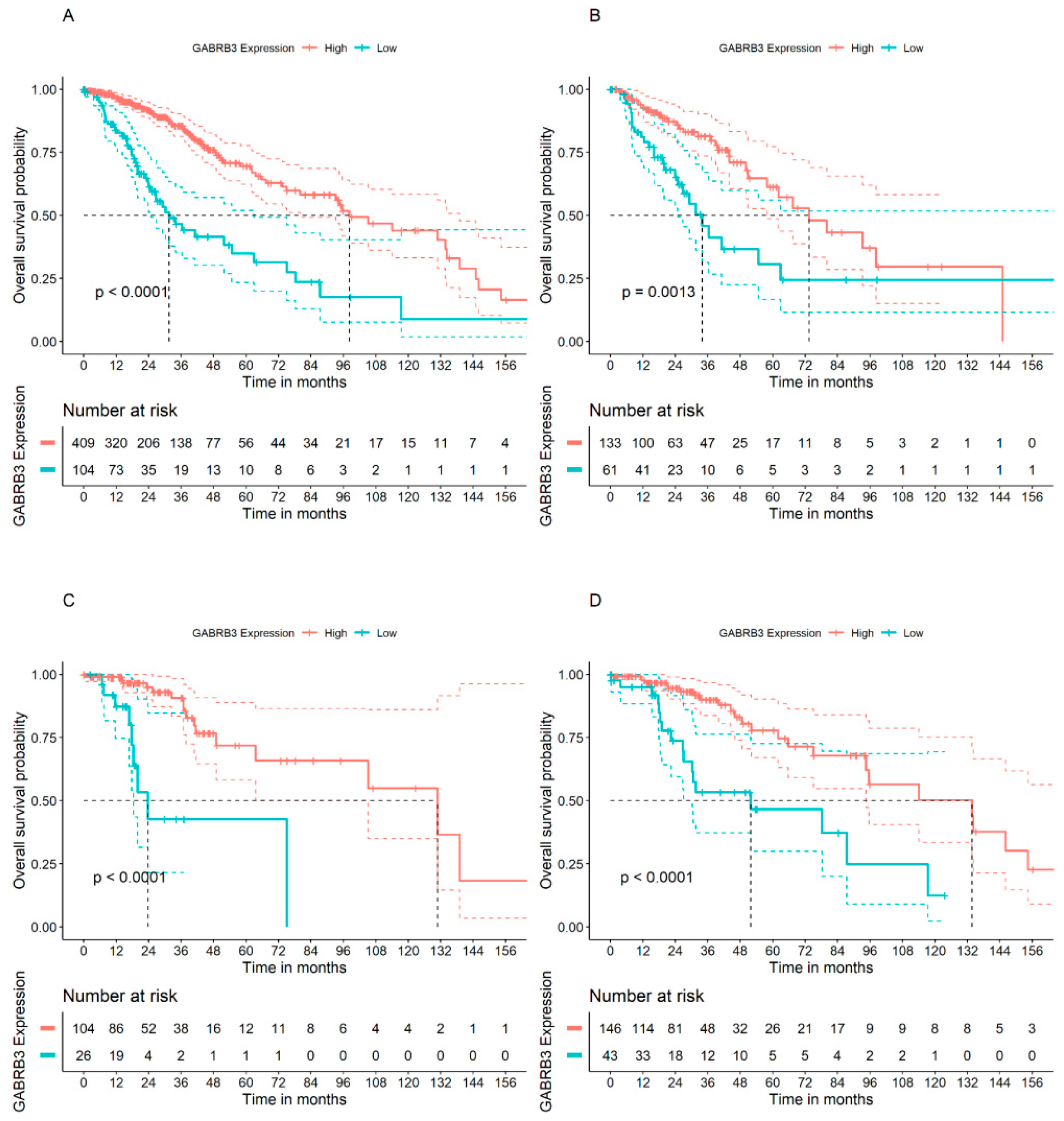
3.2. GABRA2 and GABRB3 Genes Display Opposite Patterns of Association with OS in Patients with GBM

We then selected the samples within the French cohort classified as glioblastoma (GBM) (n = 153). Genes GABRA2 and GABRB3 had a Bonferroni-adjusted p value < 0.05 in these tumor samples (Table 2). High expression of GABRA2 was associated with worse prognosis (Figure 1A,C), whereas, in contrast, high levels of GABRB3 transcripts were associated with better prognosis indicated by longer OS (Figure 1B,D). It is worth highlighting that GABRA2 was the only GABAA receptor gene associated with worse prognosis in GBM patients.

3.3. GABAA Receptor Genes and OS in Patients with Lower Grade Glioma Types

We next analyzed glioma tumors from the TCGA-LGG cohort containing 513 samples distributed across glioma subtypes astrocytoma, oligoastrocytoma, and oligodendroglioma. Using all samples in the dataset (n = 513), we carried out OS analyses for GABRA2, GABRA3, GABRB3, GABRG1, and GABRG2 genes. All genes except for GABRA2 showed significant association with OS, where higher gene expression was related to longer OS (Bonferroni-adjusted p < 0.05) (Table 3).
We went on to verify whether the GABRB3 gene, which showed significant associations with OS in GBM patients from the French cohort and also for TCGA-LGG patients when all tumor types were pooled together, would show influences on OS when lower grade tumors are analyzed separately. Higher GABRB3 expression levels were significantly associated with OS in all glioma subtypes, namely astrocytoma, oligoastrocytoma, and oligodendroglioma (Bonferroni-adjusted p < 0.05) (Figure 2).

4. Discussion

Functional GABAA receptors were initially identified in cells derived from lower grade gliomas, namely astrocytoma and oligodendroglioma, whereas GBM-derived primary cells and glioma cell lines showed no functional receptors. In tumor-derived glioma cells in acute slices or primary culture, most cells from oligodendroglioma and astrocytoma responded to GABA when responses were measured in whole-cell voltage clamp assays as inward currents under high Cl concentration. GBM-derived cells, in contrast, showed no response to GABA. The currents observed in lower grade gliomas were induced specifically by GABA through activation of GABAA receptors, given that the GABAA agonist muscimol mimicked the GABA responses, the benzodiazepine receptor agonist flunitrazepam augmented GABA-induced currents, a benzodiazepine inverse agonist reduced the currents, and the GABAA antagonists bicuculline and picrotoxin blocked GABA-induced currents. It is also noteworthy that, in this experimental setting, GABA-elicited currents could induce either hyperpolarization or depolarization, depending on the cell tested [40]. Functional GABAA receptor-activated currents in GBM cells were later demonstrated, as were findings showing that endogenous GABA continuously released by GBM cells could reduce proliferation of cells expressing progenitor and stem cells markers and negatively regulate experimental tumor growth in mouse models. Thus, shunting cellular Cl chloride ions through sustained local GABAA receptor activity reduced proliferation and tumor growth and prolonged mouse survival. These results strongly suggest that increasing GABAA receptor activity may inhibit GBM progression [41]. In U3047MG human GBM cells, GABAA currents could be pharmacologically stimulated by etomidate, propofol, or diazepam, indicating that GABA-induced currents in GBM can be enhanced by classical GABAA receptor-stimulating drugs. Expression of nRNAs for the α2, α3, α5, β1, β2, β3, δ, γ3, π, and θ GABAA receptor subunits was confirmed in U3047MG cells [37,42]. Together, these findings indicate that glioma tumors of different grades can express GABAA receptors capable of responding to endogenous GABA and other ligands to affect glioma progression.
Expression of mRNA for all 19 GABAA subunits in human glioma (n = 29) and peri-tumoral tissue (n = 5) was previously detected. Consistently with the possibility that lower GABAA receptor activity occurs in more malignant gliomas, GBM tumors showed reduced subunit levels compared to lower grade gliomas, except for the θ subunit. Expression was also found in peritumoral tissue. A consistent co-expression of ρ2 and θ subunits occurred in both astrocytomas and oligodendroglial tumors. Expression of the ρ2 subunit but not the θ subunit was shown by Kaplan–Meier analysis and Cox proportional hazards modeling to be an independent predictor of better survival in patients with astrocytomas, together with other prognostic factors [43].
Isocitrate dehydrogenase (IDH) enzymes, encoded by IDH genes, regulate cellular metabolism and homeostasis by catalyzing the oxidative decarboxylation of isocitrate. Accumulating evidence shows that IDH genes can be mutated in many human malignant cancers, gliomas, and these mutations can impact oncogenesis, tumor progression, and clinical outcome. In gliomas, IDH mutation-associated abnormal changes in cancer cell metabolism, gene expression profile and chromatin structure can lead to disruptions in normal epigenetic programming and, ultimately, resistance to therapy. Thus, increasing research efforts focus on therapeutic strategies designed to specifically target IDH-mutant gliomas [44,45,46,47]. Some IDH1 mutations in glioma are proposed as prognostic markers, with patients bearing mutated tumors showing improved survival [48]. Analysis of tumors from TCGA showed eight subunit genes significantly expressed in IDH wild-type compared with IDH-mutated tumors. Higher expression of the GABRD gene, which encodes the GABAA receptor δ subunit, was independently associated with longer patient OS in IDH wild-type LGGs. GABRD expression was negatively correlated with the extent of tumor infiltration by macrophages. These results suggest that GABRD may be a potential independent prognostic marker in patients with IDH wild-type LGG [49]. Our findings indicating that expression of most GABAA receptor subunit genes is reduced in patients with longer OS may be considered consistent with previous evidence that GABAA receptors can act as inhibitors of glioma growth [41] that display lower expression as glioma grade increases [43].
Also, consistently with an inhibitory role for GABAA receptors in brain tumors, receptor pharmacological stimulation with benzodiazepine derivatives promotes cell death in experimental MB [50]. Current consensus classifies MB tumors into four molecular subgroups, namely wingless activated (WNT), sonic hedgehog (SHH), Group 3, and Group 4, with Group 3 and Group 4 tumors being particularly aggressive [51,52]. GABRA5 and the α5 subunit are found and contribute to the assembly of functional GABAA receptors in patient-derived Group 3 MB cells and tumor tissue. In addition, a benzodiazepine preferentially targeting α5-GABAA hinders Group 3’s MB cell viability [37] with greater potency than standard-of-care chemotherapy used to treat MB patients [53]. Stimulation of GABAA receptors containing the α5 subunit with a selective agonist reduces cell survival through a mechanism involving membrane depolarization and apoptosis induction [37], highlighting the potential of the α5-GABAA receptor as a therapeutic target [54]. There is a significant correlation between expression of GABRA5 and the MYC oncogene in a subset of Group 3 and WNT MB tumors, and the same study indicated GABRA5 expression as a possible diagnostic marker for Group 3 MB [50].

5. Conclusions

In summary, the present study is the first to characterize gene expression of the different protein subunits composing the GABAA receptor in distinct types of glioma, showing that most genes are associated with better prognosis assessed by patient OS, which is consistent with an inhibitory role of GABA in glioma growth. In light of the evidence reviewed above, our findings raise the possibility that glioma tumors show a down-regulation of GABAA receptors as a mechanism to stimulate tumor growth by reducing inhibitory modulation. It should be pointed out, however, that additional functional studies are required to further validate this hypothesis, given that our findings are limited to gene expression and do not confirm that GABAA are directly implicated in determining patient outcomes. Drugs that act by stimulating GABAA receptors should be further investigated as targeted therapies for glioma.

Author Contributions

Conceptualization, R.B., M.D., O.M., J.M.R.F., R.R., M.A.C.F. and G.R.I.; methodology, R.B., M.D., O.M., J.M.R.F., R.R., M.A.C.F. and G.R.I.; formal analysis, R.B., M.D., O.M., J.M.R.F., R.R., M.A.C.F. and G.R.I.; investigation, R.B., M.D., O.M., J.M.R.F., R.R., M.A.C.F. and G.R.I.; resources, R.B., M.D., O.M., J.M.R.F., R.R., M.A.C.F. and G.R.I.; data curation, R.B., M.D., R.R., M.A.C.F. and G.R.I.; writing—original draft preparation, R.B., R.R. and G.R.I.; writing—review and editing, R.B., M.D., O.M., J.M.R.F., R.R. and G.R.I.; supervision, G.R.I.; project administration, G.R.I.; funding acquisition, R.B., O.M., J.M.R.F. and G.R.I. All authors have read and agreed to the published version of the manuscript.

Funding

This research was supported by the National Council for Scientific and Technological Development (CNPq, MCTI, Brazil) grants 305647/2019-9, 405608/2021-7, and 406484/2022-8 (INCT BioOncoPed) (R.R.), The Center for Advanced Neurology and Neurosurgery (CEANNE), the Children’s Cancer Institute, and the Mackenzie Evangelical University.

Institutional Review Board Statement

This study used public datasets and did not require ethical approval.

Informed Consent Statement

Not applicable.

Data Availability Statement

All data were generated and analyzed during this study are based on publicly available datasets and softwares, as described in the article.

Acknowledgments

Authors thank Barbara Kunzler Souza and Epigenica Biosciences for providing assistance in gene expression analysis and data interpretation.

Conflicts of Interest

G.R.I. is a founder and CEO of Spalt Therapeutics. The remaining authors declare that the research was conducted in the absence of any commercial or financial relationships that could be construed as a potential conflict of interest.

References

  1. Wen, P.Y.; Kesari, S. Malignant gliomas in adults. N. Engl. J. Med. 2008, 359, 492–507. [Google Scholar] [CrossRef]
  2. Louis, D.N.; Perry, A.; Wesseling, P.; Brat, D.J.; Cree, I.A.; Figarella-Branger, D.; Hawkins, C.; Ng, H.K.; Pfister, S.M.; Reifenberger, G.; et al. The 2021 WHO Classification of Tumors of the Central Nervous System: A summary. Neuro Oncol. 2021, 23, 1231–1251. [Google Scholar] [CrossRef]
  3. Toader, C.; Eva, L.; Costea, D.; Corlatescu, A.D.; Covache-Busuioc, R.A.; Bratu, B.G.; Glavan, L.A.; Costin, H.P.; Popa, A.A.; Ciurea, A.V. Low-grade gliomas: Histological subtypes, molecular mechanisms, and treatment strategies. Brain Sci. 2023, 13, 1700. [Google Scholar] [CrossRef]
  4. Alexander, B.M.; Cloughesy, T.F. Adult glioblastoma. J. Clin. Oncol. 2017, 35, 2402–2409. [Google Scholar] [CrossRef]
  5. Luskin, M.B. Restricted proliferation and migration of postnatally generated neurons derived from the forebrain subventricular zone. Neuron 1993, 11, 173–189. [Google Scholar] [CrossRef]
  6. Lois, C.; Alvarez-Buylla, A. Long-distance neuronal migration in the adult mammalian brain. Science 1994, 264, 1145–1148. [Google Scholar] [CrossRef] [PubMed]
  7. Doetsch, F.; Caillé, I.; Lim, D.A.; García-Verdugo, J.M.; Alvarez-Buylla, A. Subventricular zone astrocytes are neural stem cells in the adult mammalian brain. Cell 1999, 97, 703–716. [Google Scholar] [CrossRef]
  8. Lee, J.H.; Lee, J.E.; Kahng, J.Y.; Kim, S.H.; Park, J.S.; Yoon, S.J.; Um, J.Y.; Kim, W.K.; Lee, J.K.; Park, J.; et al. Human glioblastoma arises from subventricular zone cells with low-level driver mutations. Nature 2018, 560, 243–247. [Google Scholar] [CrossRef] [PubMed]
  9. Altmann, C.; Keller, S.; Schmidt, M.H.H. The role of SVZ stem cells in glioblastoma. Cancers 2019, 11, 448. [Google Scholar] [CrossRef] [PubMed]
  10. Zhang, G.L.; Wang, C.F.; Qian, C.; Ji, Y.X.; Wang, Y.Z. Role and mechanism of neural stem cells of the subventricular zone in glioblastoma. World J. Stem Cells 2021, 13, 877–893. [Google Scholar] [CrossRef] [PubMed]
  11. Beiriger, J.; Habib, A.; Jovanovich, N.; Kodavali, C.V.; Edwards, L.; Amankulor, N.; Zinn, P.O. The subventricular zone in glioblastoma: Genesis, maintenance, and modeling. Front. Oncol. 2022, 12, 790976. [Google Scholar] [CrossRef] [PubMed]
  12. Loras, A.; Gonzalez-Bonet, L.G.; Gutierrez-Arroyo, J.L.; Martinez-Cadenas, C.; Marques-Torrejon, M.A. Neural stem cells as potential glioblastoma cells of origin. Life 2023, 13, 905. [Google Scholar] [CrossRef] [PubMed]
  13. Yang, Z.; Zhao, C.; Zong, S.; Piao, J.; Zhao, Y.; Chen, X. A review on surgical treatment options in gliomas. Front. Oncol. 2023, 13, 1088484. [Google Scholar] [CrossRef] [PubMed]
  14. Stupp, R.; Mason, W.P.; van den Bent, M.J.; Weller, M.; Fisher, B.; Taphoorn, M.J.; Belanger, K.; Brandes, A.A.; Marosi, C.; Bogdahn, U.; et al. Radiotherapy plus concomitant and adjuvant temozolomide for glioblastoma. N. Engl. J. Med. 2005, 352, 987–996. [Google Scholar] [CrossRef] [PubMed]
  15. Felistia, Y.; Wen, P.Y. Molecular profiling and targeted therapies in gliomas. Curr. Neurol. Neurosci. Rep. 2023, 23, 627–636. [Google Scholar] [CrossRef] [PubMed]
  16. Picca, A.; Bruno, F.; Nichelli, L.; Sanson, M.; Rudàm, R. Advances in molecular and imaging biomarkers in lower-grade gliomas. Expert Rev. Neurother. 2023, 23, 1217–1231. [Google Scholar] [CrossRef]
  17. Mancusi, R.; Monje, M. The neuroscience of cancer. Nature 2023, 618, 467–479. [Google Scholar] [CrossRef]
  18. Prillaman, M. How cancer hijacks the nervous system to grow and spread. Nature 2024, 626, 22–24. [Google Scholar] [CrossRef]
  19. Tochitani, S.; Furukawa, T.; Bando, R.; Kondo, S.; Ito, T.; Matsushima, Y.; Kojima, T.; Matsuzaki, H.; Fukuda, A. GABAA receptors and maternally derived taurine regulate the temporal specification of progenitors of excitatory glutamatergic neurons in the mouse developing cortex. Cereb. Cortex 2021, 31, 4554–4575. [Google Scholar] [CrossRef]
  20. Bao, H.; Asrican, B.; Li, W.; Gu, B.; Wen, Z.; Lim, S.A.; Haniff, I.; Ramakrishnan, C.; Deisseroth, K.; Philpot, B.; et al. Long-range GABAergic inputs regulate neural stem cell quiescence and control adult hippocampal neurogenesis. Cell Stem Cell 2017, 21, 604–617.e5. [Google Scholar] [CrossRef]
  21. Lattanzi, D.; Di Palma, M.; Cuppini, R.; Ambrogini, P. GABAergic input affects intracellular calcium levels in developing granule cells of adult rat hippocampus. Int. J. Mol. Sci. 2020, 21, 1715. [Google Scholar] [CrossRef] [PubMed]
  22. Tian, R.; Guo, K.; Wu, B.; Wang, H. Overexpression of Shrm4 promotes proliferation and differentiation of neural stem cells through activation of GABA signaling pathway. Mol. Cell. Biochem. 2020, 463, 115–126. [Google Scholar] [CrossRef]
  23. Catavero, C.; Bao, H.; Song, J. Neural mechanisms underlying GABAergic regulation of adult hippocampal neurogenesis. Cell Tissue Res. 2018, 371, 33–46. [Google Scholar] [CrossRef]
  24. Song, D.; Chen, Y.; Chen, C.; Chen, L.; Cheng, O. GABAB receptor antagonist promotes hippocampal neurogenesis and facilitates cognitive function recovery following acute cerebral ischemia in mice. Stem Cell Res. Ther. 2021, 12, 22. [Google Scholar] [CrossRef] [PubMed]
  25. Arshad, M.N.; Oppenheimer, S.; Jeong, J.; Buyukdemirtas, B.; Naegele, J.R. Hippocampal transplants of fetal GABAergic progenitors regulate adult neurogenesis in mice with temporal lobe epilepsy. Neurobiol. Dis. 2022, 174, 105879. [Google Scholar] [CrossRef]
  26. Wang, D.D.; Krueger, D.D.; Bordey, A. GABA depolarizes neuronal progenitors of the postnatal subventricular zone via GABAA receptor activation. J. Physiol. 2003, 550 Pt 3, 785–800. [Google Scholar] [CrossRef]
  27. Young, S.Z.; Platel, J.C.; Nielsen, J.V.; Jensen, N.A.; Bordey, A. GABA(A) increases calcium in subventricular zone astrocyte-like cells through L- and T-type voltage-gated calcium channels. Front. Cell. Neurosci. 2010, 4, 8. [Google Scholar] [CrossRef]
  28. Young, S.Z.; Lafourcade, C.A.; Platel, J.C.; Lin, T.V.; Bordey, A. GABAergic striatal neurons project dendrites and axons into the postnatal subventricular zone leading to calcium activity. Front. Cell. Neurosci. 2014, 8, 10. [Google Scholar] [CrossRef]
  29. Hsieh, Y.C.; Puche, A.C. GABA modulation of SVZ-derived progenitor ventral cell migration. Dev. Neurobiol. 2015, 75, 791–804. [Google Scholar] [CrossRef]
  30. Gutiérrez-Castañeda, N.E.; González-Corona, J.; Griego, E.; Galván, E.J.; Ochoa-de la Paz, L.D. Taurine promotes differentiation and maturation of neural stem/progenitor cells from the subventricular zone via activation of GABAA receptors. Neurochem. Res. 2023, 48, 2206–2219. [Google Scholar] [CrossRef]
  31. Simon, J.; Wakimoto, H.; Fujita, N.; Lalande, M.; Barnard, E.A. Analysis of the set of GABA(A) receptor genes in the human genome. J. Biol. Chem. 2004, 279, 41422–41435. [Google Scholar] [CrossRef] [PubMed]
  32. Bhattacharya, D.; Gawali, V.S.; Kallay, L.; Toukam, D.K.; Koehler, A.; Stambrook, P.; Krummel, D.P.; Sengupta, S. Therapeutically leveraging GABAA receptors in cancer. Exp. Biol. Med. 2021, 246, 2128–2135. [Google Scholar] [CrossRef] [PubMed]
  33. Ghit, A.; Assal, D.; Al-Shami, A.S.; Hussein, D.E.E. GABAA receptors: Structure, function, pharmacology, and related disorders. J. Genet. Eng. Biotechnol. 2021, 19, 123. [Google Scholar] [CrossRef] [PubMed]
  34. Butler, K.M.; Moody, O.A.; Schuler, E.; Coryell, J.; Alexander, J.J.; Jenkins, A.; Escayg, A. De novo variants in GABRA2 and GABRA5 alter receptor function and contribute to early-onset epilepsy. Brain 2018, 141, 2392–2405. [Google Scholar] [CrossRef] [PubMed]
  35. Feng, Y.; Wei, Z.H.; Liu, C.; Li, G.Y.; Qiao, X.Z.; Gan, Y.J.; Zhang, C.C.; Deng, Y.C. Genetic variations in GABA metabolism and epilepsy. Seizure 2022, 101, 22–29. [Google Scholar] [CrossRef] [PubMed]
  36. Joghataei, M.T.; Bakhtiarzadeh, F.; Dehghan, S.; Ketabforoush, A.H.M.E.; Golab, F.; Zarbakhsh, S.; Ahmadirad, N. The role of neurotransmitters in glioblastoma multiforme-associated seizures. Int. J. Dev. Neurosci. 2023, 83, 677–690. [Google Scholar] [CrossRef]
  37. Sengupta, S.; Weeraratne, S.D.; Sun, H.; Phallen, J.; Rallapalli, S.K.; Teider, N.; Kosaras, B.; Amani, V.; Pierre-Francois, J.; Tang, Y.; et al. α5-GABAA receptors negatively regulate MYC-amplified medulloblastoma growth. Acta Neuropathol. 2014, 127, 593–603. [Google Scholar] [CrossRef]
  38. Gravendeel, L.A.; Kouwenhoven, M.C.; Gevaert, O.; de Rooi, J.J.; Stubbs, A.P.; Duijm, J.E.; Daemen, A.; Bleeker, F.E.; Bralten, L.B.; Kloosterhof, N.K.; et al. Intrinsic gene expression profiles of gliomas are a better predictor of survival than histology. Cancer Res. 2009, 69, 9065–9072. [Google Scholar] [CrossRef]
  39. Rodrigues, E.M.; Giovanini, A.F.; Ribas, C.A.P.M.; Malafaia, O.; Roesler, R.; Isolan, G.R. The nervous system development regulator neuropilin-1 as a potential prognostic marker and therapeutic target in brain cancer. Cancers 2023, 15, 4922. [Google Scholar] [CrossRef]
  40. Labrakakis, C.; Patt, S.; Hartmann, J.; Kettenmann, H. Functional GABA(A) receptors on human glioma cells. Eur. J. Neurosci. 1998, 10, 231–238. [Google Scholar] [CrossRef]
  41. Blanchart, A.; Fernando, R.; Häring, M.; Assaife-Lopes, N.; Romanov, R.A.; Andäng, M.; Harkany, T.; Ernfors, P. Endogenous GABAA receptor activity suppresses glioma growth. Oncogene 2017, 36, 777–786. [Google Scholar] [CrossRef]
  42. Babateen, O.; Jin, Z.; Bhandage, A.; Korol, S.V.; Westermark, B.; Forsberg Nilsson, K.; Uhrbom, L.; Smits, A.; Birnir, B. Etomidate, propofol and diazepam potentiate GABA-evoked GABAA currents in a cell line derived from human glioblastoma. Eur. J. Pharmacol. 2015, 748, 101–107. [Google Scholar] [CrossRef]
  43. Smits, A.; Jin, Z.; Elsir, T.; Pedder, H.; Nistér, M.; Alafuzoff, I.; Dimberg, A.; Edqvist, P.H.; Pontén, F.; Aronica, E.; et al. GABA-A channel subunit expression in human glioma correlates with tumor histology and clinical outcome. PLoS ONE 2012, 7, e37041. [Google Scholar] [CrossRef]
  44. Han, S.; Lium, Y.; Caim, S.J.; Qian, M.; Ding, J.; Larion, M.; Gilbert, M.R.; Yang, C. IDH mutation in glioma: Molecular mechanisms and potential therapeutic targets. Br. J. Cancer 2020, 122, 1580–1589. [Google Scholar] [CrossRef] [PubMed]
  45. Dono, A.; Ballester, L.Y.; Primdahl, D.; Esquenazi, Y.; Bhatia, A. IDH-mutant low-grade glioma: Advances in molecular diagnosis, management, and future directions. Curr. Oncol. Rep. 2021, 23, 20. [Google Scholar] [CrossRef] [PubMed]
  46. Kayabolen, A.; Yilmaz, E.; Bagci-Onder, T. IDH mutations in glioma: Double-edged sword in clinical applications? Biomedicines 2021, 9, 799. [Google Scholar] [CrossRef] [PubMed]
  47. Miller, J.J. Targeting IDH-mutant glioma. Neurotherapeutics 2022, 19, 1724–1732. [Google Scholar] [CrossRef] [PubMed]
  48. Sareen, H.; Ma, Y.; Becker, T.M.; Roberts, T.L.; de Souza, P.; Powter, B. Molecular biomarkers in glioblastoma: A systematic review and meta-analysis. Int. J. Mol. Sci. 2022, 23, 8835. [Google Scholar] [CrossRef] [PubMed]
  49. Zhang, H.; Zhang, L.; Tang, Y.; Wang, C.; Chen, Y.; Shu, J.; Zhang, K. Systemic screening identifies GABRD, a subunit gene of GABAA receptor as a prognostic marker in adult IDH wild-type diffuse low-grade glioma. Biomed. Pharmacother. 2019, 118, 109215. [Google Scholar] [CrossRef] [PubMed]
  50. Kallay, L.; Keskin, H.; Ross, A.; Rupji, M.; Moody, O.A.; Wang, X.; Li, G.; Ahmed, T.; Rashid, F.; Stephen, M.R.; et al. Modulating native GABAA receptors in medulloblastoma with positive allosteric benzodiazepine-derivatives induces cell death. J. Neurooncol. 2019, 142, 411–422. [Google Scholar] [CrossRef]
  51. Northcott, P.A.; Korshunov, A.; Pfister, S.M.; Taylor, M.D. The clinical implications of medulloblastoma subgroups. Nat. Rev. Neurol. 2012, 8, 340–351. [Google Scholar] [CrossRef] [PubMed]
  52. Taylor, M.D.; Northcott, P.A.; Korshunov, A.; Remke, M.; Cho, Y.J.; Clifford, S.C.; Eberhart, C.G.; Parsons, D.W.; Rutkowski, S.; Gajjar, A.; et al. Molecular subgroups of medulloblastoma: The current consensus. Acta Neuropathol. 2012, 123, 465–472. [Google Scholar] [CrossRef]
  53. Jonas, O.; Calligaris, D.; Methuku, K.R.; Poe, M.M.; Francois, J.P.; Tranghese, F.; Changelian, A.; Sieghart, W.; Ernst, M.; Krummel, D.A.; et al. First in vivo testing of compounds targeting group 3 medulloblastomas using an implantable microdevice as a new paradigm for drug development. J. Biomed. Nanotechnol. 2016, 12, 1297–1302. [Google Scholar] [CrossRef] [PubMed]
  54. Sengupta, S.; Weeraratne, S.D.; Cho, Y.J.; Pomeroy, S.L. Could α5-GABA-A receptor activation be used as a target for managing medulloblastomas? CNS Oncol. 2014, 3, 245–247. [Google Scholar] [CrossRef] [PubMed]
Figure 1. OS analysis of the genes GABRA2 and GABRB3 in patients from the French cohort. Results are derived from all glioma tumor types pooled together (n = 266) for (A) GABRA2 and (B) GABRB3; and GBM only (n = 153) for (C) GABRA2 and (D) GABRB3.
Brainsci 14 00275 g001
Figure 2. Analysis of OS in patients bearing high or low tumor levels of GABRB3 in the TCGA-LGG cohort. (A) All glioma types pooled together (n = 513), (B) astrocytoma (n = 194), (C) oligoastrocytoma (n = 130), and (D) oligodendroglioma (n = 189).
Brainsci 14 00275 g002
Table 1. Summary of patient OS analysis results conducted for all 36 probes corresponding to 18 genes that encode GABAA subunit proteins in the French dataset.
ProbeSubunitGenep ValueAdjusted p
206678_atGABAA receptor, alpha 1GABRA11.72 × 10−11
244118_atGABAA receptor, alpha 1GABRA11.68 × 10−11
1554308_s_atGABAA receptor, alpha 2GABRA21.00 × 10−33.60 × 10⁻²
207014_atGABAA receptor, alpha 2GABRA21.29 × 10−24.64 × 10⁻¹
216039_atGABAA receptor, alpha 2GABRA21.24 × 10−34.45 × 10⁻²
207210_atGABAA receptor, alpha 3GABRA31.05 × 10−73.78 × 10⁻⁶
208463_atGABAA receptor, alpha 4GABRA42.99 × 10−11
233437_atGABAA receptor, alpha 4GABRA41.83 × 10−11
206456_atGABAA receptor, alpha 5GABRA57.45 × 10−21
215531_s_atGABAA receptor, alpha 5GABRA52.69 × 10−11
217280_x_atGABAA receptor, alpha 5GABRA51.16 × 10−11
207182_atGABAA receptor, alpha 6GABRA61.42 × 10−25.10 × 10⁻¹
1557256_a_atGABAA receptor, beta 1GABRB10.010.48
207010_atGABAA receptor, beta 1GABRB12.03 × 10−27.31 × 10⁻¹
1557122_s_atGABAA receptor, beta 2GABRB29.31 × 10−33.35 × 10⁻¹
207352_s_atGABAA receptor, beta 2GABRB23.49 × 10−11
242344_atGABAA receptor, beta 2GABRB23.62 × 10−21
1569689_s_atGABAA receptor, beta 3GABRB31.51 × 10−25.45 × 10⁻¹
205850_s_atGABAA receptor, beta 3GABRB32.43 × 10−138.74 × 10⁻¹²
227690_atGABAA receptor, beta 3GABRB31.21 × 10−144.36 × 10¹³
227830_atGABAA receptor, beta 3GABRB35.55 × 10−162.00 × 10⁻¹⁴
229724_atGABAA receptor, beta 3GABRB300
208457_atGABAA receptor, deltaGABRD2.04 × 10−27.35 × 10⁻¹
230255_atGABAA receptor, deltaGABRD1.34 × 10−11
1552943_atGABAA receptor, gamma 1GABRG18.35 × 10−63.01 × 10⁻⁴
241805_atGABAA receptor, gamma 1GABRG11.43 × 10−65.16 × 10⁻⁵
1568612_atGABAA receptor, gamma 2GABRG21.63 × 10−65.88 × 10⁻⁵
206849_atGABAA receptor, gamma 2GABRG27.95 × 10−82.86 × 10⁻⁶
1555517_atGABAA receptor, gamma 3GABRG31.44 × 10−25.18 × 10⁻¹
216895_atGABAA receptor, gamma 3GABRG31.65 × 10−11
205044_atGABAA receptor, piGABRP2.78 × 10−11
220886_atGABAA receptor, thetaGABRQ3.44 × 10−11
238123_atGABAA receptor, thetaGABRQ4.06 × 10−11
206525_atGABAA receptor, rho 1GABRR14.44 × 10−31.60 × 10⁻¹
208217_atGABAA receptor, rho 2GABRR24.71 × 10−21
234410_atGABAA receptor, rho 3GABRR31.34 × 10−24.84 × 10⁻¹
206678_atGABAA receptor, alpha 1GABRA11.72 × 10−11
Table 2. Summary of the patient OS analysis results carried for five GABAA receptor subunit genes in GBM patients from the French cohort.
ProbeSubunitGenep ValueAdjusted p
1554308_s_atGABAA receptor, alpha 2GABRA25.34 × 10−32.67 × 10⁻²
207210_atGABAA receptor, alpha 3GABRA32.02 × 10−21.01 × 10⁻¹
229724_atGABAA receptor, beta 3GABRB34.39 × 10−32.19 × 10⁻²
206849_atGABAA receptor, gamma 1GABRG11.57 × 10−17.83 × 10⁻¹
241805_atGABAA receptor, gamma 2GABRG28.95 × 10−24.48 × 10⁻¹
Table 3. Summary of the patient OS analysis results carried for GABAA receptor subunit genes in lower grade glioma patients from the TCGA-LGG cohort.
SubunitGenep ValueAdjusted p
GABAA receptor, alpha 2GABRA25.37 × 10−22.69 × 10⁻¹
GABAA receptor, alpha 3GABRA36.25 × 10−143.13 × 10⁻¹³
GABAA receptor, beta 3GABRB31.63 × 10−118.13 × 10⁻¹¹
GABAA receptor, gamma 1GABRG14.13 × 10−72.07 × 10⁻⁶
GABAA receptor, gamma 2GABRG21.96 × 10−59.78 × 10⁻⁵
Disclaimer/Publisher’s Note: The statements, opinions and data contained in all publications are solely those of the individual author(s) and contributor(s) and not of MDPI and/or the editor(s). MDPI and/or the editor(s) disclaim responsibility for any injury to people or property resulting from any ideas, methods, instructions or products referred to in the content.

