Review

From Stress to Sick(le) and Back Again–Oxidative/Antioxidant Mechanisms, Genetic Modulation, and Cerebrovascular Disease in Children with Sickle Cell Anemia

1
Departamento de Genética Humana, Instituto Nacional de Saúde Doutor Ricardo Jorge (INSA), Av. Padre Cruz, 1649-016 Lisboa, Portugal
2
Grupo Ecogenética e Saúde Humana, Instituto de Saúde Ambiental (ISAMB), Faculdade de Medicina, Universidade de Lisboa, Av. Prof. Egas Moniz, 1649-028 Lisboa, Portugal
3
Laboratório Associado TERRA, Faculdade de Medicina, Universidade de Lisboa, Av. Prof. Egas Moniz, 1649-028 Lisboa, Portugal
*
Author to whom correspondence should be addressed.
Antioxidants 2023, 12(11), 1977; https://doi.org/10.3390/antiox12111977
Submission received: 4 September 2023 / Revised: 30 October 2023 / Accepted: 2 November 2023 / Published: 7 November 2023

Abstract

:
Sickle cell anemia (SCA) is a genetic disease caused by the homozygosity of the HBB:c.20A>T mutation, which results in the production of hemoglobin S (HbS). In hypoxic conditions, HbS suffers autoxidation and polymerizes inside red blood cells, altering their morphology into a sickle shape, with increased rigidity and fragility. This triggers complex pathophysiological mechanisms, including inflammation, cell adhesion, oxidative stress, and vaso-occlusion, along with metabolic alterations and endocrine complications. SCA is phenotypically heterogeneous due to the modulation of both environmental and genetic factors. Pediatric cerebrovascular disease (CVD), namely ischemic stroke and silent cerebral infarctions, is one of the most impactful manifestations. In this review, we highlight the role of oxidative stress in the pathophysiology of pediatric CVD. Since oxidative stress is an interdependent mechanism in vasculopathy, occurring alongside (or as result of) endothelial dysfunction, cell adhesion, inflammation, chronic hemolysis, ischemia-reperfusion injury, and vaso-occlusion, a brief overview of the main mechanisms involved is included. Moreover, the genetic modulation of CVD in SCA is discussed. The knowledge of the intricate network of altered mechanisms in SCA, and how it is affected by different genetic factors, is fundamental for the identification of potential therapeutic targets, drug development, and patient-specific treatment alternatives.

Graphical Abstract

1. Introduction

The hemoglobin (Hb) disorders (or hemoglobinopathies) are the most common genetic diseases worldwide. Sickle cell disease (SCD), in particular, affects about 70% of the 300,000–500,000 children born yearly with a hemoglobinopathy [1,2]. The underlying genetic defects affect Hb function, stability, or Hb levels. Furthermore, Hb abnormalities also lead to changes in the biomechanical properties of red blood cells (RBCs) [3]. The global public health burden of the disease is significant, and even though an increasing proportion of affected children now survive past five years of age, the risk of premature death remains especially high in low-income countries [1,4]. More than just a Hb disorder, its marked clinical heterogeneity renders it a disease spectrum. Different levels of anemia, intra- and extravascular hemolysis, vascular obstruction, inflammation, and metabolic and endocrine alterations, such as, growth failure, osteopenia, hypogonadism, hypothyroidism, and insulin resistance, may be observed in these patients [5,6]. The clinical manifestations of the disease range from very mild to extremely severe and life threatening (e.g., stroke, pulmonary hypertension, acute chest syndrome).
As with the phenotypes, the genotypes are also varied. First described by James Herrick, in 1910, as the presence of abnormally crescent and sickle-shaped red cells in a blood smear [7], the term SCD currently applies to a group of hemoglobinopathies caused by the presence of the βS allele–an A to T missense mutation in the β-globin gene (HBB:c.20A>T). This can occur in homozygosity βSβS—as in sickle cell anemia (SCA)—or compound heterozygosity of βS with a another mutant HBB allele, which either changes the β-globin chain of Hb or reduces HBB expression (Figure 1). Whichever the disease-causing genetic change, all patients share the same biochemical phenotype—the presence of the abnormal Hb variant S (HbS). HbS has a single amino acid difference from the normal Hb in the sixth position of the β-globin chain, a substitution of a glutamic acid by a valine (Glu6Val), that changes the protein’s properties and impacts RBC conformation [8] (Figure 1 and Figure 2). Substitution of the same glutamic acid by lysine (Glu6Lys) gives rise to another Hb variant, HbC, which, when inherited concomitantly with HbS, results in HbSC disease, a less severe form than SCA. HbS/β-thal disease is yet another form of SCD, arising from co-inheritance of β-thalassemia (β0-thal or β+-thal) and HbS.
Although SCD is a single gene disease, its clinical heterogeneity, with the involvement of multiple organs, different degrees of severity, and of environmental factors, mimics a multifactorial mode of genetic transmission. Since patients may have a combination of complications and differ from each other in disease severity, age of onset, and in rate of progression, this translates into a high variability in health-related quality of life and life expectancy [5,9,10]. The overall heterogeneity seems to result from modifying factors, ranging from environmental and sociodemographic factors (discrepancies between patients in high- and low-income settings are apparent), to genetic modulators.
Several interdependent mechanisms have been shown to contribute to vascular pathology, or vasculopathy, in SCD, occurring alongside, or as a result, of one another. These include endothelial dysfunction, cell adhesion, inflammation, chronic hemolysis, ischemia-reperfusion (I/R) injury, vaso-occlusion, and oxidative stress. Current therapies, like bone marrow transplant (curative) and gene therapy (under development), while promising, are hardly available to a high number of patients, particularly in low-income settings.
Oxidative stress, as defined by an imbalance between levels of reactive oxygen species (ROS), reactive nitrogen species (RNS), and activity/concentration of antioxidants, has been extensively reviewed in SCD [5,11,12,13]. However, it is important to understand its role in vasculopathy, as well as the interplay with other disease mechanisms and antioxidant defense, especially in cerebrovascular disease (CVD), which has a significant impact particularly in pediatric patients. The knowledge of this intricate network of mechanisms, and how it is affected by different genetic factors, is fundamental for the identification of potential therapeutic targets, drug development, and patient-specific treatment alternatives.
This review highlights the role of oxidative stress in the pathophysiology of pediatric CVD, a high impact manifestation in children with SCA. Since oxidative stress is an interdependent mechanism in vasculopathy, occurring alongside (or as result of) endothelial dysfunction, cell adhesion, inflammation, chronic hemolysis, I/R injury, and vaso-occlusion, a brief overview of the main mechanisms involved is included. Furthermore, the role of genetic modulators on CVD severity is also discussed.

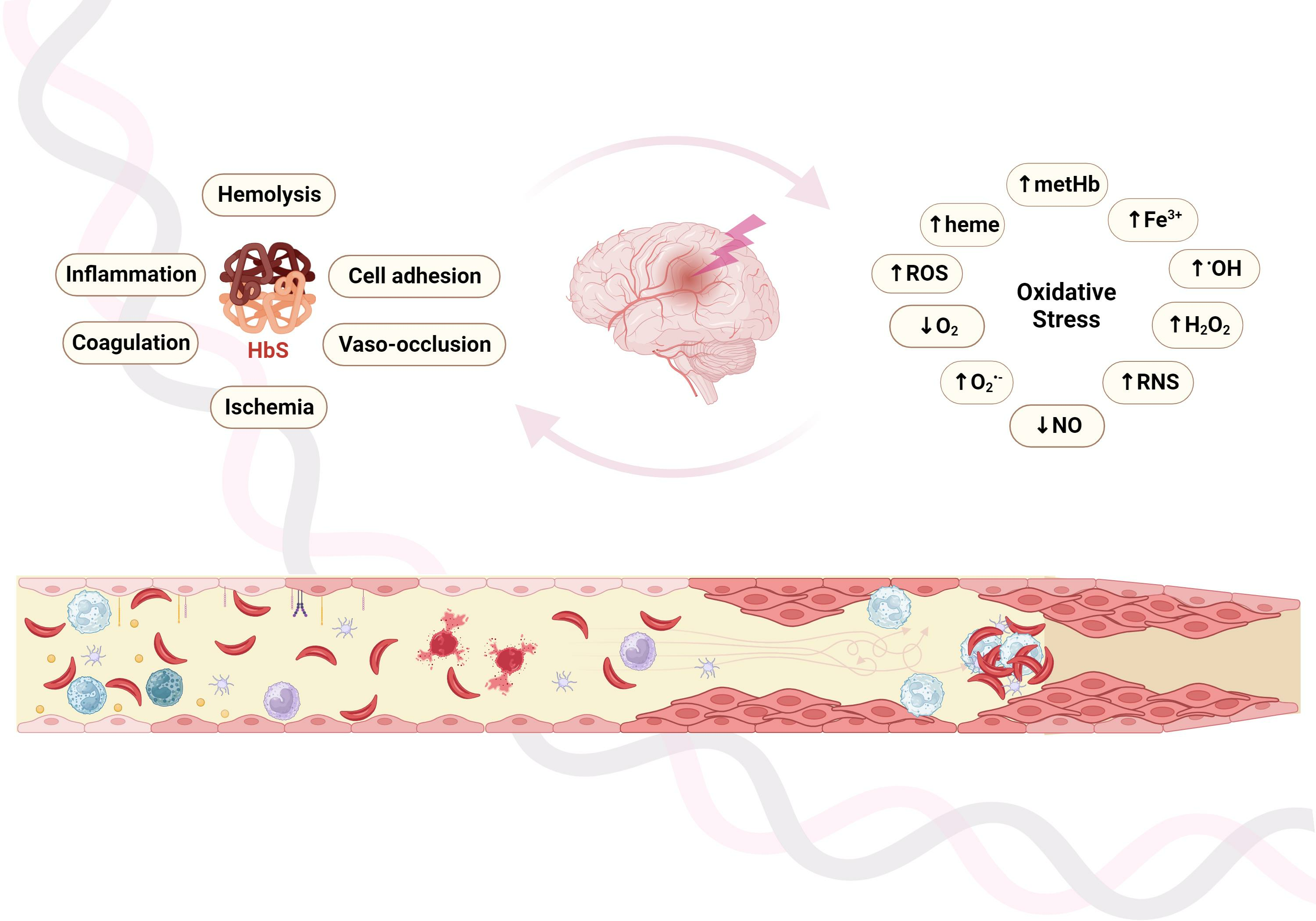
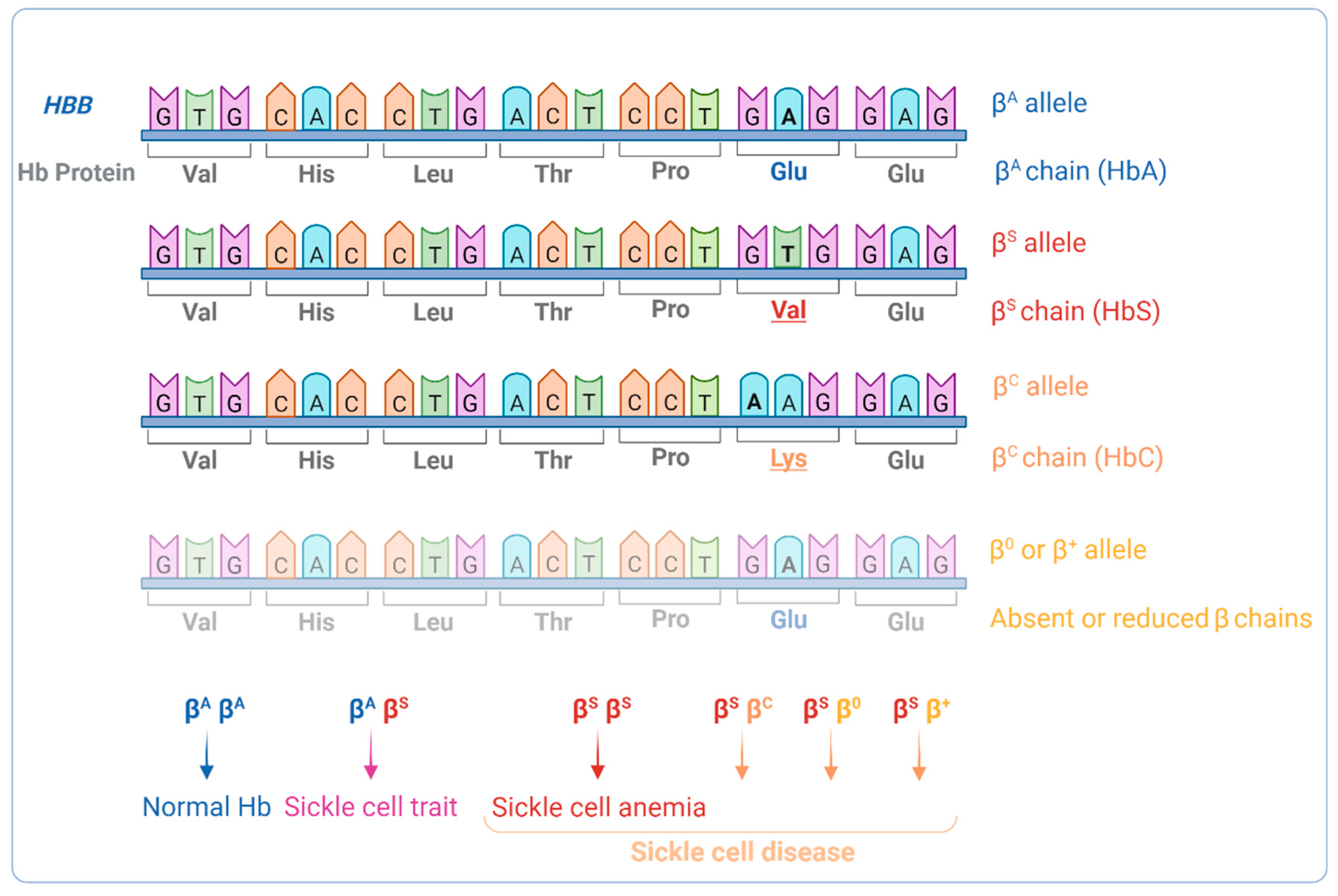
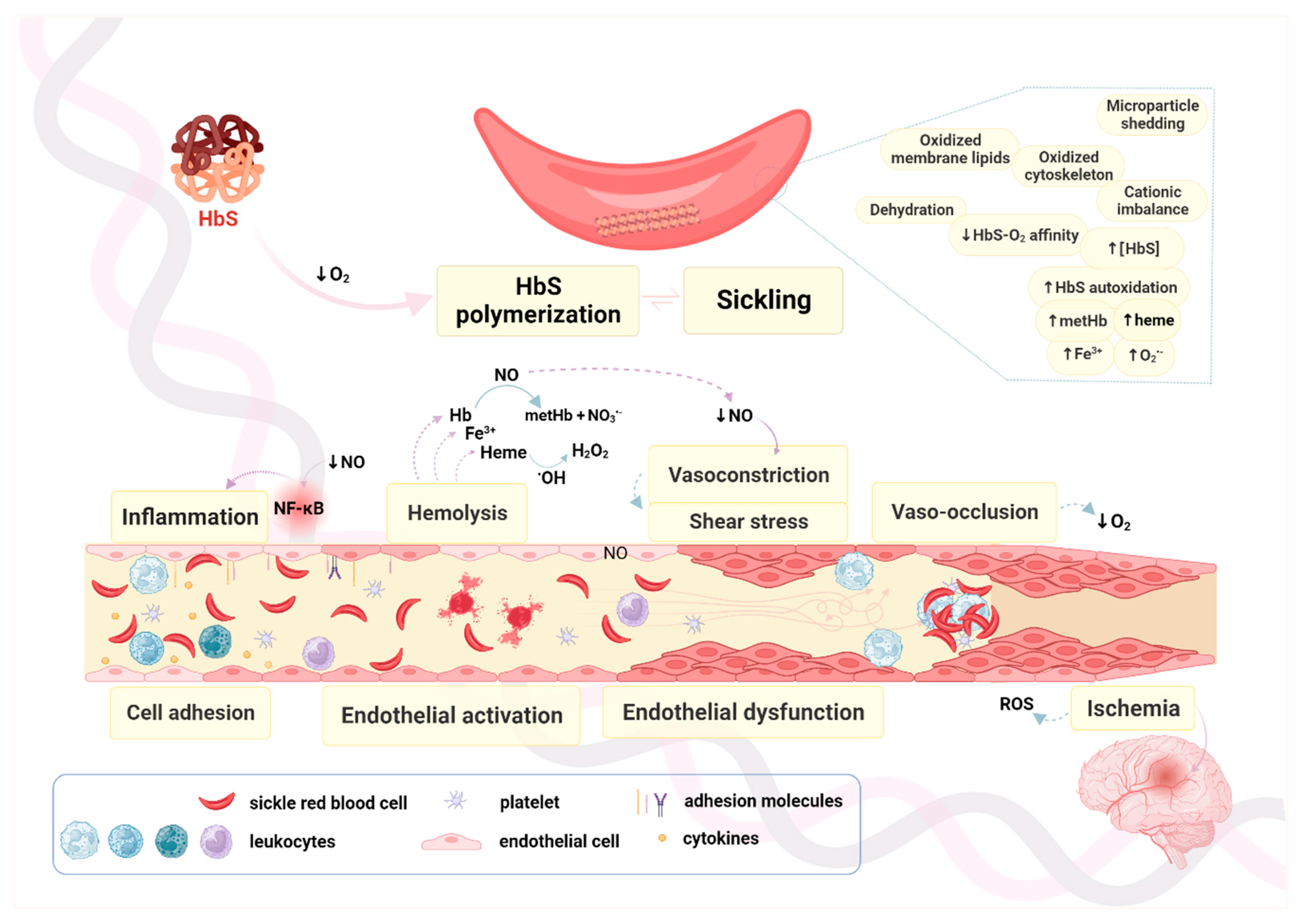
2. Oxidation in RBCs, Hemoglobin, and the Vascular Milieu in SCA

Even though SCA is the most common and severe form of SCD, the compound heterozygous genotypes also lead to the production of sufficient HbS to promote intracellular RBC sickling [14]. In those individuals, the majority of Hb is HbS, whereas in individuals with sickle cell trait (heterozygotes for the abnormal HbS), HbA is the main form present (Figure 1).
Physiologically, oxygenated Hb (ferrous state) is relatively stable. However, it can autoxidize to methemoglobin (metHb) (ferric state), particularly in low-oxygen-saturation environments, like in the microcirculation. ROS production in RBCs is almost exclusively a result of Hb autoxidation, and it is even more pronounced for unstable hemoglobins like HbS, which, contrary to HbA, has a low oxygen affinity [15]. Consequently, in SCD, Hb autoxidation is more pronounced than in normal physiological conditions, promoting a marked pro-oxidant vascular environment, especially in the microcirculation [16]. Under those hypoxic conditions, HbS polymerizes into long rigid fibers inside RBCs. Each of these fibers consists of seven intertwined double strands (with cross-linking) that distort and damage the membrane and cytoskeleton of RBCs [17]. The distortion and damage culminates in sickling, where RBCs adopt a characteristic crescent, or sickle, shape (Figure 2). The subsequent alterations in microrheological, as well as in biomechanical, properties affect their aggregability, deformability, and cell adhesion [17]. The shape changes are initially reversible, occurring in cycles of oxygenation and deoxygenation (oxy–deoxy). As the number of oxy–deoxy cycles increases, the altered RBCs become irreversibly sickled (SSRBC), a state associated with increased fragility, shorter lifespan, and higher propensity to adhere to the vessel wall [18].
Being more fragile, SSRBCs rupture easily (intravascular hemolysis) with consequent release of Hb and heme. Furthermore, extravascular hemolysis is exacerbated due to phagocytosis of SSRBCs by spleen and liver macrophages. Both types of hemolysis contribute to a reduction in the SSRBCs’ lifespan (one week, contrasting with 120 days for normal RBCs), increasing anemia and stimulating production, and an excess of circulating immature RBCs, or reticulocytes (also called stress reticulocytes). Compromised SSRBCs deformability, which impairs their normal flow through capillaries, contributes to vaso-occlusion. This process is complex and may involve multistep multicellular interactions that include endothelial activation, recruitment of adherent leukocytes, interactions of SSRBCs with adherent neutrophils, and vascular obstruction by heterotypic multicellular aggregates [19]. The overall process leads to vaso-occlusion, which in turn culminates in cumulative organ damage, and results in life-threatening crises [19].
Inflammation, and chronic hemolysis, particularly intravascular hemolysis, with the release of Hb and heme and the subsequent decrease in nitric oxide (NO) bioavailability, as well as oxidative stress, strongly contribute to vasculopathy. NO reduction shifts the balance towards vasoconstriction (Figure 3), while the activated endothelium expresses several cell adhesion molecules (CAM) like vascular cell adhesion molecule 1 (VCAM-1), intercellular cell adhesion molecule (ICAM), and/or selectins, and undergoes cell proliferation, which additionally contributes to the vaso-occlusive process [20].
These processes work in a self-sustained cycle of repeated cell activation, cell adhesion, inflammation, hemolysis, oxidative stress, vaso-occlusion, and I/R injury, ultimately leading to vasculopathy. Therefore, SCD, and particularly SCA, may be considered a vascular disease with complex pathways that can be specific to each affected organ [11].

3. The Vascular Endothelium, Endothelial Activation and Endothelial Dysfunction

Endothelial cells (EC) provide the coating (endothelium) of the luminal surface of all the vessels that constitute the elaborate circulatory network of the body. More than a passive barrier, this interface between the blood and underlying tissues plays an essential role in several cardiovascular functions, including regulation of the vascular tone and growth, fluid and solute exchange, inflammatory response, hemostatic balance, platelet leukocyte interaction, cell proliferation, and angiogenesis [21,22,23,24]. The endothelium’s sensory and effector capabilities allow it to respond to humoral, neural, and mechanical stimuli through the synthesis and release of vasoactive substances [23,24,25]. Vascular dilation occurs as a response to endothelium-derived relaxing factors (EDRFs), like NO and endothelial-derived hyperpolarizing factors (EDHFs) [26,27,28,29]. Vascular contraction, on the other hand, may be induced by the release of endothelium-derived contracting factors (EDCFs), such as endothelin, thromboxane A2, angiotensin II, and superoxide anion (O2•−) [30,31,32,33]. ECs may also produce growth inhibitors or promoters, such as heparin, and heparin sulfates, platelet derived growth factors, and thrombospondin. NO, endothelin, and angiotensin may also affect vascular growth regulation [34,35].
Although the endothelium is a highly heterogeneous tissue that varies in structure and function, space and time, health and disease, the basic functions of ECs are fundamentally the same regardless of vessel size. They provide a non-thrombogenic surface that prevents blood cells from adhering; mediate the passage of nutrients and other solutes from the blood to the tissues; produce vasoactive agents that maintain vessel patency and prevent platelet aggregation; and keep the vessel lumen open by growing as a monolayer firmly adherent to the basal membrane of the vessel wall [36,37]. On the other hand, ECs differ in morphology, mediator release, antigen presentation, or stress response, and individual cells may differ from the immediately adjacent endothelium [21]. ECs from large and small vessels differ in morphology, with large arteries and veins showing a tightly packed layer of polygonal ECs, while small capillaries and venules consist mainly of individual cylindrical ECs through which the blood cells pass in single file. Moreover, large and small vessels differ in specific markers [36,38].
Both endothelial layers—the glycocalyx and the endothelial cell layer (ESL)—are involved in blood–tissue interactions and consequently in several pathophysiological mechanisms. These include, among others, mechanical stress on blood cells, blood cell/endothelium interaction, and inflammation [21]. Conversely, several physiological, pathological, and therapeutic processes, such as oxidized low-density lipoproteins, growth factors, hypoxia, I/R, changes in plasma composition, or enzyme degrading glycocalyx or ESL components may alter the thickness, composition, and integrity of the ESL [21].
Hence, the balance between endothelial injury and recovery strongly influences endothelial function. The distinction between endothelial activation and dysfunction is often times missing, and both concepts are frequently mixed. Since the endothelium is an “active” as opposed to “quiescent” tissue, constantly reacting to maintain vascular homeostasis, activation is not necessarily detrimental. However, endothelial dysfunction is always a pathological condition [39], that occurs when the normal functions of the endothelium shift towards reduced vasodilation, a pro-inflammatory state, and pro-thrombotic properties [23]. Cytokines like interleukins 1-β (IL-1β) and 6 (IL-6) or tumor necrosis factor alpha (TNF-α) activate the endothelium and lead to a decrease in NO synthesis and a VCAM-1 overexpression [40,41,42,43,44]. On the other hand, replacement of injured ECs may occur after the production and release of circulating endothelial progenitor cells from the bone marrow [42].

3.1. Nitric Oxide Production and Regulation

In normal conditions, the endothelium produces NO and prostacyclin, in response to physical stimuli, hormones, and platelet-derived substances. This results in vascular relaxation and platelet function inhibition [23]. NO is an important element in endothelial function, namely in vascular tone modulation, leukocyte adhesion regulation, vascular smooth muscle proliferation, and platelet aggregation [45]. Once produced it diffuses to vascular smooth muscle cells, where it activates guanylate cyclase, which, in turn, leads to increased production of cyclic guanosine monophosphate (cGMP), a reduction in intracellular calcium levels, and ultimately induces vasodilation [46,47]. NO also reacts with oxygenated Hb to produce metHb and nitrate (NO3•−), and with deoxygenated Hb to produce nitrosyl-Hb [48]. In conditions where high levels of cell-free Hb are present (e.g., hemolytic states, as in SCD) this leads to NO scavenging, and to decreased NO bioavailability and endothelial dysfunction.
NO is produced by nitric oxide synthases (NOS). NOS may occur as one of three isoforms: neuronal NOS (nNOS, or NOS1), inducible NOS (iNOS, or NOS2), and endothelial NOS (eNOS, or NOS3). Each has a specific tissue location and type of expression [49]. Endothelial NOS is constitutively expressed by ECs, hence it is fundamental for the regulation of endothelial NO bioavailability, but has also been detected in other cell types such as cardiac myocytes, platelets, neurons in the brain, syncytiotrophoblasts of human placenta, and LLC-PK1 kidney tubular epithelial cells [50]. It is upregulated by calmodulin, through Ca2+ mediation [51], heat-shock protein 90 (hsp90), estrogen, vascular endothelial growth factor (VEGF), bradykinin, and fluid shear stress [52,53,54].
On the other hand, eNOS may be downregulated by caveolin-1 (produced by caveolae of ECs) [55]. The physiological functions of NO, produced by eNOS, include vasodilation and inhibition of platelet aggregation and adhesion [56,57], inhibition of leukocyte adhesion and vascular inflammation [58,59], control of vascular smooth muscle proliferation, [60,61], stimulation of angiogenesis [59,62], and activation of endothelial progenitor cells. In enhanced oxidative stress states, like the ones present in cardiovascular diseases, increased NO degradation occurs due to its reaction with O2•− [49]. Oxidative states also convert eNOS from an enzyme that synthesizes NO to an enzyme that produces O2•−—a condition called NOS uncoupling [63]. Other mechanisms that lead to eNOS uncoupling include oxidation of (6R-)5,6,7,8-tetrahydro-L-biopterin (BH4) (a critical eNOS co-factor) [64], L-arginine depletion [65], accumulation of endogenous methylarginines [66], and S-gluthationylation of eNOS [67,68].

3.2. Endothelial Dysfunction vs. Endothelial Activation

Endothelial dysfunction, particularly a reduced ability to synthesize NO and NO-mediated vasodilation, is common in patients with cardiovascular disease (e.g., coronary heart disease, atherosclerosis) and with risk factors for cardiovascular disease (e.g., hypertension, diabetes, obesity) [29,69,70].
Changes in the expression of surface adhesion molecules, leading to adhesion between SSRBCs and other blood cell types, promote multicellular aggregation, further contributing to vascular occlusion. Increased “adhesiveness” towards ECs is also apparent, especially in the microvasculature, causing an “arrest” of those aggregates inside the vessels, and contributing to proliferation of the endothelial layer and increase in-blood viscosity [71]. Furthermore, the overexpression of CAMs by the endothelium also results in shedding to the vascular lumen. High levels of soluble CAMs, especially VCAM-1, constitute strong biomarkers of endothelial activation and further contribute to progressive activation and proliferation. This ultimately results in vaso-occlusion and further intra- and extravascular hemolysis [48,71,72].

3.3. Chronic Inflammation and ROS Production

Several endothelial functions are susceptible to changes or impairment, which could lead to cell death and tissue injury. Inflammatory alterations are one cause of endothelial dysfunction already described in the pathogenesis of CVD [73,74].
Intracellular HbS polymerization increases inside RBCs under hypoxic (or acidic) conditions, and higher levels of polymerized HbS enhance RBC sickling. The cell membrane damage resulting therefrom causes an exposure of cell membrane elements, like phosphatidylserine (PS), and the production of ROS [75]. SSRBCs may cause endothelial injury, which, in addition to intravascular hemolysis, activates ECs, promoting the expression of pro-inflammatory signals, like endothelin-1 or the NF-κB pathway. Activation of NF-κB signaling triggers the upregulation of CAMs, such as E-selectin, VCAM-1, and ICAM-1 [48,76], which mediate leukocyte recruitment and adhesion [77]. Increased adhesive properties of SSRBCs, together with CAM overexpression, contribute not only to amplify adhesive interactions between SSRBCs and ECs but also with leukocytes and platelets. This stimulates a pan-cellular activation that culminates in an upregulation of cytokines, like TNF-α, granulocyte-macrophage colony-stimulating factor (GM-CSF), IL-1, IL-3, IL-6, IL-8, and macrophage colony-stimulating factor [41,44,78,79]. Additionally, high serum levels of the inflammatory modulators prostaglandins E1 and E2, as well as of the angiogenic growth factor, have been found in SCD patients.
The damaged membrane of SSRBCs makes them prone to premature cell death, which results in the release of Hb and its prosthetic moiety heme into the plasma. The cell-free Hb contributes to inflammation in SCD by scavenging NO, promoting oxidative stress, triggering apoptosis and endothelial barrier dysfunction [16,80,81]. Besides upregulating endothelial CAM expression, heme may also lead to the activation of circulating neutrophils and the formation of neutrophil extracellular traps in the vasculature, and subsequent organ injury [82]. Furthermore, studies in murine SCD models indicate that cell-free heme/hemin pro-inflammatory signaling is dependent on toll-like receptor-4 (TLR4) and NF-κB signaling [79,82,83]. Moreover, cell-free HbS, unlike HbA or heme, has been reported to elicit a significant enhancement in the expression of proinflammatory cytokines by human monocytes. This effect was found to be mediated by direct interaction with the TLR4/myeloid differentiation factor 2 (MD-2) complex, resulting in the activation of both the NF-κB and type I interferon pathways. Additionally, the authors found that in Townes SCD mice, injection of HbS, unlike HbA, was responsible for an increased production of proinflammatory cytokines, which was prevented by administration of TAK-242, a TLR4 inhibitor [84].

3.4. Intravascular RBC Hemolysis and Production of RNS

Together with inflammation, hemolytic anemia is also a chronic SCD complication which is more severe in SCA patients than any of the other SCD forms. Hb concentration varies, not only across SCD genotypes, but also among individuals with the same genotype. In SCA, the RBC survival range is two to twenty-one days. Clinical markers of hemolysis—total Hb concentration, reticulocyte count, bilirubin, and lactate dehydrogenase (LDH) levels—also mirror this variability [85,86,87,88,89].
Hemolysis results from SSRBCs’ fragility. It can occur inside or outside blood vessels (intra- or extravascular hemolysis, respectively). Chronic intravascular hemolysis results in the release of Hb, heme, and other RBC microparticles into the circulation. This strongly affects NO bioavailability and is a major underlying mechanism of several SCD complications. Cell-free Hb (oxyHb, Fe2+) binds to NO and rapidly converts it into bioactive NO3•−, shortening its half-life, and reducing its ability to diffuse across cell membranes. This process also produces metHb (containing Fe3+) [16,82]. Additionally, an arginase release depletes L-arginine, an amino acid critical for NO production. The subsequent decrease in NO bioavailability leads to vascular tone imbalance, platelet activation, and aggregation, as well as the transcriptional upregulation of VCAM-1, ICAM-1, P-selectin, and E-selectin [69]. Free Hb, heme, and heme iron catalyze the production of oxygen radicals. This further decreases NO endothelial availability and promotes endothelial dysfunction. It also disturbs the vascular tone and skews the balance towards vasoconstriction, endothelial activation, and proliferation [90].

3.5. Tissue Ischemia-Reperfusion Injury and ROS Production

Vaso-occlusion promoting events also contribute to the pathogenesis of tissue I/R injury, particularly microvascular dysfunction. I/R injury in SCA differentiates it from other chronic anemia states [91]. The resulting obstructed blood flow results in ischemic alterations in downstream tissues, exposing all ECs in the post-ischemic tissues to the same damaging effects. After the obstruction resolves, an inflammatory reaction causes local injury that may progress to systemic inflammation, damaging organs distal to the injury site, and potentially ending in life-threatening multi-organ injury or failure [92,93].
Studies in sickle murine models have demonstrated the importance of I/R injury in this disease [94,95]. Despite the common I/R insult, arterioles, capillaries, and venules react in a site-specific manner. Arterioles show a diminished acetylcholine endothelium-dependent vasodilation response [96,97]. In capillaries, the endothelial barrier function is impaired, resulting in interstitial edema, and leukocyte capillary plugging reduces the number of perfused capillaries, which enhances hypoxia [98]. Nevertheless, reperfused post-capillary venules are the vessels that bear the stronger impact of vascular response to I/R, especially through increased leukocyte–endothelial cell adhesion, platelet–leukocyte aggregation, excessive albumin extravasation, and higher oxidant production [99,100]. In SCD, vaso-occlusion precipitates the initial damage and causes local tissue hypoxia. Unable to undergo aerobic respiration, the oxygen-deprived cells become deficient in ATP, and, after mitochondrial dysfunction associated with intracellular hypercalcemia, they swell and undergo cell death [101].
Microvascular dysfunction due to I/R injury in SCD includes changes like microvascular permeability, pro-inflammatory and pro-coagulable EC activation, alterations in vasoactive mediator levels, and production of ROS [91]. Hypoxia causes necrosis, leading to increased levels of hypoxanthine, and to the conversion of xanthine dehydrogenase into xanthine oxidase (XO). Upon normalization of blood flow and oxygen delivery, those levels of hypoxanthine and XO become toxic [94,102,103]. NO deficiency also occurs as a result of inflammatory, hemolytic, and oxidant processes. Loss of NO signaling promotes activation of leukocytes, platelets, and NF-κB, while also contributing to the release of P-selectin and vWF from Weibel–Palade bodies [104].
Recurrent microvascular occlusions lead to persistent vascular damage and all organs are potentially affected by systemic I/R injury. However, animal studies indicate that different organs show different degrees of susceptibility to that damage, with the brain, heart, and kidney being more vulnerable to local ischemia and I/R injury [95].

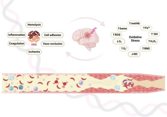
4. Oxidative Pathways, Vasculopathy, and SCA

In SCA, oxidative stress results from the participation of several players in the altered redox biology of the disease, namely SSRBCs, leukocytes, monocytes, and the vascular endothelium. SSRBCs have high baseline concentrations of ROS, hydroxyl free radical (OH), O2•−, and hydrogen peroxide (H2O2), when compared to normal RBCs, which suggests the presence of a pro-oxidant environment in SCA patients even before the onset of clinical manifestations [105]. Oxidant mediators may also take part in I/R injury and inflammation, which are frequent in SCA.
As stated above, one of the main mechanisms of oxidative stress in SCA is HbS autoxidation. Inside SSRBCs, HbS quickly oxidizes to form metHb, hemin, Fe3+, and O2•−. This affects several RBC activities, such as cytoskeletal oxidation, membrane lipid peroxidation, and PS exposure. Due to lack of mitochondria, RBCs use a glycolytic pathway (the Embder–Meyerhof pathway) to produce energy (ATP), which also produces reduced nicotinamide-adenine-dinucleotide (NADH). NADH is necessary for metHb reductase to reduce inactive metHb (containing ferric ions) to functionally active Hb (containing ferrous ions). In the case of SSRBCs, this mechanism allows metHbS reduction to its more innocuous ferrous state. However, SSRBCs also show a depletion in glutathione, an additional scavenger of ROS, thus enhancing oxidative stress in these cells [106]. In normal RBCs, the sidearm of the Embder–Meyerhof pathway (the Luebering–Rapoport shunt) generates 2,3-diphosphoglycerate (2,3-DPG), which is important in Hb-O2 affinity regulation [107,108]. In SSRBCs, 2,3-DPG levels (as well as sphingosine-1-phosphate, S1P) are increased, which reduces HbS-O2 affinity, and consequently leads to higher levels of deoxyHbS and further polymerization. Sickling and RBC oxidative damage lead to RBCs shedding microparticles. In turn, these have been shown to induce ROS production in cultured ECs, and cause vaso-occlusion in sickle mouse kidneys [109]. In SCD, PS exposure upon deoxygenation is linked to Ca2+ influx that causes Gardos channel activation and signals eryptosis (RBC cell death) [110,111]. Additionally, PS exposure increases adhesion (activating ECs and contributing to inflammation) and promotes coagulation (through direct platelet activation or by hemodynamic change at the vessel wall, indirectly leading to EC/platelet interaction) [112,113].
The constant supply of cell-free and heme/hemin provided by chronic hemolysis also promotes a pro-oxidant vascular environment. Besides the interactions between oxyHb with NO, it may also react with H2O2 through the Fenton reaction to form OH and metHb. The latter may further degrade into hemin, which is a major RBC damage-associated molecular pattern (DAMP) molecule. The hemolysis-related ROS contribute to the activation of inflammation and adhesion in endothelial cells, platelets, and neutrophils. This may ultimately result in vaso-occlusion, as demonstrated by a study by Ghosh et al. [114], where direct hemin infusion induced acute vaso-occlusive crises (VOC) in sickle mouse lungs, which was improved by inhibiting P-selectin pathways.
The oxidative damage effects are enhanced further in SCD due to the failure/downregulation of several antioxidant pathways or oxidant scavenger molecules. Hemolysis-related rapid clearing of haptoglobin and hemopexin, which scavenge cell-free Hb and hemin, respectively, ref. [115] is one example. Co-administration of haptoglobin and hemopexin was shown to restore microvascular blood flow in sickle mice that presented with acute vascular stasis due to infusion of exogenous Hb or hemin [79]. Furthermore, upregulated gene expression of hemopexin in hemin-infused Townes-SS mice (a SCA murine model) prevents microvascular occlusion, increases expression of the protective liver nuclear factor erythroid 2-related factor 2 (Nrf2) and heme-oxygenase-1 (HO-1) activity, as well as decreases pro-inflammatory NF-κB. On the other hand, venostasis is worsened in sickle cell hemopexin knock-out mice exposed to hemin [116]. Nrf2 activation has an oxidant protective effect in SCD, by removing hemolysis-derived free heme [117], and it has also been shown to induce fetal hemoglobin (HbF) by binding to γ-globin antioxidant response element [118].
HO-1 is the inducible isoform of the enzyme heme-oxygenase (HO), and its gene’s HMOX1 expression is increased by the presence of heme, which the enzyme converts to biliverdin, carbon monoxide (CO), and ferrous iron, along with several additional protective effects. Belcher and colleagues have shown that, in the Townes-SS murine model, a single co-infusion of hemopexin and haptoglobin upregulated HO-1 expression and lowered NF-κB activity in several tissues, offering protection from vaso-occlusion for up to 48 h [119]. The authors also suggested that the benefits of haptoglobin and hemopexin do not depend exclusively on rapid Hb and hemin clearance from circulation, since the effect of HO-1 on NF-κB activity and venostasis were independent of Hb and hemin plasma concentrations. In a subsequent study, using the same murine model, the administration of oral CO upregulated Nrf2 and HO-1, and downregulated NF-κB activity, soluble VCAM-1, and venostasis [120].
Other mechanisms that may further contribute to increased oxidative stress in SCD include high mobility group box 1 (HMGB1)-mediated TLR4 signaling, that activates the NF-κB pathway and may affect endothelial CAMs’ expression (e.g., P-selectin), and mitochondrial dysfunction in platelets, in addition to non-heme-dependent mechanisms [114,121,122]. The latter include NADPH oxidase, XO, and uncoupled eNOS, all of which generate oxygen free radicals, thus promoting endothelial dysfunction. Myeloperoxidase (MPO), generated by activated neutrophils, also produces oxidants that scavenge NO, further contributing to endothelial dysfunction [123,124]. Increased levels of the oxidative stress biomarkers MPO, HO, lipid peroxidation, and total thiols were recently confirmed in SCD, while antioxidants superoxide dismutase (SOD), glutathione, and catalase levels were reduced [124,125].

5. Cerebrovascular Disease and Hypoxia in SCA

Since SCD is mainly a chronic vascular disease, the majority of its pathobiological mechanisms arise due to disturbance of the homeostasis inside the micro- and macro-vasculature. Although the core mechanism results from altered properties of SSRBCs and their interactions with other blood cells or with the vascular endothelial wall, several other mechanisms are also involved (Figure 3).
In SCD, vasculopathy arises as a combined multistep process that comprises, but it is not limited to, decreased NO bioavailability, oxidant I/R injury, elevated leukocyte count, platelet activation, and increased levels of multiple inflammatory mediators [126]. Therefore, vasculopathy is central to several of the clinical complications as it often takes place before end-organ dysfunction. SCD-related vasculopathies include, among others: moyamoya (that usually precedes cerebral infarcts/hemorrhage), proliferative retinopathy (prior to eyesight loss), pulmonary vasculopathy (associated with pulmonary hypertension), and renal vasculopathy (preceding chronic renal disease) [127].
CVD is a severe SCA complication, particularly in children, with stroke constituting a highly devastating manifestation [128]. Cerebrovascular accidents are among the more frequent clinical manifestations in children with SCA, together with acute pain crises (or vaso-occlusive crises, VOCs), splenic sequestration, recurrent infections due to functional asplenia, acute chest syndrome, cholelithiasis, nocturnal enuresis, hematuria, and reduced (and delayed) growth [129]. Pediatric neurological disease risk is high in SCD and may have significant consequences, especially in the case of overt stroke. Conversely, SCA is the most common cause of pediatric stroke, as children with SCA have a 300-fold increase in stroke risk. Moreover, stroke are is more frequent in SCA than in other SCD presentations [128]. High levels of HbF, as well as co-inheritance of α-thalassemia, are associated with milder SCD phenotypes, and are thus recognized as major disease modifiers [130]. However, this modulation only accounts for a small portion of the phenotypical heterogeneity observed.
Subclinical cerebral infarctions (or silent cerebral infarcts, SCI) are even more common but require magnetic resonance imaging (MRI) for diagnosis. These lesions correlate with marked neuropsychological deficits, and approximately half of the children with SCIs often require life-long support or custodial care [131]. Significant physical and cognitive deficits limiting quality of life are frequent after an overt stroke even if the child follows the recommended therapy [132]. As with other SCD complications, cerebrovascular manifestations are varied—ranging from extensive, large vessel distributed infarcts, to more subtle lacunar infarcts—and differ in terms of epidemiology, clinical features, and pathology [133]. The risk of cerebrovascular complications is highly increased in SCA, predisposing patients to ischemic or primary hemorrhagic stroke [128,134,135]. Pediatric stroke and SCIs occur at a high cumulative rate in SCA [128,136]. A 20-year-old SCA patient has an 11% probability of having already experienced at least one stroke event, and this risk increases to 24% by age 45 [128]. Whereas ischemic stroke is the more common form in young children, intracranial hemorrhage tends to occur later in life [128]. SCIs are even more frequent, with about 37% of children suffering at least one event before reaching 14 years of age. Furthermore, children that have had previous SCIs are at increased risk of developing stroke.
Brain imaging is fundamental for confirming overt strokes (clinically apparent and with an abrupt onset of neurological manifestations) and diagnosing SCIs (present on brain MRI, but without clinical symptoms, or external signs that correlate to the neuroimaging findings) [136]. Cerebral vasculopathy is a major underlying mechanism of ischemic stroke, according to data obtained by cerebral angiography or magnetic resonance angiography (MRA) [137,138,139]. Concomitantly, the most common histopathological finding in SCD-related cerebral vasculopathy is endothelial damage in the mid- to large-sized brain arteries [140,141]. That damage occurs particularly in the branch points, and infarcts in SCA patients are more frequent where stenosis and occlusion occur, particularly in the area of the bifurcation between the internal carotid artery and the middle cerebral artery—the Circle of Willis. The infarct distribution in histopathological studies is consistent with the hypothesis that overt strokes in SCD are a consequence of mid- to large-vessel disease, affecting mainly the distal internal carotid, proximal middle cerebral, and anterior cerebral arteries [142,143].
The lower oxygen affinity of HbS and its polymerization under hypoxic conditions trigger a cascade of events from chronic hemolysis, to microvessel occlusion, endothelial activation and dysfunction, inflammation, and I/R injury. Tissue ischemia in the brain results in ischemic stroke, which in turn tends to occur in the border zones, or watershed regions, even in the absence of large-vessel vasculopathy [144,145,146].
In non-SCD ischemic stroke patients, arteriolar dilation occurs to maintain cerebral blood flow (CBF), and increases in oxygen extraction fraction (OEF, the fraction of oxygen extracted from blood by the brain tissue) contribute to keep the cerebral metabolic rate of O2 utilization (CMRO2) under conditions of decreasing perfusion pressure [147,148]. When these compensatory mechanisms are insufficient to meet the tissue’s metabolic demands, lowering the CMRO2, the result is a cerebral infarct [149]. In those adult patients who have carotid occlusive disease, elevated hemispheric OEF may be a robust indicator of stroke risk [150].
CBF is elevated in children with SCD [146,151], a compensatory mechanism for chronically low arterial oxygen content (oxygen-carrying capacity) due to anemia. Another study proposed that elevated OEF in the deep white matter reflects a metabolic stressed cerebral tissue, and is associated with increased stroke risk in those children [146]. Cerebral regional changes in CBF, OEF, and CMRO2 were observed in the deep white matter of pediatric SCD patients, with OEF peaking when CBF and CMRO2 are at their minimum. This was consistent with a failure to meet metabolic demand in regions of lower CBF and CMRO2, despite high OEF, as these were areas with higher infarct density. The overlap between areas of high infarct density and high OEF, but without low CBF, indicated that high OEF is a stronger indicator of stroke risk than low CBF [146]. Furthermore, a subsequent study showed that children with SCD cerebral metabolic stress may have their symptoms ameliorated with hydroxyurea (HU) therapy (see below). That strategy could be an adjuvant to the most used imaging tool for stroke risk prediction—the transcranial Doppler ultrasonography for measuring the time-averaged mean of maximum velocity (TAMMV) in the middle cerebral artery. This quantitative assessment of stroke risk relies on the Bernouilli’s principle of fluid dynamics. Briefly, this principle states that the speed of a fluid increases when there is a decrease in pressure. In the case of cerebral vasculopathy, that decrease takes place distally to the region where a partial occlusion (or narrowing) of the blood vessels occurs. Hence, if one measures the blood flow velocity in the large arteries mentioned earlier, this could be a strong indicator of the occurrence of a vaso-occlusive or stenotic event.
Children with SCA have a high prevalence of SCIs and these are the most frequent form of SCA-related neurological injury. They may appear very early in life and remain undetected, unless the child undergoes a brain-imaging exam, or until an overt stroke occurs. As mentioned previously, SCI risk is cumulative; up to 37% of children with SCA have suffered an SCI by age 14 years, and this increases to up to 53% in adults by the age of 32 years [128,136,152]. In addition to brain injury, SCI events are associated with increased overt stroke risk [138], failure to meet academic milestones [131], and increased cognitive impairment [132,153,154].
Contrary to strokes, SCIs do not seem to result from macrovasculopathy. Initially, studies suggested that they were spatially circumscribed to the white matter of the frontal and parietal lobes [138]. Subsequent studies demonstrated the occurrence of white matter loss [155] and disrupted matter integrity [156], as well as border zone [155,157] and cortical wedge-like infarcts [158]. The occurrence of border zone infarcts suggests that ischemic mechanisms may result from a global reduction in arterial oxygen content, cerebral hemodynamic factors, or both, while wedge-like infarcts indicate a predominance of thromboembolic factors [158]. With no apparent external manifestations, SCI diagnosis relies heavily on brain imaging technology, like MRI. A large prospective study with a pediatric SCA cohort showed that CBF decreased as infarct density increased, thus confirming that SCIs predominantly occur in the border zone vascular distribution within the white matter of the frontal and parietal lobes [158]. Additionally, the results demonstrated that 90% of children had SCIs within a relatively small border zone region, measuring 5.6% of total brain volume, and they occurred in the region of low blood flow.

6. Antioxidant Therapeutic Approaches in SCD Vasculopathy

In general, current SCA treatments target symptom relief and, where possible, primary prevention. Even though an established prevention therapy is in place for pediatric stroke prevention in high-income settings, the challenges for CVD treatment are similar to those for other SCA manifestations. Nevertheless, achieving a one-size-fits-all therapeutic solution is highly unlikely, due to the complex nature of SCA. While upstream-targeted strategies focus on the β-globin gene cluster expression, namely the Hb switching mechanism, downstream-targeted approaches address the multitude of pathophysiological mechanisms of SCD. Upstream approaches are recent and rely on circumventing the genetic defect, whether directly (by correcting the mutation) or indirectly (by reversing the physiological switch in Hb production).
Current approaches to genetic-based therapies involve collecting a patient’s hematopoietic stem cells (HSC) via bone marrow harvesting or apheresis. This is followed by genetical modification of the cells ex vivo by inserting a new transgene, or editing the gene directly. Finally, the genetically modified progenitor cells are reinfused after high-dose chemotherapy conditioning. Despite the advantages of not requiring donors, thus reducing the risk of graft-vs-host disease [159], these strategies are highly dependent on several key issues. These include: quality of viral delivery systems; quality and quantity of HSC harvested; optimization of the gene modification system in HSC; choice of recombination pathway (homologous vs. non-homologous); identification of the best gene targets; cell manufacturing; preparation regimens to allow the bone marrow to receive genetically modified cells with minimized toxicity; issues related to off-target effects; optimization of preclinical models for testing of developing gene therapy strategies; and parameters that should be used to define a cure [160].
Downstream strategies are especially challenging due to the high complexity of SCD where much is still unknown. In terms of pharmacological disease-modifying treatments, and despite numerous recent clinical trials, the European Medicines Agency (EMA) and the Food and Drug Administration (FDA, USA) have approved only four substances—HU, L-glutamine, voxelotor, and crizanlizumab.
Hydroxyurea was the first drug approved for treatment and acts on a variety of mechanisms involved in SCD. Those mechanisms include erythroid regeneration, NO-related increase in the activity of soluble guanylate cyclase and cyclic guanidine monophosphate (cGMP), which stimulates HbF-related genes expression, or NO increase through long-term post-transcriptional rise in eNOS levels [161,162]. The increase in HbF production counteracts the destructive repercussions of HbS presence, thus acting as a disease modifier. This translates into several beneficial effects in SCD, such as lower incidence of pain episodes and acute chest syndrome, as well as a reduction in the number of hospitalizations [163,164]. Furthermore, HU decreases mortality and morbidity associated with VOC events [165], cerebrovascular accidents [152,166], and proteinuria [167], thus increasing overall survival in patients with SCD [168]. This is especially important in low-resource settings where blood transfusions are not easily available, and/or are less equipped to manage the downsides of long-term transfusion therapy (e.g., risk of alloimmunization or iron overload, the need for a robust antigen matched blood supply). Therefore, HU has a similar effect on TCD velocities as blood transfusion therapy in children with SCD at high risk of stroke [164,166]. A recent study demonstrated that HU scavenges free radicals and induces the expression of antioxidant genes [169].
Of the remaining approved drugs for SCD treatment, only L-glutamine was shown to have an effect on ox–redox vascular balance. It is a NAD precursor, which provides a supplementation that will counteract NAD depletion on SSRBCs [170]. The drug has been described to reduce acute complications of SCD, both in pediatric (<5 years of age) and adult patients, namely VOC painful crises and hospitalizations [170]. Despite rapid approval, much is still unknown regarding the L-glutamine mode of action.
Voxelotor’s mode of action has been assessed in studies like the Hemoglobin Oxygen Affinity Modulation to Inhibit HbS Polymerization (HOPE) clinical trial [171]. It acts as an inhibitor of HbS polymerization, thus stabilizing HbS in the oxygenated state and inhibiting RBC sickling. Voxelotor was shown to increase Hb levels, thus reducing the number of acute anemia episodes. Levels of hemolysis markers (namely bilirubin, but also reticulocyte and LDH) were also decreased. The HOPE study did not measure stroke incidence, despite initial concerns that increasing oxygen affinity would reduce oxygen extraction in sensitive tissues like the brain [159,172].
Crizanlizumab (SelG1) is a humanized anti-P-selectin antibody that, in a study to assess safety and impact of SelG1 with or without HU therapy in SCD patients with pain crises (SUSTAIN), was shown to be effective in reducing VOC frequency and acute chest syndrome [173]. The rationale for its application was to decrease both RBC and leukocyte adhesion, which occurs by downregulating P-selectin expression and consequently inflammation-mediated cell adhesion. However, CVD was not a primary or secondary endpoint in the SUSTAIN study.
Research on further pharmacological targets is ongoing. It relies heavily on further knowledge of SCD pathobiological mechanisms. The most promising target mechanisms for pharmacotherapy identified so far, besides HbF-stimulating agents, include adhesion, I/R injury, coagulation, and hemolysis [173,174,175,176,177,178,179]. To date, and besides HU, statins like simvastatin are the ones that, besides an anti-inflammatory mode of action, also seem to act on eNOS to restore nitric oxide production [180].
Other antioxidant agents are potential adjuvants for SCD therapy though none of them have yet been approved by the FDA or the EMA. These include N-acetylcysteine, zinc supplementation, nitric oxide, L-arginine, α-lipoic acid, and acetyl-L-carnitine [13,181].
Given the multitude of effects caused by SSRBCs, the more adequate therapeutic strategy will probably be a multitargeted approach [182]. Unfortunately, the limited number of SCD patients in high-income countries and scarce resources in low-income countries make it difficult to accomplish definitive studies of many pharmacological agents [183].

7. Genetic Modulation of Cerebrovascular Disease in SCA

The standard of care in SCD pediatric stroke prevention is TCD screening, followed by regular blood transfusion therapy for children identified as being at high risk. The definition of risk depends on the TAMMV values, as follows: high risk TAMMV > 200 cm/s; moderate risk 70 cm/s < TAMMV < 200 cm/s [134]. Unfortunately, the relationship between TCD velocities and stroke incidence is not precise, nor age-independent. Approximately seven children with elevated TCD value have to be treated with regular blood transfusion to prevent one child from having a stroke [127]. Stroke risk also seems to be age-dependent, as individuals with ages above 16 years and elevated TAMMV do not appear to have a significant increase in stroke risk [184]. Furthermore, the management of hematological disease alone does not seem to prevent vasculopathy progression, which indicates that additional genetic risk factors have a role as risk modulators. This is especially important since heterogeneity in disease presentation and severity complicates prognostication, management, and clinical trials [185].
Several biomarkers of cerebral vasculopathy were already identified, including low HbF level [186], low baseline Hb, high leukocyte count, male sex, and relative high systolic pressure [128,187]. Moreover, high levels of HbF and co-inheritance of α-thalassemia are recognized modifiers of global disease severity [2,188]. Several studies indicated that gene variants co-inherited with the βS mutation are potential modifiers of stroke/stroke risk [189,190,191,192,193,194,195,196].
Research approaches that ranged from candidate genes to genome-wide association studies (GWAS) focused on identifying potential stroke/stroke risk modifying loci [193,194,197,198,199]. Those genetic modifiers, or modulators, could be potential targets for increasingly accurate (and possibly personalized) prognostic tests and therapeutic strategies. The results indicated that, besides the previously demonstrated protective role of the co-inheritance, the most powerful genetic modifiers of disease severity lie within the β-globin cluster [200]. Identification, confirmation, and functional assessment of other genes requires knowledge of their involvement in stroke pathophysiology. Several putative stroke-associated variants were identified in genes such as VCAM1 [191,193,196], IL4R [193], LDLR [193], ADRB2 [193], AGT [190], HLA [189], and TNF-α [193,195], as well as the -α3.7kb deletion that causes α-thalassemia [195] (Table 1).
Additionally, three SNPs identified by GWAS showed special significance: rs662 (Q192R) in the paraoxonase 1 gene (PON1), rs1044498 (K173Q) in the ectonucleotide pyrophosphatase/phosphodiesterase 1 (ENPP1) gene, and rs3732410 (Y1212C) in the Golgin subfamily B member 1 (GOLGB1) gene [198]. Of these, only the SNP on PON1 was previously associated with increased stroke risk in adults [201,202]. The SNP in ENPP1 was validated in another study [194]. On the other hand, our group identified a positive association between the presence of rs1044498_A allele and increased stroke risk [196]. We have also identified haplotypes of VCAM1 promoter variants as well as a specific variant (rs1409419_T) with a strong association, not only with stroke [196], but also with hemolysis [203].
In terms of NO metabolism, we found a positive association with a NOS3 gene variant with red cell distribution width (RDW) levels. Specifically, CC and TC genotypes of the rs2070744 showed a positive association with lower RDW (reduced anisocytosis) [196], a potential biomarker of lower cerebrovascular disease risk in non-SCD patients [204]. Another NOS3 variant, a variable number of tandem repeats (VNTR) on the intron 4 specific allele with five repeats of 27 bp, also showed a potential protective effect against SCIs in the pediatric group of SCA patients that we studied [196].
Table 1. Candidate genes and respective variants previously identified as putative modifiers of stroke in sickle cell anemia.
GeneVariantPredicted ModulationReference
VCAM1G1238CStroke protection[191]
VCAM1T1594CIncreased small-vessel stroke risk[193]
VCAM1rs1409419_T
Haplotype 7
Increased stroke risk
[196]
NOS3intron 4_27 bp VNTR_4b
Haplotype V
Haplotype VII
Decreased SCI risk
Decreased SCI risk
Decreased CV risk
[196]
ITGA4rs113276800_CA
rs3770138_T
Increased stroke risk
Increased CV risk
[196]
IL4RS503PIncreased large-vessel stroke risk[193]
LDLRNcol +/−Increased small-vessel stroke risk[193]
ADRB2Q27EIncreased large-vessel stroke risk[193]
AGTGT repeatsIncreased stroke risk[190]
HLADRB1*0301
DRB1*0302
DQB1*0201
DRB1*1501
DQB1*0602
DPB1*0401
DPB1*1701
-A*0102
-A*2612
-A*3301
Increased stroke risk
Decreased stroke risk
Increased small-vessel stroke risk
Increased large-vessel stroke risk
Decreased large-vessel stroke k
[189,205]
TNF-α-308G>AIncreased stroke risk[193,195,206]
GOLGB1Y1212CDecreased stroke risk[198]
ENPP1K173QDecreased stroke risk
Increased stroke risk
[198]
[194]
[199]
PON1Q192RIncreased stroke risk[198]
HBA3.7kb delDecreased stroke risk[195]
SCI—silent cerebral infarction; CV—cerebral vasculopathy; VCAM1 haplotype 7–rs1409419_T/rs3917024_C/rs3917025_CT/rs3783598_T/rs1041163_T/rs3783599_C; NOS3 intron 4_27 bp VNTR_4b–5 repeats × 27 bp; NOS3 haplotype V–rs2070744_T/VNTR_4b/rs1799983_G; NOS3 haplotype VII–rs rs2070744_T/VNTR_4b/rs1799983_T.
Assessing the biological role of the genetic variants potentially associated with SCD modulation can only be performed through functional studies. In SCD, these type of studies often rely on murine or cell models. Both types have provided important information regarding oxidative stress pathways, for example [169,196,207,208]. Studies on sickle mice used hemin (ferric heme) to simulate a sudden increase in intravascular hemolysis and blood flow stasis in post-capillary veins, due to its pro-oxidant and pro-inflammatory effects [79,209]. Heme-associated effects included the activation of the TLR-4 and NF-κB signaling pathways, that induced inflammation and vaso-occlusion, and the extent of subsequent damage was strongly associated with the presence of HbS [79]. Hemin injections also upregulated HO-1 expression. This, in turn, inhibited inflammation and adhesion.
Our studies showed that, in hemin-treated macrovascular and brain microvascular EC models, TNF-α-induced VCAM1 expression was downregulated, and HMOX1 was significantly upregulated [210]. Other authors reported that HO-1 production is also upregulated by hemin in monocytes, where it has an antioxidant effect by inhibiting apoptosis [207,208]. Furthermore, the results we obtained showed that both microvascular and macrovascular ECs present a similar HMOX1 upregulation, thus suggesting little difference in the levels by which this antioxidant system operates in the macrovascular and brain microvascular environments. HMOX1 expression also seems to be unaffected by HU not only in micro and macrovascular ECs [210] but also in peripheral blood monocytes (PBMCs) and umbilical cord vein ECs (HUVECs) [169]. This response is the opposite of what occurs with other antioxidant pathway genes’ expression, like superoxide dismutase (SOD), glutathione peroxidase (GPX), and glutathione-disulfide reductase (GSR), which were shown to be upregulated after HU treatment [169]. These observations indicate that HMOX1 activation follows an alternative mechanism of antioxidant response not susceptible to HU action.
In addition, in the endothelial setting, the cytokine TNF-α leads to reduced NO bioavailability. This results from activation of endothelial arginase (which depletes eNOS from its substrate arginine) [211] and endothelial NADPH oxidase (lowering BH4 and leading eNOS to generate superoxide) [212]. In our research, we found that NOS3 expression was downregulated in TNF-α-stimulated macrovascular ECs, and this effect was stronger in brain microvascular ECs. HU modulation of NOS3 expression seems to be differential and dose-dependent [210,213], which indicates that HU effect in NO bioavailability does not result from direct NOS3 upregulation. As proposed by several authors, the most likely mechanism by which HU increases eNOS levels is a post-translational proteasomal protection from degradation [162,213].

8. Conclusions

A clear understanding of the specific mechanisms and genetic variants modulating SCA manifestations is invaluable. This is particularly important for CVD, namely in children, due to its impact on patients’ lives. The possibility of defining a genetic marker panel associated with disease severity would allow for risk stratification of patients—together with the ability to provide timely care to high-risk patients and the identification of therapeutic targets, thus facilitating the design of new pharmacological agents, and the possibility of customizing therapeutic strategies to each patient in a precision medicine approach.

Author Contributions

M.S. conceived the idea, drafted the manuscript, designed the illustrations (created with BioRender.com (accessed on 30 October 2023)) and concurred with the final version of the review. P.F. contributed to conceive the idea, revised the manuscript, coordinated the activity, and concurred with the final version of the review. All authors have read and agreed to the published version of the manuscript.

Funding

This research received no external funding.

Institutional Review Board Statement

Not applicable.

Informed Consent Statement

Not applicable.

Data Availability Statement

Not applicable.

Conflicts of Interest

The authors declare no conflict of interest.

References

  1. Piel, F.B.; Tatem, A.J.; Huang, Z.; Gupta, S.; Williams, T.N.; Weatherall, D.J. Global Migration and the Changing Distribution of Sickle Haemoglobin: A Quantitative Study of Temporal Trends between 1960 and 2000. Lancet Glob. Health 2014, 2, e80–e89. [Google Scholar] [CrossRef] [PubMed]
  2. Piel, F.B.; Steinberg, M.H.; Rees, D.C. Sickle Cell Disease. N. Engl. J. Med. 2017, 376, 1561–1573. [Google Scholar] [CrossRef] [PubMed]
  3. Pearson, M.J.; Lipowsky, H.H. Influence of Erythrocyte Aggregation on Leukocyte Margination in Postcapillary Expansions: A Lattice Boltzmann Analysis. Am. J. Physiol. Heart Circ. Physiol. 2000, 279, H1460–H1471. [Google Scholar] [CrossRef] [PubMed]
  4. Debaun, M.R.; Kirkham, F.J. Central Nervous System Complications and Management in Sickle Cell Disease. Blood 2016, 127, 829–838. [Google Scholar]
  5. Kato, G.J.; Piel, F.B.; Reid, C.D.; Gaston, M.H.; Ohene-Frempong, K.; Krishnamurti, L.; Smith, W.R.; Panepinto, J.A.; Weatherall, D.J.; Costa, F.F.; et al. Sickle Cell Disease. Nat. Rev. Dis. Prim. 2018, 4, 18010. [Google Scholar] [CrossRef]
  6. Mandese, V.; Bigi, E.; Bruzzi, P.; Palazzi, G.; Predieri, B.; Lucaccioni, L.; Cellini, M.; Iughetti, L. Endocrine and Metabolic Complications in Children and Adolescents with Sickle Cell Disease: An Italian Cohort Study. BMC Pediatr. 2019, 19, 56. [Google Scholar] [CrossRef]
  7. Herrick, J.B. Peculiar Elongated and Sickle-Shaped Red Blood Corpuscles in a Case of Severe Anemia. Arch. Intern. Med. 1910, VI, 517–521. [Google Scholar] [CrossRef]
  8. Ingram, V.M. Gene Mutations in Human Haemoglobin: The Chemical Difference between Normal and Sickle Cell Haemoglobin. Nature 1957, 180, 326–328. [Google Scholar] [CrossRef]
  9. Dale, J.C.; Cochran, C.J.; Roy, L.; Jernigan, E.; Buchanan, G.R. Health-Related Quality of Life in Children and Adolescents with Sickle Cell Disease. J. Pediatr. Health Care 2011, 25, 208–215. [Google Scholar] [CrossRef]
  10. Kambasu, D.M.; Rujumba, J.; Lekuya, H.M.; Munube, D.; Mupere, E. Health-Related Quality of Life of Adolescents with Sickle Cell Disease in Sub-Saharan Africa: A Cross-Sectional Study. BMC Hematol. 2019, 19, 9. [Google Scholar] [CrossRef]
  11. Kato, G.J.; Hebbel, R.P.; Steinberg, M.H.; Gladwin, M.T. Vasculopathy in Sickle Cell Disease: Biology, Pathophysiology, Genetics, Translational Medicine and New Research Directions. Am. J. Hematol. 2009, 84, 618–625. [Google Scholar] [CrossRef]
  12. Voskou, S.; Aslan, M.; Fanis, P.; Phylactides, M.; Kleanthous, M. Oxidative Stress in β-Thalassaemia and Sickle Cell Disease. Redox Biol. 2015, 6, 226–239. [Google Scholar] [CrossRef] [PubMed]
  13. Vona, R.; Sposi, N.M.; Mattia, L.; Gambardella, L.; Straface, E.; Pietraforte, D. Sickle Cell Disease: Role of Oxidative Stress and Antioxidant Therapy. Antioxidants 2021, 10, 296. [Google Scholar] [CrossRef]
  14. Ware, R.E.; de Montalembert, M.; Tshilolo, L.; Abboud, M.R. Sickle Cell Disease. Lancet 2017, 390, 311–323. [Google Scholar] [CrossRef] [PubMed]
  15. van Zwieten, R.; Verhoeven, A.J.; Roos, D. Inborn Defects in the Antioxidant Systems of Human Red Blood Cells. Free Radic. Biol. Med. 2014, 67, 377–386. [Google Scholar] [CrossRef] [PubMed]
  16. Reiter, C.H.D.R.; Ang, X.U.W.; Antos, J.O.S.E.E.T.A.; Ogg, N.E.I.L.H.; Iii, R.I.O.C.A.; Chechter, A.L.A.N.N.S.; Ladwin, M.A.R.K.T.G. FreeHb Limits NO Availability in SCD. Nat. Med. 2002, 8, 1383–1389. [Google Scholar] [CrossRef] [PubMed]
  17. Barabino, G.A.; Platt, M.O.; Kaul, D.K. Sickle Cell Biomechanics. Annu. Rev. Biomed. Eng. 2010, 12, 345–367. [Google Scholar] [CrossRef]
  18. Kaul, D.K.; Fabry, M.E.; Nagel, R.L. The Pathophysiology of Vascular Obstruction in the Sickle Syndromes. Blood Rev. 1996, 10, 29–44. [Google Scholar] [CrossRef]
  19. Zhang, D.; Xu, C.; Manwani, D.; Frenette, P.S. Neutrophils, Platelets, and Inflammatory Pathways at the Nexus of Sickle Cell Disease Pathophysiology. Blood 2016, 127, 801–809. [Google Scholar] [CrossRef]
  20. Switzer, J.A.; Hess, D.C.; Nichols, F.T.; Adams, R.J. Pathophysiology and Treatment of Stroke in Sickle-Cell Disease: Present and Future. Lancet Neurol. 2006, 5, 501–512. [Google Scholar]
  21. Pries, A.R.; Kuebler, W.M. Normal Endothelium. In The Vascular Endothelium I. Handbook of Experimental Pharmacology; Moncada, S., Higgs, A., Eds.; Springer: Berlin/Heidelberg, Germany, 2006; Volume 176/I. [Google Scholar] [CrossRef]
  22. Regan, E.R.; Aird, W.C. Dynamical Systems Approach to Endothelial Heterogeneity. Circ. Res. 2012, 111, 110–130. [Google Scholar] [CrossRef] [PubMed]
  23. Rajendran, P.; Rengarajan, T.; Thangavel, J.; Nishigaki, Y.; Sakthisekaran, D.; Sethi, G.; Nishigaki, I. The Vascular Endothelium and Human Diseases. Int. J. Biol. Sci. 2013, 9, 1057–1069. [Google Scholar] [CrossRef]
  24. Behrendt, D.; Ganz, P. Endothelial Function: From Vascular Biology to Clinical Aplications. Am. J. Cardiol. 2002, 90, 40L–48L. [Google Scholar] [CrossRef]
  25. Furchgott, R.F.; Vanhoutte, P.M. Endothelium-Derived Relaxing and Contracting Factors. FASEB J. 1989, 3, 2007–2018. [Google Scholar] [CrossRef]
  26. Egashira, K.; Inou, T.; Hirooka, Y.; Kai, H.; Sugimachi, M.; Suzuki, S.; Kuga, T.; Urabe, Y.; Takeshita, A. Effects of Age on Endothelium-Dependent Vasodilation of Resistance Coronary Artery by Acetylcholine in Humans. Circulation 1993, 88, 77–81. [Google Scholar] [CrossRef]
  27. Egashira, K.; Inou, T.; Hirooka, Y.; Yamada, A.; Maruoka, Y.; Kai, H.; Sugimachi, M.; Suzuki, S.; Takeshita, A. Impaired Coronary Blood Flow Response to Acetylcholine in Patients with Coronary Risk Factors and Proximal Atherosclerotic Lesions. J. Clin. Investig. 1993, 91, 29–37. [Google Scholar] [CrossRef] [PubMed]
  28. Zeiher, A.M.; Drexler, H.; Saurbier, B.; Just, H. Endothelium-Mediated Coronary Blood Flow Modulation in Humans: Effects of Age, Atherosclerosis, Hypercholesterolemia, and Hypertension. J. Clin. Investig. 1993, 92, 652–662. [Google Scholar] [CrossRef]
  29. Nabel, E.G.; Selwyn, A.P.; Ganz, P. Large Coronary Arteries in Humans Are Responsive to Changing Blood Flow: An Endothelium-Dependent Mechanism That Fails in Patients with Atherosclerosis. J. Am. Coll. Cardiol. 1990, 16, 349–356. [Google Scholar] [CrossRef]
  30. Taddei, S.; Virdis, A.; Ghiadoni, L.; Magagna, A.; Salvetti, A. Cyclooxygenase Inhibition Restores Nitric Oxide Activity in Essential Hypertension. Hypertension 1997, 29, 274–279. [Google Scholar] [CrossRef] [PubMed]
  31. Yang, D.; Félétou, M.; Boulanger, C.M.; Wu, H.F.; Levens, N.; Zhang, J.N.; Vanhoutte, P.M. Oxygen-Derived Free Radicals Mediate Endothelium-Dependent Contractions to Acetylcholine in Aortas from Spontaneously Hypertensive Rats. Br. J. Pharmacol. 2002, 136, 104–110. [Google Scholar] [CrossRef] [PubMed]
  32. Belhassen, L.; Pelle, G.; Dubois-Rande, J.L.; Adnot, S. Improved Endothelial Function by the Thromboxane A2 Receptor Antagonist S 18886 in Patients with Coronary Artery Disease Treated with Aspirin. J. Am. Coll. Cardiol. 2003, 41, 1198–1204. [Google Scholar] [CrossRef]
  33. Flammer, A.J.; Lüscher, T.F. Human Endothelial Dysfunction: EDRFs. Pflüg. Arch. Eur. J. Physiol. 2010, 459, 1005–1013. [Google Scholar] [CrossRef]
  34. Porcu, P.; Emanueli, C.; Kapatsoris, M.; Chao, J.; Chao, L.; Madeddu, P. Reversal of Angiogenic Growth Factor Upregulation by Revascularization of Lower Limb Ischemia. Circulation 2002, 105, 67–72. [Google Scholar] [CrossRef]
  35. Tsui, J.C.S.; Baker, D.M.; Biecker, E.; Shaw, S.; Dashwood, M.R. Potential Role of Endothelin 1 in Ischaemia-Induced Angiogenesis in Critical Leg Ischaemia. Br. J. Surg. 2002, 89, 741–747. [Google Scholar] [CrossRef]
  36. Zetter, B.R. The Endothelial Cells of Large and Small Blood Vessels. Diabetes 1981, 30, 24–28. [Google Scholar] [CrossRef]
  37. Aird, W.C. Endothelial Cell Heterogeneity. Cold Spring Harb. Perspect. Med. 2012, 2, a006429. [Google Scholar] [CrossRef] [PubMed]
  38. Risau, W. Differentiation of Endothelium. FASEB J. 1995, 9, 926–933. [Google Scholar] [CrossRef] [PubMed]
  39. Aird, W.C. Endothelium in Health and Disease. Pharmacol. Rep. 2008, 60, 139–143. [Google Scholar]
  40. Marui, N.; Offermann, M.K.; Swerlick, R.; Kunsch, C.; Rosen, C.A.; Ahmad, M.; Wayne Alexander, R.; Medford, R.M.; Kunsck, C.; Rosen, C.A.; et al. Vascular Cell Adhesion Molecule-1 (VCAM-1) Gene Transcription and Expression Are Regulated through an Antioxidant-Sensitive Mechanism in Human Vascular Endothelial Cells. J. Clin. Investig. 1993, 92, 1866–1874. [Google Scholar] [CrossRef] [PubMed]
  41. Lanaro, C.; Franco-Penteado, C.F.; Albuqueque, D.M.; Saad, S.T.O.; Conran, N.; Costa, F.F. Altered Levels of Cytokines and Inflammatory Mediators in Plasma and Leukocytes of Sickle Cell Anemia Patients and Effects of Hydroxyurea Therapy. J. Leukoc. Biol. 2009, 85, 235–242. [Google Scholar] [CrossRef]
  42. Radke, R.M.; Diller, G.P.; Duck, M.; Orwat, S.; Hartmann, D.; Thum, T.; Baumgartner, H. Endothelial Function in Contemporary Patients with Repaired Coarctation of Aorta. Heart 2014, 100, 1696–1701. [Google Scholar] [CrossRef] [PubMed]
  43. Solovey, A.A.; Solovey, A.N.; Harkness, J.; Hebbel, R.P. Modulation of Endothelial Cell Activation in Sickle Cell Disease: A Pilot Study. Blood 2001, 97, 1937–1941. [Google Scholar] [CrossRef]
  44. Sakamoto, T.M.; Lanaro, C.; Ozelo, M.C.; Garrido, V.T.; Olalla-Saad, S.T.; Conran, N.; Costa, F.F. Increased Adhesive and Inflammatory Properties in Blood Outgrowth Endothelial Cells from Sickle Cell Anemia Patients. Microvasc. Res. 2013, 90, 173–179. [Google Scholar] [CrossRef] [PubMed]
  45. Wink, D.A.; Mitchell, J.B. Chemical Biology of Nitric Oxide: Insights into Regulatory, Cytotoxic, and Cytoprotective Mechanisms of Nitric Oxide. Free Radic. Biol. Med. 1998, 25, 434–456. [Google Scholar] [CrossRef]
  46. Arnold, W.P.; Mittal, C.K.; Katsuki, S.; Murad, F. Nitric Oxide Activates Guanylate Cyclase and Increases Guanosine 3 ’: 5 ’ -Cyclic Monophosphate Levels in Various Tissue Preparations Biochemistry. Proc. Natl. Acad. Sci. USA 1977, 74, 3203–3207. [Google Scholar] [CrossRef] [PubMed]
  47. Ignarro, L.J.; Byrns, R.E.; Buga, G.M.; Wood, K.S. Endothelium-Derived Relaxing Factor From Pulmonary Artery and Vein Possesses Pharmacologic and Chemical Properties Identical to Those of Nitric Oxide Radical. Circ. Res. 1987, 61, 866–879. [Google Scholar] [CrossRef]
  48. Kato, G.J.; Martyr, S.; Blackwelder, W.C.; Nichols, J.S.; Coles, W.A.; Hunter, L.A.; Brennan, M.L.; Hazen, S.L.; Gladwin, M.T. Levels of Soluble Endothelium-Derived Adhesion Molecules in Patients with Sickle Cell Disease Are Associated with Pulmonary Hypertension, Organ Dysfunction, and Mortality. Br. J. Haematol. 2005, 130, 943–953. [Google Scholar] [CrossRef]
  49. Förstermann, U.; Sessa, W.C. Nitric Oxide Synthases: Regulation and Function. Eur. Heart J. 2012, 33, 829–837. [Google Scholar] [CrossRef]
  50. Förstermann, U.; Closs, E.I.; Pollock, J.S.; Nakane, M.; Schwarz, P.; Gath, I.; Kleinert, H. Nitric Oxide Synthase Isozymes Antibodies. Hypertension 1994, 23, 1121–1131. [Google Scholar] [CrossRef]
  51. Hemmens, B.; Mayer, B. Enzymology of Nitric Oxide Synthases. In Nitric Oxide Protocols; Titheradge, M.A., Ed.; Humana Press: Totowa, NJ, USA, 1998; pp. 1–32. ISBN 978-1-59259-749-9. [Google Scholar]
  52. García-Cardeña, G.; Fan, R.; Shah, V.; Sorrentino, R.; Cirino, G.; Papapetropoulos, A.; Sessa, W.C. Dynamic Activation of Endothelial Nitric Oxide Synthase by Hsp90. Nature 1998, 392, 821–824. [Google Scholar] [CrossRef]
  53. Pritchard, K.A.; Ackerman, A.W.; Gross, E.R.; Stepp, D.W.; Shi, Y.; Fontana, J.T.; Baker, J.E.; Sessa, W.C. Heat Shock Protein 90 Mediates the Balance of Nitric Oxide and Superoxide Anion from Endothelial Nitric-Oxide Synthase. J. Biol. Chem. 2001, 276, 17621–17624. [Google Scholar] [CrossRef]
  54. Song, Y.; Cardounel, A.J.; Zweier, J.L.; Xia, Y. Inhibition of Superoxide Generation from Neuronal Nitric Oxide Synthase by Heat Shock Protein 90: Implications in NOS Regulation. Biochemistry 2002, 41, 10616–10622. [Google Scholar] [CrossRef]
  55. Sowa, G.; Pypaert, M.; Sessa, W.C. Distinction between Signaling Mechanisms in Lipid Rafts vs. Caveolae. Proc. Natl. Acad. Sci. USA 2001, 98, 14072–14077. [Google Scholar] [CrossRef] [PubMed]
  56. Rapoport, R.M.; Draznin, M.B.; Murad, F. Endothelium-Dependent Relaxation in Rat Aorta May Be Mediated through Cyclic GMP-Dependent Protein Phosphorylation. Nature 1983, 306, 174–176. [Google Scholar] [CrossRef] [PubMed]
  57. Ignarro, L.J.; Harbison, R.G.; Wood, K.S.; Kadowitz, P.J. Activation of Purified Soluble Guanylate Cyclase by Endothelium-Derived Relaxing Factor from Intrapulmonary Artery and Vein: Stimulation by Acetylcholine, Bradykinin and Arachidonic Acid. J. Pharmacol. Exp. Ther. 1986, 237, 893–900. [Google Scholar]
  58. Arndt, H.; Smith, C.W.; Granger, D.N. Leukocyte-Endothelial Cell Adhesion in Spontaneously Hypertensive and Normotensive Rats. Hypertension 1993, 21, 667–673. [Google Scholar] [CrossRef] [PubMed]
  59. Dimmeler, S.; Zeiher, A.M. Nitric Oxide—An Endothelial Cell Survival Factor. Cell Death Differ. 1999, 6, 964–968. [Google Scholar] [CrossRef]
  60. Nunokawa, Y.; Tanaka, S. Interferon-γ Inhibits Proliferation of Rat Vascular Smooth Muscle Cells by Nitric Oxide Generation. Biochem. Biophys. Res. Commun. 1992, 188, 409–415. [Google Scholar] [CrossRef]
  61. Hogan, M.; Cerami, A.; Bucala, R. Advanced Glycosylation Endproducts Block the Antiproliferative Effect of Nitric Oxide: Role in the Vascular and Renal Complications of Diabetes Mellitus. J. Clin. Investig. 1992, 90, 1110–1115. [Google Scholar] [CrossRef]
  62. Murohara, T.; Asahara, T.; Silver, M.; Bauters, C.; Masuda, H.; Kalka, C.; Kearney, M.; Chen, D.; Chen, D.; Symes, J.F.; et al. Nitric Oxide Synthase Modulates Angiogenesis in Response to Tissue Ischemia. J. Clin. Investig. 1998, 101, 2567–2578. [Google Scholar] [CrossRef]
  63. Xia, N.; Förstermann, U.; Li, H. Resveratrol and Endothelial Nitric Oxide. Molecules 2014, 19, 16102–16121. [Google Scholar] [CrossRef] [PubMed]
  64. Vásquez-Vivar, J.; Kalyanaraman, B.; Martásek, P.; Hogg, N.; Masters, B.S.S.; Karoui, H.; Tordo, P.; Pritchard, K.A. Superoxide Generation by Endothelial Nitric Oxide Synthase: The Influence of Cofactors. Proc. Natl. Acad. Sci. USA 1998, 95, 9220–9225. [Google Scholar] [CrossRef]
  65. Xu, W.; Kaneko, F.T.; Zheng, S.; Comhair, S.A.A.; Janocha, A.J.; Goggans, T.; Thunnissen, F.B.J.M.; Farver, C.; Hazen, S.L.; Jennings, C.; et al. Increased Arginase II and Decreased NO Synthesis in Endothelial Cells of Patients with Pulmonary Arterial Hypertension. FASEB J. 2004, 18, 1746–1748. [Google Scholar] [CrossRef] [PubMed]
  66. Antoniades, C.; Shirodaria, C.; Leeson, P.; Antonopoulos, A.; Warrick, N.; Van-Assche, T.; Cunnington, C.; Tousoulis, D.; Pillai, R.; Ratnatunga, C.; et al. Association of Plasma Asymmetrical Dimethylarginine (ADMA) with Elevated Vascular Superoxide Production and Endothelial Nitric Oxide Synthase Uncoupling: Implications for Endothelial Function in Human Atherosclerosis. Eur. Heart J. 2009, 30, 1142–1150. [Google Scholar] [CrossRef]
  67. Chen, C.-A.; Wang, T.-Y.; Varadharaj, S.; Reyes, L.A.; Hemann, C.; Talukder, M.A.H.; Chen, Y.-R.; Druhan, L.J.; Zweier, J.L. S-Glutathionylation Uncouples ENOS and Regulates Its Cellular and Vascular Function. Nature 2010, 468, 1115–1118. [Google Scholar] [CrossRef]
  68. Zweier, J.L.; Chen, C.-A.; Druhan, L.J. S-Glutathionylation Reshapes Our Understanding of Endothelial Nitric Oxide Synthase Uncoupling and Nitric Oxide/Reactive Oxygen Species-Mediated Signaling. Antioxid. Redox Signal. 2011, 14, 1769–1775. [Google Scholar] [CrossRef] [PubMed]
  69. Gladwin, M.T.; Sachdev, V.; Jison, M.L.; Shizukuda, Y.; Plehn, J.F.; Minter, K.; Brown, B.; Coles, W.A.; Nichols, J.S.; Ernzt, I.; et al. Pulmonary Hypertension as a Risk Factor for Death in Patients with Sickle Cell Anemia. N. Engl. J. Med. 2004, 350, 885–895. [Google Scholar] [CrossRef] [PubMed]
  70. Taddei, S.; Virdis, A.; Mattei, P.; Salvetti, A. Vasodilation to Acetylcholine in Primary and Secondary Forms of Human Hypertension. Hypertension 1993, 21, 929–933. [Google Scholar] [CrossRef]
  71. Hebbel, R.P.; Yamada, O.; Moldow, C.F.; Jacob, H.S.; White, J.G.; Eaton, J.W. Abnormal Adherence of Sickle Erythrocytes to Cultured Vascular Endothelium. Possible Mechanism for Microvascular Occlusion in Sickle Cell Disease. J. Clin. Investig. 1980, 65, 154–160. [Google Scholar] [CrossRef]
  72. Nath, K.A.; Shah, V.; Haggard, J.J.; Croatt, A.J.; Smith, L.A.; Hebbel, R.P.; Katusic, Z.S. Mechanisms of Vascular Instability in a Transgenic Mouse Model of Sickle Cell Disease. Am. J. Physiol.—Regul. Integr. Comp. Physiol. 2000, 279, 1949–1955. [Google Scholar] [CrossRef]
  73. Barone, F.C.; Feuerstein, G.Z. Inflammatory Mediators and Stroke: New Opportunities for Novel Therapeutics. J. Cereb. Blood Flow Metab. 1999, 19, 819–834. [Google Scholar] [CrossRef]
  74. del Zoppo, G.J.; Poeck, K.; Pessin, M.S.; Wolpert, S.M.; Furlan, A.J.; Ferbert, A.; Alberts, M.J.; Zivin, J.A.; Wechsler, L.; Busse, O. Recombinant Tissue Plasminogen Activator in Acute Thrombotic and Embolic Stroke. Ann. Neurol. 1992, 32, 78–86. [Google Scholar] [CrossRef]
  75. Kaul, D.K.; Finnegan, E.; Barabino, G.A. Sickle Red Cell-Endothelium Interactions. Microcirculation 2009, 16, 97–111. [Google Scholar] [CrossRef]
  76. Sultana, C.; Shen, Y.; Rattan, V.; Johnson, C.; Kalra, V.K. Interaction of Sickle Erythrocytes with Endothelial Cells in the Presence of Endothelial Cell Conditioned Medium Induces Oxidant Stress Leading to Transendothelial Migration of Monocytes. Blood 1998, 92, 3924–3935. [Google Scholar] [CrossRef]
  77. Ley, K.; Laudanna, C.; Cybulsky, M.I.; Nourshargh, S. Getting to the Site of Inflammation: The Leukocyte Adhesion Cascade Updated. Nat. Rev. Immunol. 2007, 7, 678–689. [Google Scholar] [CrossRef]
  78. Conran, N.; Saad, S.T.O.; Costa, F.F.; Ikuta, T. Leukocyte Numbers Correlate with Plasma Levels of Granulocyte-Macrophage Colony-Stimulating Factor in Sickle Cell Disease. Ann. Hematol. 2007, 86, 255–261. [Google Scholar] [CrossRef]
  79. Belcher, J.D.; Chen, C.; Nguyen, J.; Milbauer, L.; Abdulla, F.; Alayash, A.I.; Smith, A.; Nath, K.A.; Hebbel, R.P.; Vercellotti, G.M. Heme Triggers TLR4 Signaling Leading to Endothelial Cell Activation and Vaso-Occlusion in Murine Sickle Cell Disease. Blood 2014, 123, 377–390. [Google Scholar] [CrossRef] [PubMed]
  80. Schaer, C.A.; Deuel, J.W.; Bittermann, A.G.; Rubio, I.G.; Schoedon, G.; Spahn, D.R.; Wepf, R.A.; Vallelian, F.; Schaer, D.J. Mechanisms of Haptoglobin Protection against Hemoglobin Peroxidation Triggered Endothelial Damage. Cell Death Differ. 2013, 20, 1569–1579. [Google Scholar] [CrossRef] [PubMed]
  81. Singla, S.; Sysol, J.R.; Dille, B.; Jones, N.; Chen, J.; Machado, R.F. Hemin Causes Lung Microvascular Endothelial Barrier Dysfunction by Necroptotic Cell Death. Am. J. Respir. Cell Mol. Biol. 2017, 57, 307–314. [Google Scholar] [CrossRef] [PubMed]
  82. Chen, G.; Zhang, D.; Fuchs, T.A.; Manwani, D.; Wagner, D.D.; Frenette, P.S. Heme-Induced Neutrophil Extracellular Traps Contribute to the Pathogenesis of Sickle Cell Disease. Blood 2014, 123, 3818–3827. [Google Scholar] [CrossRef]
  83. Ghosh, S.; Adisa, O.A.; Chappa, P.; Tan, F.; Jackson, K.A.; Archer, D.R.; Ofori-Acquah, S.F. Extracellular Hemin Crisis Triggers Acute Chest Syndrome in Sickle Mice. J. Clin. Investig. 2013, 123, 4809–4820. [Google Scholar] [CrossRef] [PubMed]
  84. Allali, S.; Rignault-Bricard, R.; de Montalembert, M.; Taylor, M.; Bouceba, T.; Hermine, O.; Maciel, T.T. HbS Promotes TLR4-Mediated Monocyte Activation and Proinflammatory Cytokine Production in Sickle Cell Disease. Blood 2022, 140, 1972–1982. [Google Scholar] [CrossRef]
  85. Kato, G.J.; McGowan, V.; Machado, R.F.; Little, J.A.; Taylor VI, J.; Morris, C.R.; Nichols, J.S.; Wang, X.; Poljakovic, M.; Morris, S.M.; et al. Lactate Dehydrogenase as a Biomarker of Hemolysis-Associated Nitric Oxide Resistance, Priapism, Leg Ulceration, Pulmonary Hypertension, and Death in Patients with Sickle Cell Disease. Blood 2006, 107, 2279–2285. [Google Scholar] [CrossRef]
  86. Kato, G.J. Anemia, Age, Desaturation and Impaired Neurocognition in Sickle Cell Anemia. Pediatr. Blood Cancer 2012, 59, 773–774. [Google Scholar] [CrossRef] [PubMed]
  87. Zorca, S.; Freeman, L.; Hildesheim, M.; Allen, D.; Remaley, A.T.; Taylor VI, J.G.; Kato, G.J. Lipid Levels in Sickle-Cell Disease Associated with Haemolytic Severity, Vascular Dysfunction and Pulmonary Hypertension. Br. J. Haematol. 2010, 149, 436–445. [Google Scholar] [CrossRef]
  88. O’Driscoll, S.; Height, S.E.; Dick, M.C.; Rees, D.C. Serum Lactate Dehydrogenase Activity as a Biomarker in Children with Sickle Cell Disease. Br. J. Haematol. 2008, 140, 206–209. [Google Scholar] [CrossRef] [PubMed]
  89. Mecabo, G.; Yamamoto, M.; Biassi, T.P.; Figueiredo, M.S. Lactate Dehydrogenase Isoenzyme 3 and Hemolysis in Sickle Cell Anemia: A Possible Correlation. Blood 2015, 125, 3821–3822. [Google Scholar] [CrossRef]
  90. Kato, G.J.; Gladwin, M.T.; Steinberg, M.H. Deconstructing Sickle Cell Disease: Reappraisal of the Role of Hemolysis in the Development of Clinical Subphenotypes. Blood Rev. 2007, 21, 37–47. [Google Scholar] [CrossRef] [PubMed]
  91. Pecker, L.H.; Ackerman, H.C. Cardiovascular Adaptations to Anemia and the Vascular Endothelium in Sickle Cell Disease Pathophysiology. In Sickle Cell Anemia: From Basic Science to Clinical Practice; Costa, F.F., Conran, N., Eds.; Springer International Publishing: Cham, Switzerland, 2016; pp. 129–175. ISBN 978-3-319-06713-1. [Google Scholar]
  92. Eltzschig, H.K.; Eckle, T. Ischemia and Reperfusion—From Mechanism to Translation. Nat. Med. 2011, 17, 1391–1401. [Google Scholar] [CrossRef]
  93. Park, S.W.; Kim, M.; Brown, K.M.; D’Agati, V.D.; Lee, H.T. Paneth Cell-Derived IL-17A Causes Multi-Organ Dysfunction after Hepatic Ischemia and Reperfusion Injury. Hepatology 2011, 53, 1662–1675. [Google Scholar] [CrossRef]
  94. Osarogiagbon, U.R.; Choong, S.; Belcher, J.D.; Vercellotti, G.M.; Paller, M.S.; Hebbel, R.P. Reperfusion Injury Pathophysiology in Sickle Transgenic Mice. Blood 2000, 96, 314–320. [Google Scholar] [CrossRef] [PubMed]
  95. Hebbel, R.P. I Ischemia-reperfusion Injury in Sickle Cell Anemia: Relationship to Acute Chest Syndrome, Endothelial Dysfunction, Arterial Vasculopathy, and Inflammatory Pain. Hematol. Oncol. Clin. N. Am. 2014, 28, 181–198. [Google Scholar] [CrossRef]
  96. Banda, M.A.; Lefer, D.J.; Granger, D.N. Postischemic Endothelium-Dependent Vascular Reactivity Is Preserved in Adhesion Molecule-Deficient Mice. Am. J. Physiol.—Heart Circ. Physiol. 1997, 273, 2721–2725. [Google Scholar] [CrossRef] [PubMed]
  97. Harrison, D.G. Cellular and Molecular Mechanisms of Endothelial Cell Dysfunction. J. Clin. Investig. 1997, 100, 2153–2157. [Google Scholar] [CrossRef] [PubMed]
  98. Harris, N.R. Reperfusion-Induced Changes in Capillary Perfusion and Filtration: Effects of Hypercholesterolemia. Am. J. Physiol. 1999, 277, H669–H675. [Google Scholar] [CrossRef]
  99. Kurose, I.; Anderson, D.C.; Miyasaka, M.; Tamatani, T.; Paulson, J.C.; Todd, R.F.; Rusche, J.R.; Granger, D.N. Molecular Determinants of Reperfusion-Induced Leukocyte Adhesion and Vascular Protein Leakage. Circ. Res. 1994, 74, 336–343. [Google Scholar] [CrossRef]
  100. Kurose, I.; Argenbright, L.W.; Wolf, R.; Lianxi, L.; Granger, D.N. Ischemia/Reperfusion-Induced Microvascular Dysfunction: Role of Oxidants and Lipid Mediators. Am. J. Physiol. 1997, 272, H2976–H2982. [Google Scholar] [CrossRef]
  101. Hotchkiss, R.S.; Strasser, A.; McDunn, J.E.; Swanson, P.E. Cell Death in Disease: Mechanisms and Emerging Therapeutic Concepts. N. Engl. J. Med. 2009, 361, 1570–1583. [Google Scholar] [CrossRef]
  102. Ou, J.; Ou, Z.; Jones, D.W.; Holzhauer, S.; Hatoum, O.A.; Ackerman, A.W.; Weihrauch, D.W.; Gutterman, D.D.; Guice, K.; Oldham, K.T.; et al. L-4F, an Apolipoprotein A-1 Mimetic, Dramatically Improves Vasodilation in Hypercholesterolemia and Sickle Cell Disease. Circulation 2003, 107, 2337–2341. [Google Scholar] [CrossRef]
  103. Pritchard, K.A.; Ou, J.; Ou, Z.; Shi, Y.; Franciosi, J.P.; Signorino, P.; Kaul, S.; Ackland-Berglund, C.; Witte, K.; Holzhauer, S.; et al. Hypoxia-Induced Acute Lung Injury in Murine Models of Sickle Cell Disease. Am. J. Physiol.—Lung Cell. Mol. Physiol. 2004, 286, 1–4. [Google Scholar] [CrossRef]
  104. Lowenstein, C.J.; Morrell, C.N.; Yamakuchi, M. Regulation of Weibel-Palade Body Exocytosis. Trends Cardiovasc. Med. 2005, 15, 302–308. [Google Scholar] [CrossRef] [PubMed]
  105. Aslan, M.; Freeman, B.A. Redox-Dependent Impairment of Vascular Function in Sickle Cell Disease. Free Radic. Biol. Med. 2007, 43, 1469–1483. [Google Scholar] [CrossRef] [PubMed]
  106. Nolfi-Donegan, D.; Pradhan-Sundd, T.; Pritchard, K.A.; Hillery, C.A. Redox Signaling in Sickle Cell Disease. Curr. Opin. Physiol. 2019, 9, 26–33. [Google Scholar] [CrossRef] [PubMed]
  107. Van Wijk, R.; Van Solinge, W.W. The Energy-Less Red Blood Cell Is Lost: Erythrocyte Enzyme Abnormalities of Glycolysis. Blood 2005, 106, 4034–4042. [Google Scholar] [CrossRef]
  108. Mairbäurl, H.; Weber, R.E. Oxygen Transport by Hemoglobin. Compr. Physiol. 2012, 2, 1463–1489. [Google Scholar]
  109. Camus, S.M.; Gausserès, B.; Bonnin, P.; Loufrani, L.; Grimaud, L.; Charue, D.; De Moraes, J.A.; Renard, J.M.; Tedgui, A.; Boulanger, C.M.; et al. Erythrocyte Microparticles Can Induce Kidney Vaso-Occlusions in a Murine Model of Sickle Cell Disease. Blood 2012, 120, 5050–5058. [Google Scholar] [CrossRef]
  110. Wesseling, M.C.; Wagner-Britz, L.; Nguyen, D.B.; Asanidze, S.; Mutua, J.; Mohamed, N.; Hanf, B.; Ghashghaeinia, M.; Kaestner, L.; Bernhardt, I. Novel Insights in the Regulation of Phosphatidylserine Exposure in Human Red Blood Cells. Cell. Physiol. Biochem. 2016, 39, 1941–1954. [Google Scholar] [CrossRef] [PubMed]
  111. Weiss, E.; Rees, D.C.; Gibson, J.S. Role of Calcium in Phosphatidylserine Externalisation in Red Blood Cells from Sickle Cell Patients. Anemia 2011, 2011, 379894. [Google Scholar] [CrossRef]
  112. Kaestner, L.; Minetti, G. The Potential of Erythrocytes as Cellular Aging Models. Cell Death Differ. 2017, 24, 1475–1477. [Google Scholar] [CrossRef]
  113. Setty, Y.B.N.; Kulkarni, S.; Stuart, M.J. Role of Erythrocyte Phosphatidylserine in Sickle Red Cell-Endothelial Adhesion. Blood 2002, 99, 1564–1571. [Google Scholar] [CrossRef]
  114. Ghosh, S.; Flage, B.; Weidert, F.; Ofori-Acquah, S.F. P-Selectin Plays a Role in Haem-Induced Acute Lung Injury in Sickle Mice. Br. J. Haematol. 2019, 186, 329–333. [Google Scholar] [CrossRef] [PubMed]
  115. Kato, G.J.; Steinberg, M.H.; Gladwin, M.T. Intravascular Hemolysis and the Pathophysiology of Sickle Cell Disease. J. Clin. Investig. 2017, 127, 750–760. [Google Scholar] [CrossRef] [PubMed]
  116. Vercellotti, G.M.; Zhang, P.; Nguyen, J.; Abdulla, F.; Chen, C.; Nguyen, P.; Nowotny, C.; Steer, C.J.; Smith, A.; Belcher, J.D. Hepatic Overexpression of Hemopexin Inhibits Inflammation and Vascular Stasis in Murine Models of Sickle Cell Disease. Mol. Med. 2016, 22, 437–451. [Google Scholar] [CrossRef] [PubMed]
  117. Ghosh, S.; Hazra, R.; Ihunnah, C.A.; Weidert, F.; Flage, B.; Ofori-Acquah, S.F. Augmented NRF2 Activation Protects Adult Sickle Mice from Lethal Acute Chest Syndrome. Br. J. Haematol. 2018, 182, 271–275. [Google Scholar] [CrossRef] [PubMed]
  118. Li, B.; Zhu, X.; Ward, C.M.; Starlard-Davenport, A.; Takezaki, M.; Berry, A.; Ward, A.; Wilder, C.; Neunert, C.; Kutlar, A.; et al. MIR-144-Mediated NRF2 Gene Silencing Inhibits Fetal Hemoglobin Expression in Sickle Cell Disease. Exp. Hematol. 2019, 70, 85–96. [Google Scholar] [CrossRef]
  119. Belcher, J.D.; Chen, C.; Nguyen, J.; Abdulla, F.; Zhang, P.; Nguyen, H.; Nguyen, P.; Killeen, T.; Miescher, S.M.; Brinkman, N.; et al. Haptoglobin and Hemopexin Inhibit Vaso-Occlusion and Inflammation in Murine Sickle Cell Disease: Role of Heme Oxygenase-1 Induction. PLoS ONE 2018, 13, e0196455. [Google Scholar] [CrossRef]
  120. Belcher, J.D.; Gomperts, E.; Nguyen, J.; Chen, C.; Abdulla, F.; Kiser, Z.M.; Gallo, D.; Levy, H.; Otterbein, L.E.; Vercellotti, G.M. Oral Carbon Monoxide Therapy in Murine Sickle Cell Disease: Beneficial Effects on Vasoocclusion, Inflammation and Anemia. PLoS ONE 2018, 13, e0205194. [Google Scholar] [CrossRef]
  121. Xu, H.; Wandersee, N.J.; Guo, Y.H.; Jones, D.W.; Holzhauer, S.L.; Hanson, M.S.; Machogu, E.; Brousseau, D.C.; Hogg, N.; Densmore, J.C.; et al. Sickle Cell Disease Increases High Mobility Group Box 1: A Novel Mechanism of Inflammation. Blood 2014, 124, 3978–3981. [Google Scholar] [CrossRef]
  122. Bennewitz, M.F.; Tutuncuoglu, E.; Gudapati, S.; Brzoska, T.; Watkins, S.C.; Monga, S.P.; Pradhan-Sundd, T.; Sundd, P. P-Selectin-Deficient Mice to Study Pathophysiology of Sickle Cell Disease. Blood Adv. 2020, 4, 266–273. [Google Scholar] [CrossRef]
  123. Zhang, H.; Xu, H.; Weihrauch, D.; Jones, D.W.; Jing, X.; Shi, Y.; Gourlay, D.; Oldham, K.T.; Hillery, C.A.; Pritchard, K.A. Inhibition of Myeloperoxidase Decreases Vascular Oxidative Stress and Increases Vasodilatation in Sickle Cell Disease Mice. J. Lipid Res. 2013, 54, 3009–3015. [Google Scholar] [CrossRef]
  124. Biswal, S.; Rizwan, H.; Pal, S.; Sabnam, S.; Parida, P.; Pal, A. Oxidative Stress, Antioxidant Capacity, Biomolecule Damage, and Inflammation Symptoms of Sickle Cell Disease in Children. Hematology 2019, 24, 1–9. [Google Scholar] [CrossRef]
  125. Castilhos, L.G.; de Oliveira, J.S.; Adefegha, S.A.; Magni, L.P.; Doleski, P.H.; Abdalla, F.H.; de Andrade, C.M.; Leal, D.B.R. Increased Oxidative Stress Alters Nucleosides Metabolite Levels in Sickle Cell Anemia. Redox Rep. 2017, 22, 451–459. [Google Scholar] [CrossRef]
  126. Turhan, A.; Weiss, L.A.; Mohandas, N.; Coller, B.S.; Frenette, P.S. Primary Role for Adherent Leukocytes in Sickle Cell Vascular Occlusion: A New Paradigm. Proc. Natl. Acad. Sci. USA 2002, 99, 3047–3051. [Google Scholar] [CrossRef]
  127. Kassim, A.A.; DeBaun, M.R. Sickle Cell Disease, Vasculopathy, and Therapeutics. Annu. Rev. Med. 2013, 64, 451–466. [Google Scholar] [CrossRef]
  128. Ohene-Frempong, K.; Weiner, S.J.; Sleeper, L.A.; Miller, S.T.; Embury, S.; Moohr, J.W.; Wethers, D.L.; Pegelow, C.H.; Gill, F.M. Cerebrovascular Accidents in Sickle Cell Disease: Rates and Risk Factors. Blood 1998, 91, 288–294. [Google Scholar] [PubMed]
  129. Nickel, R.S.; Hsu, L.L. Clinical Manifestations of Sickle Cell Anemia: Infants and Children. In Sickle Cell Anemia: From Basic Science to Clinical Practice; Costa, F.F., Conran, N., Eds.; Springer International Publishing: Cham, Switzerland, 2016; pp. 213–229. ISBN 978-3-319-06713-1. [Google Scholar]
  130. Steinberg, M.H. Genetic Etiologies for Phenotypic Diversity in Sickle Cell Anemia. Sci. World J. 2009, 9, 46–67. [Google Scholar] [CrossRef]
  131. Schatz, J.; White, D.A.; Moinuddin, A.; Armstrong, M.; DeBaun, M.R. Lesion Burden and Cognitive Morbidity in Children with Sickle Cell Disease. J. Child Neurol. 2002, 17, 891–895. [Google Scholar] [CrossRef]
  132. Armstrong, F.D.; Thompson, R.J.; Wang, W.; Zimmerman, R.; Pegelow, C.H.; Miller, S.; Moser, F.; Bello, J.; Hurtig, A. Cognitive Functioning and Brain Magnetic Resonance Imaging in Children With Sickle Cell Disease. Pediatrics 1996, 97, 864–870. [Google Scholar] [CrossRef] [PubMed]
  133. Hoppe, C. Defining Stroke Risk in Children with Sickle Cell Anaemia. Br. J. Haematol. 2004, 128, 751–766. [Google Scholar] [CrossRef]
  134. Powars, D.; Wilson, B.; Imbus, C.; Pegelow, C.; Allen, J. The Natural History of Stroke in Sickle Cell Disease. Am. J. Med. 1978, 65, 461–471. [Google Scholar] [CrossRef] [PubMed]
  135. Powars, D.R.; Chan, L.S.; Hiti, A.; Ramicone, E.; Johnson, C. Outcome of Sickle Cell Anemia: A 4-Decade Observational Study of 1056 Patients. Medicine 2005, 84, 363–376. [Google Scholar] [CrossRef] [PubMed]
  136. DeBaun, M.R.; Sarnaik, S.A.; Rodeghier, M.J.; Minniti, C.P.; Howard, T.H.; Iyer, R.V.; Inusa, B.; Telfer, P.T.; Kirby-Allen, M.; Quinn, C.T.; et al. Associated Risk Factors for Silent Cerebral Infarcts in Sickle Cell Anemia: Low Baseline Hemoglobin, Sex, and Relative High Systolic Blood Pressure. Blood 2012, 119, 3684–3690. [Google Scholar] [CrossRef]
  137. Moser, F.G.; Miller, S.T.; Bello, J.A.; Pegelow, C.H.; Zimmerman, R.A.; Wang, W.C.; Ohene-Frempong, K.; Schwartz, A.; Vichinsky, E.P.; Gallagher, D.; et al. The Spectrum of Brain MR Abnormalities in Sickle-Cell Disease: A Report from the Cooperative Study of Sickle Cell Disease. Am. J. Neuroradiol. 1996, 17, 965–972. [Google Scholar]
  138. Pegelow, C.H.; Macklin, E.A.; Moser, F.G.; Wang, W.C.; Bello, J.A.; Miller, S.T.; Vichinsky, E.P.; DeBaun, M.R.; Guarini, L.; Zimmerman, R.A.; et al. Longitudinal Changes in Brain Magnetic Resonance Imaging Findings in Children with Sickle Cell Disease. Blood 2002, 99, 3014–3018. [Google Scholar] [CrossRef]
  139. Steen, R.G.; Hankins, G.M.; Xiong, X.; Wang, W.C.; Beil, K.; Langston, J.W.; Helton, K.J. Prospective Brain Imaging Evaluation of Children with Sickle Cell Trait: Initial Observations. Radiology 2003, 228, 208–215. [Google Scholar] [CrossRef] [PubMed]
  140. Adams, R.J.; Nichols, F.T.; Figueroa, R.; McKie, V.; Lott, T. Transcranial Doppler Correlation with Cerebral Angiography in Sickle Cell Disease. Stroke 1992, 23, 1073–1077. [Google Scholar] [CrossRef]
  141. Deane, C.R.; Goss, D.; Bartram, J.; Pohl, K.R.E.; Height, S.E.; Sibtain, N.; Jarosz, J.; Thein, S.L.; Rees, D.C. Extracranial Internal Carotid Arterial Disease in Children with Sickle Cell Anemia. Haematologica 2010, 95, 1287–1292. [Google Scholar] [CrossRef] [PubMed]
  142. Stockman, J.A.; Nigro, M.A.; Mishkin, M.M.; Oski, F.A. Occlusion of Large Cerebral Vessels in Sickle-Cell Anemia. N. Engl. J. Med. 1972, 287, 846–849. [Google Scholar] [CrossRef]
  143. Russell, M.O.; Goldberg, H.I.; Reis, L.; Friedman, S.; Slater, R.; Reivich, M.; Schwartz, E. Transfusion Therapy for Cerebrovascular Abnormalities in Sickle Cell Disease. J. Pediatr. 1976, 88, 382–387. [Google Scholar] [CrossRef]
  144. Momjian-Mayor, I.; Baron, J.C. The Pathophysiology of Watershed Infarction in Internal Carotid Artery Disease: Review of Cerebral Perfusion Studies. Stroke 2005, 36, 567–577. [Google Scholar] [CrossRef]
  145. Guilliams, K.P.; Fields, M.E.; Ragan, D.K.; Chen, Y.; Eldeniz, C.; Hulbert, M.L.; Binkley, M.M.; Rhodes, J.N.; Shimony, J.S.; McKinstry, R.C.; et al. Large-Vessel Vasculopathy in Children With Sickle Cell Disease: A Magnetic Resonance Imaging Study of Infarct Topography and Focal Atrophy. Pediatr. Neurol. 2017, 69, 49–57. [Google Scholar] [CrossRef]
  146. Fields, M.E.; Guilliams, K.P.; Ragan, D.K.; Binkley, M.M.; Eldeniz, C.; Chen, Y.; Hulbert, M.L.; McKinstry, R.C.; Shimony, J.S.; Vo, K.D.; et al. Regional Oxygen Extraction Predicts Border Zone Vulnerability to Stroke in Sickle Cell Disease. Neurology 2018, 90, e1134–e1144. [Google Scholar] [CrossRef]
  147. Powers, W.J.; Grubb, R.L.; Darriet, D.; Raichle, M.E. Cerebral Blood Flow and Cerebral Metabolic Rate of Oxygen Requirements for Cerebral Function and Viability in Humans. J. Cereb. Blood Flow Metab. 1985, 5, 600–608. [Google Scholar] [CrossRef]
  148. Derdeyn, C.P.; Videen, T.O.; Yundt, K.D.; Fritsch, S.M.; Carpenter, D.A.; Grubb, R.L.; Powers, W.J. Variability of Cerebral Blood Volume and Oxygen Extraction: Stages of Cerebral Haemodynamic Impairment Revisited. Brain 2002, 125, 595–607. [Google Scholar] [CrossRef]
  149. Heiss, W.D.; Huber, M.; Fink, G.R.; Herholz, K.; Pietrzyk, U.; Wagner, R.; Wienhard, K. Progressive Derangement of Periinfarct Viable Tissue in Ischemic Stroke. J. Cereb. Blood Flow Metab. 1992, 12, 193–203. [Google Scholar] [CrossRef]
  150. Grubb Robert, L., Jr.; Derdeyn, C.P.; Fritsch, S.M.; Carpenter, D.A.; Yundt, K.D.; Videen, T.O.; Spitznagel, E.L.; Powers, W.J. Importance of Hemodynamic Factors in the Prognosis of Symptomatic Carotid Occlusion. JAMA 1998, 280, 1055–1060. [Google Scholar] [CrossRef] [PubMed]
  151. Gevers, S.; Nederveen, A.J.; Fijnvandraat, K.; Van Den Berg, S.M.; Van Ooij, P.; Heijtel, D.F.; Heijboer, H.; Nederkoorn, P.J.; Engelen, M.; Van Osch, M.J.; et al. Arterial Spin Labeling Measurement of Cerebral Perfusion in Children with Sickle Cell Disease. J. Magn. Reson. Imaging 2012, 35, 779–787. [Google Scholar] [CrossRef] [PubMed]
  152. Kassim, A.A.; Pruthi, S.; Day, M.; Rodeghier, M.; Gindville, M.C.; Brodsky, M.A.; Debaun, M.R.; Jordan, L.C. Silent Cerebral Infarcts and Cerebral Aneurysms Are Prevalent in Adults with Sickle Cell Anemia. Blood 2016, 127, 2038–2040. [Google Scholar] [CrossRef]
  153. DeBaun, M.R.; Schatz, J.; Siegel, M.J.; Koby, M.; Craft, S.; Resar, L.; Chu, J.-Y.; Launius, G.; Dadash-Zadeh, M.; Lee, R.B.; et al. Cognitive Screening Examinations for Silent Cerebral Infarcts in Sickle Cell Disease. Neurology 1998, 50, 1678–1682. [Google Scholar] [CrossRef]
  154. Bernaudin, F.; Verlhac, S.; Fréard, F.; Roudot-Thoraval, F.; Benkerrou, M.; Thuret, I.; Mardini, R.; Vannier, J.P.; Ploix, E.; Romero, M.; et al. Multicenter Prospective Study of Children With Sickle Cell Disease: Radiographic and Psychometric Correlation. J. Child Neurol. 2000, 15, 333–343. [Google Scholar] [CrossRef] [PubMed]
  155. Baldeweg, T.; Hogan, A.M.; Saunders, D.E.; Telfer, P.; Gadian, D.G.; Vargha-Khadem, F.; Kirkham, F.J. Detecting White Matter Injury in Sickle Cell Disease Using Voxel-Based Morphometry. Ann. Neurol. 2006, 59, 662–672. [Google Scholar] [CrossRef]
  156. Balci, A.; Karazincir, S.; Beyoglu, Y.; Cingiz, C.; Davran, R.; Gali, E.; Okuyucu, E.; Egilmez, E. Quantitative Brain Diffusion-Tensor MRI Findings in Patients with Sickle Cell Disease. Am. J. Roentgenol. 2012, 198, 1167–1174. [Google Scholar] [CrossRef] [PubMed]
  157. Rothman, S.M.; Fulling, K.H.; Nelson, J.S. Sickle Cell Anemia and Central Nervous System Infarction: A Neuropathological Study. Ann. Neurol. 1986, 20, 684–690. [Google Scholar] [CrossRef]
  158. Ford, A.L.; Ragan, D.K.; Fellah, S.; Binkley, M.M.; Fields, M.E.; Guilliams, K.P.; An, H.; Jordan, L.C.; McKinstry, R.C.; Lee, J.M.; et al. Silent Infarcts in Sickle Cell Disease Occur in the Border Zone Region and Are Associated with Low Cerebral Blood Flow. Blood 2018, 132, 1714–1723. [Google Scholar] [CrossRef]
  159. Light, J.; Boucher, M.; Baskin-Miller, J.; Winstead, M. Managing the Cerebrovascular Complications of Sickle Cell Disease: Current Perspectives. J. Blood Med. 2023, 14, 279–293. [Google Scholar] [CrossRef]
  160. Telen, M.J. Curative vs Targeted Therapy for SCD: Does It Make More Sense to Address the Root Cause than Target Downstream Events? Blood 2020, 4, 3457–3465. [Google Scholar] [CrossRef] [PubMed]
  161. Cokic, V.P.; Smith, R.D.; Beleslin-Cokic, B.B.; Njoroge, J.M.; Miller, J.L.; Gladwin, M.T.; Schechter, A.N. Hydroxyurea Induces Fetal Hemoglobin by the Nitric Oxide-Dependent Activation of Soluble Guanylyl Cyclase. J. Clin. Investig. 2003, 111, 231–239. [Google Scholar] [CrossRef]
  162. Cokic, V.P.; Beleslin-Cokic, B.B.; Noguchi, C.T.; Schechter, A.N. Hydroxyurea Increases ENOS Protein Levels through Inhibition of Proteasome Activity. Nitric Oxide—Biol. Chem. 2007, 16, 371–378. [Google Scholar] [CrossRef]
  163. Charache, S.; Terrin, M.L.; Moore, R.D.; Dover, G.J.; Barton, F.B.; Eckert, S.; McMahon, R.P.; Bonds, D.R. Effect of Hydroxyurea on the Frequency of Painful Crises in Sickle Cell Anemia. N. Engl. J. Med. 1995, 332, 1317–1322. [Google Scholar] [CrossRef] [PubMed]
  164. Thornburg, C.D.; Files, B.A.; Luo, Z.; Miller, S.T.; Kalpatthi, R.; Iyer, R.; Seaman, P.; Lebensburger, J.; Alvarez, O.; Thompson, B.; et al. Impact of Hydroxyurea on Clinical Events in the BABY HUG Trial. Blood J. Am. Soc. Hematol. 2012, 120, 4304–4310. [Google Scholar] [CrossRef]
  165. Steinberg, M.; Barton, F.B.; Castro, O.; Ballas, S.K. Effect of Hydroxyurea on Mortality and Morbidity in Adult Sickle Cell Anemia: Risks and Benefits up to 9 Years of Treatment. JAMA 2003, 289, 1645–1651. [Google Scholar] [CrossRef]
  166. Ware, R.E.; Davis, B.R.; Schultz, W.H.; Brown, R.C.; Aygun, B.; Sarnaik, S.; Odame, I.; Fuh, B.; George, A.; Owen, W.; et al. Hydroxycarbamide versus Chronic Transfusion for Maintenance of Transcranial Doppler Flow Velocities in Children with Sickle Cell Anaemia—TCD with Transfusions Changing to Hydroxyurea (TWiTCH): A Multicentre, Open-Label, Phase 3, Non-Inferiority Trial. Lancet 2016, 387, 661–670. [Google Scholar] [CrossRef] [PubMed]
  167. Fitzhugh, C.D.; Wigfall, D.R.; Ware, R.E. Enalapril and Hydroxyurea Therapy for Children with Sickle Nephropathy. Pediatr. Blood Cancer 2005, 45, 982–985. [Google Scholar] [CrossRef] [PubMed]
  168. Voskaridou, E.; Christoulas, D.; Bilalis, A.; Plata, E.; Varvagiannis, K.; Stamatopoulos, G.; Sinopoulou, K.; Balassopoulou, A.; Loukopoulos, D.; Terpos, E.; et al. The Effect of Prolonged Administration of Hydroxyurea on Morbidity and Mortality in Adult Patients with Sickle Cell Syndromes: Results of a 17-Year, Single-Center Trial (LaSHS). Blood 2010, 115, 2354–2363. [Google Scholar] [CrossRef] [PubMed]
  169. Santana, S.S.; Pitanga, T.N.; de Santana, J.M.; Zanette, D.L.; Vieira, J.d.J.; Yahouédéhou, S.C.M.A.; Adanho, C.S.A.; Viana, S. de M.; Luz, N.F.; Borges, V.M.; et al. Hydroxyurea Scavenges Free Radicals and Induces the Expression of Antioxidant Genes in Human Cell Cultures Treated with Hemin. Front. Immunol. 2020, 11, 1488. [Google Scholar] [CrossRef]
  170. Niihara, Y.; Miller, S.T.; Kanter, J.; Lanzkron, S.; Smith, W.R.; Hsu, L.L.; Gordeuk, V.R.; Viswanathan, K.; Sarnaik, S.; Osunkwo, I.; et al. A Phase 3 Trial of l -Glutamine in Sickle Cell Disease. N. Engl. J. Med. 2018, 379, 226–235. [Google Scholar] [CrossRef] [PubMed]
  171. Vichinsky, E.; Hoppe, C.C.; Ataga, K.I.; Ware, R.E.; Nduba, V.; El-Beshlawy, A.; Hassab, H.; Achebe, M.M.; Alkindi, S.; Brown, R.C.; et al. A Phase 3 Randomized Trial of Voxelotor in Sickle Cell Disease. N. Engl. J. Med. 2019, 381, 509–519. [Google Scholar] [CrossRef]
  172. Hebbel, R.P.; Hedlund, B.E. Sickle Hemoglobin Oxygen Affinity-Shifting Strategies Have Unequal Cerebrovascular Risks. Am. J. Hematol. 2018, 93, 321–325. [Google Scholar] [CrossRef]
  173. Ataga, K.I.; Kutlar, A.; Kanter, J.; Liles, D.; Cancado, R.; Friedrisch, J.; Guthrie, T.H.; Knight-Madden, J.; Alvarez, O.A.; Gordeuk, V.R.; et al. Crizanlizumab for the Prevention of Pain Crises in Sickle Cell Disease. N. Engl. J. Med. 2017, 376, 429–439. [Google Scholar] [CrossRef]
  174. Gabbianelli, M.; Testa, U.; Massa, A.; Pelosi, E.; Sposi, N.M.; Riccioni, R.; Luchetti, L.; Peschle, C. Hemoglobin Switching in Unicellular Erythroid Culture of Sibling Erythroid Burst-Forming Units: Kit Ligand Induces a Dose-Dependent Fetal Hemoglobin Reactivation Potentiated by Sodium Butyrate. Blood 2000, 95, 3555–3561. [Google Scholar] [CrossRef]
  175. Matsui, N.M.; Varki, A.; Embury, S.H. Heparin Inhibits the Flow Adhesion of Sickle Red Blood Cells to P-Selectin. Blood 2002, 100, 3790–3796. [Google Scholar] [CrossRef] [PubMed]
  176. Chang, J.; Patton, J.T.; Sarkar, A.; Ernst, B.; Magnani, J.L.; Frenette, P.S. GMI-1070, a Novel Pan-Selectin Antagonist, Reverses Acute Vascular Occlusions in Sickle Cell Mice. Blood 2010, 116, 1779–1786. [Google Scholar] [CrossRef]
  177. Wun, T.; Soulieres, D.; Frelinger, A.L.; Krishnamurti, L.; Novelli, E.M.; Kutlar, A.; Ataga, K.I.; Knupp, C.L.; Mcmahon, L.E.; Strouse, J.J.; et al. A Double-Blind, Randomized, Multicenter Phase 2 Study of Prasugrel versus Placebo in Adult Patients with Sickle Cell Disease. J. Hematol. Oncol. 2013, 6, 17. [Google Scholar] [CrossRef] [PubMed]
  178. Styles, L.; Heiselman, D.; Heath, L.E.; Moser, B.A.; Small, D.S.; Jakubowski, J.A.; Zhou, C.; Redding-Lallinger, R.; Heeney, M.M.; Quinn, C.T.; et al. Prasugrel in Children with Sickle Cell Disease: Pharmacokinetic and Pharmacodynamic Data from an Open-Label, Adaptive-Design, Dose-Ranging Study. J. Pediatr. Hematol. Oncol. 2015, 37, 1–9. [Google Scholar] [CrossRef] [PubMed]
  179. White, J.; Krishnamoorthy, S.; Gupta, D.; Lancelot, M.; Moore, N.; Sarnaik, S.; Hobbs, W.E.; Light, D.R.; Hines, P. VLA-4 Blockade by Natalizumab Inhibits Sickle Reticulocyte and Leucocyte Adhesion during Simulated Blood Flow. Br. J. Haematol. 2016, 174, 970–982. [Google Scholar] [CrossRef]
  180. Hoppe, C.; Kuypers, F.; Hagar, W.; Vichinsky, E. A Pilot Study of the Short-Term Use of Simvastatin in Sickle Cell Disease: Effects on Markers of Vascular Dysfunction. Br. J. Haematol. 2011, 153, 655–663. [Google Scholar] [CrossRef]
  181. Kapoor, S.; Little, J.A.; Pecker, L.H. Advances in the Treatment of Sickle Cell Disease. Mayo Clin. Proc. 2018, 93, 1810–1824. [Google Scholar] [CrossRef]
  182. Telen, M.J.; Malik, P.; Vercellotti, G.M. Therapeutic Strategies for Sickle Cell Disease: Towards a Multi-Agent Approach. Nat. Rev. Drug Discov. 2019, 18, 139–158. [Google Scholar] [CrossRef]
  183. Telen, M.J. Beyond Hydroxyurea: New and Old Drugs in the Pipeline for Sickle Cell Disease. Blood 2016, 127, 810–819. [Google Scholar] [CrossRef]
  184. Valadi, N.; Silva, G.S.; Bowman, L.S.; Ramsingh, D.; Vicari, P.; Filho, A.C.; Massaro, A.R.; Kutlar, A.; Nichols, F.T.; Adams, R.J. Transcranial Doppler Ultrasonography in Adults with Sickle Cell Disease. Neurology 2006, 67, 572–574. [Google Scholar] [CrossRef]
  185. Ngo, D.A.; Steinberg, M.H. Genomic Approaches to Identifying Targets for Treating β Hemoglobinopathies. BMC Med. Genom. 2015, 8, 44. [Google Scholar] [CrossRef] [PubMed]
  186. Sommet, J.; Alberti, C.; Couque, N.; Verlhac, S.; Haouari, Z.; Mohamed, D.; Missud, F.; Holvoet, L.; Elmaleh, M.; Ithier, G.; et al. Clinical and Haematological Risk Factors for Cerebral Macrovasculopathy in a Sickle Cell Disease Newborn Cohort: A Prospective Study. Br. J. Haematol. 2016, 172, 966–977. [Google Scholar] [CrossRef]
  187. Jordan, L.C.; Casella, J.F.; Debaun, M.R. Prospects for Primary Stroke Prevention in Children with Sickle Cell Anaemia. Br. J. Haematol. 2012, 157, 14–25. [Google Scholar] [CrossRef] [PubMed]
  188. Gardner, K.; Thein, S.L. Genetic Factors Modifying Sickle Cell Disease Severity. In Sickle Cell Anemia: From Basic Science to Clinical Practice; Costa, F.F., Conran, N., Eds.; Springer International Publishing: Cham, Switzerland, 2016; pp. 371–397. ISBN 978-3-319-06713-1. [Google Scholar]
  189. Styles, L.A.; Hoppe, C.; Klitz, W.; Vichinsky, E.; Lubin, B.; Trachtenberg, E. Evidence for HLA-Related Susceptibility for Stroke in Children with Sickle Cell Disease. Blood J. Am. Soc. Hematol. 2000, 95, 3562–3567. [Google Scholar]
  190. Tang, D.C.; Prauner, R.; Liu, W.; Kim, K.; Hirsch, R.P.; Driscoll, M.C.; Rodgers, G.P. Polymorphisms within the Angiotensinogen Gene (GT-Repeat) and the Risk of Stroke in Pediatric Patients with Sickle Cell Disease: A Case-Control Study. Am. J. Hematol. 2001, 68, 164–169. [Google Scholar] [CrossRef] [PubMed]
  191. Taylor, J.G.; Tang, D.C.; Savage, S.A.; Leitman, S.F.; Heller, S.I.; Serjeant, G.R.; Rodgers, G.P.; Chanock, S.J. Variants in the VCAM1 Gene and Risk for Symptomatic Stroke in Sickle Cell Disease. Blood 2002, 100, 4303–4309. [Google Scholar] [CrossRef]
  192. Driscoll, C.C.; Hurlet, A.; Styles, L.; McKie, V.; Files, B.; Olivieri, N.; Pegelow, C.; Berman, B.; Drachtman, R.; Patel, K.; et al. Stroke Risk in Siblings with Sickle Cell Anemia. Blood 2003, 101, 2401–2404. [Google Scholar] [CrossRef]
  193. Hoppe, C.; Klitz, W.; Cheng, S.; Apple, R.; Steiner, L.; Robles, L.; Girard, T.; Vichinsky, E.; Styles, L. Gene Interactions and Stroke Risk in Children with Sickle Cell Anemia. Blood 2004, 103, 2391–2396. [Google Scholar] [CrossRef]
  194. Belisário, A.R.; Sales, R.R.; Toledo, N.E.; Velloso-Rodrigues, C.; Silva, C.M.; Viana, M.B. Association between ENPP1 K173Q and Stroke in a Newborn Cohort of 395 Brazilian Children with Sickle Cell Anemia. Blood 2015, 126, 1259–1260. [Google Scholar] [CrossRef]
  195. Belisário, A.R.; Nogueira, F.L.; Rodrigues, R.S.; Toledo, N.E.; Cattabriga, A.L.M.; Velloso-Rodrigues, C.; Duarte, F.O.C.; Silva, C.M.; Viana, M.B. Association of Alpha-Thalassemia, TNF-Alpha (-308G>A) and VCAM-1 (c.1238G>C) Gene Polymorphisms with Cerebrovascular Disease in a Newborn Cohort of 411 Children with Sickle Cell Anemia. Blood Cells Mol. Dis. 2015, 54, 44–50. [Google Scholar] [CrossRef]
  196. Silva, M.; Vargas, S.; Coelho, A.; Ferreira, E.; Mendonça, J.; Vieira, L.; Maia, R.; Dias, A.; Ferreira, T.; Morais, A.; et al. Biomarkers and Genetic Modulators of Cerebral Vasculopathy in Sub-Saharan Ancestry Children with Sickle Cell Anemia. Blood Cells Mol. Dis. 2020, 83, 102436. [Google Scholar] [CrossRef] [PubMed]
  197. Flanagan, J.M.; Frohlich, D.M.; Howard, T.A.; Schultz, W.H.; Driscoll, C.; Nagasubramanian, R.; Mortier, N.A.; Kimble, A.C.; Aygun, B.; Adams, R.J.; et al. Genetic Predictors for Stroke in Children with Sickle Cell Anemia. Blood 2011, 117, 6681–6684. [Google Scholar] [CrossRef] [PubMed]
  198. Flanagan, J.M.; Sheehan, V.; Linder, H.; Howard, T.A.; Wang, Y.D.; Hoppe, C.C.; Aygun, B.; Adams, R.J.; Neale, G.A.; Ware, R.E. Genetic Mapping and Exome Sequencing Identify 2 Mutations Associated with Stroke Protection in Pediatric Patients with Sickle Cell Anemia. Blood 2013, 121, 3237–3245. [Google Scholar] [CrossRef] [PubMed]
  199. Martella, M.; Quaglia, N.; Frigo, A.C.; Basso, G.; Colombatti, R.; Sainati, L. Association between a Combination of Single Nucleotide Polymorphisms and Large Vessel Cerebral Vasculopathy in African Children with Sickle Cell Disease. Blood Cells Mol. Dis. 2016, 61, 1–3. [Google Scholar] [CrossRef]
  200. Bitoungui, V.J.N.; Pule, G.D.; Hanchard, N.; Ngogang, J.; Wonkam, A. Beta-Globin Gene Haplotypes Among Cameroonians and Review of the Global Distribution: Is There a Case for a Single Sickle Mutation Origin in Africa? Omi. J. Integr. Biol. 2015, 19, 171–179. [Google Scholar] [CrossRef] [PubMed]
  201. Bhattacharya, T.; Nicholls, S.J.; Topol, E.J.; Zhang, R.; Yang, X.; Schmitt, D.; Fu, X.; Shao, M.; Brennan, D.M.; Ellis, S.G.; et al. Relationship of Paraoxonase 1 (PON1) Gene Polymorphisms and Functional Activity With Systemic Oxidative Stress and Cardiovascular Risk. JAMA 2008, 299, 1265–1276. [Google Scholar] [CrossRef] [PubMed]
  202. Luu, H.N.; Kingah, P.L.; North, K.; Boerwinkle, E.; Volcik, K.A. Interaction of Folate Intake and the Paraoxonase Q192R Polymorphism with Risk of Incident Coronary Heart Disease and Ischemic Stroke: The Atherosclerosis Risk in Communities Study. Ann. Epidemiol. 2011, 21, 815–823. [Google Scholar] [CrossRef]
  203. Coelho, A.; Dias, A.; Morais, A.; Nunes, B.; Ferreira, E.; Picanço, I.; Faustino, P.; Lavinha, J. Genetic Variation in CD36, HBA, NOS3 and VCAM1 Is Associated with Chronic Haemolysis Level in Sickle Cell Anaemia: A Longitudinal Study. Eur. J. Haematol. 2014, 92, 237–243. [Google Scholar] [CrossRef]
  204. Li, N.; Zhou, H.; Tang, Q. Red Blood Cell Distribution Width: A Novel Predictive Indicator for Cardiovascular and Cerebrovascular Diseases. Dis. Markers 2017, 2017. [Google Scholar] [CrossRef]
  205. Hoppe, C.; Klitz, W.; Noble, J.; Vigil, L.; Vichinsky, E.; Styles, L. Distinct HLA Associations by Stroke Subtype in Children with Sickle Cell Anemia. Blood J. Am. Soc. Hematol. 2003, 101, 2865–2869. [Google Scholar] [CrossRef] [PubMed]
  206. Hoppe, C.; Klitz, W.; Harlingue, K.D.; Cheng, S.; Grow, M.; Steiner, L.; Noble, J.; Adams, R.; Styles, L. Confirmation of an Association Between the TNF (-308 ) Promoter Polymorphism and Stroke Risk in Children With Sickle Cell Anemia. Stroke 2007, 38, 2241–2246. [Google Scholar] [CrossRef]
  207. Lang, D.; Reuter, S.; Buzescu, T.; August, C.; Heidenreich, S. Heme-Induced Heme Oxygenase-1 (HO-1) in Human Monocytes Inhibits Apoptosis despite Caspase-3 up-Regulation. Int. Immunol. 2005, 17, 155–165. [Google Scholar] [CrossRef]
  208. Belcher, J.D.; Mahaseth, H.; Welch, T.E.; Otterbein, L.E.; Hebbel, R.P.; Vercellotti, G.M. Heme Oxygenase-1 Is a Modulator of Inflammation and Vaso-Occlusion in Transgenic Sickle Mice. J. Clin. Investig. 2006, 116, 808–816. [Google Scholar] [CrossRef] [PubMed]
  209. Belcher, J.D.; Bryant, C.J.; Nguyen, J.; Bowlin, P.R.; Kielbik, M.C.; Bischof, J.C.; Hebbel, R.P.; Vercellotti, G.M. Transgenic Sickle Mice Have Vascular Inflammation. Blood 2003, 101, 3953–3959. [Google Scholar] [CrossRef]
  210. Silva, M.; Coelho, A.; Vargas, S.; Faustino, P. VCAM1, HMOX1 and NOS3 Differential Endothelial Expression May Impact Sickle Cell Anemia Vasculopathy. Blood Cells Mol. Dis. 2022, 93, 102639. [Google Scholar] [CrossRef] [PubMed]
  211. Gao, X.; Xu, X.; Belmadani, S.; Park, Y.; Tang, Z.; Feldman, A.M.; Chilian, W.M.; Zhang, C. TNF-α Contributes to Endothelial Dysfunction by Upregulating Arginase in Ischemia/Reperfusion Injury. Arterioscler. Thromb. Vasc. Biol. 2007, 27, 1269–1275. [Google Scholar] [CrossRef] [PubMed]
  212. Crabtree, M.J.; Channon, K.M. Synthesis and Recycling of Tetrahydrobiopterin in Endothelial Function and Vascular Disease. Nitric Oxide—Biol. Chem. 2011, 25, 81–88. [Google Scholar] [CrossRef]
  213. da Guarda, C.C.; Santiago, R.P.; Pitanga, T.N.; Santana, S.S.; Zanette, D.L.; Borges, V.M.; Goncalves, M.S. Heme Changes HIF-α, ENOS and Nitrite Production in HUVECs after Simvastatin, HU, and Ascorbic Acid Therapies. Microvasc. Res. 2016, 106, 128–136. [Google Scholar] [CrossRef]
Figure 1. β-globin genotypes and the respective β-globin alterations in sickle cell disease. Partial HBB gene and protein sequences are shown. The βS allele results from an A to T mutation in the 6th triplet of the HBB gene. This causes substitution of a glutamic acid residue to a valine residue in the 6th position of the mature β-globin chain and gives rise to the production of hemoglobin S (HbS). Mutation in both HBB alleles, whether in homozygosity or in compound heterozygosity, results in sickle cell disease (SCD). When the βS allele is present in homozygosity sickle cell anemia (SCA), the most severe form of SCD, arises.
Antioxidants 12 01977 g001
Figure 2. Hemoglobin and red blood cell changes resulting from the different genotypes in sickle cell disease. Heterozigosity for the normal β-globin and the βS alleles underlies a condition called sickle cell trait, which is mostly an asymptomatic carrier state. Compound heterozygosity of βS and other β allele mutation leads to HbS production. HbS has a lower O2 affinity and tends to polymerize into rigid fibers inside red blood cells (RBC), under hypoxic conditions. This HbS polymerization leads to RBC sickling due to distortion, increased rigidity, and fragility. Initially, sickling is a reversible process occurring in cycles of oxygenation and deoxygenation. Increased and continuous oxy–deoxy cycles lead to irreversibly sickled RBCs, the hallmark of sickle cell disease.
Antioxidants 12 01977 g002
Figure 3. Oxidative mechanisms in the pathobiology of sickle cell disease. Oxidative stress in SCD is not an isolated mechanism. Several pathobiological mechanisms unfold inside blood vessels, especially in those with lower oxygen pressure, like arterioles, capillaries, and post-capillary venules. Those mechanisms range from hemolysis to vaso-occlusion, culminate in ischemia, and ultimately, in tissue damage. Hemoglobin S (HbS) damages and causes membrane dysfunction on the sickle red blood cell (SSRBC) membrane, which leads to hemolysis. Oxidized membrane proteins expose phosphatidylserine. SSRBCs rupture and their content is released into the circulation through the intravascular hemolysis. This results in NO scavenging by cell-free Hb, enhanced by depletion of L-arginine, the nitric oxide synthases’ (NOS) substrate, and asymmetric dimethylarginine (ADMA) NOS inhibition. Reactive oxygen and nitrogen species (ROS and RNS, respectively) also deplete NO even further. The overall decrease in NO content elicits vasoconstriction which, together with endothelial proliferation, leads to vascular remodeling. Decreased NO and adenine dinucleotides levels lead to activation of platelets and blood clotting factors. Hemolysis also elicits activation of the innate immune system through heme release and other damage-associated molecular pattern (DAMP) molecules. Leukocytes are activated to release inflammatory cytokines which results in inflammation and activation of endothelial cells (EC). Enhanced circulating blood cells’ adhesion to each other promotes formation of multicellular aggregates. This blood cell adhesion, together with adhesion to the activated endothelium, strongly contributes to vaso-occlusion. While vasoconstriction increases blood flow velocity downstream from the constriction site, enhances shear stress, and further contributes to endothelial activation and dysfunction, vaso-occlusion causes flow blockage. The blockage ultimately results in (transient or permanent) ischemia and end-organ damage. Ischemic events are one of the main causes of cerebrovascular disease, namely silent cerebral infarction and stroke, in children with SCD. The interplay of all these mechanisms underlies the clinical manifestations of SCD, the severity of which may be modulated by variants in genes other than HBB. HbS: hemoglobin S; metHb: methemoglobin; NF-kB: nuclear factor kappa B.
Antioxidants 12 01977 g003
Disclaimer/Publisher’s Note: The statements, opinions and data contained in all publications are solely those of the individual author(s) and contributor(s) and not of MDPI and/or the editor(s). MDPI and/or the editor(s) disclaim responsibility for any injury to people or property resulting from any ideas, methods, instructions or products referred to in the content.

