Article

Anti-Inflammatory Effect of Caffeine on Muscle under Lipopolysaccharide-Induced Inflammation

by
Tuany Eichwald
1,
Alexandre Francisco Solano
1,
Jennyffer Souza
1,
Taís Browne de Miranda
2,
Liebert Bernardes Carvalho
2,
Paula Lemes dos Santos Sanna
2,
Rodrigo A. Foganholi da Silva
2,3,† and
Alexandra Latini
1,*,†
1
Laboratory of Bioenergetics and Oxidative Stress—LABOX, Department of Biochemistry, Federal University of Santa Catarina, Florianópolis 88037-100, Brazil
2
Epigenetic Study Center and Gene Regulation—CEEpiRG, Program in Environmental and Experimental Pathology, Paulista University—UNIP, São Paulo 05508-070, Brazil
3
School of Dentistry, University of Taubaté, Taubaté 12020-3400, Brazil
*
Author to whom correspondence should be addressed.
These authors contributed equally to this work.
Antioxidants 2023, 12(3), 554; https://doi.org/10.3390/antiox12030554
Submission received: 19 January 2023 / Revised: 13 February 2023 / Accepted: 16 February 2023 / Published: 23 February 2023

Abstract

:
Evidence has shown that caffeine administration reduces pro-inflammatory biomarkers, delaying fatigue and improving endurance performance. This study examined the effects of caffeine administration on the expression of inflammatory-, adenosine receptor- (the targets of caffeine), epigenetic-, and oxidative metabolism-linked genes in the vastus lateralis muscle of mice submitted to lipopolysaccharide (LPS)-induced inflammation. We showed that caffeine pre-treatment before LPS administration reduced the expression of Il1b, Il6, and Tnfa, and increased Il10 and Il13. The negative modulation of the inflammatory response induced by caffeine involved the reduction of inflammasome components, Asc and Casp1, promoting an anti-inflammatory scenario. Caffeine treatment per se promoted the upregulation of adenosinergic receptors, Adora1 and Adora2A, an effect that was counterbalanced by LPS. Moreover, there was observed a marked Adora2A promoter hypermethylation, which could represent a compensatory response towards the increased Adora2A expression. Though caffeine administration did not alter DNA methylation patterns, the expression of DNA demethylating enzymes, Tet1 and Tet2, was increased in mice receiving Caffeine+LPS, when compared with the basal condition. Finally, caffeine administration attenuated the LPS-induced catabolic state, by rescuing basal levels of Ampk expression. Altogether, the anti-inflammatory effects of caffeine in the muscle can be mediated by modifications on the epigenetic landscape.

1. Introduction

Regular, moderate-intensity exercise has been proven to promote an anti-inflammatory state that helps prevent the development of chronic diseases (for a review see [1]). Strenuous exercise can lead to increased levels of blood proinflammatory cytokines, which are linked to fatigue, and therefore, to reduced performance [2]. This scenario has led to caffeine administration being used to increase alertness [3], to accelerate metabolism [4], and to delay fatigue development in aerobic and anaerobic exercises, including muscular strength [5], running [6], cycling [7], and team sports [8], among others.
While it is unclear what molecular mechanisms are behind caffeine consumption and its ergogenic responses, evidence is mounting that caffeine may induce anti-inflammatory effects in both humans, and animals. For example, it has been demonstrated that caffeine supplementation reduced inflammatory markers in the blood of athletes [9,10,11]. In the case of animal models, reduced pro-inflammatory and increased anti-inflammatory markers were not only seen in the blood of trained rats, but also in key tissues linked to exercise performance, such as the brain, the lung, the heart and the skeleton, of rodents exposed to caffeine [12,13,14,15,16]. Furthermore, an elegant study involving 114 participants showed that caffeine intake is associated with lower inflammation and activation of the inflammasome, which resulted in less production of the pro-inflammatory cytokine interleukin-1 beta (IL-1b) [17].
In addition, caffeine supplementation has been shown to cause changes in gene expression that could be linked to improved exercise performance [18,19,20]. These modifications have been related to altered epigenetics, a term conceived to describe the possible causal processes acting on genes that regulate phenotype [21]. Some of the reported effects of caffeine are associated with DNA methylation [22], a major epigenetic factor influencing gene activities. Considering that epigenetics can change the activity of a DNA segment without changing the sequence, it is plausible that caffeine can modulate inflammatory processes by changing the epigenetic landscape. When DNA methylation is increased in a gene promoter, it will typically act to repress gene transcription, including the expression of inflammatory mediators. Altogether, we aimed to understand whether caffeine can modulate epigenetics to induce an anti-inflammatory scenario in the mouse skeletal muscle.

2. Materials and Methods

2.1. Animals

Adult Swiss male mice (3–5 months of age; body mass 45–50 g) from the central animal house of the Centre for Biological Sciences, Universidade Federal de Santa Catarina (Brazil) were kept in a controlled environment (22 ± 1 °C, 12 h light/dark cycle) with water and food ad libitum, for ten days (acclimatation period). The experimental protocols were approved by the Ethics Committee for Animal Research (PP00760/CEUA) of the Federal University of Santa Catarina (Brazil). All efforts were made to minimize the number of animals used and their suffering. Five mice were included per experimental group, unless otherwise stated.

2.2. LPS-Induced Inflammation

Acclimatized mice were randomly divided into the following 4 groups (5 animals per group): Vehicle: Animals that received an intraperitoneal (i.p.) injection of 0.9 % sodium chloride (injection volume of 0.1 mL for every 10 g of body weight); Caffeine: Animals that received an i.p. injection of caffeine (6 mg/kg of body weight); LPS: Animals that received an i.p. injection of LPS (0.33 mg/kg of body mass; E. coli LPS, serotype 0127:B8), and Caffeine+LPS: Animals that received an i.p. injection of caffeine and 15 min later received the injection of LPS. Twenty-four hours after the treatment mice were euthanized by cervical dislocation and the vastus lateralis muscle was immediately collected and processed in Trizol as previously published by our group [23]. The dosage of LPS used was based on previously published data [24,25].

2.3. RNA Extraction and cDNA Synthesis

For total RNA extraction, the vastus lateralis muscle was collected 24 h after the administration of the different treatments (vehicle, caffeine, LPS and Caffeine+LPS), and immediately homogenized with Ambion TRIzol Reagent (Life Sciences, Fisher Scientific Inc., Waltham, MA, USA). After adding 200 μL of chloroform, and followed by centrifugation at 17,000× g for 15 min at 4 °C, the upper aqueous layer containing the RNA was collected and transferred to a new tube. Then, 800 μL of chilled isopropanol were added and after light agitation, the RNA was precipitated by centrifugation at 17,000× g for 15 min at 4 °C. The supernatant was removed by inversion and the precipitated RNA was washed with 1 mL of 70 % alcohol and again centrifuged at 17,000× g for 5 min at 4 °C. Supernatants were discarded and 50 μL of nuclease-free H2O was added to the tube. The quantity and purity of the extracted RNA was estimated by using the NanoDrop spectrophotometer, at 260 and 280 nm. The synthesis of the cDNA was performed after treating the total RNA with DNase I (Invitrogen, Carlsbad, CA, USA), and with high-capacity cDNA Reverse Transcription Kit (Applied Biosystems, Foster City, CA, USA), according to the manufacturer’s instructions.

2.4. Real-Time Reverse Transcription and Quantitative PCR (RT-qPCR)

Real-time reverse transcription and quantitative PCR (RT-qPCR) reactions were performed using SYBR Green Master Mix (PowerUp™ SYBR™ Green Master Mix-Applied Biosystems, Foster City, CA, USA) with specific primers shown in Table 1. All the reactions were carried out in a total of 10 μL, containing 5 μL of specific primers (0.4 μM of each primer), 50 ng of cDNA and nuclease-free H2O in a QuantStudio® 3 Real-Time PCR (Thermo Fisher Scientific, Waltham, MA, USA).

2.5. DNA Extraction

Genomic DNA (gDNA) was extracted from the mouse muscle 24 h after the treatments. Tissues were homogenized in extraction buffer (10 mM Tris pH 3.0; 0.5% SDS, 5 mM EDTA) and then digested with proteinase K solution at 65 °C for 16 h. Additionally, 500 μL of equilibrium phenol was transferred to the tube and thus the mixture was spun down at 17,000× g for 15 min. The upper aqueous layer containing the target DNA was preserved and mixed with 200 μL of chloroform. The mixture was centrifuged at 17,000× g for 15 min and the supernatant was collected and transferred to a new tube. Then, 800 μL of isopropanol and 150 μL sodium acetate 3 M was added to the mixture. Next, the mixture was centrifuged at 17,000× g for 15 min. The supernatant was removed, and the pellet was washed with 500 μL of 70 % alcohol, centrifuged at 17,000× g for 5 min. The supernatant was then completely discarded and 50 μL of nuclease free H2O was added to the tube. The quantity and purity of extracted gDNA was estimated by using the spectrophotometer apparatus NanoDrop, at 260 and 280 nm.

2.6. Enzymatic gDNA Treatment

After confirming the quantity and purity by spectrophotometry (OD 260/280 ≥ 1.8 and OD 260/230 ≥ 1.0), the gDNA was treated with T4-β-glucosyltransferase (T4-BGT) and subsequently with MspI and HpaII (New England BioLabs, Beverly, MA, USA). For this, three tubes (A, B and C) containing 400 ng gDNA of each sample were treated with 40 mM UDP glucose and T4-BGT (1 unit) for 1 h at 37 °C, followed by enzyme inactivation for 10 min at 65 °C. Next, the samples were digested with H2O (tube A), MspI (tube B) and HpaII (tube C) for 2 h at 37 °C according to the manufacturer’s instructions.

2.7. Methylation-Specific qPCR (MS-qPCR)

MS-qPCR methylation data were derived from 5 independent animals and a technical duplicate. The pattern of methylation (5-meC) and hydroxymethylation (5-hmeC) of the promoter regions of Adora1 (island 1 (F: 5′ AAG GAG CTC ACC ATC CTG 3′); (R: 5′ GTG GGT GGG CAC AGG GTA G 3′) and island 2 (F: 5′ CGA GAC TCC ACT CTG GC 3′); (R: 5′ CAC CTC GGT ACT GTC CCT GT 3′)) and Adora2A (F: 5′ AGG GTG CGC CCA TGA GCG GC 3′); (R: 5′ CAA CCC GAG AGT CTG ACC CGC CT 3′) were determined in qPCR reactions containing 2x SYBR Green I Master (5 µL), 0.4 µM specific primers (1 µL), 25 ng of treated gDNA (1.5 µL-3 conditions: H2O, MspI and HpaII) and q.s.p of nuclease-free H2O (2.5 µL). Primer sequences were designed in regulatory regions with CpG islands within regions of hypersensitivity to DnaseI, regulated by histone modification markers and with transcription factor binding sites using the Primer3 Input program (version 0.4.0) [26]. All primers sequences were blasted to confirm chromosomal location by the in-silico PCR tool (https://genome.ucsc.edu/, accessed on 15 June 2022) and the secondary structures and annealing temperatures analyzed using the Beacon Designer program (http://www.premierbiosoft.com/, accessed on 15 June 2022).

2.8. Statistical Analysis

Data are presented as mean ± standard error of mean (SEM). Data were analyzed by two-way ANOVA followed by the post hoc test of Tukey when F was significant. When comparing two independent groups, Student’s t-test for independent samples was used. The accepted level of significance for the tests was p < 0.05. Statistics and all graphs were performed by using GraphPad Prism 9®.

3. Results

3.1. Caffeine Administration Reduced LPS-Mediated Inflammation in the Mouse Muscle

Figure 1 shows the effect of caffeine and/or LPS administration (i.p.) after twenty-four h on pro-inflammatory cytokines gene expression in the mouse vastus lateralis muscle (Figure 1A). LPS exposure significantly increased the expression of the pro-inflammatory cytokine Il1b (F(1,16) = 6.46, p < 0.01) (Figure 1B). Moreover, the expression of the anti-inflammatory cytokines Il10 (F(1,16) = 6.73, p < 0.05) (Figure 1E) and Il13 (F(1,16) = 5.36, p < 0.01) (Figure 1F) were also upregulated, possibly as a physiological compensatory response elicited by LPS-induced inflammation. Figure 1B shows that the expressions of Il1b and Il6 (Figure 1C) were downregulated, and Il10 (Figure 1E) upregulated when caffeine was administered in association with LPS. Furthermore, caffeine per se positively modulated the expression of Tnfa (F(1,16) = 19.32, p < 0.001) (Figure 1D) and decreased the Il6 levels (F(1,16) = 2.20, p < 0.05) (Figure 1C). However, no differences were observed in the levels of Il1b (Figure 1B), Il10 (Figure 1E), and Il13 (Figure 1F).

3.2. The Anti-Inflammatory Effect of Caffeine Was Mediated by Downregulating Nrlp3 Inflammasome Components

Figure 2 shows the effect of the administration of caffeine and/or LPS (i.p.) on NLRP3 inflammasome components gene expression in the mouse vastus lateralis muscle (Figure 2A). Figure 2 shows that LPS administration elicited the upregulation of the inflammasome assembly components Asc (F(1,15) = 28.90, p < 0.001) (Figure 2C) and Casp1 (F(1,15) = 58.57, p < 0.001) (Figure 2D) in the mouse muscle, which was prevented by the administration of caffeine (Caffeine+LPS experimental group). Although, LPS treatment per se did not alter the levels of Nlrp3 expression 24 h after the administration, the combination with caffeine provoked its upregulation (F(1,16) = 34.78, p < 0.001) (Figure 2B) in the mouse muscle. No differences were induced by caffeine administration alone. Similar results were found in the absolute gene expression (Figure S1).

3.3. Caffeine Administration Enhanced the Expression of Adenosinergic Receptors in the Vastus Lateralis Muscle Mice

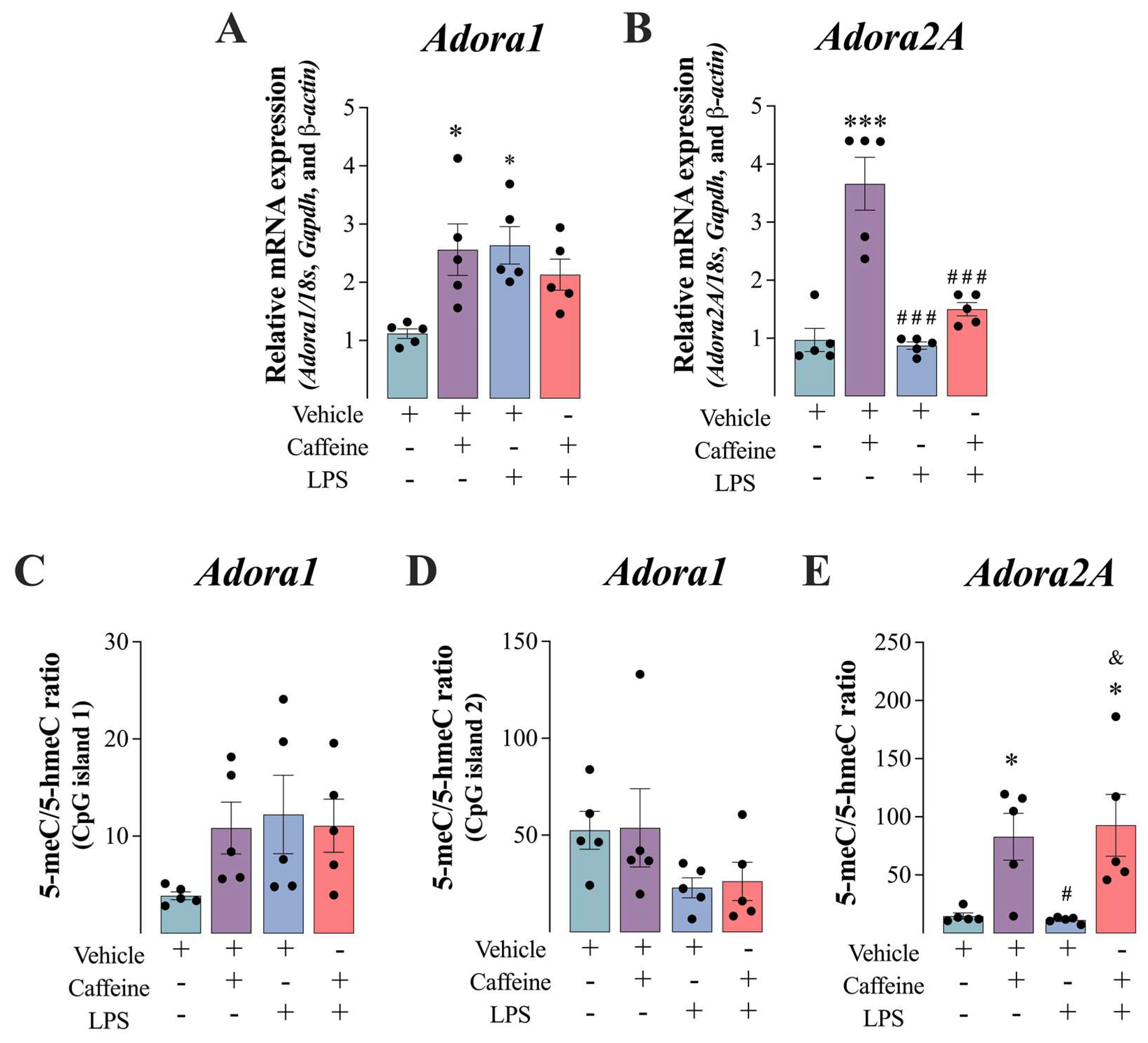
Figure 3 shows the effect of caffeine administration on adenosinergic receptors gene expression and gene methylation. Caffeine administration per se increased the expression of Adora1 (F(1,16) = 10.03, p < 0.05) (Figure 3A) and Adora2A (F(1,16) = 16.26, p < 0.001) (Figure 3B) in the mouse muscle, which was prevented by the administration of LPS alone and combined with caffeine. LPS per se increased the levels of Adora1 (F(1,16) = 10.03, p < 0.01) (Figure 3B). Considering that gene expression can be controlled by epigenetics, the levels of DNA methylation and demethylation of Adora1 and Adora2A were analyzed. While global DNA methylation was not modified by caffeine and/or LPS administration (Figure 3C,D), 5-meC/5-hmeC ratio, used as an index of DNA methylation, was increased under caffeine and Caffeine+LPS administration (F(1,16) = 0.16, p < 0.05) (Figure 3E). The higher methylation of the Adora2A promoter might represent a homeostatic mechanism to control the upregulation of the gene. Figure S2 shows that the absolute gene expression of the two ARs, Adora1 and Adora2A, were similar in basal conditions.

3.4. Caffeine+LPS Exposure Enhanced the De Novo DNA Methylation in the Vastus Lateralis Muscle Mice

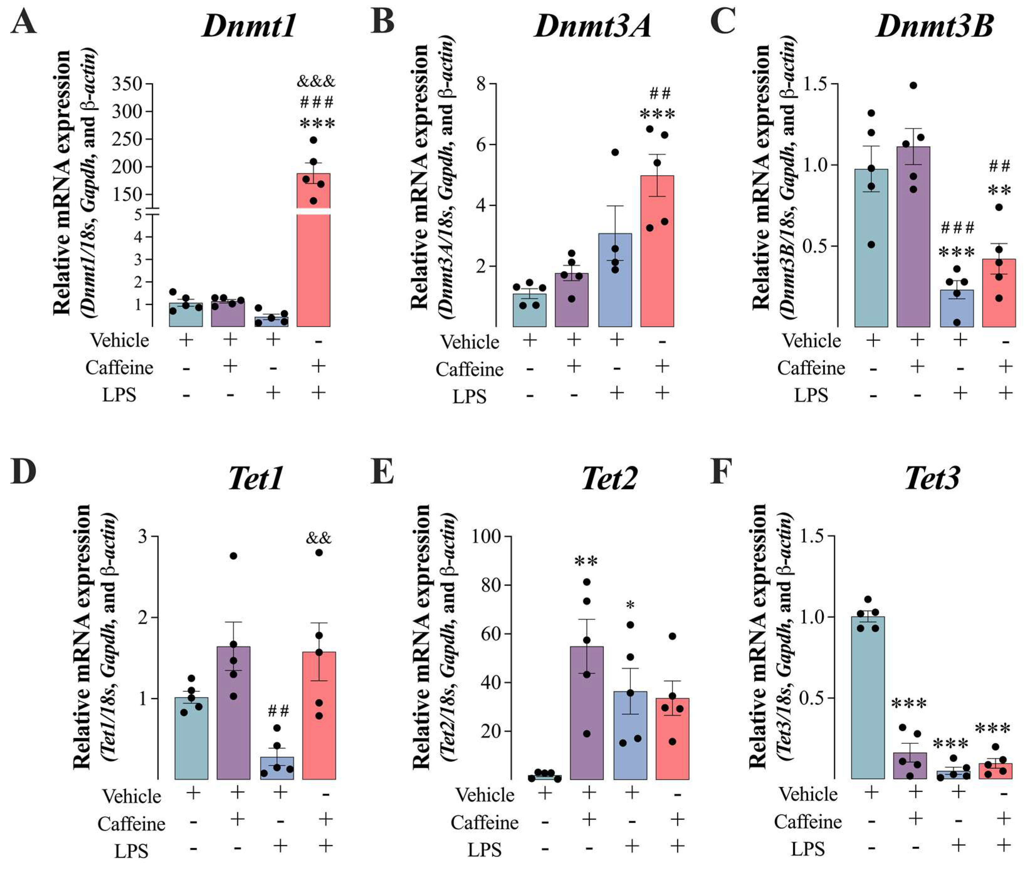
Figure 4 shows the effects of caffeine and/or LPS administration on the epigenetic profile in the mouse vastus lateralis muscle. Caffeine+LPS significantly upregulated the expression of the maintenance methylation gene Dnmt1 (F(1,16) = 101.05, p < 0.001) (Figure 4A), and de novo methylation gene Dnmt3A (F(1,15) = 1.26, p < 0.001) (Figure 4B), while the expression of de novo methylation gene Dnmt3B was significantly downregulated (F(1,16) = 0.06, p < 0.001) (Figure 4C) in the mouse muscle, when compared with the Vehicle and LPS groups. Caffeine+LPS treatment also inhibited the expression of the gene encoding the DNA demethylation enzyme Tet3 (F(1,16) = 133.5, p < 0.001) (Figure 4F), while no effect was observed on Tet1 and Tet2 when compared with the basal condition (vehicle), or under inflammation (LPS group). In addition, LPS modulated the expression of Tet genes; while LPS positively modulated the expression of Tet2 (F(1,16) = 11.84, p < 0.05) (Figure 4E), LPS administration negatively modulated the expression of Tet3 (F(1,16) = 133.50, p < 0.001), when compared with the vehicle condition. LPS treatment also compromised the expression of Dnmt3B, which was partially attenuated by caffeine co-administration (F(1,16) = 0.06, p < 0.01) (Figure 4C). Caffeine treatment per se increased the levels of Tet2 (Figure 4E), while it inhibited Tet3 expression (Figure 4F).

3.5. Caffeine Administration Attenuated the Catabolic State Induced by LPS Administration in the Mouse VASTUS lateralis

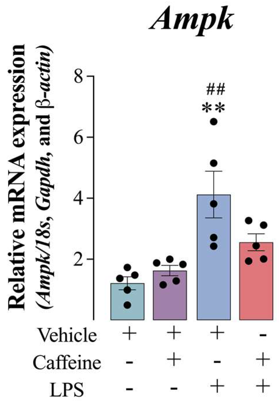
Figure 5 shows the effects of caffeine and/or LPS catabolism in the mouse muscle. LPS treatment significantly elicited the upregulation of the energy status sensor gene Ampk (F(1,16) = 5.35, p < 0.01) (Figure 5). The coadministration of caffeine significantly reverted the effect induced by LPS.

4. Discussion

Caffeine is a stimulant drug widely known and used due to its psychoactive and ergogenic effects [27]. The effects of caffeine on physical exercise, endurance performance, and fatigue stalling are well documented [28,29,30]. However, the molecular mechanisms behind these modulations are still under study. To the best of our knowledge, this is the first study to show that epigenetics is involved in the anti-inflammatory effects of caffeine on the vastus lateralis muscle of resting mice. Here, we showed that treatment with caffeine prevented an increase of the gene expression of LPS-induced pro-inflammatory cytokines Il1b and Il6 and promoted the upregulation of the anti-inflammatory genes Il10 and Il13 in the mouse muscle. The anti-inflammatory state observed in the caffeine experimental group occurred with decreased gene expression of the NLRP3 inflammasome components, Asc and Casp1. Indeed, the activation of caspase 1 mediates the cleavage of pro-IL-1β to generate and release its biologically pro-inflammatory active form, IL-1β [31]. Moreover, caffeine administration promoted the upregulation of the adenosinergic receptors Adora1 and Adora2A, the signaling of which is known to induce vasodilatation, healing and anti-inflammation, promotion of tissue blood flow and cellular homeostasis in different cell types [32]. Thus, in order to maintain homeostasis, the upregulation of Adora2A might have been responsible of triggering the methylation of its promoter. While DNA methylation patterns were not altered by caffeine treatment, the DNA methylating status was increased after Caffeine+LPS administration, suggesting that the observed adaptation to inflammation induced by caffeine was due to epigenetics.
Caffeine is the most commonly consumed social drug to increase alertness, arousal and energy [33]. Its consumption has been related to improvement in cognitive performance and mood in healthy population [34,35], and is the main ergogenic resource used by athletes to enhance exercise performance, extend time to exhaustion, and to delay fatigue [36].
Moreover, it has been shown that caffeine upregulated dopamine metabolism and signaling, and increased the synthesis and turnover of noradrenaline, being closely associated with an improvement in both peripheral and central fatigue [37]. Furthermore, the ergogenic effects of caffeine consumption have also been shown to be more evident in fatigued than in well-rested subjects [38,39]. These effects are proposed to be mediated by the non-selective antagonism of Adora1 and Adora2A [33]. Adora1 are widely expressed in the cortex, hippocampus, cerebellum, and thalamus [33], and in the adipose tissue, stomach, kidney, and heart [40]. Due to the capacity for lowering cAMP intracellular levels, the activation of Adora1 promotes bradycardia, inhibition of lipolysis, antinociception, reduction of sympathetic and parasympathetic activity, neuronal hyperpolarization, among others [41]. In contrast, Adora2A has a more restricted distribution, being more expressed in the striatum, nucleus accumbens, and olfactory tubercle [33]. Skeletal muscle, bladder, and the immune system are the tissues with the highest density of these receptors in the periphery [42]. The activation of Adora2A triggers neurotransmitter release, anti-inflammatory immune responses, and vascular smooth muscle cell relaxation, due to the activation of signaling pathways mediated by increased cAMP intracellular levels [36,41]. Therefore, the typical effects of adenosine, the natural agonist of Adoras and the final catabolite of ATP, that are associated with tiredness and drowsiness are counterbalanced by caffeine.
The effective dose of caffeine to antagonize Adoras and to lead to increased exercise time to fatigue ranges from 3 to 9 mg/kg in humans [28,43,44] and rodents [45,46]. These doses have been shown to increase performance in endurance, intermittent and resistance exercises in humans [5,6,7,8,47,48,49,50].
These effects have been associated with enhanced peripheral energy metabolism, activation of ryanodine channels for quicker release of calcium, and oxidant system in the muscle, improving muscle speed and strength (for a review see [16]). However, a large body of evidence suggests that caffeine can also mediate its ergogenic effects by inducing an anti-inflammatory status, preventing excessive endogenous catabolism and oxidative stress [1,16,51]. The anti-inflammatory effect of caffeine is also supported by the fact that individuals who suffer from cancer, obesity or liver, metabolic or neurodegenerative disorders and for whom persistent inflammation has been reported, developed fatigue when the symptoms appeared [52,53,54,55,56,57,58]. Moreover, high circulating levels of caffeine have been associated with delayed onset or reduced risk of dementia in individuals with mild cognitive impairment [59]. Furthermore, healthy individuals receiving an acute dose of caffeine showed reduced levels of pro-inflammatory markers and delayed development of fatigue [54,60]. Indeed, one of the first and most common symptoms associated with system immune activation is fatigue [61]. In addition, regular coffee consumption has also been associated with a reduced risk of low-grade inflammation in clinical conditions such as type 2 diabetes mellitus [62], and metabolic syndrome [63].
Considering that fatigue is characterized by temporary reductions in voluntary muscular force production, and cognitive and motivational changes that induces poor physical performance [64], we aimed to study whether caffeine could induce anti-inflammatory effects in the inflamed mouse muscle, and whether these effects are associate with epigenetic modifications.
It has been suggested that the development of fatigue may activate pathways that promote the activation of nuclear factor kappa b (NF-κB), which is considered a prototypical pro-inflammatory signaling pathway (for a review see [65]). NF-κB is known to be activated by a wide array of mediators, including LPS, inflammatory cytokines such as IL-1b and TNF-a, and reactive oxygen species, which in turn activates several signal transduction cascades and induces changes in transcription factors that promote a pro-inflammatory status. NF-κB’s activation induces the synthesis of pro-IL-1β and the activation of caspase 1 that induces the proteolytic maturation of pro-IL-1β [31]. Caspase 1 and ASC are key components of NLRP3 inflammasome, a multiproteic complex that induces inflammatory responses and cell death in response to various danger signals [66], including pro-inflammatory cytokines, reactive species, oxidized compounds that are known to accumulate in the muscle and blood during exhaustive physical exercise [67,68,69]. Therefore, the effect of caffeine on the inflammatory response induced by LPS that we observed in the mouse muscle suggests that part of its ergogenic effects might be mediated by the inhibition of the inflammasome assembly.
The anti-inflammatory effect might also be related to the positive effects we observed on Adoras’ increased expression in the inflamed muscle. Thus, increasing the antagonism of Adoras will potentiate the anti-inflammatory effect on different immune cell populations, that are cells known for their expression of high levels of Adora2A. Accordingly, it has been proposed that caffeine is an immunosuppressor since it has shown to inhibit proliferation, activation, and cytokine secretion by lymphocytes [70]. For example, it has been shown that caffeine reduced TNF-a secretion and enhanced the expression of Adora2A in LPS-activated human macrophages [70]. In addition, reduced ATP/AMP ratio, which occurs during the inflammatory response [71], is a key modulator of enhanced AMPK signaling, which denotes energy deficit. Although, we did not measure the levels of phosphorylated AMPK, the restoration of basal Ampk gene expression suggests that caffeine also protects the inflamed muscle by improving energy metabolism as previously proposed [72].
DNA methylation is also known to repress gene expression by blocking the promoter sites at which activating transcription factors are bound [73]. The reduced global DNA demethylation could be responsible for the upregulation of Ampk under LPS treatment, which was rescued when caffeine was also administered. This is also in agreement with the increased expression of pro-inflammatory cytokines, which was negatively modulated after the coadministration of caffeine in our study. This effect has also been reported in other tissues and cells. For example, LPS-challenged human peripheral blood mononuclear cells exposed to caffeine at different concentrations (10–100 μM) for 24 h, negatively modulated the production of TNF-α [74]. Similarly, mouse splenocyte cultures stimulated with concanavalin A (a pro-inflammatory agent) showed reduced production of TNF-α, IL-2, and IFN-γ when co-treated with 3.75 and 10 mM of caffeine for 24 h [75].
The DNA methylation machinery requires DNMT3a and DNMT3b for the de novo [76], and DNMT1 for the maintenance [77] of DNA methylation. In general, when methylation occurs in the promoter region of a particular gene, the gene expression is expected to be repressed. DNA can also be demethylated by the action of ten-eleven translocation (TET) enzymes TET1, TET2, and TET3 [78], which may result in enhanced gene expression. Therefore, the balance of these processes may regulate the expression of different genes, including the ones involved in inflammation and adenosine signaling as shown here. Indeed, genome-wide meta-analyses identified several genes positively associating caffeine consumption and DNA methylation [22,79,80]. While previous studies have shown that caffeine intake is positively correlated with higher DNA methylation [22], we have shown in this study that caffeine per se can be responsible for the negative modulation of the expression of inflammatory genes in animals submitted to acute inflammation.

5. Conclusions

This study provides evidence for the anti-inflammatory effect of caffeine in the mouse muscle. The immune system activated by LPS induced the release of pro-inflammatory cytokines that was prevented by caffeine administration, an effect also observed in the reduction of the inflammasome components, possibly by a modulatory effect of caffeine on epigenetics.

Supplementary Materials

The following supporting information can be downloaded at: https://www.mdpi.com/article/10.3390/antiox12030554/s1, Figure S1: Caffeine administration prevented lipopolysaccharide (LPS)-induced Nrlp3 inflammasome components upregulation in the vastus lateralis muscle of mice.; Figure S2: Absolute gene expression of adenosine receptors in the mouse vastus lateralis muscle.

Author Contributions

T.E., A.F.S., R.A.F.d.S. and A.L. conceived and designed the study, performed the experiments, and wrote the first draft and the edited versions. J.S.; T.B.d.M., L.B.C. and P.L.d.S.S. contributed significantly to the literature review and the writing of the manuscript; All authors have read and agreed to the published version of the manuscript.

Funding

This research was supported by CNPq (Conselho Nacional de Desenvolvimento Científico e Tecnológico, Brazil; CNPq Universal #422488/2021-6), and FAPESC (Fundação de Amparo a Pesquisa e Inovação do Estado de Santa Catarina; FAPESC TO #2021TR000452). A.L. is a CNPq fellow (CNPq PQ #312854/2019-6). L.B.C. is a Fapeti (Research, Technology and Innovation Support Foundation of the University of Taubaté) scholar. P.L.d.S.S. is a FAPESC (São Paulo Research Foundation scholar; FAPESP: 2020/13436-5) scholar. T.E. is a FAPESC scholar (FAPESC/0599/2018).

Institutional Review Board Statement

The study was conducted in accordance with Ethics Committee for Animal Research (PP00760/CEUA) of the Universidade Federal de Santa Catarina (Brazil).

Data Availability Statement

The data presented in this study are available on request from the corresponding author.

Acknowledgments

We thank LAMEB/CCB-UFSC technicians for their helpful assistance, and to Ted Griswold for the English editing.

Conflicts of Interest

The authors declare no conflict of interest.

References

  1. da Luz Scheffer, D.; Latini, A. Exercise-Induced Immune System Response: Anti-Inflammatory Status on Peripheral and Central Organs. Biochim. Biophys. Acta Mol. Basis Dis. 2020, 1866, 165823. [Google Scholar] [CrossRef]
  2. Hargreaves, M.; Spriet, L.L. Skeletal Muscle Energy Metabolism during Exercise. Nat. Metab. 2020, 2, 817–828. [Google Scholar] [CrossRef]
  3. Vital-Lopez, F.G.; Ramakrishnan, S.; Doty, T.J.; Balkin, T.J.; Reifman, J. Caffeine Dosing Strategies to Optimize Alertness during Sleep Loss. J. Sleep Res. 2018, 27, e12711. [Google Scholar] [CrossRef] [Green Version]
  4. Harpaz, E.; Tamir, S.; Weinstein, A.; Weinstein, Y. The Effect of Caffeine on Energy Balance. J. Basic Clin. Physiol. Pharmacol. 2017, 28, 1–10. [Google Scholar] [CrossRef]
  5. Astorino, T.A.; Rohmann, R.L.; Firth, K. Effect of Caffeine Ingestion on One-Repetition Maximum Muscular Strength. Eur. J. Appl. Physiol. 2007, 102, 127–132. [Google Scholar] [CrossRef]
  6. Graham, T.E.; Spriet, L.L. Performance and Metabolic Responses to a High Caffeine Dose during Prolonged Exercise. J. Appl. Physiol. 1991, 71, 2292–2298. [Google Scholar] [CrossRef] [Green Version]
  7. Spriet, L.L.; MacLean, D.A.; Dyck, D.J.; Hultman, E.; Cederblad, G.; Graham, T.E. Caffeine Ingestion and Muscle Metabolism during Prolonged Exercise in Humans. Am. J. Physiol. Metab. 1992, 262, E891–E898. [Google Scholar] [CrossRef]
  8. Schneiker, K.T.; Bishop, D.; Dawson, B.; Hackett, L.P. Effects of Caffeine on Prolonged Intermittent-Sprint Ability in Team-Sport Athletes. Med. Sci. Sport. Exerc. 2006, 38, 578–585. [Google Scholar] [CrossRef]
  9. Rodas, L.; Martinez, S.; Aguilo, A.; Tauler, P. Caffeine Supplementation Induces Higher IL-6 and IL-10 Plasma Levels in Response to a Treadmill Exercise Test. J. Int. Soc. Sports Nutr. 2020, 17, 47. [Google Scholar] [CrossRef]
  10. Tauler, P.; Martinez, S.; Martinez, P.; Lozano, L.; Moreno, C.; Aguiló, A. Effects of Caffeine Supplementation on Plasma and Blood Mononuclear Cell Interleukin-10 Levels After Exercise. Int. J. Sport Nutr. Exerc. Metab. 2016, 26, 8–16. [Google Scholar] [CrossRef]
  11. Tauler, P.; Martínez, S.; Moreno, C.; Monjo, M.; Martínez, P.; Aguiló, A. Effects of Caffeine on the Inflammatory Response Induced by a 15-Km Run Competition. Med. Sci. Sport. Exerc. 2013, 45, 1269–1276. [Google Scholar] [CrossRef]
  12. Farokhi-Sisakht, F.; Farhoudi, M.; Mahmoudi, J.; Farajdokht, F.; Kahfi-Ghaneh, R.; Sadigh-Eteghad, S. Effect of Intranasal Administration of Caffeine on MPFC Ischemia-induced Cognitive Impairment in BALB/c Mice. Acta Neurobiol. Exp. 2022, 82, 295–303. [Google Scholar] [CrossRef]
  13. Yang, L.; Yu, X.; Zhang, Y.; Liu, N.; Xue, X.; Fu, J. Caffeine Treatment Started before Injury Reduces Hypoxic-Ischemic White-Matter Damage in Neonatal Rats by Regulating Phenotypic Microglia Polarization. Pediatr. Res. 2022, 92, 1543–1554. [Google Scholar] [CrossRef]
  14. Jia, H.; Aw, W.; Egashira, K.; Takahashi, S.; Aoyama, S.; Saito, K.; Kishimoto, Y.; Kato, H. Coffee Intake Mitigated Inflammation and Obesity-Induced Insulin Resistance in Skeletal Muscle of High-Fat Diet-Induced Obese Mice. Genes Nutr. 2014, 9, 389. [Google Scholar] [CrossRef] [Green Version]
  15. Endesfelder, S.; Strauß, E.; Bendix, I.; Schmitz, T.; Bührer, C. Prevention of Oxygen-Induced Inflammatory Lung Injury by Caffeine in Neonatal Rats. Oxidative Med. Cell. Longev. 2020, 2020, 3840124. [Google Scholar] [CrossRef]
  16. Barcelos, R.P.; Lima, F.D.; Carvalho, N.R.; Bresciani, G.; Royes, L.F. Caffeine Effects on Systemic Metabolism, Oxidative-Inflammatory Pathways, and Exercise Performance. Nutr. Res. 2020, 80, 1–17. [Google Scholar] [CrossRef]
  17. Furman, D.; Chang, J.; Lartigue, L.; Bolen, C.R.; Haddad, F.; Gaudilliere, B.; Ganio, E.A.; Fragiadakis, G.K.; Spitzer, M.H.; Douchet, I.; et al. Expression of Specific Inflammasome Gene Modules Stratifies Older Individuals into Two Extreme Clinical and Immunological States. Nat. Med. 2017, 23, 174–184. [Google Scholar] [CrossRef]
  18. Egan, B.; O’Connor, P.L.; Zierath, J.R.; O’Gorman, D.J. Time Course Analysis Reveals Gene-Specific Transcript and Protein Kinetics of Adaptation to Short-Term Aerobic Exercise Training in Human Skeletal Muscle. PLoS ONE 2013, 8, e74098. [Google Scholar] [CrossRef]
  19. Egan, B.; Zierath, J.R. Exercise Metabolism and the Molecular Regulation of Skeletal Muscle Adaptation. Cell Metab. 2013, 17, 162–184. [Google Scholar] [CrossRef] [Green Version]
  20. Seaborne, R.A.; Strauss, J.; Cocks, M.; Shepherd, S.; O’Brien, T.D.; van Someren, K.A.; Bell, P.G.; Murgatroyd, C.; Morton, J.P.; Stewart, C.E.; et al. Human Skeletal Muscle Possesses an Epigenetic Memory of Hypertrophy. Sci. Rep. 2018, 8, 1898. [Google Scholar] [CrossRef] [Green Version]
  21. Waddington, C.H. The Epigenotype. Int. J. Epidemiol. 2012, 41, 10–13. [Google Scholar] [CrossRef] [Green Version]
  22. Chuang, Y.-H.; Quach, A.; Absher, D.; Assimes, T.; Horvath, S.; Ritz, B. Coffee Consumption Is Associated with DNA Methylation Levels of Human Blood. Eur. J. Hum. Genet. 2017, 25, 608–616. [Google Scholar] [CrossRef] [Green Version]
  23. Remor, A.P.; da Silva, R.A.; de Matos, F.J.; Glaser, V.; de Paula Martins, R.; Ghisoni, K.; da Luz Scheffer, D.; Andia, D.C.; Portinho, D.; de Souza, A.P.; et al. Chronic Metabolic Derangement-Induced Cognitive Deficits and Neurotoxicity Are Associated with REST Inactivation. Mol. Neurobiol. 2018, 56, 1539–1557. [Google Scholar] [CrossRef]
  24. Ghisoni, K.; Martins, R.D.P.R.; de, P.; Barbeito, L.; Latini, A. Neopterin as a Potential Cytoprotective Brain Molecule. J. Psychiatr. Res. 2015, 71, 134–139. [Google Scholar] [CrossRef]
  25. De Paula Martins, R.; Glaser, V.; Aguiar, A.S.; de Paula Ferreira, P.M.; Ghisoni, K.; da Luz Scheffer, D.; Lanfumey, L.; Raisman-Vozari, R.; Corti, O.; De Paul, A.L.; et al. De Novo Tetrahydrobiopterin Biosynthesis Is Impaired in the Inflammed Striatum of Parkin(-/-) Mice. Cell Biol. Int. 2018, 42, 725–733. [Google Scholar] [CrossRef]
  26. Untergasser, A.; Cutcutache, I.; Koressaar, T.; Ye, J.; Faircloth, B.C.; Remm, M.; Rozen, S.G. Primer3—New Capabilities and Interfaces. Nucleic Acids Res. 2012, 40, e115. [Google Scholar] [CrossRef] [Green Version]
  27. Spriet, L.L. Exercise and Sport Performance with Low Doses of Caffeine. Sport. Med. 2014, 44, 175–184. [Google Scholar] [CrossRef] [Green Version]
  28. Graham, T.E.; Spriet, L.L. Metabolic, Catecholamine, and Exercise Performance Responses to Various Doses of Caffeine. J. Appl. Physiol. 1995, 78, 867–874. [Google Scholar] [CrossRef]
  29. Davis, J.K.; Green, J.M. Caffeine and Anaerobic Performance: Ergogenic Value and Mechanisms of Action. Sport. Med. 2009, 39, 813–832. [Google Scholar] [CrossRef]
  30. Van Soeren, M.H.; Sathasivam, P.; Spriet, L.L.; Graham, T.E. Caffeine Metabolism and Epinephrine Responses during Exercise in Users and Nonusers. J. Appl. Physiol. 1993, 75, 805–812. [Google Scholar] [CrossRef]
  31. Martinon, F.; Burns, K.; Tschopp, J. The Inflammasome. Mol. Cell 2002, 10, 417–426. [Google Scholar] [CrossRef]
  32. Liu, H.; Xia, Y. Beneficial and Detrimental Role of Adenosine Signaling in Diseases and Therapy. J. Appl. Physiol. 2015, 119, 1173–1182. [Google Scholar] [CrossRef] [Green Version]
  33. Fredholm, B.B.; Bättig, K.; Holmén, J.; Nehlig, A.; Zvartau, E.E. Actions of Caffeine in the Brain with Special Reference to Factors That Contribute to Its Widespread Use. Pharmacol. Rev. 1999, 51, 83–133. [Google Scholar]
  34. Pasman, W.J.; Boessen, R.; Donner, Y.; Clabbers, N.; Boorsma, A. Effect of Caffeine on Attention and Alertness Measured in a Home-Setting, Using Web-Based Cognition Tests. JMIR Res. Protoc. 2017, 6, e6727. [Google Scholar] [CrossRef] [Green Version]
  35. Smith, A.P. Caffeine, Extraversion and Working Memory. J. Psychopharmacol. 2013, 27, 71–76. [Google Scholar] [CrossRef]
  36. Mielgo-Ayuso, J.; Marques-Jiménez, D.; Refoyo, I.; Del Coso, J.; León-Guereño, P.; Calleja-González, J. Effect of Caffeine Supplementation on Sports Performance Based on Differences Between Sexes: A Systematic Review. Nutrients 2019, 11, 2313. [Google Scholar] [CrossRef] [Green Version]
  37. Fisone, G.; Borgkvist, A.; Usiello, A. Caffeine as a Psychomotor Stimulant: Mechanism of Action. Cell. Mol. Life Sci. 2004, 61, 857–872. [Google Scholar] [CrossRef]
  38. Lorist, M.M.; Snel, J.; Kok, A.; Mulder, G. Influence of Caffeine on Selective Attention in Well-Rested and Fatigued Subjects. Psychophysiology 1994, 31, 525–534. [Google Scholar] [CrossRef]
  39. Childs, E.; de Wit, H. Enhanced Mood and Psychomotor Performance by a Caffeine-Containing Energy Capsule in Fatigued Individuals. Exp. Clin. Psychopharmacol. 2008, 16, 13–21. [Google Scholar] [CrossRef]
  40. Ralevic, V.; Burnstock, G. Receptors for Purines and Pyrimidines. Pharmacol. Rev. 1998, 50, 413–492. [Google Scholar]
  41. Fredholm, B.B.; IJzerman, A.P.; Jacobson, K.A.; Klotz, K.N.; Linden, J. International Union of Pharmacology. XXV. Nomenclature and Classification of Adenosine Receptors. Pharmacol. Rev. 2001, 53, 527–552. [Google Scholar]
  42. Ongini, E.; Fredholm, B.B. Pharmacology of Adenosine A2A Receptors. Trends Pharmacol. Sci. 1996, 17, 364–372. [Google Scholar] [CrossRef]
  43. Pasman, W.; van Baak, M.; Jeukendrup, A.; de Haan, A. The Effect of Different Dosages of Caffeine on Endurance Performance Time. Int. J. Sport. Med. 1995, 16, 225–230. [Google Scholar] [CrossRef] [Green Version]
  44. Costill, D.L.; Dalsky, G.P.; Fink, W.J. Effects of Caffeine Ingestion on Metabolism and Exercise Performance. Med. Sci. Sports 1978, 10, 155–158. [Google Scholar]
  45. Lim, B.-V.; Jang, M.-H.; Shin, M.-C.; Kim, H.-B.; Kim, Y.-J.; Kim, Y.-P.; Chung, J.-H.; Kim, H.; Shin, M.-S.; Kim, S.-S.; et al. Caffeine Inhibits Exercise-Induced Increase in Tryptophan Hydroxylase Expression in Dorsal and Median Raphe of Sprague–Dawley Rats. Neurosci. Lett. 2001, 308, 25–28. [Google Scholar] [CrossRef]
  46. Solano, A.F.; da Luz Scheffer, D.; de Bem Alves, A.C.; Silva Aguiar Jr, A.; Latini, A. Potential Pitfalls When Investigating the Ergogenic Effects of Caffeine in Mice. J. Syst. Integr. Neurosci. 2017, 3, 1–4. [Google Scholar] [CrossRef] [Green Version]
  47. Mohr, M.; Nielsen, J.J.; Bangsbo, J. Caffeine Intake Improves Intense Intermittent Exercise Performance and Reduces Muscle Interstitial Potassium Accumulation. J. Appl. Physiol. 2011, 111, 1372–1379. [Google Scholar] [CrossRef]
  48. Duncan, M.J.; Stanley, M.; Parkhouse, N.; Cook, K.; Smith, M. Acute Caffeine Ingestion Enhances Strength Performance and Reduces Perceived Exertion and Muscle Pain Perception during Resistance Exercise. Eur. J. Sport Sci. 2013, 13, 392–399. [Google Scholar] [CrossRef]
  49. Skulachev, M.V.; Antonenko, Y.N.; Anisimov, V.N.; Chernyak, B.V.; Cherepanov, D.A.; Chistyakov, V.A.; Egorov, M.V.; Kolosova, N.G.; Korshunova, G.A.; Lyamzaev, K.G.; et al. Mitochondrial-Targeted Plastoquinone Derivatives. Effect on Senescence and Acute Age-Related Pathologies. Curr. Drug Targets 2011, 12, 800–826. [Google Scholar] [CrossRef]
  50. Del Coso, J.; Muñoz, G.; Muñoz-Guerra, J. Prevalence of Caffeine Use in Elite Athletes Following Its Removal from the World Anti-Doping Agency List of Banned Substances. Appl. Physiol. Nutr. Metab. 2011, 36, 555–561. [Google Scholar] [CrossRef] [Green Version]
  51. Rodak, K.; Kokot, I.; Kratz, E.M. Caffeine as a Factor Influencing the Functioning of the Human Body—Friend or Foe? Nutrients 2021, 13, 3088. [Google Scholar] [CrossRef]
  52. Capuron, L.; Gumnick, J.F.; Musselman, D.L.; Lawson, D.H.; Reemsnyder, A.; Nemeroff, C.B.; Miller, A.H. Neurobehavioral Effects of Interferon-Alpha in Cancer Patients: Phenomenology and Paroxetine Responsiveness of Symptom Dimensions. Neuropsychopharmacology 2002, 26, 643–652. [Google Scholar] [CrossRef]
  53. Heesen, C.; Nawrath, L.; Reich, C.; Bauer, N.; Schulz, K.-H.; Gold, S.M. Fatigue in Multiple Sclerosis: An Example of Cytokine Mediated Sickness Behaviour? J. Neurol. Neurosurg. Psychiatr. 2006, 77, 34–39. [Google Scholar] [CrossRef] [Green Version]
  54. Cho, H.J.; Seeman, T.E.; Bower, J.E.; Kiefe, C.I.; Irwin, M.R. Prospective Association Between C-Reactive Protein and Fatigue in the Coronary Artery Risk Development in Young Adults Study. Biol. Psychiatr. 2009, 66, 871–878. [Google Scholar] [CrossRef] [Green Version]
  55. Goulart, F.O.; Godke, B.A.; Borges, V.; Azevedo-Silva, S.M.C.; Mendes, M.F.; Cendoroglo, M.S.; Ferraz, H.B. Fatigue in a Cohort of Geriatric Patients with and without Parkinson’s Disease. Brazilian J. Med. Biol. Res. 2009, 42, 771–775. [Google Scholar] [CrossRef] [Green Version]
  56. Kempf, K.; Herder, C.; Erlund, I.; Kolb, H.; Martin, S.; Carstensen, M.; Koenig, W.; Sundvall, J.; Bidel, S.; Kuha, S.; et al. Effects of Coffee Consumption on Subclinical Inflammation and Other Risk Factors for Type 2 Diabetes: A Clinical Trial. Am. J. Clin. Nutr. 2010, 91, 950–957. [Google Scholar] [CrossRef] [Green Version]
  57. Xiao, C.; Beitler, J.J.; Higgins, K.A.; Conneely, K.; Dwivedi, B.; Felger, J.; Wommack, E.C.; Shin, D.M.; Saba, N.F.; Ong, L.Y.; et al. Fatigue Is Associated with Inflammation in Patients with Head and Neck Cancer before and after Intensity-Modulated Radiation Therapy. Brain. Behav. Immun. 2016, 52, 145–152. [Google Scholar] [CrossRef] [Green Version]
  58. Lee, A.; Lim, W.; Kim, S.; Khil, H.; Cheon, E.; An, S.; Hong, S.; Lee, D.H.; Kang, S.-S.; Oh, H.; et al. Coffee Intake and Obesity: A Meta-Analysis. Nutrients 2019, 11, 1274. [Google Scholar] [CrossRef] [Green Version]
  59. Cao, C.; Loewenstein, D.A.; Lin, X.; Zhang, C.; Wang, L.; Duara, R.; Wu, Y.; Giannini, A.; Bai, G.; Cai, J.; et al. High Blood Caffeine Levels in MCI Linked to Lack of Progression to Dementia. J. Alzheimer’s Dis. 2012, 30, 559–572. [Google Scholar] [CrossRef] [Green Version]
  60. Jokela, M.; Virtanen, M.; Batty, G.D.; Kivimäki, M. Inflammation and Specific Symptoms of Depression. JAMA Psychiatr. 2016, 73, 87–88. [Google Scholar] [CrossRef] [Green Version]
  61. DellaGioia, N.; Devine, L.; Pittman, B.; Hannestad, J. Bupropion Pre-Treatment of Endotoxin-Induced Depressive Symptoms. Brain. Behav. Immun. 2013, 31, 197–204. [Google Scholar] [CrossRef]
  62. Ding, M.; Bhupathiraju, S.N.; Chen, M.; van Dam, R.M.; Hu, F.B. Caffeinated and Decaffeinated Coffee Consumption and Risk of Type 2 Diabetes: A Systematic Review and a Dose-Response Meta-Analysis. Diabetes Care 2014, 37, 569–586. [Google Scholar] [CrossRef] [Green Version]
  63. Shang, F.; Li, X.; Jiang, X. Coffee Consumption and Risk of the Metabolic Syndrome: A Meta-Analysis. Diabetes Metab. 2016, 42, 80–87. [Google Scholar] [CrossRef]
  64. Taylor, J.L.; Todd, G.; Gandevia, S.C. Evidence for a Supraspinal Contribution to Human Muscle Fatigue. Clin. Exp. Pharmacol. Physiol. 2006, 33, 400–405. [Google Scholar] [CrossRef]
  65. Lawrence, T. The Nuclear Factor NF-KappaB Pathway in Inflammation. Cold Spring Harb. Perspect. Biol. 2009, 1, a001651. [Google Scholar] [CrossRef] [Green Version]
  66. Schroder, K.; Tschopp, J. The Inflammasomes. Cell 2010, 140, 821–832. [Google Scholar] [CrossRef] [Green Version]
  67. Abkenar, I.K.; Rahmani-nia, F.; Lombardi, G. The Effects of Acute and Chronic Aerobic Activity on the Signaling Pathway of the Inflammasome NLRP3 Complex in Young Men. Medicina 2019, 55, 105. [Google Scholar] [CrossRef] [Green Version]
  68. Li, H.; Miao, W.; Ma, J.; Xv, Z.; Bo, H.; Li, J.; Zhang, Y.; Ji, L.L. Acute Exercise-Induced Mitochondrial Stress Triggers an Inflammatory Response in the Myocardium via NLRP3 Inflammasome Activation with Mitophagy. Oxidative Med. Cell. Longev. 2016, 2016, 1987149. [Google Scholar] [CrossRef] [Green Version]
  69. Ostrowski, K.; Rohde, T.; Asp, S.; Schjerling, P.; Pedersen, B.K. Pro- and Anti-inflammatory Cytokine Balance in Strenuous Exercise in Humans. J. Physiol. 1999, 515, 287–291. [Google Scholar] [CrossRef]
  70. Kovács, E.G.; Alatshan, A.; Budai, M.M.; Czimmerer, Z.; Bíró, E.; Benkő, S. Caffeine Has Different Immunomodulatory Effect on the Cytokine Expression and NLRP3 Inflammasome Function in Various Human Macrophage Subpopulations. Nutrients 2021, 13, 2409. [Google Scholar] [CrossRef]
  71. Pandit, M.; Timilshina, M.; Gu, Y.; Acharya, S.; Chung, Y.; Seo, S.-U.; Chang, J.-H. AMPK Suppresses Th2 Cell Responses by Repressing MTORC2. Exp. Mol. Med. 2022, 54, 1214–1224. [Google Scholar] [CrossRef]
  72. Egawa, T.; Hamada, T.; Kameda, N.; Karaike, K.; Ma, X.; Masuda, S.; Iwanaka, N.; Hayashi, T. Caffeine Acutely Activates 5′adenosine Monophosphate–Activated Protein Kinase and Increases Insulin-Independent Glucose Transport in Rat Skeletal Muscles. Metabolism 2009, 58, 1609–1617. [Google Scholar] [CrossRef] [Green Version]
  73. Moore, L.D.; Le, T.; Fan, G. DNA Methylation and Its Basic Function. Neuropsychopharmacology 2013, 38, 23–38. [Google Scholar] [CrossRef] [Green Version]
  74. Horrigan, L.A.; Kelly, J.P.; Connor, T.J. Caffeine Suppresses TNF-α Production via Activation of the Cyclic AMP/Protein Kinase A Pathway. Int. Immunopharmacol. 2004, 4, 1409–1417. [Google Scholar] [CrossRef]
  75. Ritter, M.; Hohenberger, K.; Alter, P.; Herzum, M.; Tebbe, J.; Maisch, M. Caffeine Inhibits Cytokine Expression in Lymphocytes. Cytokine 2005, 30, 177–181. [Google Scholar] [CrossRef]
  76. Okano, M.; Xie, S.; Li, E. Cloning and Characterization of a Family of Novel Mammalian DNA (Cytosine-5) Methyltransferases. Nat. Genet. 1998, 19, 219–220. [Google Scholar] [CrossRef]
  77. Hermann, A.; Goyal, R.; Jeltsch, A. The Dnmt1 DNA-(Cytosine-C5)-Methyltransferase Methylates DNA Processively with High Preference for Hemimethylated Target Sites. J. Biol. Chem. 2004, 279, 48350–48359. [Google Scholar] [CrossRef] [Green Version]
  78. Ito, S.; D’Alessio, A.C.; Taranova, O.V.; Hong, K.; Sowers, L.C.; Zhang, Y. Role of Tet Proteins in 5mC to 5hmC Conversion, ES-Cell Self-Renewal and Inner Cell Mass Specification. Nature 2010, 466, 1129–1133. [Google Scholar] [CrossRef] [Green Version]
  79. Cornelis, M.C.; Monda, K.L.; Yu, K.; Paynter, N.; Azzato, E.M.; Bennett, S.N.; Berndt, S.I.; Boerwinkle, E.; Chanock, S.; Chatterjee, N.; et al. Genome-Wide Meta-Analysis Identifies Regions on 7p21 (AHR) and 15q24 (CYP1A2) As Determinants of Habitual Caffeine Consumption. PLoS Genet. 2011, 7, e1002033. [Google Scholar] [CrossRef]
  80. Cornelis, M.C.; Byrne, E.M.; Esko, T.; Nalls, M.A.; Ganna, A.; Paynter, N.; Monda, K.L.; Amin, N.; Fischer, K.; Renstrom, F.; et al. Genome-Wide Meta-Analysis Identifies Six Novel Loci Associated with Habitual Coffee Consumption. Mol. Psychiatr. 2015, 20, 647–656. [Google Scholar] [CrossRef] [Green Version]
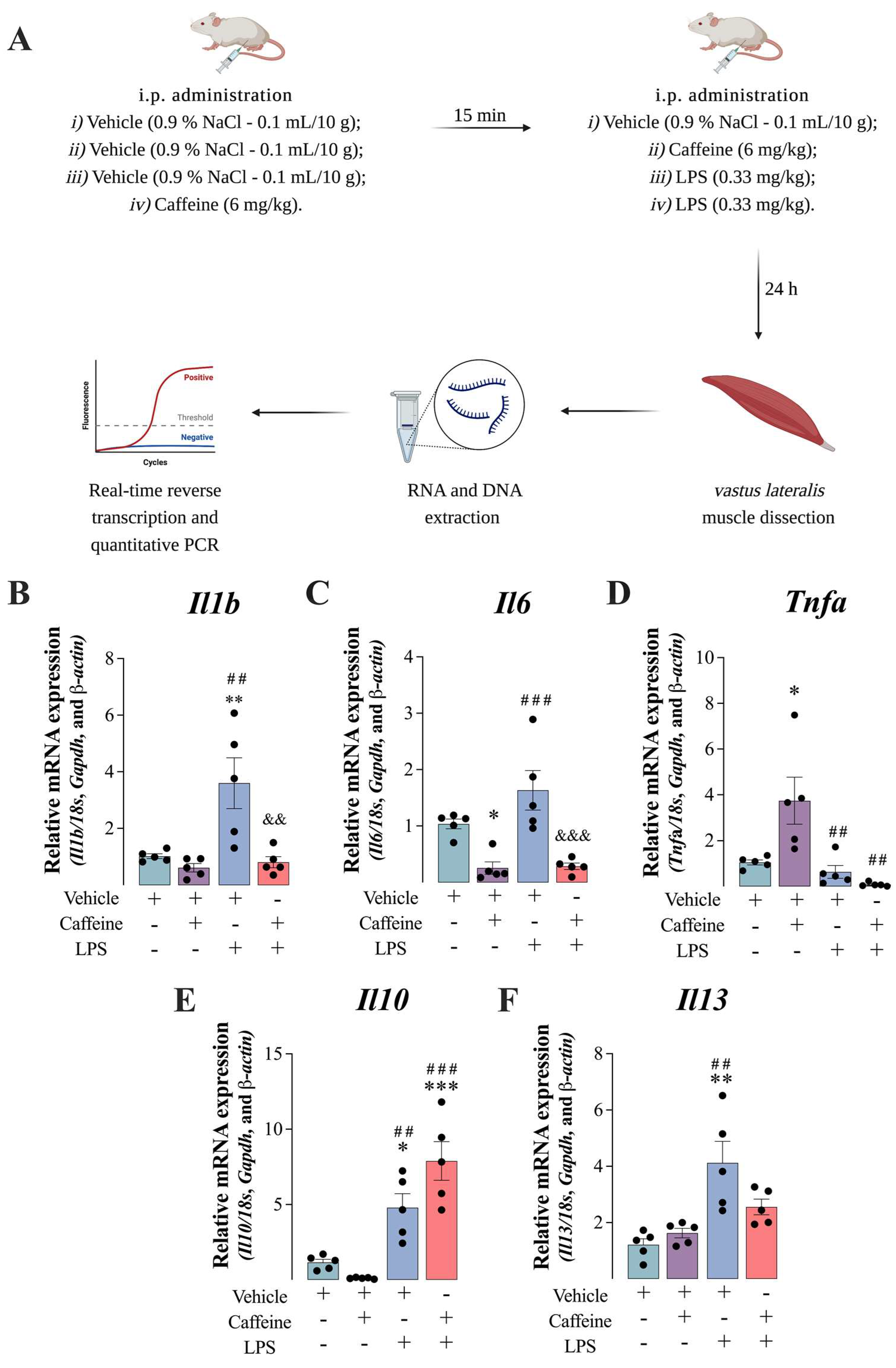
Figure 1. Caffeine administration reduced lipopolysaccharide (LPS)-mediated inflammation in the mouse vastus lateralis muscle. Adult Swiss male mice (3–5 months of age; body mass 45–50 g) received a single intraperitoneal (i.p.) injection of caffeine and/or LPS (See Materials and Methods for details). Schematic representation of the experimental protocol used to induced LPS-mediated inflammation in mouse, the muscle dissection and RNA/DNA extraction for the PCR analysis (A). The transcriptional profile of the cytokines Il1b (B), Il6 (C), Tnfa (D), Il10 (E) and Il13 (F) were evaluated by qPCR after the total RNA extraction (TRIzol®/Chloroform/Isopropanol method) from the muscle. Gene expression raw data were normalized by the average of the Ct of the 18s, Gapdh and β-actin genes and calculated by the method (2−ΔCt). Bars represent the mean ± standard error of mean of five independent experiments (animals) performed in technical duplicates. * p < 0.05; ** p < 0.01; and *** p < 0.001 vs. vehicle; ## p < 0.01; ### p < 0.001 vs. to caffeine, and && p < 0.01 and &&& p < 0.001 vs. LPS. Two-way ANOVA followed by Tukey’s test.
Antioxidants 12 00554 g001
Figure 2. Caffeine administration rescued lipopolysaccharide (LPS)-induced Nrlp3 inflammasome components upregulation in the vastus lateralis muscle of mice. Adult Swiss male mice (3–5 months of age; body mass 45–50 g) received a single intraperitoneal (i.p.) injection of caffeine and/or LPS (See Materials and Methods for details). NLRP3 inflammasome consists of a sensor (Nlrp3), an adaptor (Asc) and an effector (Caspase-1) (A). The transcriptional profile of the components of the NLRP3 inflammasome Nlrp3 (B), Asc1 (C), and Casp1 (D). Gene expression raw data were normalized by the average of the Ct of the 18s, Gapdh and β-actin genes and calculated by the method (2−ΔCt). Bars represent the mean ± standard error of the mean of five independent experiments (animals) performed in technical duplicates. *** p < 0.001 vs. vehicle; ### p < 0.001 vs. to caffeine, and &&& p < 0.001 vs. LPS. Two-way ANOVA followed by Tukey’s test.
Antioxidants 12 00554 g002
Figure 3. Caffeine administration induced the upregulation of genes encoding adenosinergic receptors in the vastus lateralis muscle of mice. Adult Swiss male mice (3–5 months of age; body mass 45–50 g) received a single intraperitoneal (i.p.) injection of caffeine and/or LPS (See Materials and Methods for details). The transcriptional profile of the components of the adenosine receptors Adora1 (A), and Adora2A (B). DNA methylation and demethylation of Adora1 CpG island 1 (C), Adora1 Cpg island 2 (D), and Adora2A (E). Gene expression raw data were normalized by the average of the Ct of the 18s, Gapdh and β-actin genes and calculated by the method (2−ΔCt). Bars represent the mean ± standard error of the mean of five independent experiments (animals) performed in technical duplicates. * p < 0.05; *** p < 0.001 vs. vehicle; # p < 0.05; ### p < 0.001 vs. to caffeine, and & p < 0.05 vs. LPS. Two-way ANOVA followed by Tukey’s test.
Antioxidants 12 00554 g003
Figure 4. Caffeine plus lipopolysaccharide (LPS) administration induced de novo DNA methylation in the vastus lateralis muscle of mice. Adult Swiss male mice (3–5 months of age; body mass 45–50 g) received a single intraperitoneal (i.p.) injection of caffeine and/or LPS (See Materials and Methods for details). The transcriptional profile of the de novo DNA methylation of Dnmt1 (A), Dnmt2A (B), and Dnmt3B (C). The status of the DNA demethylation enzymes Tet1 (D), Tet2 (E), and Tet3 (F). Gene expression raw data were normalized by the average of the Ct of the 18s, Gapdh and β-actin genes and calculated by the method (2−ΔCt). Bars represent the mean ± standard error of the mean of five independent experiments (animals) performed in technical duplicates. * p < 0.05; ** p < 0.01; *** p < 0.001 vs. vehicle; ## p < 0.01; ### p < 0.001 vs. caffeine and && p < 0.01 and &&& p < 0.001 vs. LPS. Two-way ANOVA followed by Tukey’s test.
Antioxidants 12 00554 g004
Figure 5. Caffeine administration reverted the catabolic effect induced by the administration of lipopolysaccharide (LPS) in the vastus lateralis muscle of mice. Adult Swiss male mice (3–5 months of age; body mass 45–50 g) received a single intraperitoneal (i.p.) injection of caffeine and/or LPS (See Materials and Methods for details). Ampk gene expression raw data were normalized by the average of the Ct of the 18s, Gapdh and β-actin genes and calculated by the method (2−ΔCt). Bars represent the mean ± standard error of the mean of five independent experiments (animals) performed in technical duplicates. ** p < 0.01 vs. vehicle; ## p < 0.01 vs. caffeine. Two-way ANOVA followed by Tukey’s test.
Antioxidants 12 00554 g005
Table 1. Oligonucleotide primers and PCR conditions used in quantitative real-time PCR.
Gene (ID)Primer5′-3′SequenceReaction ConditionProduct Size (bp)
Il1b (16176)ForwardGAC CTT GGA TGA GGA CA95 °C-15 s; 60 °C-30 s;
72 °C-30 s
183
ReverseAGC TCA TAT GGG TCC GAC AG
Il6 (16193)ForwardAGT TCG CTT CTT GGG ACT GA95 °C-15 s; 60 °C-30 s;
72 °C-30 s
191
ReverseCAG AAT TGC CAT TGC ACA AC
Tnfa (11647)ForwardCCA CAT CTC CCT CCA GAA AA95 °C-15 s; 60 °C-30 s;
72 °C-30 s
259
ReverseAGG GTC TGG GCC ATA GAA CT
Il10 (21926)ForwardCCA AGC CCT TAT CGG AAA TGA95 °C-15 s; 60 °C-30 s;
72 °C-30 s
163
ReverseTTT TCA CAG GGG AGA AAT CG
Il13 (16163)ForwardCAG TCC TGG CTC TTG CTT G95 °C-15 s; 60 °C-30 s;
72 °C-30 s
165
ReverseCCA GGT CCA CAC TCC ATA CC
Adora1 (11539)ForwardAGA ACC ACC TCC ACC CTT CT95 °C-15 s; 63 °C-30 s;
72 °C-30 s
227
ReverseTAC TCT GGG TGG TGG TCA CA
Adora2A (11540)ForwardATC CCT CAGAGA AGG GAA GC95 °C-15 s; 63 °C-30 s;
72 °C-30 s
300
ReverseAGC TTC CCA AAG GCT TTC TC
Dnmt1 (13433)ForwardCCT TTG TGG GAA CCT GGA A95 °C-15 s; 63 °C-30 s;
72 °C-30 s
240
ReverseCTG TCG TCT GCG GTG ATT
Dnmt3A (13435)ForwardGAG GGA ACT GAG ACC CCA C95 °C-15 s; 63 °C-30 s;
72 °C-30 s
216
ReverseCTG GAA GGT GAG TCT TGG CA
Dnmt3B (113436)ForwardAGC GGG TAT GAG GAG TGC AT95 °C-15 s; 63 °C-30 s;
72 °C-30 s
91
ReverseGGG AGC ATC CTT CGT GTC TG
Tet1 (52463)ForwardGAG CCT GTT CCT CGA TGT GG95 °C-15 s; 65 °C-30 s;
72 °C-30 s
367
ReverseCAA ACC CAC CTG AGG CTG TT
Tet2 (214133)ForwardAAC CTG GCT ACT GTC ATT GCT CCA95 °C-15 s; 65 °C-30 s;
72 °C-30 s
211
ReverseATG TTC TGC TGG TCT CTG TGG GAA
Tet3 (194388)ForwardGTC TCC CCA AGT CCT ACC TCC G95 °C-15 s; 63 °C-30 s;
72 °C-30 s
137
ReverseGTC AGT GCC CCA CGC TTC A
b-actin (11461)ForwardTCT TGG GTA TGG AAT CCT GTG95 °C-15 s; 58 °C-30 s;
72 °C-30 s
82
ReverseAGG TCT TTA CGG ATG TCA ACG
Gapdh (14433)ForwardAGG CCG GTG CTG AGT ATG TC95 °C-15 s; 58 °C-30 s;
72 °C-30 s
530
ReverseTGC CTG CTT CAC CAC CTT CT
Disclaimer/Publisher’s Note: The statements, opinions and data contained in all publications are solely those of the individual author(s) and contributor(s) and not of MDPI and/or the editor(s). MDPI and/or the editor(s) disclaim responsibility for any injury to people or property resulting from any ideas, methods, instructions or products referred to in the content.