Article

Nanoemulsions of Phoenix dactylifera L. (Decaffeinated) and Coffea arabica L. Extracts as a Novel Approach for the Treatment of Carbon Tetrachloride-Mediated Liver Fibrosis

by
Eman S. Alamri
1,*,
Hala M. Bayomy
1,*,
Mohamed A. Mohamady Hussein
2,
Nawal A. Ozaybi
1,
Seham E. Almasoudi
1,
Nahla S. Zidan
1,
Renad A. Albalwi
3,
Hebatallah H. Atteia
4 and
Fayza M. EL-Ezaly
5
1
Food Science and Nutrition Department, Faculty of Science, University of Tabuk, Tabuk 71491, Saudi Arabia
2
Department of Pharmacology, Medical Research and Clinical Studies Institute, National Research Centre, Dokki, Giza 12622, Egypt
3
Department of Nutrition and Food Science, University of Tabuk, Tabuk 71491, Saudi Arabia
4
Department of Pharmaceutical Chemistry, Faculty of Pharmacy, University of Tabuk, Tabuk 71491, Saudi Arabia
5
Department of Home Economics, Faculty of Specific Education, Mansoura University, Mansoura 35516, Egypt
*
Authors to whom correspondence should be addressed.
Antioxidants 2024, 13(3), 355; https://doi.org/10.3390/antiox13030355
Submission received: 4 February 2024 / Revised: 1 March 2024 / Accepted: 13 March 2024 / Published: 16 March 2024

Abstract

:
Liver fibrosis is a condition characterized by the excessive buildup of scar tissue in the liver. This scarring occurs as a result of chronic liver damage, often caused by conditions such as hepatitis, alcohol abuse, certain metabolic disorders, genetic abnormalities, autoimmunity, and noninfectious diseases such as fatty liver which leads to liver fibrosis. Nanoparticles have gained attention in recent years as potential therapeutic agents for liver fibrosis. They offer unique advantages due to their small size, large surface area, and ability to carry drugs or target specific cells or tissues. Studies have suggested that nanoemulsions may enhance drug delivery systems, enabling targeted drug delivery to specific sites in the liver and improving therapeutic outcomes. In this study, we explore the protective and therapeutic values with phytochemical profiling of the used agro-wastes decaffeinated palm date seeds (Phoenix dactylifera L., PSC) coffee and caffeinated Arabic coffee seeds (Coffea arabica L.; ACS). Both ACS and PSC extracts were converted into nanoemulsion (NE) forms using the oleic acid/Tween 80 system, which was recruited for the purpose of treating a rat model with liver fibrosis. Transmission electron microscopy (TEM) and dynamic light scattering (DLS) were used to record the sizes, morphologies, hydrodynamic diameters, and ζ-potentials of the prepared NE-ACSE and NE-PSCE. Accordingly, the NE-ACSE and NE-PSCE imaged via TEM and their ζ-potentials were recorded at 20.7, 23.3 nm and −41.4, −28.0 mV, respectively. The antioxidant properties were determined with a DPPH scavenging assay. The synthesized NE-PSCE and NE-ACSE were employed to treat a rat model with CCl4-induced liver fibrosis, to estimate the role of each emulsion-based extract in the treatment of liver fibrosis through recording inflammatory parameters, liver functions, antioxidant enzymes, and histopathological analysis results. The nanoemulsion forms of both ACSE and PSCE provided significant increases in antioxidant enzymes, reducing inflammatory parameters, compared to other groups, where liver functions were decreased with values close to those of the control group. In conclusion, both nanoemulsions, ACSE and PSCE, provided a new avenue as therapeutic approaches for liver diseases, and further studies are encouraged to obtain maximum efficiency of treatment via the combination of both extracts.

1. Introduction

Chronic liver disease encompasses a wide range of conditions, including non-alcoholic fatty liver disease (NAFLD), hepatitis B and C, cirrhosis, and autoimmune liver diseases [1]. In liver fibrosis, fibrous scar tissues are accumulated in the liver with an excessive pattern [2]. This scarring occurs as a result of chronic liver damage, often caused by conditions such as hepatitis, alcohol abuse, certain metabolic disorders, genetic abnormalities, autoimmunity, and noninfectious diseases such as fatty liver which leads to liver fibrosis [3]. In advanced liver fibrosis, impaired blood flow can cause fluid to accumulate in the abdominal cavity, resulting in a condition called ascites [2]. Liver fibrosis can impair the liver’s ability to remove toxins from the bloodstream, leading to hepatic encephalopathy, a condition characterized by confusion, cognitive impairment, and even coma [4,5]. Prolonged liver fibrosis increases the risk of hepatocellular carcinoma [6].
Nanoparticles have gained attention in recent years as potential therapeutic agents for liver fibrosis. They provide unique properties due to their small size, large surface area, and ability to carry drugs or target specific cells or tissues. Several types of nanoparticles have been investigated for the treatment tof CCl4-induced liver fibrosis. These include liposomes [7], and polymeric [8], gold [9], and magnetic nanoparticles [10]. For example, liposomal formulations containing antioxidants like vitamin E, N-acetylcysteine, or curcumin have shown promising results in animal models of CCl4-induced liver fibrosis [11].
It is worth mentioning that nanotechnology and nanoemulsions are a rapidly evolving field of research, and there is ongoing exploration of their potential applications in liver disease treatment. Emulsions are colloidal systems comprising dispersed oil droplets in an aqueous bulk phase. This system needs to be stabilized via a surfactant/emulsifier and a co-surfactant [12]. Various techniques such as homogenization and ultrasonication were used to disperse the oleic phase into the aqueous phase, which can help to provide oil droplets in nanoscale in the range of 10 nm to microscale. In nanoemulsions, both the continuous aqueous phase and the dispersed oleic phase are immiscible enough with droplet sizes in the scale of 10 to 500 nm [13,14]. Studies have suggested that nanoemulsions may enhance drug delivery systems, enabling targeted drug delivery to specific sites in the liver and improving therapeutic outcomes [15,16].
However, further research and clinical trials are needed to understand their safety, efficacy, and potential benefits in the treatment of chronic liver disease. The treatment of chronic liver disease depends on the specific underlying cause and severity of the condition. One of the notable aspects of using nanoemulsions is their ability to encapsulate and deliver natural components effectively. These natural components can include bioactive compounds obtained from plants, such as polyphenols, flavonoids, essential oils, and many therapeutic molecules. By encapsulating these components in nanoemulsions, their stability, solubility, and bioavailability can be greatly enhanced. While nanoemulsions of natural components may have some potential in the field of liver disease treatment, it is important to note that they are still under investigation and not yet widely used in clinical practice.
Oleic acid (OA), a naturally occurring fatty acid, is derived from plant and animal sources. OA nanoemulsions are produced through using high-pressure homogenization technology [17]. OA exhibits an inefficient packaging effect in the bulk phase due to a double bond in the cis configuration in the middle of its 18-carbon chain. Thus, OA is very fluid and not viscous or dense at room temperature. This results in the generation of liquid droplets as cores in nanosystems [18]. Thus, using Tween 80 as a surfactant in addition to polyethylene glycol (PEG) as a co-surfactant provides a perfect complement for achieving the NE structure.
Phoenix dactylifera is sub-classified under the family of Arecaceae, which is known as Palmaceae, and it contains the date seed [19]. Palm date seed coffee has gained popularity in recent years, primarily for its potential health benefits. While more studies are still required to fully understand the extent of such health values, preliminary studies suggest several positive effects of consuming palm date seed coffee due to its antioxidant, anti-inflammatory, and digestive-health-promoting properties, nutritional value, and energy-boosting properties [20,21]. Palm date seed coffee contains phenolic compounds and flavonoids, which exhibit potent antioxidant activity [22,23]. Palm date seed coffee contains dietary fiber, which is important for maintaining a healthy digestive system. Furthermore, coffee with palm date seeds is a good source of important nutrients [24]. It has significant concentrations of iron, calcium, magnesium, potassium, and other elements that are necessary to sustain physiological processes [25].
The Arabian coffee (Coffea arabica) plant is a member of the Coffea genus, which belongs to the family Rubiaceae. Coffee has been investigated for its protective effect against many chronic diseases, such as cancer [26], liver diseases, Parkinson’s disease [27], and diabetes mellitus T2. Regular consumption of coffee can help maintain liver function activity, with a reduction in the risk of chronic liver disease [28,29]. Furthermore, coffee phytochemicals are claimed to improve mitochondrial function and oxidation of lipids, and minimize steatosis risk in liver tissues [30].
Inedible parts of vegetables and fruits are usually thrown away as wastes. However, such agro-wastes contain bioactive compounds with health values, such as phenolic compounds, which display antioxidant properties [22]. In this study, we explore the protective and therapeutic values with phytochemical profiling of the used agro-wastes palm date seed coffee (PSC) and Arabic coffee seeds (ACS). Both ACS extract (ACSE) and PSC extract (PSCE) were converted into nanoemulsion (NE) forms (NE-PSCE, NE-ACSE), and the antioxidant properties were analyzed. An in vivo study was carried out in which the NEs of both ACSE and PSCE were given to animal models with chronic liver disease. In the current study, a green nano-delivery system was fabricated for the efficient delivery of PSCE and ACSE, which were recruited for the purpose of treating a mouse model of liver fibrosis. The composite systems composed of ACSE or PSCE were emulsified into the oil phase of oleic acid. The synthesized NE-PSCE and NE-ACSE were employed to treat a CCl4-induced liver fibrosis model to estimate the role of each emulsion-based extract in the treatment of liver fibrosis through recording inflammatory parameters, liver functions, and antioxidant enzymes.

2. Materials and Methods

2.1. Materials

The date palm (Phoenix dactylifera L.) and Arabica seed coffee (Coffea arabica L.) used in this study were purchased from a local market, Malaz Dist., Riyadh, Riyadh Province, Saudi Arabia. Oleic acid, Tween 80, and polyethylene glycol-2000 (PEG-2000) were purchased from Sisco Research Laboratories Pvt. Ltd., Mumbai, India. Carbon tetra chloride (CCL4) of 100% concentration and olive oil were purchased from the Algomhoria Company, Cairo, Egypt.

Animals

Healthy male rats (body weight = 190 ± 10 gm) were used in this study. Rats were divided into 6 groups, where each group contained 6 rats. The animals were purchased from the Agricultural Research Center, Giza, Egypt. The experiments were conducted for 2 months and rats were housed in suitable conditions, on a regular light/dark cycle and fed on a basal diet, and the animals were provided with ad libitum access to water for the pharmacokinetic experiments.

2.2. Extraction of Date Palm Seed Coffee (PSC) and Arabica Seed Coffee (ACS)

Palm date seeds were taken out and carefully washed in water. At room temperature, the gathered seeds were allowed to dry for two to three days. The dried hard seeds were then ground into a coarse powder using a coffee seed grinder. Coffee seeds were ground as well. Ethanol extracts of PSC and ACS were obtained using the following method. Around 250 g of PSC or ACS was soaked in 10 volumes of ethanol with occasional stirring for 48 h. The macerates were separated by the filtration technique. Then, the filtrate was placed in a rotary evaporator at 50 °C to obtain a condensed extract. The raw extracts were obtained, placed in darkened bottles, and stocked in a deep freezer until utilization.

2.3. Gas Chromatography–Mass Spectrometry (GC-MS) Analysis

Using a direct capillary column TG–5MS (30 m × 0.25 mm × 0.25 µm film thickness), a GC-TSQ mass spectrometer (Thermo Scientific, Austin, TX, USA) was used to analyze the chemical composition of the samples. The temperature of the column oven was first maintained at 60 °C, then raised by 5 °C/min to 250 °C and held for 2 min, then raised to 280 °C at a rate of 25 °C/min. At 270 °C, the injector temperature was maintained. As a carrier gas, helium was employed at a steady flow rate of 1 mL/min. Diluted samples containing 1 µL were automatically injected using an Autosampler AS3000 connected to a GC in split mode, with a 4 min solvent delay. In full scan mode, EI mass spectra were obtained at 70 eV ionization voltages covering m/z 50–650. The ion source and transfer line were respectively set at a temperature of 200 °C. By comparing the mass spectra of the constituent parts with those of the NIST14 and WILEY 09 mass spectral databases, the components were identified.

2.4. Preparation of PSCE Nanoemulsions (NE-PSCE) and ACSE Nanoemulsions (NE-ACSE)

NE-PSCE and NE-ACSE were prepared using a water/oil nanoemulsion with some modifications [31,32]. An aqueous phase was achieved by adding Tween 80 (5.5%) to an aqueous solution (87.5%) with seed extract (2.5%), which was then stirred for 30 min at room temperature. The oil phase, containing oleic acid (~4.45%) with PEG (0.05%) as a co-surfactant, was then stirred for 20 min. After complete dissolution, the oil phase was added dropwise into the aqueous phase and stirred for 30 min. Finally, the total emulsion was ultrasonicated for 30 min using a 24 kHz 400 W ultrasonicator (Model UP400S, Ultrasound Technology, Teltow, Berlin, Germany).

2.5. Characterization of Nanoemulsions

The particle shapes of NE-PSCE and NE-ACSE were investigated using a transmission electron microscope (TEM; JEM model 1400, 100 kV). The particle size distributions and ζ–potentials of both NE-ACSE and NE-PSCE were recorded using dynamic light scattering (DLS) measurements (Malvern Instruments Ltd., Malvern, UK). DLS analyses were performed at 633 nm. Scattering intensity was recorded using a photodiode detector at a 173° angle relative to backscattering. Each sample was measured 3 times, in 10 runs for each measurement.

2.6. Antioxidant Activity Using DPPH Radical Scavenging Assay

In the DPPH assay, the free radical DPPH (DPPH) reacts with antioxidants to provide 2,2-diphenyl-l-picryl hydrazine, which is colorless, where the more colorless it is, the greater its antioxidant properties. Various concentrations of extracts prepared in ethanol were mixed with a determined amount of freshly prepared DPPH in ethanol as described previously, with some modifications [33]. The prepared mixture was vortexed and then incubated at room temperature in the dark for 0.5 h. The absorbance of the mixtures was recorded at 517 nm using a spectrophotometer. The inhibition % was calculated using the following equation [34]:
Inhibition (%) = [(Acontrol − Asample)/Acontrol] × 100
where Acontrol is the absorbance of the control and Asample is the absorbance of the tested sample. The efficient concentration that is required to obtain 50% antioxidant activity (EC50) was recorded as well.

2.7. Experimental Conditions

2.7.1. Experimental Animal Protocol

There were six sets of Wistar albino rats, each containing 6 animals. The animals were fasted for 24 h before being treated with carbon tetrachloride (CCl4). Intraperitoneal (IP) injection of carbon tetrachloride (CCl4), diluted in olive oil, was administered twice a week at a concentration of 0.5 mg/kg body weight of the rat to cause liver fibrosis [35,36]. All animal groups were given CCl4 except the first group (group 1), which was maintained as a normal control and received normal saline at 5 mL/kg orally. Liver fibrosis groups were divided as follows: group 2 was maintained as a positive group left without treatment, group 3 was treated with a diet of ACSE at 100 mg/kg daily, group 4 was treated with a diet of PSCE at 100 mg/kg daily, group 5 was treated with NE-PSCE at 100 mg/kg daily, and group 6 was treated with NE-ACSE at 100 mg/kg daily.

2.7.2. Calculation of Nutritional Parameters

The rats’ daily food intake was tracked, and once a week, their weight was assessed to determine how much weight they had acquired (body weight gain). At the conclusion of the experiment, the body weight gain, body weight gain percentage, and relative organ weight were determined using the following equations:
Body weight gain % = [Final weight (gm) − Initial weight (gm)/Initial weight (gm)] × 100
Relative organs weight = (organ weight (g)/final body weight (g)) × 100

2.7.3. Biochemical and Enzyme Activities

Glutathione peroxidase (GPx) and malondialdehyde (MDA) were measured according to a previous study [37,38]. Hydrogen peroxide (H2O2) was measured using a H2O2 assay, and catalase was recorded with a catalase assay with kits from Biodiagnostic, according to Aebi [39]. Reduced glutathione (GSH) was measured according to [40]. SOD and catalase were recorded according to Nishikimi et al. [41], using specific kits in accordance with the manufacturer’s instructions. Interleukin-6 (IL-6) was assessed and quantified using an enzyme-linked immunosorbent assay (ELISA) according to the manufacturer’s instructions, in accordance with the method of [42]. The C-reactive protein level (CRP) was measured using an immunoturbidimetric assay [43]. Rat TNF-α (tumor necrosis factor alpha), was measured by using the manufacturer’s protocol for a Mouse/Rat Dopamine ELISA Assay Kit (No. 438204 (5 plates); BioLegend, Inc., San Diego, CA, USA). Evaluation of liver functions was calculated through utilization of kits for the following analyses: Alanine aminotransferase (ALT) and aspartate aminotransferase (AST) activities were analyzed according to the method of [44], using kits bought from Randox Company (Crumlin, UK). Serum albumin levels (Alb) were determined according to the method of [45], using kits purchased from Diamond (Germany). Serum globulin (Glb) values were detected by subtracting the albumin from the total proteins. Serum total bilirubin (T-Bil) was determined using the enzymatic colorimetric method according to Balistreri WF and Shaw LM (1987), using kits purchased from Diamond (Hildesheim, Germany).

2.7.4. Histopathological Examination of Liver

Tissue specimens from the liver were collected from the rats, and then they were instantly fixed in 10% buffered formaldehyde solution for 24 h. Then, the fixed tissues were dehydrated with ethanol solutions in differing concentrations and inserted into paraffin wax blocks. Then, paraffin blocks were made by cutting them into slices of 4 microns thickness; then, the cut specimens were placed on glass slides and stained with hematoxylin and eosin staining (H&E) to be photographed under a light microscope, according to [46].

2.8. Statistical Analysis

The results were analyzed with the software GraphPad Prism 8.0.2. The data are expressed as means ± SD, as statistical significance was evaluated using one-way ANOVA followed by Tukey’s correction (* p < 0.0332; ** p < 0.0021; *** p < 0.0002; **** p < 0.0001; versus control).

3. Results

3.1. Chemical Compositions of Coffea arabica and Phoenix dactylifera Extracts Obtained Using Gas Chromatography–Mass Spectrometry (GC-MS)

In gas chromatography–mass spectrometry (GC-MS), the features of gas–liquid chromatography along with mass spectrometry are combined in a technical method to identify various substances within a tested sample [47]. Lately, GC-MS has become the primary technical platform for characterizing secondary metabolites in plants and organisms that are not plants [48]. According to the GC-MS analysis, thirty-five (35) compounds were identified in ACSE. The active principles, retention times (RTs), molecular formulas, molecular weights (MWs), and concentrations (%) are displayed in Table 1 and Figure S1. The prevailing compounds were 1h-purine-2,6-dione, 3,7-dihydro-1,3,7-trimethyl-(caffeine), and palmitic acid, TMS derivative, as shown in Table 1. A total of 42 compounds were identified in PSCE. Triethylene glycol, palmitic acid, and oleic acid are the active principles that are shown in Table 2 and Figure S2, together with their RTs, MWs, molecular formulas, and concentration percentages. Because of its greater ability to be extracted, ethanol may yield a variety of active ingredients that are involved in a wide range of biological processes. In order to use them for the creation of traditional medicines, more research is required to extract unique active components from the medicinal plants, which could lead to the development of a new treatment for a number of incurable illnesses [49]. The fatty acid profiles of 14 different types of date seeds that were examined using GC-MS were recorded, as the fat contents were located in the range of 5% to 9% (w/w). A total of 11 fatty acids were recorded, and oleic acid and palmitic acid were found in most varieties [50]. This was found in a previous study as well [51].

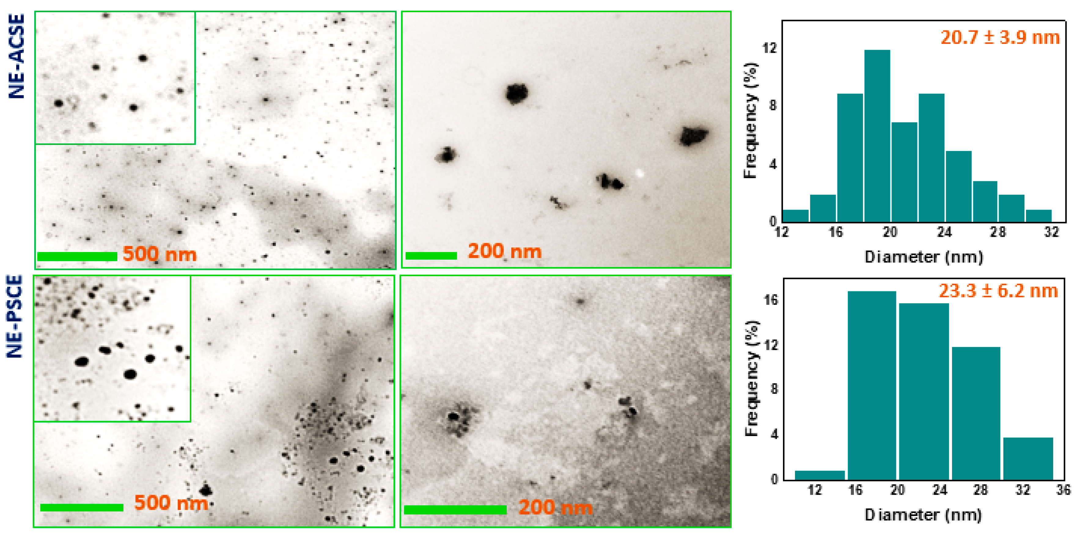
3.2. Morphological Investigation of NE-ACSE and NE-PSCE

The sizes and shapes of the as-prepared NE-ACSE and NE-PSCE were recorded with the help of a transmission electron microscope (TEM), as shown in Figure 1. The images of the NEs recorded with the TEM depict that the prepared NE-ACSE and NE-PSCE are spherical, with nanosizes of 20.7 ± 3.9 nm and 23.3 ± 6.2 nm, respectively.

3.3. Distribution of Particle Sizes and ζ-Potentials of Nanoemulsions NE-PSCE and NE-ACSE

DLS was used to measure the prepared NEs’ particle size distributions and ζ-potentials, as shown in Figure 2. According to the obtained results, NE-ACSE showed a hydrodynamic diameter of 68.3 ± 26.6 nm, whereas NE-PSCE exhibited 69.9 ± 27.9 nm. Moreover, both nanoemulsion samples were imaged as displayed in Figure S3a,b. For the ζ-potentials, as shown in Figure 2c (Figure S4), NE-ACSE displayed −41.4 ± 7.5 mV, whereas NE-PSCE showed −28.0 ± 5.8 mV. The negative charges of the NEs could be attributed to the polyphenols which are available in both ACS and PSC extracts, and also to using Tween 80 (a non-ionic surfactant) and oleic acid as an anionic surfactant in addition to PEG [52]. It was reported that, for nanomaterials with ζ-potentials over ±30 mV, it is necessary to stabilize the NPs in the aqueous suspension [53,54,55]. And it can be observed that NE-ACSE is more stable than NE-PSCE. The particle sizes depicted by the TEM analysis are smaller than in the DLS measurements, as in DLS, nanoparticles could be aggregated to form larger particles. Furthermore, the suspended nanoparticle solution swelled in the solvent used to prepare samples for DLS analysis. The values of the polydispersity index (PDI) were 0.332 and 0.321 (<0.5), which indicates that the NE particles are distributed uniformly when suspended in the solution.

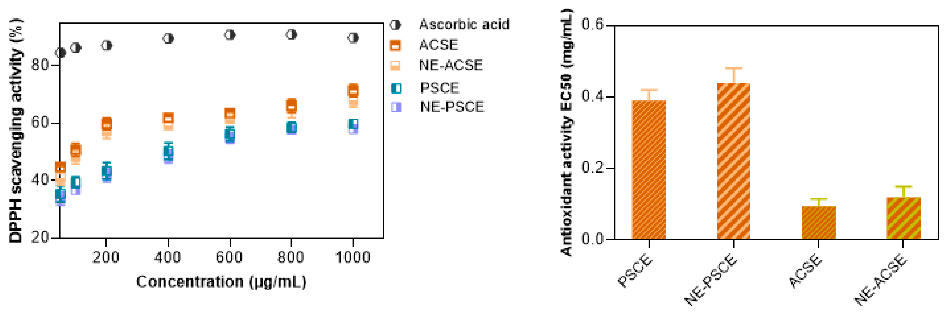
3.4. DPPH Scavenging Activity Measurements of Nanoemulsions

The antioxidant activities of the tested samples were evaluated as shown in Figure 3 using a DPPH radical scavenging assay at various concentrations of the extracts and their nanoemulsions, while recording the efficiency concentration required to obtain a 50% antioxidant effect (EC50). The antioxidant activities of the encapsulated ACSE and PSCE were slightly lower than those of the corresponding free extracts. It is better for bioactive materials to be bio-accessible and released in a sustained manner when displaying the related bioactivity. Though the antioxidant properties of the encapsulated extracts are less than free, the nanoemulsified components are protected against rapid degradation and loss, and their antioxidant activities are maintained efficiently [56]. Such protection could be achieved against rapid pepsin digestion or during storage against light, heat, and oxygen, which encourage the chemical breakdown of the bioactive compounds, lowering their biological value [57,58,59].
Such findings are consistent with a previous study which found that a nanoemulsion of curcumin solid lipid particles provided antioxidant activity using a FRAP assay, with a value of 0.996 ± 0.07 mM L-ascorbic acid equivalent; in addition, the free curcumin displayed antioxidant activity with a value of 2.504 ± 0.06 mM L-ascorbic acid equivalent [60]. The antioxidant properties of ACS extract could be attributed to it being rich in antioxidants including phenolic compounds and flavonoids [61]. The same is true for PSC extract, as its antioxidant activity is a result of high polyphenol content [21].

3.5. Effects of NE-PSCE and NE-ACSE on CCL4-Induced Liver Fibrosis in Rat Model

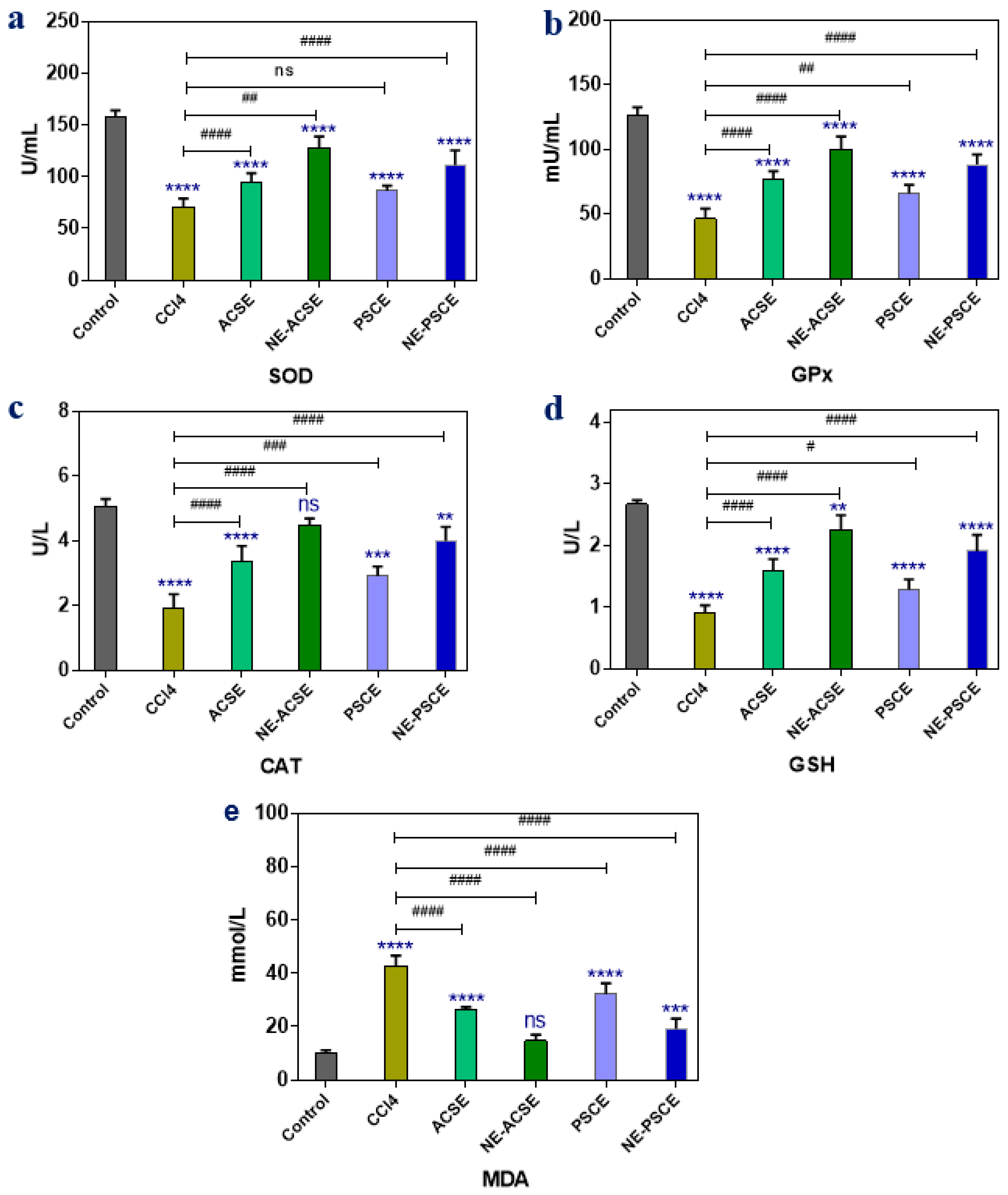
3.5.1. Antioxidant Enzymes and MDA

The effects of PSCE, ACSE, and their nanoemulsion forms, NE-PSCE and NE-ACSE, on chronic liver disease-afflicted rats’ antioxidant enzyme and free radical levels, including those of malondialdehyde (MDA), glutathione peroxidase (GPx), catalase (CAT), reduced glutathione (GSH), and superoxide dismutase (SOD), were observed, as illustrated in Figure 4. When compared to the control group, the CCL4 group demonstrated a significant decrease in MDA levels and a significant decrease in the values of SOD, GPX, GSH, and CAT. Treatment with both PSCE and ACSE increased the levels of SOD, GPx, CAT, and GSH while decreasing MDA in comparison to the chronic liver group (CCl4). The administration of the NE-ACSE and NE-PSCE nanoemulsion forms to ill animals resulted in significant improvements in SOD, GPx, GSH, and CAT levels. In this case, NE-ACSE may be able to improve things more than the other treatments.

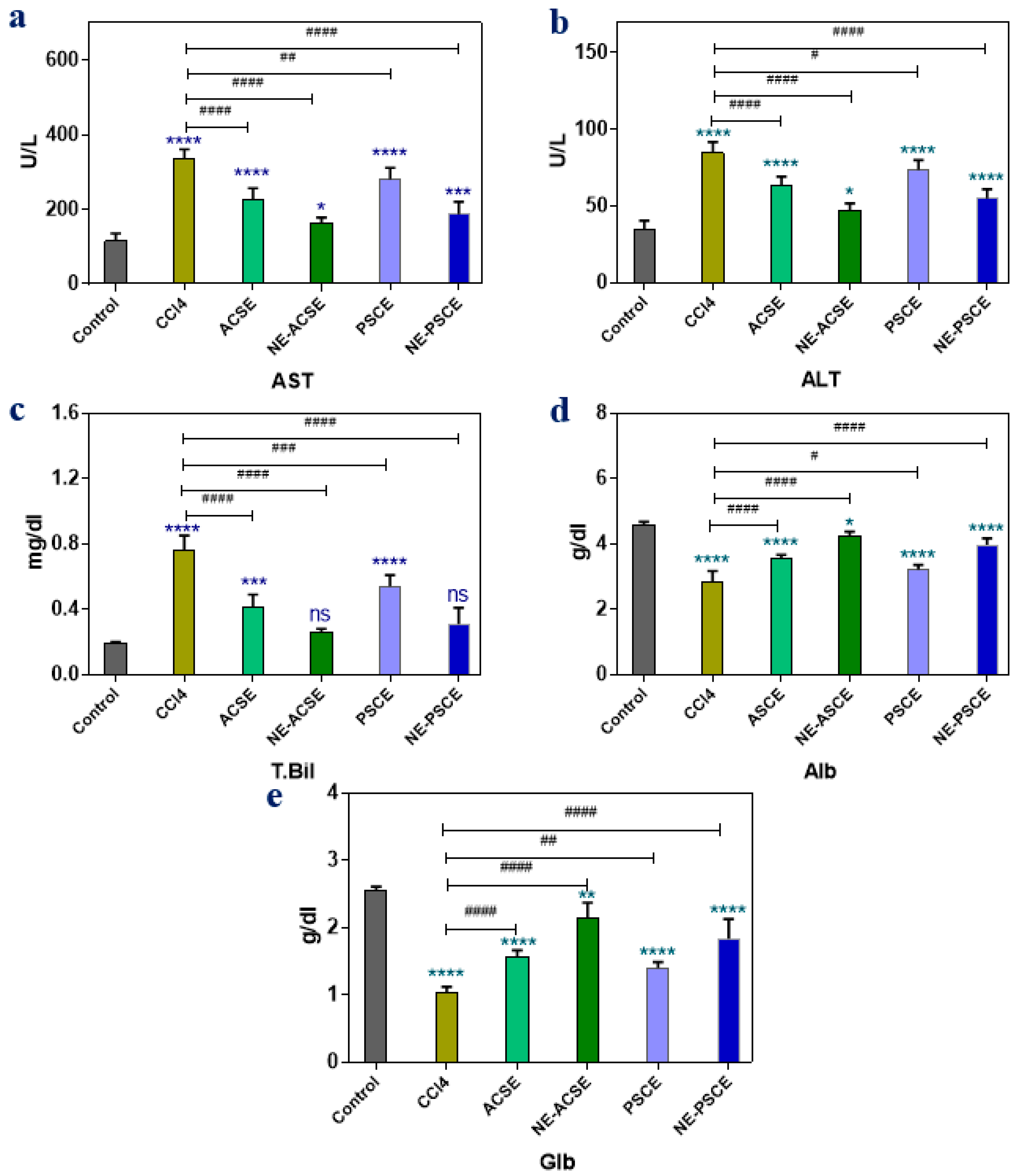
3.5.2. Liver Functions

The activities of alanine aminotransferases (ALTs), aspartate aminotransferases (ASTs), albumin (Alb), globulin (GLb), and total bilirubin (T. Bil) are among the liver’s activities that are tracked in the chronic liver disease rat groups treated with date palm seed coffee extract (PSCE), Arabica seed coffee extract (ACSE), and their corresponding nanoemulsions, including nanoemulsions of date palm seed coffee extract (NE-PSCE) and of Arabica seed coffee extract (NE-ACSE), as displayed in Figure 5. The control rat group showed significant decreases in AST, ALT, and T. Bil while showing significant increases in Alb and Glb compared with the group of non-treated diseased animals (CCl4). Rats with persistent liver damage treated with PSCE and ACSE had significantly lower levels of AST, ALT, and T. Bil than those in the group that was not treated (CCl4), while the levels of Alb and Glb were increased significantly in the PSCE and ACSE groups compared to the CCl4 group. In animals treated with nanoemulsions, including NE-PSCE and NE-ACSE, the levels of AST, ALT, and T. Bil decreased more than in either the PSCE or ACSE groups, whereas levels of Alb and Glb increased more than in the PSCE and ACSE groups. It can be observed that NE-ACSE and NE-PSCE were more effective in improving the levels of AST, ALT, T-Bil, Alb, and Glb for animals with chronic liver disease. In particular, NE-ACSE provided more effective treatment than other groups.

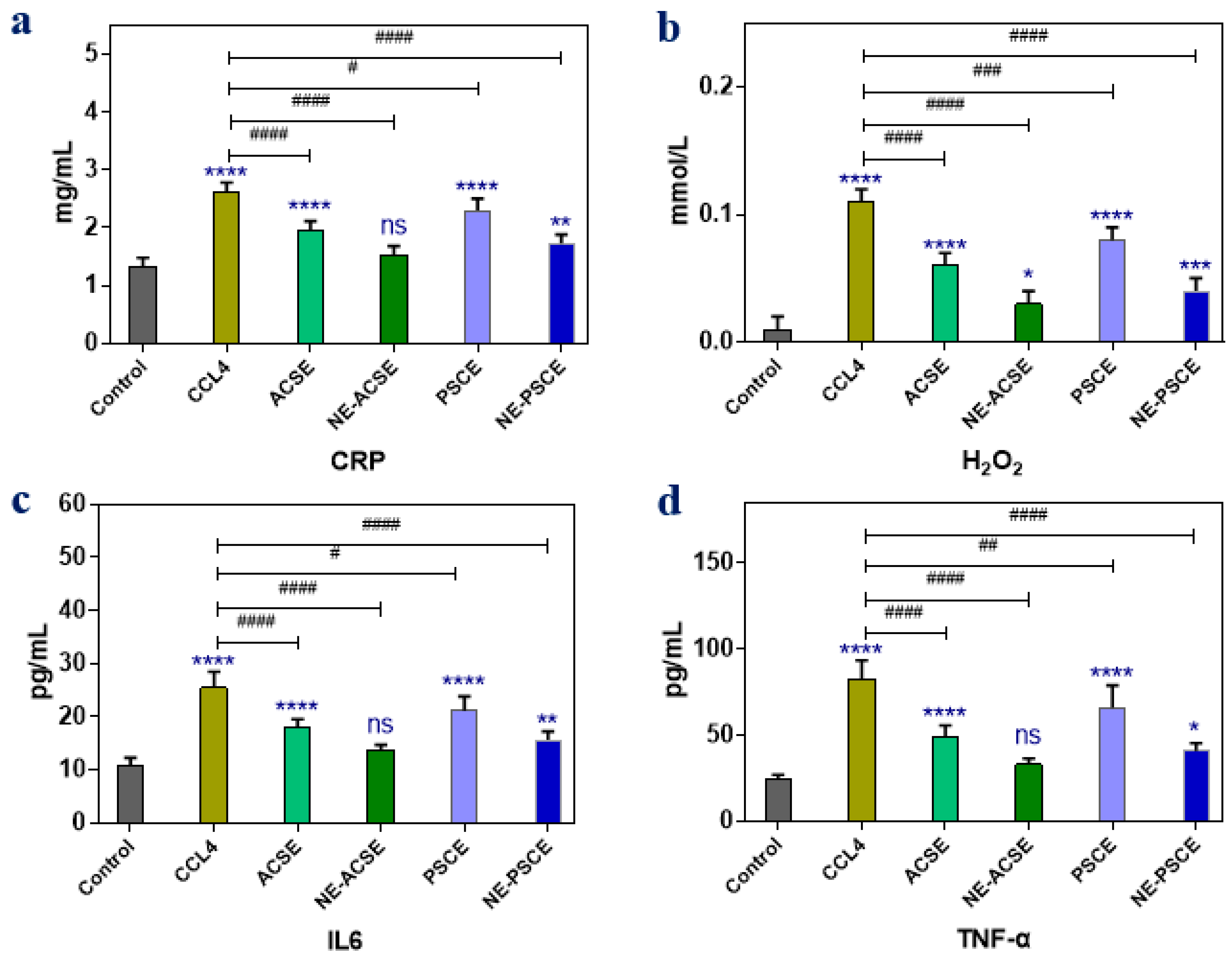
3.5.3. Anti-Inflammatory Parameters

Anti-inflammatory parameters including C-reactive protein (CRP), hydrogen peroxide (H2O2), interleukin-6 (IL6), and tumor necrosis factor alpha (TNF-α) were recorded to show the effects of extracts in normal forms or nanoemulsion forms on the levels of such inflammatory parameters, as displayed in Figure 6. It can be observed that the inflammatory markers CRP, H2O2, IL6, and TNF-α were increased in the diseased liver group compared to control group, whereas the administration of extracts as ACSE and PSCE could minimize the levels of such parameters compared to those of the non-treated diseased liver group. In addition, the reduction in inflammatory markers was clearly observed when animals were treated with the nanoemulsion forms of the extracts, NE-PSCE and NE-ACSE. In accordance with the above results, NE-ACSE exhibited lower levels of the inflammatory parameters, as in the control.

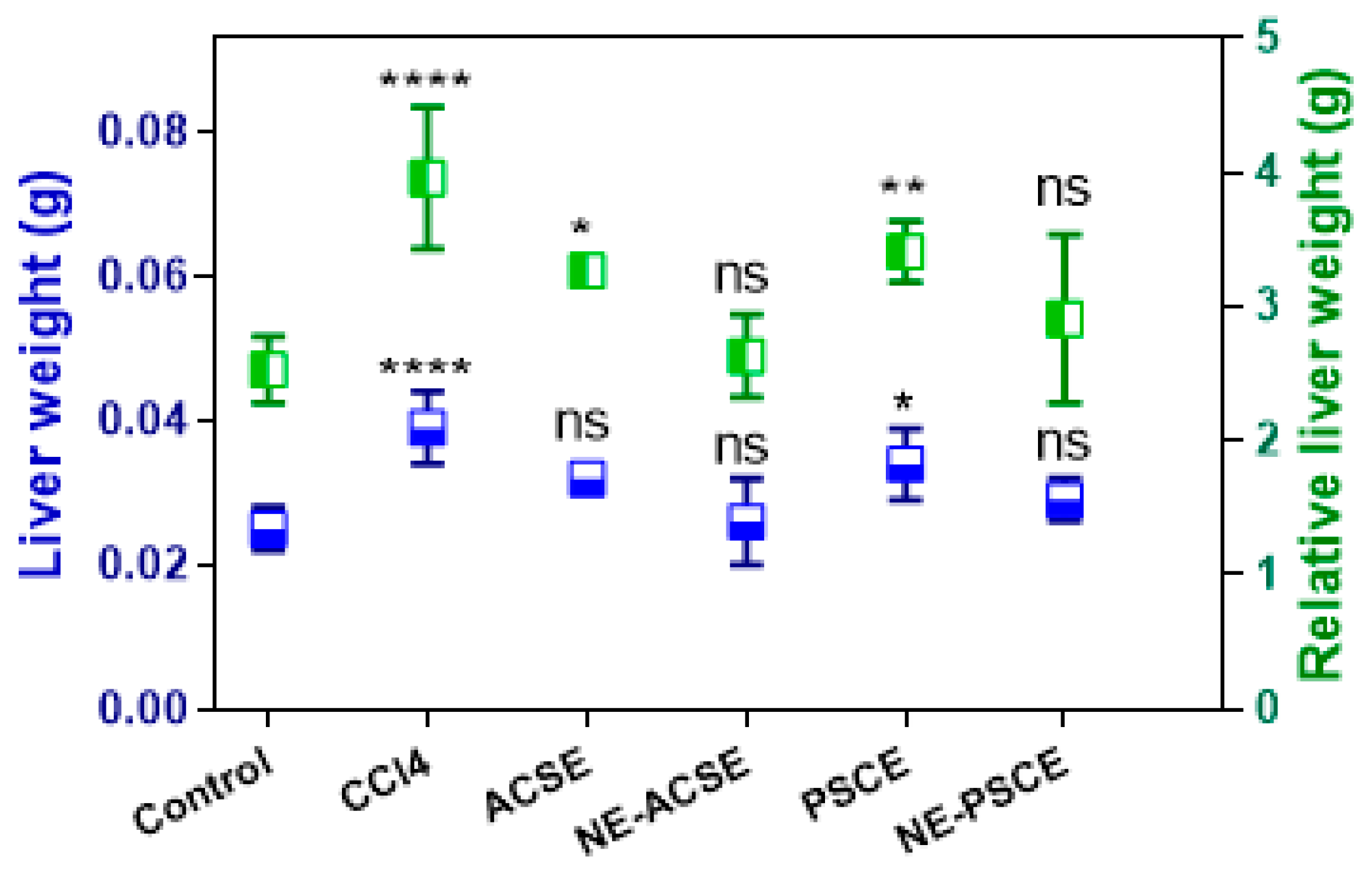
3.5.4. Liver Weight, Relative Liver Weight, Weight Gain, and Food Intake

Liver weight and relative liver weight were recorded, as can be observed in Figure 7. According to the obtained results, the liver weight increased upon giving CCl4 to the diseased animal group with chronic liver disease in comparison with the control group. This was obvious in terms of the relative liver weight as well. As a result of treatment with extracts of both PSCE and ACSE, both liver weight and relative weight were decreased compared to the diseased CCl4 group. In addition, these decreases in the liver weight and relative liver weight were more obvious in diseased animals treated with NE-PSCE and NE-ACSE compared to the diseased non-treated group. Consistent with above results, NE-ACSE provided a more prominent effect.
The effects of ACSE, PSCE, and their nanoemulsion forms NE-ACSE and NE-PSCE on body weight gain and food intake in the chronic liver disease rat groups were determined as exhibited in Table 3. The effects of chronic liver disease on the diseased non-treated animals were negative in terms of health, feeding intake, and consequently weight gain, and such parameters were decreased significantly compared to the control group. On the other hand, weight gain and food intake were increased upon provision of the diseased animals with extracts of ACSE and PSCE. Moreover, the nanoemulsions of NE-ACSE and NE-PSCE could significantly improve weight gain and food intake compared to the diseased non-treated group. The nanoemulsions were more effective than the free extracts. Similar to the above results, NE-ACSE improved the status of the diseased animals, making it close to that of the control group.

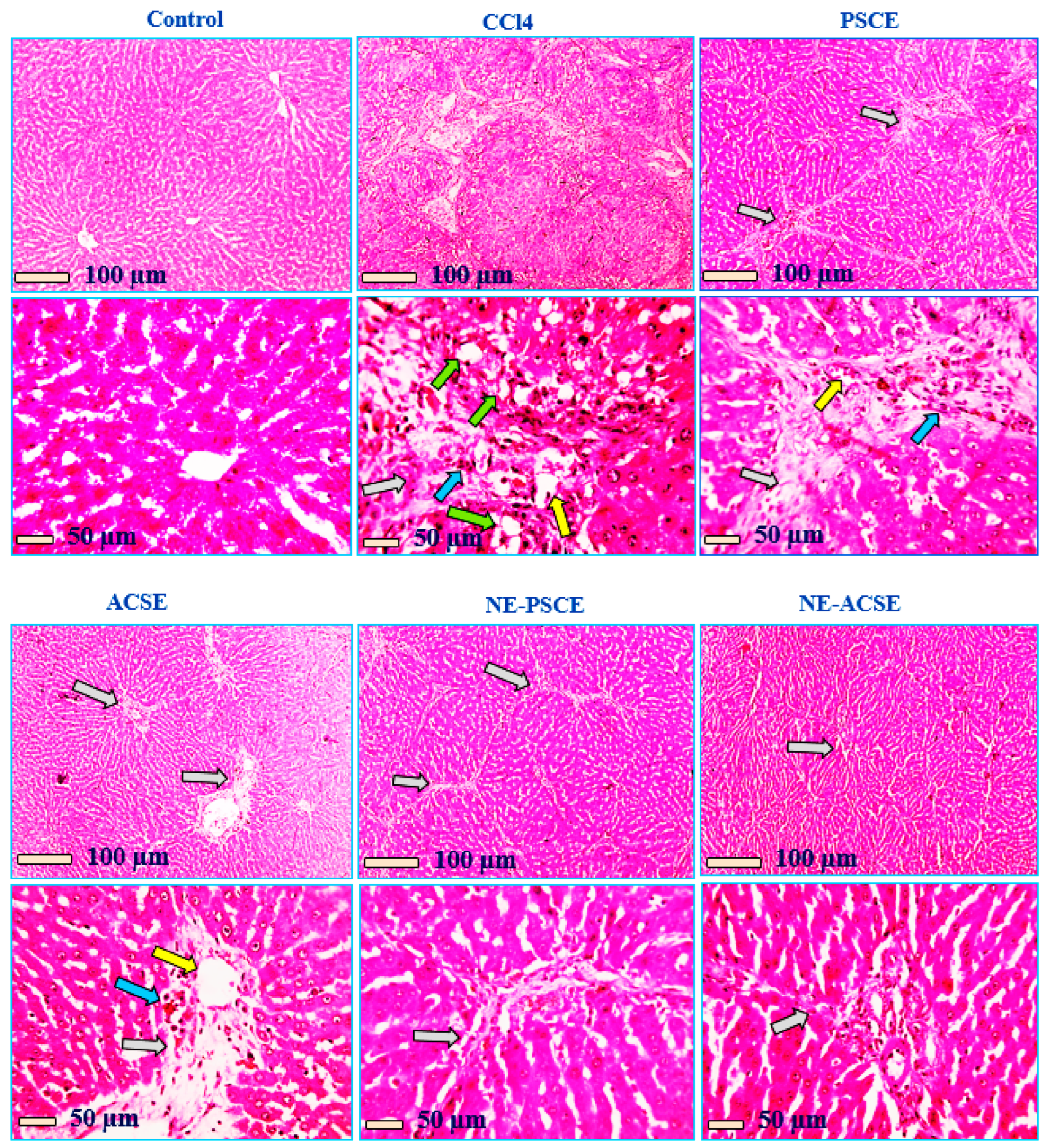
3.5.5. Histopathological Analysis

Histopathological analysis was performed for all groups—control, CCl4, PSCE, ACSE, NE-ACSE, and NE-PSCE—as can be observed in Figure 8. Under a microscope, hepatic slices stained with HE reveal a regular configuration of hepatic cords around central veins, together with normal portal regions and sinusoids in the normal control group. Hepatic sections from the positive group that received CCL4 show marked distension of portal areas due to fibrosis, leukocyte cell and hemosiderin-laden macrophage infiltration (blue arrow), congested blood vessels (yellow arrow), and thick anastomosing fibrous tissue extension from portal areas (grey arrow). Green arrows indicate ballooning degeneration in hepatocytes. Hepatic slices from the PSCE-treated group exhibit reduced portal area distension (grey arrow), a small infiltration of leukocyte cells and hemosiderin-loaded macrophages (blue arrow), less congested blood vessels (yellow arrows), and a long, thin fibrous tissue extension that anastomoses from the portal areas. Hepatic sections from the ACSE-treated group show greatly decreased distension of portal areas (grey arrows), fewer leukocytic cell infiltrations (blue arrows), few dilated blood vessels (yellow arrow), and few short non-anastomosing fibrous tissue extensions from portal areas. Hepatic sections from the NE-PSCE-treated group show few very short and very thin non-anastomosing fibrous tissue extensions from portal areas (blue arrows). Hepatic sections from the NE-ACSE-treated group show very few short and very thin non-anastomosing fibrous tissue extensions from portal areas (grey arrow).

4. Discussion

Regular coffee’s beneficial components have led to its classification as a functional food [62,63,64]. Particularly in wealthy nations, coffee is commonly consumed. Coffees are classified into broad categories according to how they are roasted or processed, which has an impact on the finished product’s chemical makeup. There are thus various varieties, including filtered and unfiltered coffee, as well as brewed, expresso, infused, instant, boiling, and decaffeinated coffee [65].
Coffea canephora (robusta) and Coffea arabica (C. arabica) are regarded as the two main species of coffee consumed globally. C. arabica is grown extensively, accounting for 70% of the world’s coffee production. One possible explanation for C. arabica’s exceptional quality is its organoleptic characteristics [66]. In Yunnan Province, China, C. arabica is widely used as a plant with significant medicinal value. Many of the chemical components of coffee have been separated thanks to advancements in contemporary technology and experimental methodologies. These components comprise sterols, taste compounds, alkaloids, flavonoids, terpenes, phenolic acids and their derivatives, and other substances. Numerous pharmacological actions, including those that are antioxidative, anti-inflammatory, anticancer, antidiabetic, liver-protecting, and neuroprotective, are a result of the chemical variety of its constituents. Caffeine, as a main constituent of C. arabica, was reported for its antioxidant properties, and in a wide range of dosages, caffeine is a safe xanthine alkaloid for human consumption [67]. The Food and Drug Administration classified caffeine as safe because the average adult’s hazardous amount is greater than 10 g [68]. Additionally, it has been reported that caffeine reduces oxidative stress, which may lessen liver damage and enhance neurological symptoms in rat models of hepatic encephalopathy [69]. Moreover, previous studies demonstrated that caffeine can partially prevent alcoholic liver fibrosis in rats, a condition that is assumed to be mediated by the cAMP-PKA-CREB signaling pathway [70,71]. It should be noted that the primary goal of the research is to identify the chemicals in C. arabica [72]. C. arabica beans are medium-roasted to produce Arabian coffee. It is a well-liked beverage in most Middle Eastern Arab nations [73]. Natural active substances found in abundance in C. arabica include sucrose, trigonelline, and chlorogenic acid [74]. C. arabica provides several pharmacological properties such as anti-inflammatory [75], antimicrobial [76], anticancer [26,77,78], and antioxidant properties [79]. Coffee attracts attention due to its significant application in clinical trials, in particular in inhibition of and protection against both alcoholic and non-alcoholic liver cirrhosis [80,81,82].
Date seed extract shows health benefits due to its antioxidant bioactive compounds including flavonoids, phenolic compounds, and vitamins, which have the capability to scavenge free radicals [83] and provide protection from hepatorenal toxicity [84]. GC/MS was used to examine Al-Baha date palm kernel (AB-DPK) extract. Antioxidants like 1-Trilinolein and (Z,Z) 3Dioctadecenoyl Glycerol as well as anti-tumor, anti-inflammatory, and antiviral medicines like botulin were among the compounds discovered in the investigation [85,86]. Extracts of the flesh and pits of dates were evaluated to ameliorate CCl4-induced hepatotoxicity in a rat model, and the extracts displayed significant hepatoprotective activity for animals that received the date extracts [87]. In a different study, when thioacetamide-treated animals were compared to control rats, there were notable increases in biochemical indicators of liver injury, such as ALT, AST, ALP, and total bilirubin, and a decrease in albumin levels. In addition, there was a substantial decrease in liver enzymes and an increase in serum albumin (p < 0.05) after treatment with extracts from palm dates. The hepatoprotective properties of palm date extracts may be due to their concentration of quercetin, which possesses potent antioxidant properties [88]. The power of P. dactylifera seed extract to scavenge DPPH could be attributed to the high antioxidant capacity of the extract contents. It was demonstrated that the antioxidant properties of date palms could have emerged from available compounds such as selenium, vitamin E, ascorbic acid, flavonoids, tannins, and other phenolic compounds [89,90].
The levels of intracellular enzymes, including AST and ALT as well as T. Bil, are biomarkers and significantly sensitive to hepatic injuries [91]. As a result of leakage of the cell membrane associated with a loss of membrane integrity, the activities of ALT and AST are increased; meanwhile, the impairment in the biliary function of the liver and the associated damages to hepatic tissues result in an increase in the level of T-Bil [92,93]. ALT, AST, and T-Bil serum levels in rats exposed to PSCE or ACSE were reduced compared to those in the chronic liver disease group in this study. Such results attest to the safety profile and protective therapy of both ACSE and PSCE in restoring liver functions. In a previous study, methanolic and aqueous extracts of Phoenix dactylifera were demonstrated as protective products against oxidative and hepatic injuries induced by paracetamol. According to the study, the extracts worked through blocking the oxidative stress caused by the paracetamol, thus ameliorating the induced hepatocellular injury and elevated serum ALT and AST [94]. In another study which used aqueous Ajwa date fruit extract as an anticancer agent to protect against and treat hepatocellular carcinoma induced using diethylnitrosamine in a rat model, the extract could improve liver functions due to its antioxidant activity, and the levels of ALT and AST were reduced [95]. As a result of the capability to improve kidney and liver functions that emerges from their potent antioxidant properties, there is the possibility of using date seed extracts as anticancer agents against colorectal and hepatocellular carcinoma cell lines, but further investigations are still required [96].
According to the obtained results of this study, the Coffea arabica and Phoenix dactylifera nanoemulsions NE-ACSE and NE-PSCE provided more significant therapeutic effects against CCl4-induced chronic liver disease in a rat model compared to the free extracts. These results were obvious due to reductions in liver intracellular enzymes such as AST, ALT, and T. Bil. Also, the inflammatory parameters, such as IL6, TNF alpha, CRP, and H2O2, were significantly decreased for diseased animals that received either nanoemulsion, NE-ACSE or NE-PSCE, compared to the CCl4 group and those provided with free extracts. Also, antioxidant enzymes such as SOD, CAT, GSH, and GPx were improved significantly. Furthermore, the health status parameters, including food intake, weight gain, and food efficiency rate, were improved more by using nanoemulsions than by using free extracts. Such results were confirmed and were consistent, as displayed in histopathological examinations, where the nanoemulsions NE-ACSE and NE-PSCE could decrease the cellular infiltrations and damages more than the free extracts. The obtained results, which indicate the high efficiency of nanoemulsions of NE-ACSE or NE-PSCE as protective agents in induced chronic liver disease, could be attributed to the high stability of the extracts when they are nanoemulsified, and the slow and sustained release of the extracts over a longer period than that of the free extracts. Because of the special qualities of the involved nanosized droplets, such as large surface area, nanoemulsions are being studied in the fields of food and healthcare [97]. Furthermore, texture, stability, absorption, absorbability, fortification, and bioavailability will all be improved by the extracts’ active components in nanoemulsified latex [98,99,100]. Additionally, nanoemulsions can protect natural components from degradation, thereby extending their shelf life and preserving their efficacy. Bioactive substances can eventually deteriorate due to direct exposure to external influences including heat, light, and oxygen, and this is prevented by encapsulation. Furthermore, nanoemulsions can improve the targeted delivery of the natural components of PSCE and ACSE to the targeted affected organ/tissue within the body.

5. Conclusions

The present study reported on nanoformulations of ACSE and PSCE with the aid of oleic acid and the Tween 80 system. To the best of our knowledge, the influence of the developed nanoemulsions NE-PSCE and NE-ACSE on induced liver fibrosis in a rat model was investigated herein for the first time. The major findings reveal that both NE-ACSE and NE-PSCE were allocated in nanoscales with negative ζ-potentials. The antioxidant activity was determined, and ACS and PSC extracts and their nanoemulsions displayed antioxidant properties. In a rat model with liver fibrosis induced via CCl4, the influence of both nanoemulsions was demonstrated in the aspects of inflammatory parameters, liver functions, antioxidant enzymes, and histopathological analysis. In comparison to normal extracts of ACSE and PSCE, the nanoemulsions NE-ACSE and NE-PSCE provided significant improvements in antioxidant enzymes and decreases in inflammatory parameters, and the liver functions of ALT and AST were decreased to be close to those of the control group. It can be concluded that nanoemulsions of ACSE and PSCE provide a new avenue as a therapeutic approach to liver diseases, and further studies are encouraged to obtain maximum efficiency of treatments via the combination of both extracts in nanoforms.

Supplementary Materials

The following supporting information can be downloaded at: https://www.mdpi.com/article/10.3390/antiox13030355/s1. Figure S1. Compounds Identified in the ACS extract using GC-MS; Figure S2. Compounds Identified in the PSC extract using GC-MS; Figure S3. Nanoemulsion of (a) NE-ACSE and (b) NE-PSCE; Figure S4. ζ-potential (a, b) of the developed NE-ACSE and NE-PSCE, respectively.

Author Contributions

Conceptualization, N.S.Z. and H.H.A.; Methodology, E.S.A., M.A.M.H., R.A.A. and F.M.E.-E.; Software, M.A.M.H., N.A.O., S.E.A. and R.A.A.; Formal analysis, E.S.A., H.M.B., M.A.M.H. and F.M.E.-E.; Investigation, N.A.O., N.S.Z. and H.H.A.; Resources, H.M.B., N.A.O., S.E.A., N.S.Z. and F.M.E.-E.; Data curation, E.S.A., H.M.B., M.A.M.H. and S.E.A.; Writing—original draft, N.A.O., S.E.A., N.S.Z., H.H.A. and F.M.E.-E.; Writing—review & editing, E.S.A., H.M.B. and M.A.M.H.; Visualization, R.A.A. and F.M.E.-E.; Supervision, E.S.A., M.A.M.H., H.H.A. and F.M.E.-E.; Project administration, H.M.B. and N.S.Z. All authors have read and agreed to the published version of the manuscript.

Funding

This research was funded by the Deanship of Scientific Research at the University of Tabuk in Saudi Arabia, grant number S-1443-0030.

Institutional Review Board Statement

Not applicable.

Informed Consent Statement

Not applicable.

Data Availability Statement

Data is contained within the article and Supplementary Materials.

Acknowledgments

The authors extend their appreciation to the Deanship of Scientific Research at the University of Tabuk in Saudi Arabia for funding this research work through the project number S-1443-0030.

Conflicts of Interest

The authors declare no conflicts of interest.

References

  1. Xu, J.-H.; Yu, Y.-Y.; Xu, X.-Y. Management of chronic liver diseases and cirrhosis: Current status and future directions. Chin. Med. J. 2020, 133, 2647–2649. [Google Scholar] [CrossRef] [PubMed]
  2. Manka, P.; Zeller, A.; Syn, W.-K. Fibrosis in Chronic Liver Disease: An Update on Diagnostic and Treatment Modalities. Drugs 2019, 79, 903–927. [Google Scholar] [CrossRef] [PubMed]
  3. Kisseleva, T.; Brenner, D. Molecular and cellular mechanisms of liver fibrosis and its regression. Nat. Rev. Gastroenterol. Hepatol. 2021, 18, 151–166. [Google Scholar] [CrossRef]
  4. Dai, C.; Xiao, X.; Li, D.; Tun, S.; Wang, Y.; Velkov, T.; Tang, S. Chloroquine ameliorates carbon tetrachloride-induced acute liver injury in mice via the concomitant inhibition of inflammation and induction of apoptosis. Cell Death Dis. 2018, 9, 1164. [Google Scholar] [CrossRef]
  5. Li, J.; Tuo, B. Current and Emerging Approaches for Hepatic Fibrosis Treatment. Gastroenterol. Res. Pract. 2021, 2021, 6612892. [Google Scholar] [CrossRef]
  6. Li, Q.; Zhang, J.; Xiao, S.; Hu, M.; Cheng, J.; Yao, C.; Zhuang, Q. The impact of liver fibrosis on the progression of hepatocellular carcinoma via a hypoxia-immune-integrated prognostic model. Int. Immunopharmacol. 2023, 125, 111136. [Google Scholar] [CrossRef] [PubMed]
  7. Ullah, A.; Chen, G.; Yibang, Z.; Hussain, A.; Shafiq, M.; Raza, F.; Liu, D.; Wang, K.; Cao, J.; Qi, X. A new approach based on CXCR4-targeted combination liposomes for the treatment of liver fibrosis. Biomater. Sci. 2022, 10, 2650–2664. [Google Scholar] [CrossRef]
  8. Poilil Surendran, S.; Thomas, R.; Moon, M.-J.; Jeong, Y. Nanoparticles for the treatment of liver fibrosis. Int. J. Nanomed. 2017, 12, 6997–7006. [Google Scholar] [CrossRef]
  9. Bai, X.; Su, G.; Zhai, S. Recent Advances in Nanomedicine for the Diagnosis and Therapy of Liver Fibrosis. Nanomaterials 2020, 10, 1945. [Google Scholar] [CrossRef]
  10. Eftekhari, A.; Arjmand, A.; Asheghvatan, A.; Švajdlenková, H.; Šauša, O.; Abiyev, H.; Ahmadian, E.; Smutok, O.; Khalilov, R.; Kavetskyy, T.; et al. The Potential Application of Magnetic Nanoparticles for Liver Fibrosis Theranostics. Front. Chem. 2021, 9, 674786. [Google Scholar] [CrossRef]
  11. Suntres, Z.E. Liposomal Antioxidants for Protection against Oxidant-Induced Damage. J. Toxicol. 2011, 2011, 152474. [Google Scholar] [CrossRef]
  12. Kumar, N.; Verma, A.; Mandal, A. Formation, characteristics and oil industry applications of nanoemulsions: A review. J. Pet. Sci. Eng. 2021, 206, 109042. [Google Scholar] [CrossRef]
  13. Kumar, N.; Mandal, A. Surfactant Stabilized Oil-in-Water Nanoemulsion: Stability, Interfacial Tension, and Rheology Study for Enhanced Oil Recovery Application. Energy Fuels 2018, 32, 6452–6466. [Google Scholar] [CrossRef]
  14. Tadros, T.; Izquierdo, P.; Esquena, J.; Solans, C. Formation and stability of nano-emulsions. Adv. Colloid Interface Sci. 2004, 108–109, 303–318. [Google Scholar] [CrossRef]
  15. Wilson, R.J.; Li, Y.; Yang, G.; Zhao, C.-X. Nanoemulsions for drug delivery. Particuology 2022, 64, 85–97. [Google Scholar] [CrossRef]
  16. Ansari, V.; Ahmad, U.; Upadhyay, T.; Sultana, N.; Akhtar, J. Exploring Nanoemulsion for Liver Cancer Therapy. Curr. Cancer Ther. Rev. 2020, 16, 260–268. [Google Scholar] [CrossRef]
  17. Sravanthi, V.; Preethi Pallavi, M.C.; Bonam, S.R.; Sathyabama, S.; Sampath Kumar, H.M. Oleic acid nanoemulsion for nasal vaccination: Impact on adjuvanticity based immune response. J. Drug Deliv. Sci. Technol. 2015, 28, 56–63. [Google Scholar] [CrossRef]
  18. de Lourdes Pérez-González, M.L.; González-de la Rosa, C.H.; Pérez-Hernández, G.; Beltrán, H.I. Nanostructured oleic acid/polysorbate 80 emulsions with diminished toxicity in NL-20 cell line: Insights of potential drug carriers. Colloids Surf. B Biointerfaces 2020, 187, 110758. [Google Scholar] [CrossRef] [PubMed]
  19. Metoui, M.; Essid, A.; Bouzoumita, A.; Ferchichi, A. Chemical Composition, Antioxidant and Antibacterial Activity of Tunisian Date Palm Seed. Pol. J. Environ. Stud. 2018, 28, 267–274. [Google Scholar] [CrossRef] [PubMed]
  20. Fikry, M.; Yusof, Y.; Al-awaadh, A.; Abdul Rahman, R.; Chin, N. Prediction of the shelf-life of date seeds brew by integration of acceptability and quality indices. J. Food Meas. Charact. 2020, 14, 1158–1171. [Google Scholar] [CrossRef]
  21. Souda, B.; Rami, R.; Jalloul, B.; Mohamed, D. Roasted date palm seeds (Phoenix dactylifera) as an alternative coffee: Chemical composition and bioactive properties. Biomass Convers. Biorefinery 2022, 12, 3771–3781. [Google Scholar] [CrossRef]
  22. Niazi, S.; Khan, I.; Rasheed, S.; Niazi, F.; Shoaib, M.; Raza, H.; Iqbal, M. An Overview: Date Palm Seed Coffee, a Functional Beverage. Int. J. Public Health 2017, 2, 18–25. [Google Scholar]
  23. Fikry, M.; Yusof, Y.A.; Al-Awaadh, A.M.; Abdul Rahman, R.; Chin, N.L.; Ghazali, H.M. Antioxidative and Quality Properties of Full-Fat Date Seeds Brew as Influenced by the Roasting Conditions. Antioxidants 2019, 8, 226. [Google Scholar] [CrossRef] [PubMed]
  24. Bentrad, N.; Hamida-Ferhat, A. Chapter 22—Date palm fruit (Phoenix dactylifera): Nutritional values and potential benefits on health. In The Mediterranean Diet, 2nd ed.; Preedy, V.R., Watson, R.R., Eds.; Academic Press: Cambridge, MA, USA, 2020; pp. 239–255. [Google Scholar] [CrossRef]
  25. Dghaim, R.; Hammami, Z.; Al Ghali, R.; Smail, L.; Haroun, D. The Mineral Composition of Date Palm Fruits (Phoenix dactylifera L.) under Low to High Salinity Irrigation. Molecules 2021, 26, 7361. [Google Scholar] [CrossRef] [PubMed]
  26. Nigra, A.D.; Teodoro, A.J.; Gil, G.A. A Decade of Research on Coffee as an Anticarcinogenic Beverage. Oxid. Med. Cell. Longev. 2021, 2021, 4420479. [Google Scholar] [CrossRef] [PubMed]
  27. Lee, L.K.; Mhd Rodzi, N.A. Addressing the Neuroprotective Actions of Coffee in Parkinson’s Disease: An Emerging Nutrigenomic Analysis. Antioxidants 2022, 11, 1587. [Google Scholar] [CrossRef]
  28. Degertekin, B. Regular coffee intake improves liver enzyme levels and liver histology in patients with chronic alcohol consumption, non-alcoholic fatty liver and non-alcoholic steatohepatitis: Report of 259 cases. Hepatol. Forum 2020, 1, 88–96. [Google Scholar] [CrossRef]
  29. Heath, R.; Brahmbhatt, M.; Tahan, A.; Ibdah, J.; Tahan, V. Coffee: The magical bean for liver diseases. World J. Hepatol. 2017, 9, 689. [Google Scholar] [CrossRef]
  30. Kolb, H.; Martin, S.; Kempf, K. Coffee and Lower Risk of Type 2 Diabetes: Arguments for a Causal Relationship. Nutrients 2021, 13, 1144. [Google Scholar] [CrossRef]
  31. Yildirim, S.T.; Oztop, M.H.; Soyer, Y. Cinnamon oil nanoemulsions by spontaneous emulsification: Formulation, characterization and antimicrobial activity. LWT 2017, 84, 122–128. [Google Scholar] [CrossRef]
  32. Wang, S.; Liang, X.; Zhao, W.; Mi, X.; Zhang, C.; Zhang, W.; Cheng, Y.; Wang, L.; Jiang, Y. Preparation of nanoemulsion of grapefruit seed extract and evaluation of its antibacterial activity. J. Food Process. Preserv. 2022, 46, e16197. [Google Scholar] [CrossRef]
  33. Singh, R.; Murthy, K.; Jayaprakasha, G. Studies on the Antioxidant Activity of Pomegranate (Punica granatum) Peel and Seed Extracts Using In Vitro Models. J. Agric. Food Chem. 2002, 50, 81–86. [Google Scholar] [CrossRef] [PubMed]
  34. Brem, B.; Seger, C.; Pacher, T.; Hartl, M.; Hadacek, F.; Hofer, O.; Vajrodaya, S.; Greger, H. Antioxidant dehydrotocopherols as a new chemical character of Stemona species. Phytochemistry 2004, 65, 2719–2729. [Google Scholar] [CrossRef] [PubMed]
  35. Iredale, J.P.; Benyon, R.C.; Pickering, J.; McCullen, M.; Northrop, M.; Pawley, S.; Hovell, C.; Arthur, M.J. Mechanisms of spontaneous resolution of rat liver fibrosis. Hepatic stellate cell apoptosis and reduced hepatic expression of metalloproteinase inhibitors. J. Clin. Investig. 1998, 102, 538–549. [Google Scholar] [CrossRef] [PubMed]
  36. Elzoheiry, A.; Ayad, E.; Omar, N.; Elbakry, K.; Hyder, A. Anti-liver fibrosis activity of curcumin/chitosan-coated green silver nanoparticles. Sci. Rep. 2022, 12, 18403. [Google Scholar] [CrossRef]
  37. Ozden, M.; Maral, H.; Akaydın, D.; Cetınalp, P.; Kalender, B. Erythrocyte glutathione peroxidase activity, plasma malondialdehyde and erythrocyte glutathione levels in hemodialysis and CAPD patients. Clin. Biochem. 2002, 35, 269–273. [Google Scholar] [CrossRef]
  38. Paglia, D.E.; Valentine, W.N. Studies on the quantitative and qualitative characterization of erythrocyte glutathione peroxidase. J. Lab. Clin. Med. 1967, 70, 158–169. [Google Scholar] [CrossRef]
  39. Aebi, H. [13] Catalase in vitro. In Oxygen Radicals in Biological Systems; Academic Press: Cambridge, MA, USA, 1984; Volume 105, pp. 121–126. [Google Scholar] [CrossRef]
  40. Beutler, E.; Duron, O.; Kelly, B.M. Improved method for the determination of blood glutathione. J. Lab. Clin. Med. 1963, 61, 882–888. [Google Scholar]
  41. Nishikimi, M.; Appaji Rao, N.; Yagi, K. The occurrence of superoxide anion in the reaction of reduced phenazine methosulfate and molecular oxygen. Biochem. Biophys. Res. Commun. 1972, 46, 849–854. [Google Scholar] [CrossRef]
  42. Mousavizadeh, K.; Rahimian, R.; Fakhfouri, G.; Aslani, F.S.; Ghafourifar, P. Anti-inflammatory effects of 5-HT3 receptor antagonist, tropisetron on experimental colitis in rats. Eur. J. Clin. Investig. 2009, 39, 375–383. [Google Scholar] [CrossRef] [PubMed]
  43. Panahi, Y.; Sahebkar, A.; Parvin, S.; Saadat, A. A randomized controlled trial on the anti-inflammatory effects of curcumin in patients with chronic sulphur mustard-induced cutaneous complications. Ann. Clin. Biochem. 2012, 49, 580–588. [Google Scholar] [CrossRef]
  44. Pappas, N.J. Enhanced Cardiac Enzyme Profile. Clin. Lab. Med. 1989, 9, 689–716. [Google Scholar] [CrossRef] [PubMed]
  45. Doumas, B.T.; Biggs, H.G.; Arends, R.L.; Pinto, P.V.C. Determination of Serum Albumin. In Standard Methods of Clinical Chemistry; Cooper, G.R., Ed.; Elsevier: Amsterdam, The Netherlands, 1972; pp. 175–188. [Google Scholar] [CrossRef]
  46. Suvarna, K.S.; Layton, C.; Bancroft, J.D. Bancroft’s Theory and Practice of Histological Techniques E-Book, 8th ed.; Elsevier: Amsterdam, The Netherlands, 2020; pp. 4–7. [Google Scholar]
  47. Kell, D.B.; Brown, M.; Davey, H.M.; Dunn, W.B.; Spasic, I.; Oliver, S.G. Metabolic footprinting and systems biology: The medium is the message. Nat. Rev. Microbiol. 2005, 3, 557–565. [Google Scholar] [CrossRef] [PubMed]
  48. Fernie, A.R.; Trethewey, R.N.; Krotzky, A.J.; Willmitzer, L. Metabolite profiling: From diagnostics to systems biology. Nat. Rev. Mol. Cell Biol. 2004, 5, 763–769. [Google Scholar] [CrossRef] [PubMed]
  49. Vijayabaskar, G.; Elango, V. Determination of phytocompounds in Withania somnifera and Smilax china using GC-MS technique. J. Pharmacogn. Phytochem. 2018, 7, 554–557. [Google Scholar]
  50. Al-Shahib, W.; Marshall, R. Fatty acid content of the seeds from 14 varieties of date palm Phoenix dactylifera L. Int. J. Food Sci. Technol. 2003, 38, 709–712. [Google Scholar] [CrossRef]
  51. Al-Hooti, S.; Sidhu, J.S.; Qabazard, H. Chemical composition of seeds of date fruit cultivars of United Arab Emirates. J. Food Sci. Technol. 1998, 35, 44–46. [Google Scholar]
  52. Santamaría, E.; Maestro, A.; Vilchez, S.; González, C. Study of nanoemulsions using carvacrol/MCT-(Oleic acid-potassium oleate)/Tween 80®—Water system by low energy method. Heliyon 2023, 9, e16967. [Google Scholar] [CrossRef]
  53. Farid, R.M.; El-Salamouni, N.S.; El-Kamel, A.H.; El-Gamal, S.S. Chapter 16—Lipid-based nanocarriers for ocular drug delivery. In Micro and Nano Technologies; Andronescu, E., Grumezescu, A.M., Eds.; Elsevier: Amsterdam, The Netherlands, 2017; pp. 495–522. [Google Scholar] [CrossRef]
  54. Ajitha, B.; Kumar Reddy, Y.A.; Reddy, P.S.; Jeon, H.J.; Ahn, C.W. Role of capping agents in controlling silver nanoparticles size, antibacterial activity and potential application as optical hydrogen peroxide sensor. RSC Adv. 2016, 6, 36171–36179. [Google Scholar] [CrossRef]
  55. Mohamady Hussein, M.A.; Baños, F.G.D.; Grinholc, M.; Abo Dena, A.S.; El-Sherbiny, I.M.; Megahed, M. Exploring the physicochemical and antimicrobial properties of gold-chitosan hybrid nanoparticles composed of varying chitosan amounts. Int. J. Biol. Macromol. 2020, 162, 1760–1769. [Google Scholar] [CrossRef]
  56. Bouayed, J.; Hoffmann, L.; Bohn, T. Total phenolics, flavonoids, anthocyanins and antioxidant activity following simulated gastro-intestinal digestion and dialysis of apple varieties: Bioaccessibility and potential uptake. Food Chem. 2011, 128, 14–21. [Google Scholar] [CrossRef]
  57. Sari, T.P.; Mann, B.; Kumar, R.; Singh, R.R.B.; Sharma, R.; Bhardwaj, M.; Athira, S. Preparation and characterization of nanoemulsion encapsulating curcumin. Food Hydrocoll. 2015, 43, 540–546. [Google Scholar] [CrossRef]
  58. Lv, S.; Zhang, Y.; Tan, H.; Zhang, R.; McClements, D.J. Vitamin E Encapsulation within Oil-in-Water Emulsions: Impact of Emulsifier Type on Physicochemical Stability and Bioaccessibility. J. Agric. Food Chem. 2019, 67, 1521–1529. [Google Scholar] [CrossRef] [PubMed]
  59. Qian, C.; Decker, E.A.; Xiao, H.; McClements, D.J. Inhibition of β-carotene degradation in oil-in-water nanoemulsions: Influence of oil-soluble and water-soluble antioxidants. Food Chem. 2012, 135, 1036–1043. [Google Scholar] [CrossRef]
  60. Donsì, F.; Sessa, M.; Mediouni, H.; Mgaidi, A.; Ferrari, G. Encapsulation of bioactive compounds in nanoemulsion-based delivery systems. Procedia Food Sci. 2011, 1, 1666–1671. [Google Scholar] [CrossRef]
  61. Ahmed, G.M.; El-Ghamery, H.E.; Samy, M.F. Effect of Green and Degree of Roasted Arabic Coffee on Hyperlipidemia and Antioxidant Status in Diabetic Rats. Adv. J. Food Sci. Technol. 2013, 5, 619–626. [Google Scholar] [CrossRef]
  62. Esquivel, P.; Jiménez, V.M. Functional properties of coffee and coffee by-products. Food Res. Int. 2012, 46, 488–495. [Google Scholar] [CrossRef]
  63. Chu, Y.-F. Coffee: Emerging Health Effects and Disease Prevention; John Wiley & Sons: Hoboken, NJ, USA, 2012. [Google Scholar] [CrossRef]
  64. Farah, A. Coffee Constituents. In Coffee; Wiley-Blackwell: Hoboken, NJ, USA, 2012; pp. 21–58. [Google Scholar] [CrossRef]
  65. Saeed, M.; Naveed, M.; BiBi, J.; Ali Kamboh, A.; Phil, L.; Chao, S. Potential nutraceutical and food additive properties and risks of coffee: A comprehensive overview. Crit. Rev. Food Sci. Nutr. 2019, 59, 3293–3319. [Google Scholar] [CrossRef] [PubMed]
  66. López-Froilán, R.; Ramírez-Moreno, E.; Podio, N.S.; Pérez-Rodríguez, M.L.; Cámara, M.; Baroni, M.V.; Wunderlin, D.A.; Sánchez-Mata, M.C. In vitro assessment of potential intestinal absorption of some phenolic families and carboxylic acids from commercial instant coffee samples. Food Funct. 2016, 7, 2706–2711. [Google Scholar] [CrossRef]
  67. Ősz, B.-E.; Jîtcă, G.; Ștefănescu, R.-E.; Pușcaș, A.; Tero-Vescan, A.; Vari, C.-E. Caffeine and Its Antioxidant Properties—It Is All about Dose and Source. Int. J. Mol. Sci. 2022, 23, 13074. [Google Scholar] [CrossRef] [PubMed]
  68. Heckman, M.A.; Weil, J.; De Mejia, E.G. Caffeine (1, 3, 7-trimethylxanthine) in Foods: A Comprehensive Review on Consumption, Functionality, Safety, and Regulatory Matters. J. Food Sci. 2010, 75, R77–R87. [Google Scholar] [CrossRef]
  69. Guth, I.; Matos-Pardal, C.F.; Ferreira-Lima, R.; Loureiro-Rebouças, R.; Sobral, A.C.; Moraes-Marques, C.A.; Kubrusly, L.F. Caffeine attenuates liver damage and improves neurologic signs in a rat model of hepatic encephalopathy. Rev. Gastroenterol. México (Engl. Ed.) 2022, 87, 159–169. [Google Scholar] [CrossRef]
  70. Wang, Q.; Dai, X.; Yang, W.; Wang, H.; Zhao, H.; Yang, F.; Yang, Y.; Li, J.; Lv, X. Caffeine protects against alcohol-induced liver fibrosis by dampening the cAMP/PKA/CREB pathway in rat hepatic stellate cells. Int. Immunopharmacol. 2015, 25, 340–352. [Google Scholar] [CrossRef]
  71. Yang, Y.; Wang, H.; Lv, X.; Wang, Q.; Zhao, H.; Yang, F.; Yang, Y.; Li, J. Involvement of cAMP-PKA pathway in adenosine A1 and A2A receptor-mediated regulation of acetaldehyde-induced activation of HSCs. Biochimie 2015, 115, 59–70. [Google Scholar] [CrossRef] [PubMed]
  72. Shen, X.-J.; Zhou, Z.-B.; Nie, F.-Q.; Zi, C.-T.; Fan, J.P. Progress in Phytochemical and Bioactivities of Coffea arabica L. Med. Res. 2020, 4, 200012. [Google Scholar] [CrossRef]
  73. Rezk, N.; Sameh, A.; Muzaffar, I.; Omar, A.; Ahmed, M. Comparative evaluation of caffeine content in Arabian coffee with other caffeine beverages. Afr. J. Pharm. Pharmacol. 2018, 12, 19–26. [Google Scholar] [CrossRef]
  74. Ky, C.-L.; Louarn, J.; Dussert, S.; Guyot, B.; Hamon, S.; Noirot, M. Caffeine, trigonelline, chlorogenic acids and sucrose diversity in wild Coffea arabica L. and C. canephora P. accessions. Food Chem. 2001, 75, 223–230. [Google Scholar] [CrossRef]
  75. Moreira, M.E.D.C.; Pereira, R.G.F.A.; Dias, D.F.; Gontijo, V.S.; Vilela, F.C.; de Moraes, G.D.O.I.; Giusti-Paiva, A.; dos Santos, M.H. Anti-inflammatory effect of aqueous extracts of roasted and green Coffea arabica L. J. Funct. Foods 2013, 5, 466–474. [Google Scholar] [CrossRef]
  76. Duangjai, A.; Suphrom, N.; Wungrath, J.; Ontawong, A.; Nuengchamnong, N.; Yosboonruang, A. Comparison of antioxidant, antimicrobial activities and chemical profiles of three coffee (Coffea arabica L.) pulp aqueous extracts. Integr. Med. Res. 2016, 5, 324–331. [Google Scholar] [CrossRef]
  77. Makino, T.; Izumi, K.; Hiratsuka, K.; Kano, H.; Shimada, T.; Nakano, T.; Kadomoto, S.; Naito, R.; Iwamoto, H.; Yaegashi, H.; et al. Anti-proliferative and anti-migratory properties of coffee diterpenes kahweol acetate and cafestol in human renal cancer cells. Sci. Rep. 2021, 11, 675. [Google Scholar] [CrossRef]
  78. Eldesouki, S.; Qadri, R.; Abu Helwa, R.; Barqawi, H.; Bustanji, Y.; Abu-Gharbieh, E.; El-Huneidi, W. Recent Updates on the Functional Impact of Kahweol and Cafestol on Cancer. Molecules 2022, 27, 7332. [Google Scholar] [CrossRef]
  79. Komes, D.; Bušić, A. Chapter 3—Antioxidants in Coffee. In Processing and Impact on Antioxidants in Beverages; Preedy, V., Ed.; Academic Press: San Diego, CA, USA, 2014; pp. 25–32. [Google Scholar] [CrossRef]
  80. Gallus, S.; Tavani, A.; Negri, E.; La Vecchia, C. Does Coffee Protect Against Liver Cirrhosis? Ann. Epidemiol. 2002, 12, 202–205. [Google Scholar] [CrossRef]
  81. Kennedy, O.J.; Roderick, P.; Buchanan, R.; Fallowfield, J.A.; Hayes, P.C.; Parkes, J. Systematic review with meta-analysis: Coffee consumption and the risk of cirrhosis. Aliment. Pharmacol. Ther. 2016, 43, 562–574. [Google Scholar] [CrossRef]
  82. Liu, F.; Xiwei, W.; Wu, G.; Chen, L.; Hu, P.; Ren, H.; Hu, H. Coffee Consumption Decreases Risks for Hepatic Fibrosis and Cirrhosis: A Meta-Analysis. PLoS ONE 2015, 10, e0142457. [Google Scholar] [CrossRef]
  83. Vayalil, P.K. Date Fruits (Phoenix dactylifera Linn): An Emerging Medicinal Food. Crit. Rev. Food Sci. Nutr. 2012, 52, 249–271. [Google Scholar] [CrossRef] [PubMed]
  84. Ahmed, A.; Alqahtani, J.; Al-Youssef, H.; Al-Said, M.; Ashour, A.; Al-Sohaibani, M.; Rafatullah, S. Proanthocyanidin-Rich Date Seed Extract Protects against Chemically Induced Hepatorenal Toxicity. J. Med. Food 2015, 18, 280–289. [Google Scholar] [CrossRef] [PubMed]
  85. Zhang, D.-M.; Xu, H.-G.; Wang, L.; Li, Y.-J.; Sun, P.-H.; Wu, X.-M.; Wang, G.-J.; Chen, W.-M.; Ye, W.-C. Betulinic Acid and its Derivatives as Potential Antitumor Agents. Med. Res. Rev. 2015, 35, 1127–1155. [Google Scholar] [CrossRef]
  86. Pang, K.; Vijayaraghavan, K.; Alsayed, B.; Seyed, M. Betulinic acid-induced expression of nicotinamide adenine dinucleotide phosphate-diaphorase in the immune organs of mice: A possible role of nitric oxide in immunomodulation. Mol. Med. Rep. 2017, 17, 3035–3041. [Google Scholar] [CrossRef]
  87. Al-Qarawi, A.A.; Mousa, H.; Ali, B.; Abdel-Rahman, H.; El-Mougy, S.A. Protective effect of extracts from dates (Phoenix dactylifera L.) on carbon tetrachloride-induced hepatotoxicity in rats. Int. J. Appl. Res. Vet. Med. 2009, 2, 176–180. [Google Scholar]
  88. Okwuosa, C.; Theophilus Kachi, U.; Umeifekwem, J.; Onuba, A.C.; Anioke, I.; Madubueze, R. Hepatoprotective Effect of Methanolic Fruit Extracts of Phoenix dactylifera (Arecaceae) on Thioacetamide Induced Liver Damage in Rats. Am. J. Phytomedicine Clin. Ther. 2014, 2, 290–300. [Google Scholar]
  89. Guyton, K.Z.; Kensler, T.W. Oxidative mechanisms in carcinogenesis. Br. Med. Bull. 1993, 49, 523–544. [Google Scholar] [CrossRef]
  90. Fain, O. Carences en vitamine C. La Rev. Médecine Interne 2004, 25, 872–880. [Google Scholar] [CrossRef]
  91. Sherman, K.E. Chapter 14—Evaluation of Abnormal Liver Tests. In GI/Liver Secrets Plus, 4th ed.; McNally, P.R., Ed.; Mosby: Philadelphia, PA, USA, 2010; pp. 94–99. [Google Scholar] [CrossRef]
  92. Boone, L.; Meyer, D.; Cusick, P.; Ennulat, D.; Bolliger, A.; Everds, N.; Meador, V.; Elliott, G.; Honor, D.; Bounous, D.; et al. Selection and interpretation of clinical pathology indicators of hepatic injury in preclinical studies. Vet. Clin. Pathol. 2005, 34, 182–188. [Google Scholar] [CrossRef] [PubMed]
  93. Kelly, D. 77—Liver Failure. In Pediatric Gastrointestinal Disease, 4th ed.; Wyllie, R., Hyams, J.S., Eds.; W.B. Saunders: St. Louis, MO, USA, 2011; pp. 840–852.e3. [Google Scholar] [CrossRef]
  94. Salem, G.A.; Shaban, A.; Diab, H.A.; Elsaghayer, W.A.; Mjedib, M.D.; Hnesh, A.M.; Sahu, R.P. Phoenix dactylifera protects against oxidative stress and hepatic injury induced by paracetamol intoxication in rats. Biomed. Pharmacother. 2018, 104, 366–374. [Google Scholar] [CrossRef] [PubMed]
  95. Khan, F.; Khan, T.J.; Kalamegam, G.; Pushparaj, P.N.; Chaudhary, A.; Abuzenadah, A.; Kumosani, T.; Barbour, E.; Al-Qahtani, M. Anti-cancer effects of Ajwa dates (Phoenix dactylifera L.) in diethylnitrosamine induced hepatocellular carcinoma in Wistar rats. BMC Complement. Altern. Med. 2017, 17, 418. [Google Scholar] [CrossRef]
  96. Alothaid, H. Evaluation of date palm kernels’ biological activities and possible role in improving liver and kidney functions. J. King Saud Univ. Sci. 2022, 34, 102211. [Google Scholar] [CrossRef]
  97. Ashaolu, T.J. Nanoemulsions for health, food, and cosmetics: A review. Environ. Chem. Lett. 2021, 19, 3381–3395. [Google Scholar] [CrossRef] [PubMed]
  98. Mohammadi, A.; Jafari, S.M.; Esfanjani, A.F.; Akhavan, S. Application of nano-encapsulated olive leaf extract in controlling the oxidative stability of soybean oil. Food Chem. 2016, 190, 513–519. [Google Scholar] [CrossRef] [PubMed]
  99. Azmi, N.A.; Elgharbawy, A.A.M.; Motlagh, S.R.; Samsudin, N.; Salleh, H.M. Nanoemulsions: Factory for Food, Pharmaceutical and Cosmetics. Processes 2019, 7, 617. [Google Scholar] [CrossRef]
  100. Dasgupta, N.; Ranjan, S.; Gandhi, M. Nanoemulsion ingredients and components. Environ. Chem. Lett. 2019, 17, 917–928. [Google Scholar] [CrossRef]
Figure 1. TEM images and particle size distribution histograms of NE-ACSE and NE-PSCE.
Antioxidants 13 00355 g001
Figure 2. DLS measurements displaying hydrodynamic diameters (particle size distributions) (a,b) of the developed NE-ACSE and NE-PSCE, respectively. ζ-potentials (c) of the developed NE-ACSE and NE-PSCE.
Antioxidants 13 00355 g002
Figure 3. Antioxidant activity of ACS and PSC extracts and their nanoemulsions NE-ACSE and NE-PSCE.
Antioxidants 13 00355 g003
Figure 4. Effects of NE-PSCE and NE-ACSE on oxidative stress biomarkers including antioxidant enzymes and MDA (ae) in treated rats with induced liver fibrosis compared to normal and CCl4-induced liver fibrosis groups. Data are expressed as average ± SD (n = 6). Where, ns indicates not significant, ** p < 0.0021, *** p < 0.0002, **** p < 0.0001, versus control. # p < 0.0332, ## p < 0.0021, ### p < 0.0002, #### p < 0.0001, versus CCl4 group.
Antioxidants 13 00355 g004
Figure 5. Effects of NE-PSCE and NE-ACSE on liver enzymes; AST, ALT, and T-Bil, Alb, and Glb (ae) in treated rats with induced liver fibrosis compared to normal and CCl4-induced liver fibrosis groups. Data are expressed as average ± SD (n = 6). Where, ns indicates not significant, * p < 0.0332, ** p < 0.0021, *** p < 0.0002, **** p < 0.0001, versus control. # p < 0.0332, ## p < 0.0021, ### p < 0.0002, #### p < 0.0001, versus CCl4 group.
Antioxidants 13 00355 g005
Figure 6. Effects of NE-PSCE and NE-ACSE on inflammatory parameters in treated rats with induced liver fibrosis compared to normal and CCl4-induced liver fibrosis groups with measurements of CRP (a), H2O2 (b), IL6 (c), and TNF-α (d). Data are expressed as average ± SD (n = 6). Where, ns indicates not significant, * p < 0.0332, ** p < 0.0021, *** p < 0.0002, **** p < 0.0001, versus control. # p < 0.0332, ## p < 0.0021, ### p < 0.0002, #### p < 0.0001, versus CCl4 group.
Antioxidants 13 00355 g006
Figure 7. Effects of NE-PSCE and NE-ACSE on liver weight and relative liver weight in treated rats with induced liver fibrosis compared to the normal and CCl4-induced liver fibrosis groups. Data are expressed as average ± SD (n = 6). Where, ns indicates not significant, * p < 0.0332, ** p < 0.0021, **** p < 0.0001, versus control.
Antioxidants 13 00355 g007
Figure 8. Histopathological analysis of rat model with induced liver fibrosis treated with PSCE, ACSE, and their nanoemulsions, NE-ACSE and NE-PSCE.
Antioxidants 13 00355 g008
Table 1. GC-MS of ACS extract.
S. No.RTName of the CompoundMolecular FormulaM.W.Area %
19.33Trimethylsilyl)oxy]phenolC9H14O2Si1820.67
211.36Niacin, TMS derivativeC9H13NO2Si1950.78
311.65Methoxy-4-vinylphenolC9H10O21500.71
414.0110,11-Dimethylbicyclo [6.3.0]undec-1(8)-en-9-oneC13H20O1920.68
514.782,6-Dicarbomethoxy-4-chloroterahydropyranC13H20O1920.58
616.132,2-Dimethyl-5-[2-(2-trimethylsilylethoxymethoxy)-propyl]-[1,3]dioxolane-4-carboxaldehydeC15H30O5Si3181.27
716.95Triethylene glycol, 2TMS derivativeC12H30O4Si22940.63
817.346-D1-androst-5-en-3á-olC19H31DO2271.43
917.992,2,18,18-Tetramethyl-3,6,10,13,17-pentaoxa-2,18-disilaneonadecaneC16H38O5Si23660.37
1018.132,2,18,18-Tetramethyl-3,6,10,13,17-pentaoxa-2,18-disilaneonadecaneC16H38O5Si23660.49
1118.23Tripropylene glycol monomethyl ether, TMS derivativeC13H30O4Si2780.34
1218.63à-L-Galactopyranoside, methyl 6-deoxy-2-O-(trimethylsilyl)-, cyclic methylboronateC11H23BO5Si2740.97
1320.30Dodecanoic acid, TMS derivativeC15H32O2Si720.54
1421.60á-D-Galactopyranoside, methyl 2,6-bis-O-(trimethylsilyl)-, cyclic methylboronateC14H31BO6Si23620.56
1523.221H-Purine-2,6-dione, 3,7-dihydro-1,3,7-trimethyl-C8H10N4O219456.36
1624.43Myristic acid, TMS derivativeC17H36O2Si3000.73
1725.68Pentadecanoic acid, 14-methyl-, methyl esterC17H34O22700.32
1826.47Hexadecanoic acidC16H32O22562.53
1928.22Palmitic Acid, TMS derivativeC19H40O2Si32810.81
2029.449,12-Octadecadienoic acid (Z,Z)-C18H32O22801.29
2129.62trans-13-Octadecenoic acidC18H34O22821.65
2230.08Octadecanoic acidC18H36O22840.41
2330.999,12-Octadecadienoic acid (Z,Z)-, TMS derivativeC21H40O2Si3520.94
2431.149-Octadecenoic acid, (E)-, TMS derivativeC21H42O2Si3451.49
2531.67Stearic acid, TMS derivativeC21H44O2Si3560.63
2634.08CreatindialC20H24O22960.81
2734.535,16,20-Pregnatriene-3beta,20-diol diacetateC25H34O43893.23
2834.78(20R)-18,20-Epoxypregn-5-en-3á-yl acetateC21H30O2890.71
2935.149-Anthracenol, 1,4,8-trimethoxy-2-methyl-C18H18O42983.37
3035.63Aarda-5,20(22)-dienolide, 3,14,19-trihydroxy-, (3á)-C23H32O53881.05
3136.205-Iodo-6-formyl-3,4-dimethoxy-2,2’-bipyridineC13H11IN2O33701.10
3240.44Aucubin, hexakis(trimethylsilyl) etherC33H70O9Si67780.33
3344.22Stigmasta-5,22-dien-3-olC29H48O4120.58
3444.74á-SitosterolC29H50O4140.93
3545.239,12-Octadecadienoic acid (z,z)-, 2,3-bis[(trimethylsilyl)oxy]propyl esterC27H54O4Si24980.69
Table 2. GC-MS of PSC extract.
S. No.RTName of the CompoundMolecular FormulaM.W.Area %
19.32à-Phenylbenzenemethyl 4-nitrobenzoateateC20H15NO43331.03
210.62Diethylene glycol, 2TMS derivativeC10H26O3Si22500.76
313.16Tetraethylene glycol, TMS derivativeC11H26O5Si2665.23
414.84Tripropylene glycol monomethyl ether, TMS derivativeC13H30O4Si2780.80
514.94Tripropylene glycol monomethyl ether, TMS derivativeC13H30O4Si2781.30
615.031-(1-Butoxy-2-propoxy)-2-propanol, TMS derivativeC13H30O3Si2621.44
715.121-(1-Butoxy-2-propoxy)-2-propanol, TMS derivativeC13H30O3Si2620.97
816.96Triethylene glycol, 2TMS derivativeC12H30O4Si22949.98
917.75Tripropylene glycol monomethyl ether, TMS derivativeC13H30O4Si2781.20
1017.99Tripropylene glycol monomethyl ether, TMS derivativeC13H30O4Si 1.56
1118.13Tripropylene glycol mono-n-butyl ether, TMS derivativeC16H36O4Si3201.99
1218.24Tripropylene glycol monomethyl ether, TMS derivativeC13H30O4Si2782.23
1320.30Dodecanoic acid, TMS derivativeC15H32O2Si2724.37
1421.59Cyclopentanetridecanoic acid, methyl esterC19H36O22960.68
1524.42Myristic acid, TMS derivativeC17H36O2Si3003.62
1625.68Pentadecanoic acid, 14-methyl-, methyl esterC17H34O22700.67
1726.41Hexadecanoic acidC16H32O22560.62
1826.49d-Galactose, 2,3,4,5,6-pentakis-O-(trimethylsilyl)-, o-methyloxyme, (1Z)-C22H55NO6Si55690.67
1928.22Palmitic Acid, TMS derivativeC19H40O2Si32815.32
2028.8610-Octadecenoic acid, methyl esterC19H36O22961.27
2129.61cis-13-Octadecenoic acidC18H34O22822.87
2229.74Benzoxepino [5,4-b]pyridine-3-carbonitrile, 5,6-dihydro-2-methyl-4-(methylthio)-C16H14N2OS2820.86
2329.909-octadecenamideC18H35NO2810.48
2430.91Decaethylene glycol, 2TMS derivativeC26H58O11Si26020.40
2530.99Linoelaidic acid, trimethylsilyl esterC21H40O2Si3522.03
2631.16Oleic Acid, (Z)-, TMS derivativeC21H42O2Si35412.63
2731.67Stearic acid, TMS derivativeC21H44O2Si3561.90
2832.08Glycidyl palmitateC19H36O33120.58
2932.849-octadecenamideC18H35NO2811.16
3034.87Glycidyl oleateC21H38O33381.10
3135.86Diisooctyl phthalateC24H38O43901.22
3236.571,3-Dipalmitin, TMS derivativeC38H76O5Si6401.02
3337.949-Octadecenoic acid (Z)-, 2-hydroxy-1-(hydroxymethyl)ethyl esterC21H40O43561.30
3438.989-Octadecenoic acid (z)-, 2-[(trimethylsilyl)oxy]-1-[[(trimethylsilyl)oxy]methyl]ethyl esterC27H56O4Si25000.88
3539.109-Octadecenoic acid (z)-, 2-[(trimethylsilyl)oxy]-1-[[(trimethylsilyl)oxy]methyl]ethyl esterC27H56O4Si25002.32
3640.45D-(+)-Trehalose, octakis(trimethylsilyl) etherC36H86O11Si89181.52
3743.209,12,15-Octadecatrienoic acid, 2,3-bis[(trimethylsilyl)oxy]propyl ester, (z,z,z)-C27H52O4Si24961.14
3843.432-(dodecanoyloxy)-1-(hydroxymethyl)ethyl laurate #C27H52O54560.88
3943.92Ethyl iso-allocholateC26H44O54360.69
4044.15Lup-20(29)-en-3-oneC30H48O4241.09
4144.75ç-SitosterolC29H50O4145.87
4244.87Cholest-5-en-3-ol, 24-propylidene-, (3á)-C30H50O4261.26
Table 3. Effects of NE-PSCE and NE-ACSE on weight gain (%) and food intake in treated rats with induced liver fibrosis compared to the normal and CCl4-induced liver fibrosis groups. Data are expressed as average ± SD (n = 6). Where, ns indicates not significant, ** p < 0.0021, *** p < 0.0002, **** p < 0.0001, versus control.
GroupWeight Gain
%
Food Intake
(g)
Control30.29 ± 6.524.48 ± 3.71
CCl417.48 ± 5.24 ***16.32 ± 0.32 ****
ACSE24.65 ± 3.92 ns24.40 ± 0.49 ns
NE-ACSE29.07 ± 4.37 ns25.89 ± 0.34 ns
PSCE24.21 ± 3.36 ns20.39 ± 0.84 **
NE-PSCE25.63 ± 1.31 ns24.49 ± 0.21 ns
Disclaimer/Publisher’s Note: The statements, opinions and data contained in all publications are solely those of the individual author(s) and contributor(s) and not of MDPI and/or the editor(s). MDPI and/or the editor(s) disclaim responsibility for any injury to people or property resulting from any ideas, methods, instructions or products referred to in the content.

