Review

Dose-Dependent Effects of Lipopolysaccharide on the Endothelium—Sepsis versus Metabolic Endotoxemia-Induced Cellular Senescence

by
Dennis Merk
1,†,
Fiona Frederike Cox
1,2,†,
Philipp Jakobs
1,
Simone Prömel
3,
Joachim Altschmied
1,4,* and
Judith Haendeler
1,4,*
1
Environmentally-Induced Cardiovascular Degeneration, Clinical Chemistry and Laboratory Diagnostics, Medical Faculty, University Hospital and Heinrich-Heine-University, 40225 Düsseldorf, Germany
2
Medical Faculty, Institute for Translational Pharmacology, University Hospital and Heinrich-Heine-University, 40225 Düsseldorf, Germany
3
Department of Biology, Institute of Cell Biology, Heinrich-Heine-University, 40225 Düsseldorf, Germany
4
Medical Faculty, Cardiovascular Research Institute Düsseldorf, CARID, University Hospital and Heinrich-Heine-University Düsseldorf, 40225 Düsseldorf, Germany
*
Authors to whom correspondence should be addressed.
These authors contributed equally to this work.
Antioxidants 2024, 13(4), 443; https://doi.org/10.3390/antiox13040443
Submission received: 19 February 2024 / Revised: 3 April 2024 / Accepted: 8 April 2024 / Published: 9 April 2024
(This article belongs to the Special Issue Oxidative Stress-Mediated Vascular Disease)

Abstract

:
The endothelium, the innermost cell layer of blood vessels, is not only a physical barrier between the bloodstream and the surrounding tissues but has also essential functions in vascular homeostasis. Therefore, it is not surprising that endothelial dysfunction is associated with most cardiovascular diseases. The functionality of the endothelium is compromised by endotoxemia, the presence of bacterial endotoxins in the bloodstream with the main endotoxin lipopolysaccharide (LPS). Therefore, this review will focus on the effects of LPS on the endothelium. Depending on the LPS concentration, the outcomes are either sepsis or, at lower concentrations, so-called low-dose or metabolic endotoxemia. Sepsis, a life-threatening condition evoked by hyperactivation of the immune response, includes breakdown of the endothelial barrier resulting in failure of multiple organs. A deeper understanding of the underlying mechanisms in the endothelium might help pave the way to new therapeutic options in sepsis treatment to prevent endothelial leakage and fatal septic shock. Low-dose endotoxemia or metabolic endotoxemia results in chronic inflammation leading to endothelial cell senescence, which entails endothelial dysfunction and thus plays a critical role in cardiovascular diseases. The identification of compounds counteracting senescence induction in endothelial cells might therefore help in delaying the onset or progression of age-related pathologies. Interestingly, two natural plant-derived substances, caffeine and curcumin, have shown potential in preventing endothelial cell senescence.

1. Cardiovascular Diseases and Endothelial Dysfunction
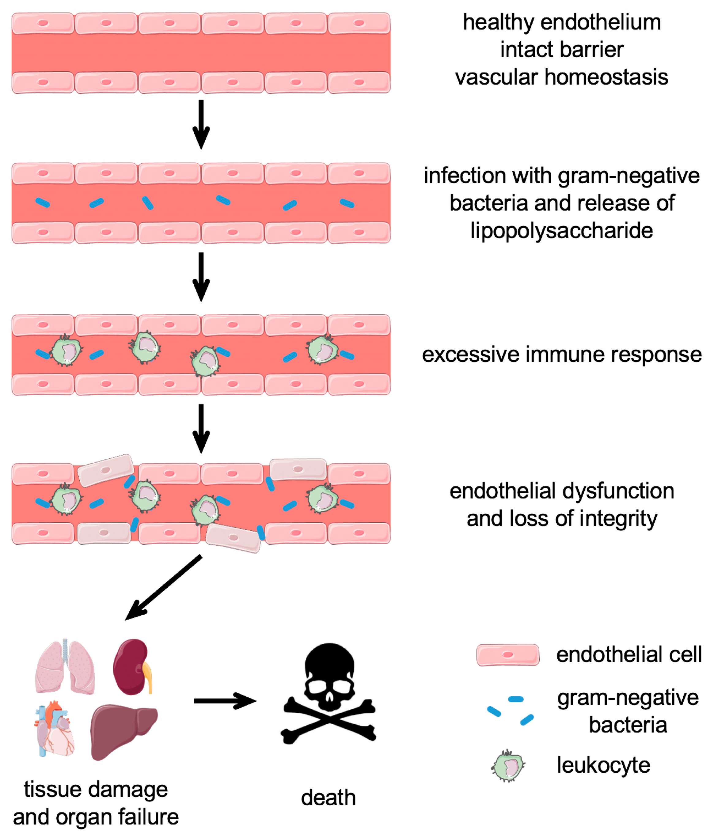
Cardiovascular diseases (CVDs) are the leading cause of morbidity and mortality in the world and were responsible for 17.9 million deaths in 2019 (https://www.who.int/news-room/fact-sheets/detail/cardiovascular-diseases-(cvds) (accessed on 7 February 2024). Their development and progression are intimately linked to endothelial dysfunction caused by factors such as oxidative stress. The vascular endothelium consists of a monolayer of endothelial cells, which constitutes the inner lining of all blood vessels. Endothelial cells are attached to the basal lamina, which acts as a scaffold, with pericytes and smooth muscle cells situated on the outside. The endothelium thereby forms a barrier between the bloodstream and the surrounding tissue and maintains vascular homeostasis. It secretes multiple factors that influence coagulation, vascular tone, adhesion of leukocytes, and inflammation, underscoring the importance of the endothelium for vascular homeostasis [1]. Endothelial cells are also important for the exchange of nutrients and molecules between the blood and the surrounding tissue. Their functionality requires energy to maintain a healthy endothelium [2].
Endothelial dysfunction is defined as the failure of endothelial cells to carry out their normal functions and is a consequence of their excessive activation and apoptosis [3]. The activation is induced by proinflammatory cytokines, including Tumor Necrosis Factor (TNF)-α and Interleukin-6 (IL-6) [4]. It is characterized by the expression of cell-surface adhesion molecules, such as Intercellular Adhesion Molecule 1 (ICAM1) or Vascular Cell Adhesion Molecule 1 (VCAM1). These adhesion molecules facilitate the recruitment and attachment of leukocytes to the endothelium, leading to increased endothelial leakiness, allowing diapedesis—the transmigration of leukocytes through the endothelial cell layer [3]. Prolonged endothelial cell activation and the unrestricted secretion of inflammatory cytokines by leukocytes favor endothelial cell apoptosis. Apoptosis is executed by caspases, which cleave proteins critical for cell survival [5]. Tight control of apoptosis is a prerequisite for maintaining a healthy endothelium, especially because endothelial cells are the first cells exposed to invading pathogens. Their dysfunction is a precursor to tissue and organ damage and is a major contributor to certain lethal pathologies, e.g., sepsis. Thus, the next two chapters will introduce the mechanisms in sepsis/high-dose endotoxemia with a specific focus on the endothelium.

2. Sepsis

Sepsis and septic shock are the leading cause of mortality in critically ill patients, accounting for approximately 20% of all-cause deaths globally. It was estimated that sepsis had affected 49 million individuals and was related to 11 million deaths worldwide in 2017 [6]. Sepsis is defined as a life-threatening organ dysfunction caused by a dysregulated host response to infection [7]. Its pathogenesis includes various steps, such as infection, hyperactivation of the immune system, endothelial dysfunction leading to loss of barrier integrity, followed by tissue damage, and finally, multi-organ failure and death [8] (Figure 1).
Infection with gram-negative bacteria, including Escherichia coli and Pseudomonas aeruginosa, is the prevailing cause of severe sepsis in humans [9]. Under normal circumstances, the innate immune system reacts to local infections and protects the organism from further damage. However, the hyperactivity of the immune response in sepsis occurs when the host loses control of the local containment of an infection, such that it becomes systemic. This leads to the production of proinflammatory cytokines, reactive oxygen species (ROS), and cellular injury, mediated by the recognition of pathogens by pattern recognition receptors (PRRs) [10,11]. Those PRRs, which are expressed on the surface of immune and phagocytic cells, and also on some non-immune cells, recognize pathogen-associated molecular patterns (PAMPs) that are expressed on both harmless and pathogenic microbes [11]. During tissue damage, intracellular components are released, which are recognized as damage-associated molecular patterns (DAMPs), and activate the immune system in concert with PAMPs [12].
The most important PAMP in the context of sepsis is lipopolysaccharide (LPS), an outer membrane component of gram-negative bacteria. LPS consists of a proximal oligosaccharide core, the distal O antigen, a glycan polymer, and a lipid A hydrophobic anchor—a glucosamine disaccharide decorated with multiple fatty acids [13]. Its immunostimulatory activity is mostly derived from the lipid A moiety, which in humans and mice binds to toll-like receptors (TLRs) and thereby activates the innate immune response [14]. LPS can exert its effect either locally or, after entering the blood circulation, systemically. LPS also affects the endothelial cell transcriptome by regulating transcripts of protein coding and non-coding RNAs [15]. Endothelial cells treated with LPS showed a significant increase in upregulated genes with gene ontology (GO) terms related to immune response to bacteria and TNF signaling [16]. Moreover, LPS concentrations above approximately 500 pg/mL are known to be lethal. This was shown in a study with 253 patients at the onset of severe sepsis and/or septic shock, where the median endotoxin concentration in the plasma was 300 pg/mL. More importantly, in survivors over a 28-day period, the mean level at study entry was 230 pg/mL, while it was 515 pg/mL in non-survivors [17].
Although TLR signaling is well described (see chapter 3), LPS effects are more complex and multifaceted, and the underlying mechanisms are far from understood. This explains why there are still no FDA-approved treatment options available that improve sepsis survival, despite significant advancements in therapeutic management and over 100 therapeutic trials [18,19]. Further exploration of the endothelial cell response to LPS and possible interference with the negative effects might provide clues for new targets for adjuvant therapies aimed at protecting the endothelium against damage and preventing the breakdown of the endothelial barrier.

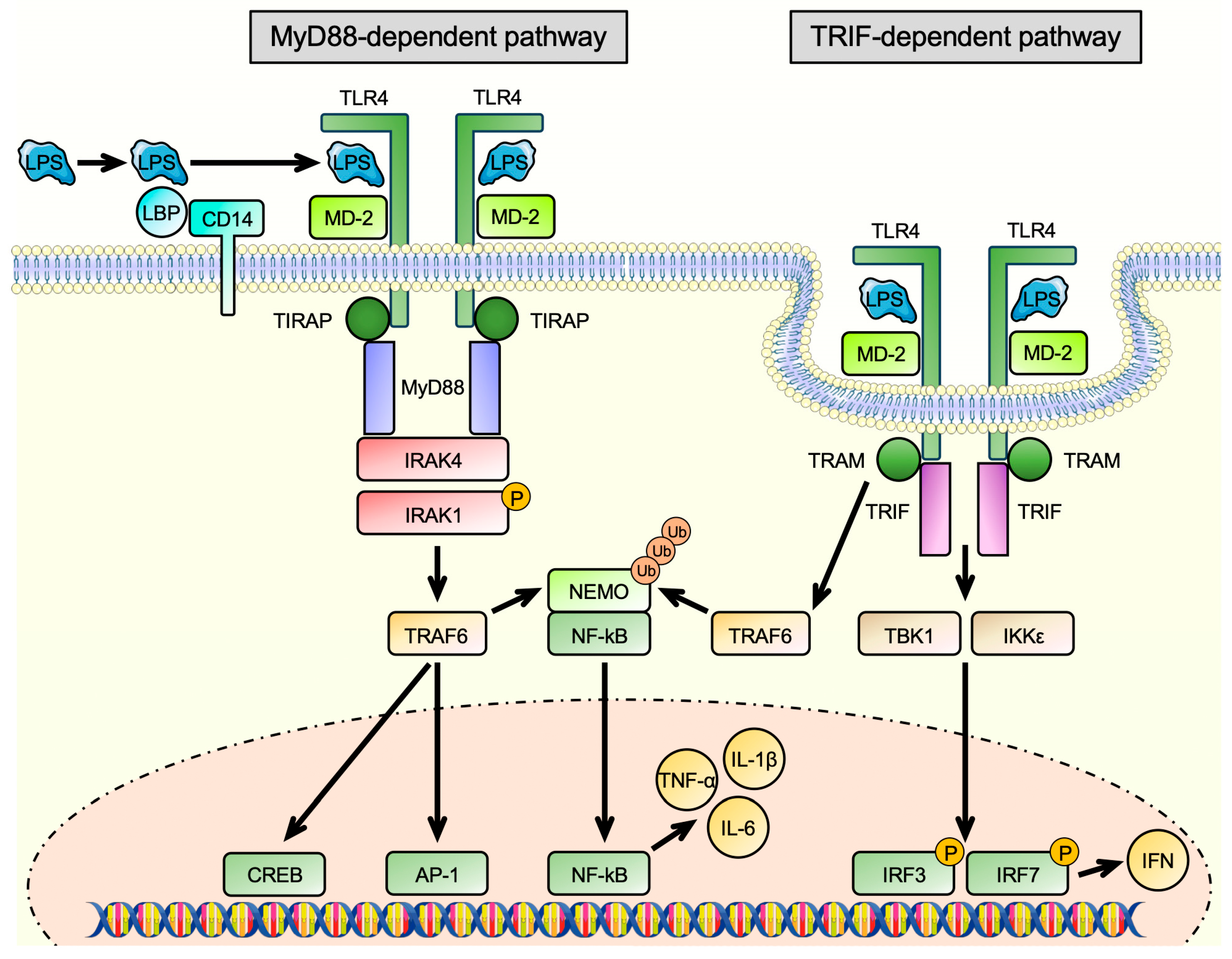
3. Toll-like Receptors

TLRs are a family of transmembrane glycoproteins, which, in humans, is comprised of 10 members. They are located on macrophages, T-cells, B-cells, dendritic cells, and also non-immune cells, like endothelial cells [20,21]. They recognize and bind to a wide range of different molecules, including microbial components, such as LPS, lipopeptides, or double-stranded RNA, via a horseshoe-shaped ligand binding domain built from leucine-rich repeats [22]. The binding of ligands induces a rearrangement of the receptor complex and induces the innate immune response and inflammation [14]. They detect different microbial components, such as LPS, lipopeptides, or double-stranded RNA, and induce the innate immune response and inflammation [14]. The most important TLR in LPS signaling is TLR4 [23]. According to the publicly available databases Genecards [24], Human Protein Atlas [25], and GTEx Portal [26], its transcript is present in nearly all human tissues. TLR4 can not only be found on blood cells but also on the cell surface of endothelial cells [27], smooth muscle cells [28], skeletal muscle cells [29], cardiac myocytes [30], and cells of the central nervous system [31]. The lipid A moiety of LPS is recognized and first bound by lipopolysaccharide-binding protein (LBP) and transferred to CD14 molecules. Membrane-bound CD14 (mCD14) is a glycosylphosphatidylinositol (GPI)-anchored protein located on the surface of myeloid–lineage cells like monocytes, macrophages, and dendritic cells [32]. After binding to CD14, LPS is subsequently transferred to Myeloid Differentiation Protein 2 (MD-2), and this transfer is catalyzed by CD14 [33]. MD-2 is essential for the activation of TLR4 by LPS, which was shown by the lack of response of macrophages derived from MD-2 knockout mice after LPS treatment [34]. In addition to mCD14, a soluble form of CD14 (sCD14), which lacks the GPI anchor, exists, which is also released from non-myeloid cells, including hepatocytes, adipocytes, and intestinal epithelial cells [35]. It is produced by shedding or secretion and is found in serum of normal individuals [36]. It has long been known that sCD14 can make CD14-negative cells like epithelial and endothelial cells responsive to LPS stimulation [37,38]. This is achieved by the high-affinity binding of LPS to sCD14 and the transport of this complex through the bloodstream. Soluble CD14 can then transfer LPS to MD-2 as efficiently as mCD14 [33], such that this reaction can also take place on cells devoid of CD14 themselves [39,40]. At this step, a complex of TLR4/MD2/LPS is formed, which builds a homodimer and triggers two consecutive pathways: (1) The MyD88-dependent pathway, which is induced in the plasma membrane, and (2) the TRIF-dependent pathway, which starts in early endosomes after the endocytosis of the receptor complex [41] (Figure 2).
During the MyD88-dependent pathway, the membrane-anchored TLR4 receptor interacts with the TIR domain-containing adapter protein (TIRAP) and recruits MyD88, triggering a signaling cascade. This cascade includes the recruitment of IL-1 Receptor Associated Kinases (IRAKs) 1 and 4. IRAK4 was shown to phosphorylate and thereby activates IRAK1 upon stimulation [42]. Following phosphorylation, IRAK1 associates with TNF Receptor Associated Factor 6 (TRAF6), which is a mediator of cytokine signaling and stress response pathways [43]. These include the activation of the transcription factors cAMP Response Element Binding Protein (CREB) and Activator Protein-1 (AP-1) [44,45,46]. TRAF6 also promotes the polyubiquitination of NEMO, an inhibitor of nuclear factor-κB (NF-κB) nuclear translocation. This triggers the degradation of NEMO, allowing nuclear entry of NF-κB, which then initiates the production of pro-inflammatory mediators, such as IL-6, IL-1β, and TNF-α, leading to an even further enhanced inflammatory response [41]. Following the dissociation of membrane-bound TLR4 from TIRAP and MyD88, TLR4 undergoes endocytosis and binds TIR Domain Containing Adaptor Molecule 1 (TRIF) and TRIF-Related Adapter Molecule (TRAM). It was shown that TRAM recruits TRIF to the plasma membrane and acts as a bridging adaptor, enabling the interaction of TRIF with TLR4 [47]. LPS promotes the complex building of TLR4 and TRAM and their following translocation into the endosome [48]. The LPS-induced internalization of TLR4 is controlled by CD14, whose importance is highlighted by the increased localization of TRAM to regions enriched with CD14 [49]. Endocytosis terminates the MyD88-dependent signaling, and endosome maturation as well as lysosomal degradation of TLR4 define the extent of the TRIF-dependent signaling [50]. TRIF-dependent signaling activates two kinases, TANK Binding Kinase 1 (TBK1) and IκB Kinase ε (IKKε), which phosphorylate the transcription factors Interferon Regulatory Factor (IRF) 3 and 7, leading to the expression of type I interferons, enhancing the inflammatory response even further [41]. In addition, TRAM also interacts with TRAF6, supporting NF-κB activation through the degradation of NEMO [51].
However, it has to be mentioned that the LPS signal cannot only be transmitted via TLR4. An intracellular inflammasome complex, which acts as a molecular signaling platform in the cytoplasm, has been found to recognize LPS intracellularly and activate a human caspase-4/murine caspase-11-dependent pathway that leads to increase in IL-1β and IL-18 and is sufficient to induce pyroptosis [52,53,54]. TLR4 is dispensable for this response, as demonstrated in TLR4-deficient mice, which were primed with a TLR3 agonist to activate caspase 11 and were as susceptible to LPS-triggered sepsis as wildtype animals [52]. After these initial studies, which used LPS-loaded macrophages, it was later demonstrated that outer membrane vesicles produced by gram-negative bacteria can deliver LPS into the cytosol and trigger caspase-11-dependent effector responses in vitro and in vivo [55].
Additionally, it was shown that the shape of LPS can vary, activating different TLRs; for example, TLR2 [56]. This is highlighted by the alternative formation of specific TLR receptor clusters in response to different bacterial products [57]. Moreover, the potency of LPS varies, with the hexa-acylated and phosphorylated LPS of Escherichia coli being one of the most potent species, whereas under-acylated, dephosphorylated LPS species have weaker pro-inflammatory activity [58]. This also contributes to the aforementioned structural diversity among LPS and influences its functionality [59].
Since TLR4 is the dominant PRR in LPS signaling, it seems reasonable to inhibit TLR-4. However, Eritoran, an analog of lipid A and TLR4 antagonist, which inhibits LPS-triggered TLR4 signaling [60], did not result in reduced 28-day mortality in a phase III clinical trial [17].
Septic patients also exhibit overwhelming oxidative stress, which results from the uncontrolled production of reactive oxygen and nitrogen species from activated immune cells. This explains why antioxidant therapies, using vitamin C, selenium, or N-acetylcysteine, were investigated in sepsis models [61]. However, they did not show any positive effects in humans [11].
In contrast to the acute effects of high-dose LPS on the vasculature, the effects of low-dose LPS are usually chronic and related to stress-induced senescence and aging, with aging being a major risk factor for the development of CVDs. In the following chapters of the review, we focus on endothelial senescence, its induction by low-dose endotoxemia, the role of the gut microbiome, and highlight the potential of natural dietary compounds to counteract senescence induction.

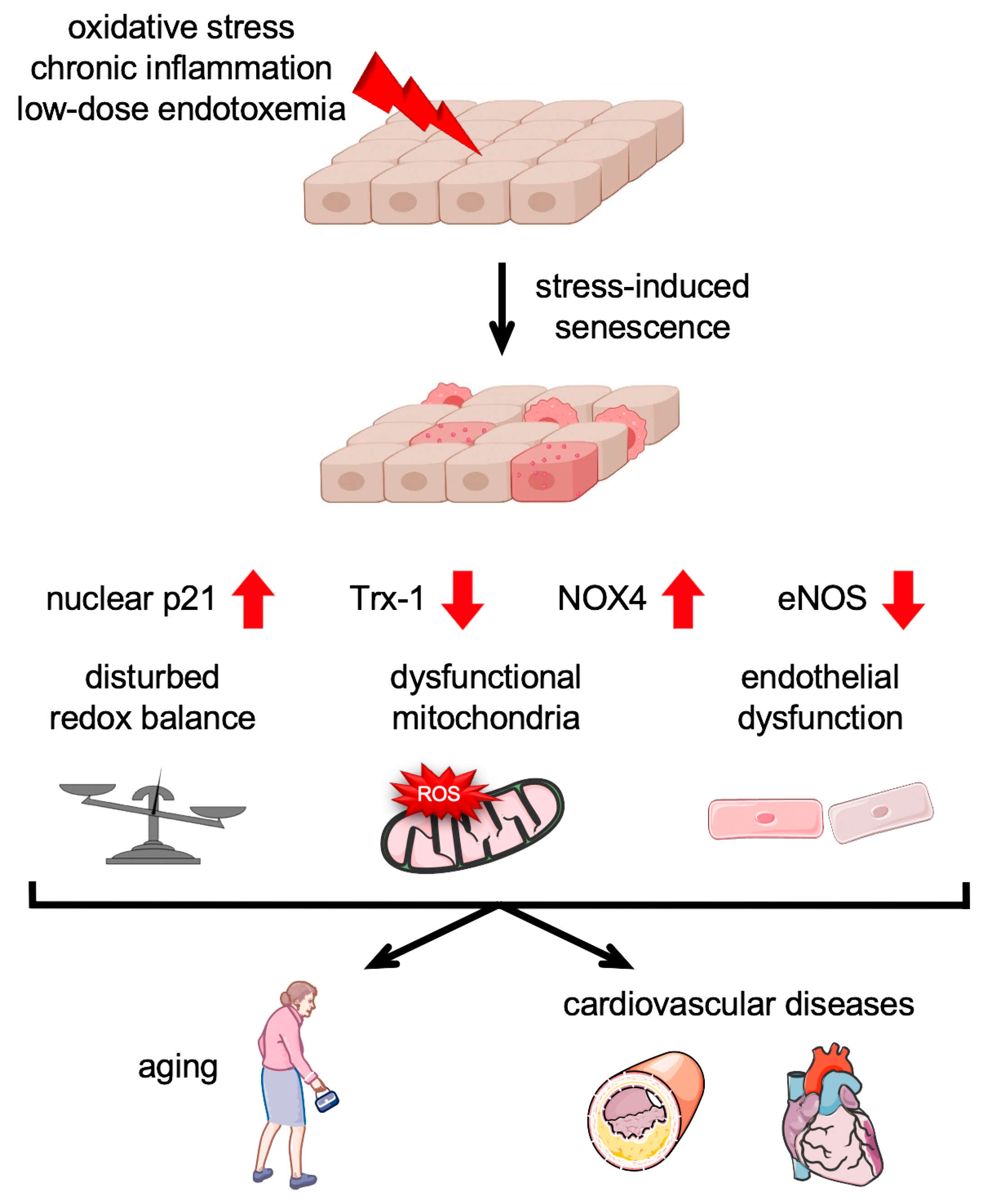
4. Stress-Induced Senescence in the Endothelium

Stress-induced cellular senescence is a phenomenon that occurs in response to various stressors including oxidative stress, xenobiotics [62], or low-dose endotoxemia [63] and goes along with increased intracellular ROS production. It can have detrimental effects on tissue function and homeostasis, and thus, it plays a significant role in age-related dysfunction of the vascular system and the development of cardiovascular diseases. Cells undergoing senescence sometimes exhibit a senescence-associated secretory phenotype (SASP) [64,65]. The SASP-associated inflammatory molecules expressed by vascular cells in atherosclerotic lesions and LPS-induced cellular senescence exaggerate the pathogenesis of CVDs [66]. Understanding the molecular mechanisms underlying stress-induced senescence may provide valuable insights for the development of therapeutic strategies aimed at ameliorating age-related pathologies.
In an oxidative stress-induced senescence model in primary human endothelial cells, Goy et al. showed that senescent endothelial cells have decreased levels of the antioxidative enzyme Thioredoxin-1 (Trx-1), while the levels of the ROS-producing NADPH Oxidase 4 (NOX4) were elevated [67]. In mice overexpressing NOX4 specifically in the endothelium, in which ROS levels are increased [68], the protein levels of Trx-1 negatively correlated with the expression of NOX4, providing an explanation for the disturbed redox balance in senescent endothelial cells [67] (Figure 3). The importance of redox homeostasis was emphasized by the finding that senescence induction could be inhibited by permanent Trx-1 expression [67]. Induction of endothelial cell senescence with other stimuli was demonstrated by different studies, in which ultrafine carbon nanoparticles [69], low density lipoprotein (LDL) [69,70,71], or concentrations of LPS found in low-dose endotoxemia [63] were used.
Cellular senescence in general is inter alia characterized by the upregulation of senescence-associated beta-Galactosidase, an increase in nuclear p21, and a disturbed redox balance. An additional, specific hallmark of endothelial cell senescence is a decreased production of NO. NO is a key vasodilator that has also been observed to be reduced in endothelial dysfunction [72]. The loss of NO is due to downregulation of endothelial NO Synthase (eNOS), whose levels also decline with age. Moreover, there is less activity of the enzyme, caused by reduced levels of the essential cofactor tetrahydrobiopterin (BH4) and the substrate L-arginine [72,73] (Figure 3).
One source of intracellular ROS in senescent cells, among others, is dysfunctional mitochondria [74]. Mitochondrial dysfunction is induced by altered mitochondrial structure due to reduced fusion and fission events, and mitochondrial DNA damage occurs during endothelial cell aging [75]. It is characterized by a reduced respiratory chain capacity per mitochondrion, a reduced mitochondrial membrane potential, and an increased production of ROS. It can lead to senescence and is simultaneously exacerbated by the initiation and maintenance of the senescent phenotype (Figure 3).

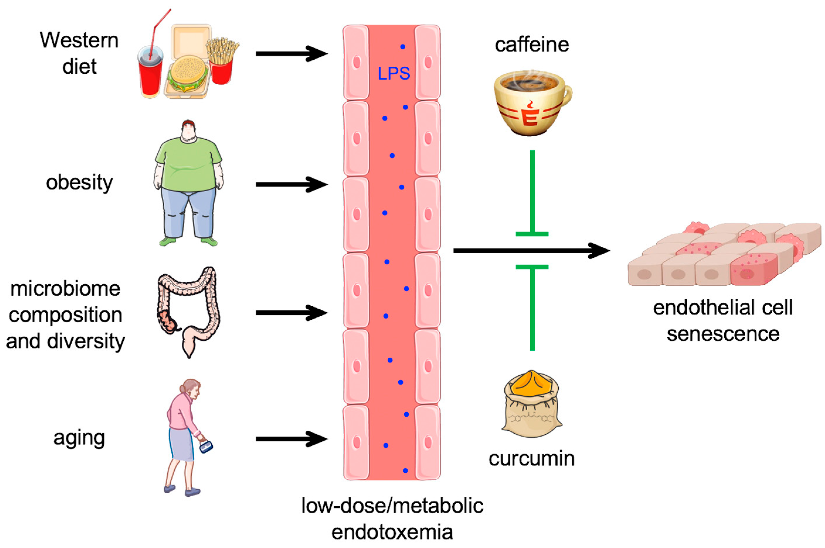
5. Low-Dose or Metabolic Endotoxemia—Role of the Gut Microbiome and the Endothelium

LPS is present—among other places—in the gastrointestinal tract, where it is ingested through the diet or on the gram-negative bacteria that are naturally present in the gut microbiota. It has been estimated that the human gut is populated with approximately 1014 commensal microorganisms, with the majority of them being present in the colon [76]. These include gram-negative bacteria, making them the major source of LPS in the intestinal lumen. Normally, the intestinal epithelium provides an effective barrier against LPS penetration into the bloodstream, such that the median plasma levels are below 15 pg/mL [77,78,79,80]. Consequently, an LPS concentration above 20 pg/mL in the circulation is considered a so-called low-dose endotoxemia, which is set apart from sepsis, where the plasma concentration of LPS is much higher [17].
Various factors, including an unhealthy diet, obesity, diet-induced changes in microbial diversity, and aging, increase the transfer of LPS from the gut to the bloodstream [81,82,83] (Figure 4). This results in low-dose endotoxemia, which—for the reasons explained below—is also called metabolic endotoxemia.
It has been shown that unbalanced, hyperlipidic meals have a short-term effect on LPS plasma levels, leading to a post-prandial increase already within one hour after a high-fat meal or drink [80,84,85,86]. This rapid increase suggests that dietary lipids facilitate the transfer of LPS from the gut to the bloodstream, and there is evidence that chylomicrons could contribute to the adsorption and transport of LPS [86,87,88].
However, these effects are only transient; an unhealthy diet, on the contrary, leads to a persistent increase in blood LPS levels, which was first demonstrated in rodent models. 4 weeks of a high-fat diet in mice persistently increased the plasma LPS concentration, which usually fluctuates diurnally with the feeding cycles, reaching the highest levels at the end of the feeding period, approximately threefold. As the same effect was obtained by subcutaneous infusion of LPS and the increase in plasma LPS was 10 to 50 times lower than in sepsis, the term metabolic endotoxemia was coined for this phenomenon. In both settings, the increase in circulating LPS was paralleled by the upregulation of inflammatory cytokines, indicating that metabolic endotoxemia entails a low-grade inflammation [89]. An increase in plasma LPS was also observed in mice on high-glucose or high-fructose diets [90] and rats on Western [91] and high-fat/high-sugar diets [92], respectively. Furthermore, metabolic endotoxemia was associated with obesity and insulin resistance [89,90], both being risk factors for the development of cardiovascular diseases. Interestingly, endotoxemia seems to induce these phenomena [89].
Unhealthy diets also induce changes in the gut microbiome [89,90], which seem to be at least in part responsible for the observed metabolic alterations, as dietary interventions that positively affect the composition of the microbiota or treatment with antibiotics can revert some of the effects [93,94]. In that respect, it is noteworthy that not simply the sheer number of gut microorganisms but rather the composition of the microbiome and, thus, the source of LPS is critical for the outcome. While hexa-acylated E. coli-derived LPS promotes dysglycemia and inflammation, lower acylated LPS from another bacterial species does not evoke these effects. Moreover, it can even antagonize the dysglycemia caused by E. coli LPS in lean mice and improve insulin sensitivity in obese animals [95]. In summary, these data show that diet-induced metabolic endotoxemia is tightly linked to gut microbiome dysbiosis.
Although animal models allow for experimental manipulations not possible in humans, similar correlations and effects have been observed in epidemiological studies and some small-scale clinical trials. A study based on a dietary survey revealed an association between energy intake and endotoxemia in healthy men, and it was suspected that fat is the critical component for the transfer of LPS into the bloodstream [96]. A correlation between dietary habits and metabolic endotoxemia has also been inferred from another study, in which healthy food choices have been associated with lower serum LPS activity [97]. Direct experimental proof for the impact of an unhealthy diet on serum LPS levels was obtained in crossover studies with healthy subjects. Similar to mice, a meal rich in saturated fats induced a post-prandial increase in LPS in the blood [98]. Also, the long-term effects could be recapitulated by demonstrating an increase in plasma endotoxin activity of over 70% after a 4-week Western diet, which returned to baseline after a washout period and another 4 weeks on a prudent diet [99]. Interestingly, experimental endotoxemia in humans induced insulin resistance [100], similar to what has been observed in mice, again underscoring that low-dose endotoxemia leads to metabolic states that increase the risk for cardiovascular diseases.
As in mice, unhealthy diets also have a major impact on the human gut microbiome. Work in humanized gnotobiotic mice, i.e., germ-free animals transplanted with human fecal microbial communities, elegantly showed that a Western diet shifts the gut microbiome already within a day. Interestingly, mice colonized with microbiota from obese Western diet-fed humanized donors developed adiposity [101], similar to what has been described for animals on a 4-week high-fat diet. A correlation between dietary patterns and changes in gut microbiome composition has also been directly demonstrated in humans. Interestingly, controlled consumption of a high-fat/low-fiber or low-fat/high-fiber diet induced alterations already within 24 h, the same time frame as in the gnotobiotic mice [102]. Diet can not only change the ratio between different bacterial species in the gut microbiome, but also has a profound effect on its complexity, which is directly connected to metabolic and inflammatory parameters. A microbiome analysis of nearly 300 non-obese and obese subjects revealed that lower bacterial richness goes along with adiposity, insulin resistance, and more pronounced inflammation [103]. The same observations were made in a parallel study, which additionally demonstrated that weight loss and weight stabilization interventions in obese and overweight individuals can increase microbiome diversity and improve clinical phenotypes [104].
The connection between the intestinal microbiome and chronic diseases or risk factors for their development is also reflected in the elderly. The analysis of fecal microbiota in elderly subjects revealed major changes, which are related to clinical parameters like inflammatory markers and frailty [105,106]. Notably, although a part of these alterations in the microbiome is due to lifestyle, there also seem to be intrinsic changes in the gut microbiota during the aging process, which has been shown in rodent models [107] as well as in humans [108]. An unfavorable composition of the microbiome impairs the barrier function of the gut epithelium, leading to increased transfer of LPS into the bloodstream [109]. Actually, a nearly two-fold increase in plasma LPS levels, and thus metabolic endotoxemia, was demonstrated in aged subjects free of chronic age-related diseases and physical disabilities, when compared to young individuals [29].
As described before, metabolic endotoxemia entails a systemic low-grade inflammation and thereby affects several cell types, also including those in the vasculature, e.g., leukocytes, platelets, and endothelial cells. Therefore, low-grade endotoxemia increases the risk for cardiovascular diseases [79,110]. One of the most critical targets for this metabolic endotoxemia-associated low-grade inflammation is the endothelium, as it plays a central role in vascular homeostasis, such that its dysfunction is associated with nearly all cardiovascular diseases. In fact, a low-grade systemic inflammation triggered by infusion of 2 ng/kg LPS in humans was paralleled by a significant decrease in peripheral endothelial progenitor cells, which are important for the repair of endothelial injuries [111]. However, not only these progenitor cells are affected by metabolic endotoxemia but also mature endothelial cells in the vascular wall. While treatment of primary human endothelial cells with 150 ng/mL of LPS led to activation and apoptosis [16], exposure to 1 ng/mL over a period of 2 weeks induced cellular senescence similarly to treatment with low doses of H2O2 [63], clearly demonstrating dose-dependent effects of LPS on the endothelium. The induction of endothelial cell senescence by low-dose endotoxemia also explains the association between metabolic endotoxemia and cardiovascular diseases.
Based on the notion that senescence impairs mitochondrial functionality, which is also essential for endothelial cells, it seems attractive to use compounds that positively affect the mitochondria to counteract senescence induction. One such compound is caffeine, a widely consumed drug found in coffee, tea, soft drinks, and chocolate. Large epidemiological studies have demonstrated an association between habitual caffeine intake and a decrease in mortality, including death as consequence of cardiovascular disorders [112,113]. We have previously shown that the consumption of four cups of coffee translates into caffeine serum concentrations of approximately 30 µM [114]. These physiological concentrations improved the migration of endothelial progenitor cells and mature endothelial cells and repair after vascular injury [114]. Moreover, in mice, they improved outcomes after myocardial infarction [115]. This positive effect is due to increased translocation of p27 into the mitochondria and improved electron transport chain activity. Moreover, caffeine reduced age-associated decline in mitochondrial functionality [115].
Another interesting natural compound in this context could be curcumin. This polyphenol is the main active ingredient in the root of Curcuma longa, which has been used as a medicinal plant in Asia for over 4000 years. Multiple studies have shown a protective effect of curcumin in cardiovascular diseases (for review, see [116]).
While there is some evidence that curcumin and caffeine play a role in the reduction of the effects of endotoxemia, information on detailed mechanisms underlying these are sparse to date. As these dietary factors have to pass through the digestive tract, one could assume that they interact with and have an impact on the gut microbiome. Some studies hint towards curcumin altering the gut microbiome composition with an increase in beneficial versus pathogenic bacteria (for review, see [117]). Based on the above-discussed impact of the microbiome on plasma LPS levels, it can be speculated that this effect might reduce LPS concentrations in the circulation. Although caffeine can alter the gut microbiome and thereby ameliorate the metabolic syndrome in obese mice [118], this is only very indirect evidence that it might also affect plasma LPS levels, as metabolic syndrome is associated with metabolic endotoxemia.
However, the direct effects of curcumin and caffeine on the endothelium have been investigated in some more detail and will be discussed in the following chapter.

6. Impact of Curcumin and Caffeine on High-Dose and Metabolic Endotoxemia with Respect to the Endothelium

As outlined above, upon leakage of the gut, LPS enters the circulation, which in high doses leads to so-called “leaky vessels”, resulting in circulatory and organ failure, finally leading to death within hours or days in mice and humans. The role of curcumin and caffeine treatment in high-dose endotoxemia/sepsis with respect to the endothelium has barely been investigated.
With respect to curcumin, the expression of ICAM-1 was inhibited ex vivo and in vivo in mice, resulting in reduced adhesion of neutrophils and monocytes/macrophages to the endothelium [119]. In rats, curcumin infusion preserved endothelial barrier function in a high-dose LPS model [120]. Of note, these studies only investigated the preventive effects of the compound, as it was administered prior to LPS infusion or treatment. In 2022, the first double-blinded placebo controlled clinical trial with curcumin supplementation in critically ill sepsis patients was performed. Although, the curcumin supplementation was only for 10 days in 20 patients and 20 placebo controls, curcumin significantly improved inflammatory markers, such as Interleukin-1beta, and reduced markers of endothelial activation including ICAM-1. Moreover, it decreased the sepsis-related organ failure assessment score and the duration of mechanical ventilation [121]. Thus, one could assume that curcumin supplementation could improve endothelial functionality in sepsis and may lead to reduced mortality. However, further trials are needed to assess the potential of curcumin with respect to endothelial function and mortality in sepsis patients.
For caffeine, there are currently no studies available that investigate its impact on the endothelium in high-dose endotoxemia/sepsis.
As described before, low-dose endotoxemia entails endothelial cell senescence. However, the impact of curcumin on metabolic endotoxemia and endothelial cell senescence has not been investigated directly. Nevertheless, there is circumstantial evidence for potential protective effects. Curcumin improved the barrier function in two colon carcinoma cell lines [122], suggesting that it might reduce LPS transfer from the gut to the circulation and thus prevent systemic inflammation, one of the consequences of metabolic endotoxemia. A direct impact of curcumin on inflammation in a disease setting characterized by metabolic endotoxemia has been shown by the reduction of inflammatory cytokine levels in subjects with metabolic syndrome in a randomized controlled trial [123]. In this study, a daily dose of 1 g curcumin was administered over a period of 8 weeks; however, the curcumin concentrations in the blood were not measured. Several studies have measured the serum concentration after administration of crude curcumin ranging from 1 to 3200 ng/mL depending on dose (2 to 12 g) and subject physiology (for review, see [124]). Moreover, short-term treatment of human endothelial cells with curcumin induced stabilization and nuclear translocation of the transcription factor Nrf-2 and enhanced expression of antioxidative defense systems, including Thioredoxin-1 and Superoxide Dismutase 2 [125,126] (Figure 5). This could also be relevant in the context of endothelial cell senescence, which is associated with increased ROS production but has not been studied so far.
In contrast, caffeine effects have been investigated not only in healthy endothelial cells and with respect to vascular repair after injury but also in low-dose endotoxemia-induced endothelial senescence, which was counteracted by caffeine. This was shown by the treatment of endothelial cells with caffeine during senescence induction with low-dose LPS. The co-incubation with both substances blocked the upregulation of nuclear p21, a general senescence marker. Moreover, it also conserved endothelial functionality by maintaining the levels of Trx-1 and eNOS and preserving the migratory capacity of endothelial cells. Mechanistically, this is again due to the translocation of p27 into the mitochondria, as permanent expression of mitochondrially targeted p27 also blocked senescence induction (Figure 5). Interestingly, the same effects were observed when caffeine was applied to already senescent endothelial cells [63]. These data suggest that caffeine can reverse or at least delay endothelial cell senescence induced by metabolic endotoxemia and might, therefore, be an additional protective dietary factor in the context of aging.
This last section highlights that caffeine and curcumin—natural food components—could offer options to combat the negative senescence-inducing effects of low-dose or metabolic endotoxemia.

7. Conclusions

In conclusion, dependent on the concentration of LPS in the blood, high-dose or low-dose endotoxemia is induced. High-dose endotoxemia, also called sepsis, often leads to septic shock, endothelial cell death, organ failure, and death. In contrast, low-dose or metabolic endotoxemia leads to chronic inflammation, which results in endothelial cell senescence. Two natural compounds, caffeine and curcumin, seem to inhibit or at least delay low-dose endotoxemia-induced endothelial cell senescence. Thus, natural compounds may have the potential to protect against the negative senescence-inducing effects of metabolic endotoxemia.

Author Contributions

Conceptualization, P.J., S.P., J.A. and J.H.; writing—original draft preparation, D.M., F.F.C., J.A. and J.H.; writing—review and editing, F.F.C., P.J., S.P., J.A. and J.H.; visualization, P.J. and J.A.; supervision, J.A. and J.H.; funding acquisition, S.P. and J.H. All authors have read and agreed to the published version of the manuscript.

Funding

This research was funded in part by Bayer Grant4Target (2019-08-2427) to J.H. and by intramural funding (9772888) of the University Hospital of the Heinrich-Heine-University to S.P. and J.H.

Institutional Review Board Statement

Not applicable.

Informed Consent Statement

Not applicable.

Data Availability Statement

Not applicable.

Acknowledgments

Single graphical elements for figures were taken from Servier Medical Art (smart.servier.com accessed on 2 April 2024), which is licensed under a Creative Commons Attribution 3.0 Unported License.

Conflicts of Interest

The authors declare no conflicts of interest.

References

  1. Gao, J.; Pan, X.; Li, G.; Chatterjee, E.; Xiao, J. Physical Exercise Protects Against Endothelial Dysfunction in Cardiovascular and Metabolic Diseases. J. Cardiovasc. Transl. Res. 2022, 15, 604–620. [Google Scholar] [CrossRef]
  2. Hadi, H.A.; Carr, C.S.; Al Suwaidi, J. Endothelial dysfunction: Cardiovascular risk factors, therapy, and outcome. Vasc. Health Risk Manag. 2005, 1, 183–198. [Google Scholar]
  3. Pober, J.S.; Min, W.; Bradley, J.R. Mechanisms of endothelial dysfunction, injury, and death. Annu. Rev. Pathol. 2009, 4, 71–95. [Google Scholar] [CrossRef]
  4. Liao, J.K. Linking endothelial dysfunction with endothelial cell activation. J. Clin. Investig. 2013, 123, 540–541. [Google Scholar] [CrossRef]
  5. Boatright, K.M.; Salvesen, G.S. Mechanisms of caspase activation. Curr. Opin. Cell Biol. 2003, 15, 725–731. [Google Scholar] [CrossRef]
  6. World Health Organization. Global Report on the Epidemiology and Burden of Sepsis: Current Evidence, Identifying Gaps and Future Directions; World Health Organization: Geneva, Switzerland, 2020; p. 55. [Google Scholar]
  7. Singer, M.; Deutschman, C.S.; Seymour, C.W.; Shankar-Hari, M.; Annane, D.; Bauer, M.; Bellomo, R.; Bernard, G.R.; Chiche, J.D.; Coopersmith, C.M.; et al. The Third International Consensus Definitions for Sepsis and Septic Shock (Sepsis-3). JAMA 2016, 315, 801–810. [Google Scholar] [CrossRef]
  8. Virzi, G.M.; Mattiotti, M.; de Cal, M.; Ronco, C.; Zanella, M.; De Rosa, S. Endotoxin in Sepsis: Methods for LPS Detection and the Use of Omics Techniques. Diagnostics 2022, 13, 79. [Google Scholar] [CrossRef]
  9. Mayr, F.B.; Yende, S.; Angus, D.C. Epidemiology of severe sepsis. Virulence 2014, 5, 4–11. [Google Scholar] [CrossRef]
  10. Dauphinee, S.M.; Karsan, A. Lipopolysaccharide signaling in endothelial cells. Lab. Investig. 2006, 86, 9–22. [Google Scholar] [CrossRef]
  11. Delano, M.J.; Ward, P.A. The immune system’s role in sepsis progression, resolution, and long-term outcome. Immunol. Rev. 2016, 274, 330–353. [Google Scholar] [CrossRef]
  12. Medzhitov, R.; Janeway, C., Jr. Innate immunity. N. Engl. J. Med. 2000, 343, 338–344. [Google Scholar] [CrossRef]
  13. Page, M.J.; Kell, D.B.; Pretorius, E. The Role of Lipopolysaccharide-Induced Cell Signalling in Chronic Inflammation. Chronic Stress 2022, 6, 24705470221076390. [Google Scholar] [CrossRef]
  14. Molinaro, A.; Holst, O.; Di Lorenzo, F.; Callaghan, M.; Nurisso, A.; D’Errico, G.; Zamyatina, A.; Peri, F.; Berisio, R.; Jerala, R.; et al. Chemistry of lipid A: At the heart of innate immunity. Chemistry 2015, 21, 500–519. [Google Scholar] [CrossRef]
  15. Ho, J.; Chan, H.; Wong, S.H.; Wang, M.H.; Yu, J.; Xiao, Z.; Liu, X.; Choi, G.; Leung, C.C.; Wong, W.T.; et al. The involvement of regulatory non-coding RNAs in sepsis: A systematic review. Crit. Care 2016, 20, 383. [Google Scholar] [CrossRef]
  16. Merk, D.; Ptok, J.; Jakobs, P.; von Ameln, F.; Greulich, J.; Kluge, P.; Semperowitsch, K.; Eckermann, O.; Schaal, H.; Ale-Agha, N.; et al. Selenoprotein T Protects Endothelial Cells against Lipopolysaccharide-Induced Activation and Apoptosis. Antioxidants 2021, 10, 1427. [Google Scholar] [CrossRef]
  17. Opal, S.M.; Laterre, P.F.; Francois, B.; LaRosa, S.P.; Angus, D.C.; Mira, J.P.; Wittebole, X.; Dugernier, T.; Perrotin, D.; Tidswell, M.; et al. Effect of eritoran, an antagonist of MD2-TLR4, on mortality in patients with severe sepsis: The ACCESS randomized trial. JAMA 2013, 309, 1154–1162. [Google Scholar] [CrossRef]
  18. Marshall, J.C. Why have clinical trials in sepsis failed? Trends Mol. Med. 2014, 20, 195–203. [Google Scholar] [CrossRef]
  19. Guarino, M.; Perna, B.; Cesaro, A.E.; Maritati, M.; Spampinato, M.D.; Contini, C.; De Giorgio, R. 2023 Update on Sepsis and Septic Shock in Adult Patients: Management in the Emergency Department. J. Clin. Med. 2023, 12, 3188. [Google Scholar] [CrossRef]
  20. Duan, T.; Du, Y.; Xing, C.; Wang, H.Y.; Wang, R.F. Toll-Like Receptor Signaling and Its Role in Cell-Mediated Immunity. Front. Immunol. 2022, 13, 812774. [Google Scholar] [CrossRef]
  21. Vaure, C.; Liu, Y. A comparative review of toll-like receptor 4 expression and functionality in different animal species. Front. Immunol. 2014, 5, 316. [Google Scholar] [CrossRef]
  22. Gay, N.J.; Gangloff, M. Structure and function of Toll receptors and their ligands. Annu. Rev. Biochem. 2007, 76, 141–165. [Google Scholar] [CrossRef]
  23. Poltorak, A.; He, X.; Smirnova, I.; Liu, M.Y.; Van Huffel, C.; Du, X.; Birdwell, D.; Alejos, E.; Silva, M.; Galanos, C.; et al. Defective LPS signaling in C3H/HeJ and C57BL/10ScCr mice: Mutations in Tlr4 gene. Science 1998, 282, 2085–2088. [Google Scholar] [CrossRef]
  24. Stelzer, G.; Rosen, N.; Plaschkes, I.; Zimmerman, S.; Twik, M.; Fishilevich, S.; Stein, T.I.; Nudel, R.; Lieder, I.; Mazor, Y.; et al. The GeneCards Suite: From Gene Data Mining to Disease Genome Sequence Analyses. Curr. Protoc. Bioinform. 2016, 54, 1.30.1–1.30.33. [Google Scholar] [CrossRef]
  25. Uhlen, M.; Fagerberg, L.; Hallstrom, B.M.; Lindskog, C.; Oksvold, P.; Mardinoglu, A.; Sivertsson, A.; Kampf, C.; Sjostedt, E.; Asplund, A.; et al. Tissue-based map of the human proteome. Science 2015, 347, 1260419. [Google Scholar] [CrossRef]
  26. Consortium, G.T. The Genotype-Tissue Expression (GTEx) project. Nat. Genet. 2013, 45, 580–585. [Google Scholar] [CrossRef]
  27. Taylor, K.R.; Trowbridge, J.M.; Rudisill, J.A.; Termeer, C.C.; Simon, J.C.; Gallo, R.L. Hyaluronan fragments stimulate endothelial recognition of injury through TLR4. J. Biol. Chem. 2004, 279, 17079–17084. [Google Scholar] [CrossRef]
  28. Sasu, S.; LaVerda, D.; Qureshi, N.; Golenbock, D.T.; Beasley, D. Chlamydia pneumoniae and chlamydial heat shock protein 60 stimulate proliferation of human vascular smooth muscle cells via toll-like receptor 4 and p44/p42 mitogen-activated protein kinase activation. Circ. Res. 2001, 89, 244–250. [Google Scholar] [CrossRef]
  29. Ghosh, S.; Lertwattanarak, R.; Garduno Jde, J.; Galeana, J.J.; Li, J.; Zamarripa, F.; Lancaster, J.L.; Mohan, S.; Hussey, S.; Musi, N. Elevated muscle TLR4 expression and metabolic endotoxemia in human aging. J. Gerontol. A Biol. Sci. Med. Sci. 2015, 70, 232–246. [Google Scholar] [CrossRef]
  30. Frantz, S.; Kobzik, L.; Kim, Y.D.; Fukazawa, R.; Medzhitov, R.; Lee, R.T.; Kelly, R.A. Toll4 (TLR4) expression in cardiac myocytes in normal and failing myocardium. J. Clin. Investig. 1999, 104, 271–280. [Google Scholar] [CrossRef]
  31. Kielian, T. Toll-like receptors in central nervous system glial inflammation and homeostasis. J. Neurosci. Res. 2006, 83, 711–730. [Google Scholar] [CrossRef]
  32. Plociennikowska, A.; Hromada-Judycka, A.; Borzecka, K.; Kwiatkowska, K. Co-operation of TLR4 and raft proteins in LPS-induced pro-inflammatory signaling. Cell Mol. Life Sci. 2015, 72, 557–581. [Google Scholar] [CrossRef]
  33. Ryu, J.K.; Kim, S.J.; Rah, S.H.; Kang, J.I.; Jung, H.E.; Lee, D.; Lee, H.K.; Lee, J.O.; Park, B.S.; Yoon, T.Y.; et al. Reconstruction of LPS Transfer Cascade Reveals Structural Determinants within LBP, CD14, and TLR4-MD2 for Efficient LPS Recognition and Transfer. Immunity 2017, 46, 38–50. [Google Scholar] [CrossRef]
  34. Meng, J.; Gong, M.; Bjorkbacka, H.; Golenbock, D.T. Genome-wide expression profiling and mutagenesis studies reveal that lipopolysaccharide responsiveness appears to be absolutely dependent on TLR4 and MD-2 expression and is dependent upon intermolecular ionic interactions. J. Immunol. 2011, 187, 3683–3693. [Google Scholar] [CrossRef]
  35. Funda, D.P.; Tuckova, L.; Farre, M.A.; Iwase, T.; Moro, I.; Tlaskalova-Hogenova, H. CD14 is expressed and released as soluble CD14 by human intestinal epithelial cells in vitro: Lipopolysaccharide activation of epithelial cells revisited. Infect. Immun. 2001, 69, 3772–3781. [Google Scholar] [CrossRef]
  36. Haziot, A.; Chen, S.; Ferrero, E.; Low, M.G.; Silber, R.; Goyert, S.M. The monocyte differentiation antigen, CD14, is anchored to the cell membrane by a phosphatidylinositol linkage. J. Immunol. 1988, 141, 547–552. [Google Scholar] [CrossRef]
  37. Frey, E.A.; Miller, D.S.; Jahr, T.G.; Sundan, A.; Bazil, V.; Espevik, T.; Finlay, B.B.; Wright, S.D. Soluble CD14 participates in the response of cells to lipopolysaccharide. J. Exp. Med. 1992, 176, 1665–1671. [Google Scholar] [CrossRef]
  38. Pugin, J.; Schurer-Maly, C.C.; Leturcq, D.; Moriarty, A.; Ulevitch, R.J.; Tobias, P.S. Lipopolysaccharide activation of human endothelial and epithelial cells is mediated by lipopolysaccharide-binding protein and soluble CD14. Proc. Natl. Acad. Sci. USA 1993, 90, 2744–2748. [Google Scholar] [CrossRef]
  39. Golenbock, D.T.; Bach, R.R.; Lichenstein, H.; Juan, T.S.; Tadavarthy, A.; Moldow, C.F. Soluble CD14 promotes LPS activation of CD14-deficient PNH monocytes and endothelial cells. J. Lab. Clin. Med. 1995, 125, 662–671. [Google Scholar]
  40. Haziot, A.; Rong, G.W.; Silver, J.; Goyert, S.M. Recombinant soluble CD14 mediates the activation of endothelial cells by lipopolysaccharide. J. Immunol. 1993, 151, 1500–1507. [Google Scholar] [CrossRef]
  41. Zhang, Y.; Liang, X.; Bao, X.; Xiao, W.; Chen, G. Toll-like receptor 4 (TLR4) inhibitors: Current research and prospective. Eur. J. Med. Chem. 2022, 235, 114291. [Google Scholar] [CrossRef]
  42. Li, S.; Strelow, A.; Fontana, E.J.; Wesche, H. IRAK-4: A novel member of the IRAK family with the properties of an IRAK-kinase. Proc. Natl. Acad. Sci. USA 2002, 99, 5567–5572. [Google Scholar] [CrossRef]
  43. Arch, R.H.; Gedrich, R.W.; Thompson, C.B. Tumor necrosis factor receptor-associated factors (TRAFs)—A family of adapter proteins that regulates life and death. Genes. Dev. 1998, 12, 2821–2830. [Google Scholar] [CrossRef]
  44. Shaywitz, A.J.; Greenberg, M.E. CREB: A stimulus-induced transcription factor activated by a diverse array of extracellular signals. Annu. Rev. Biochem. 1999, 68, 821–861. [Google Scholar] [CrossRef]
  45. Zhao, D.; Kwon, S.H.; Chun, Y.S.; Gu, M.Y.; Yang, H.O. Anti-Neuroinflammatory Effects of Fucoxanthin via Inhibition of Akt/NF-kappaB and MAPKs/AP-1 Pathways and Activation of PKA/CREB Pathway in Lipopolysaccharide-Activated BV-2 Microglial Cells. Neurochem. Res. 2017, 42, 667–677. [Google Scholar] [CrossRef]
  46. Lu, Y.; Li, B.; Xu, A.; Liang, X.; Xu, T.; Jin, H.; Xie, Y.; Wang, R.; Liu, X.; Gao, X.; et al. NF-kappaB and AP-1 are required for the lipopolysaccharide-induced expression of MCP-1, CXCL1, and Cx43 in cultured rat dorsal spinal cord astrocytes. Front. Mol. Neurosci. 2022, 15, 859558. [Google Scholar] [CrossRef]
  47. Marongiu, L.; Gornati, L.; Artuso, I.; Zanoni, I.; Granucci, F. Below the surface: The inner lives of TLR4 and TLR9. J. Leukoc. Biol. 2019, 106, 147–160. [Google Scholar] [CrossRef]
  48. Tanimura, N.; Saitoh, S.; Matsumoto, F.; Akashi-Takamura, S.; Miyake, K. Roles for LPS-dependent interaction and relocation of TLR4 and TRAM in TRIF-signaling. Biochem. Biophys. Res. Commun. 2008, 368, 94–99. [Google Scholar] [CrossRef]
  49. Jiang, Z.; Georgel, P.; Du, X.; Shamel, L.; Sovath, S.; Mudd, S.; Huber, M.; Kalis, C.; Keck, S.; Galanos, C.; et al. CD14 is required for MyD88-independent LPS signaling. Nat. Immunol. 2005, 6, 565–570. [Google Scholar] [CrossRef]
  50. Ciesielska, A.; Matyjek, M.; Kwiatkowska, K. TLR4 and CD14 trafficking and its influence on LPS-induced pro-inflammatory signaling. Cell Mol. Life Sci. 2021, 78, 1233–1261. [Google Scholar] [CrossRef]
  51. Verstak, B.; Stack, J.; Ve, T.; Mangan, M.; Hjerrild, K.; Jeon, J.; Stahl, R.; Latz, E.; Gay, N.; Kobe, B.; et al. The TLR signaling adaptor TRAM interacts with TRAF6 to mediate activation of the inflammatory response by TLR4. J. Leukoc. Biol. 2014, 96, 427–436. [Google Scholar] [CrossRef]
  52. Kayagaki, N.; Wong, M.T.; Stowe, I.B.; Ramani, S.R.; Gonzalez, L.C.; Akashi-Takamura, S.; Miyake, K.; Zhang, J.; Lee, W.P.; Muszynski, A.; et al. Noncanonical inflammasome activation by intracellular LPS independent of TLR4. Science 2013, 341, 1246–1249. [Google Scholar] [CrossRef]
  53. Hagar, J.A.; Powell, D.A.; Aachoui, Y.; Ernst, R.K.; Miao, E.A. Cytoplasmic LPS activates caspase-11: Implications in TLR4-independent endotoxic shock. Science 2013, 341, 1250–1253. [Google Scholar] [CrossRef]
  54. Molloy, S. Host response: LPS goes non-canonical. Nat. Rev. Microbiol. 2013, 11, 599. [Google Scholar] [CrossRef]
  55. Vanaja, S.K.; Russo, A.J.; Behl, B.; Banerjee, I.; Yankova, M.; Deshmukh, S.D.; Rathinam, V.A.K. Bacterial Outer Membrane Vesicles Mediate Cytosolic Localization of LPS and Caspase-11 Activation. Cell 2016, 165, 1106–1119. [Google Scholar] [CrossRef]
  56. Hirschfeld, M.; Weis, J.J.; Toshchakov, V.; Salkowski, C.A.; Cody, M.J.; Ward, D.C.; Qureshi, N.; Michalek, S.M.; Vogel, S.N. Signaling by toll-like receptor 2 and 4 agonists results in differential gene expression in murine macrophages. Infect. Immun. 2001, 69, 1477–1482. [Google Scholar] [CrossRef]
  57. Palsson-McDermott, E.M.; O’Neill, L.A. Signal transduction by the lipopolysaccharide receptor, Toll-like receptor-4. Immunology 2004, 113, 153–162. [Google Scholar] [CrossRef]
  58. Steimle, A.; Autenrieth, I.B.; Frick, J.S. Structure and function: Lipid A modifications in commensals and pathogens. Int. J. Med. Microbiol. 2016, 306, 290–301. [Google Scholar] [CrossRef]
  59. Mohr, A.E.; Crawford, M.; Jasbi, P.; Fessler, S.; Sweazea, K.L. Lipopolysaccharide and the gut microbiota: Considering structural variation. FEBS Lett. 2022, 596, 849–875. [Google Scholar] [CrossRef]
  60. Barochia, A.; Solomon, S.; Cui, X.; Natanson, C.; Eichacker, P.Q. Eritoran tetrasodium (E5564) treatment for sepsis: Review of preclinical and clinical studies. Expert. Opin. Drug Metab. Toxicol. 2011, 7, 479–494. [Google Scholar] [CrossRef]
  61. Abelli, J.; Mendez-Valdes, G.; Gomez-Hevia, F.; Bragato, M.C.; Chichiarelli, S.; Saso, L.; Rodrigo, R. Potential Antioxidant Multitherapy against Complications Occurring in Sepsis. Biomedicines 2022, 10, 3088. [Google Scholar] [CrossRef]
  62. Liang, Y.; Liang, N.; Yin, L.; Xiao, F. Cellular and molecular mechanisms of xenobiotics-induced premature senescence. Toxicol. Res. 2020, 9, 669–675. [Google Scholar] [CrossRef]
  63. Merk, D.; Greulich, J.; Vierkant, A.; Cox, F.; Eckermann, O.; von Ameln, F.; Dyballa-Rukes, N.; Altschmied, J.; Ale-Agha, N.; Jakobs, P.; et al. Caffeine Inhibits Oxidative Stress- and Low Dose Endotoxemia-Induced Senescence-Role of Thioredoxin-1. Antioxidants 2023, 12, 1244. [Google Scholar] [CrossRef]
  64. Coppé, J.P.; Patil, C.K.; Rodier, F.; Sun, Y.; Muñoz, D.P.; Goldstein, J.; Nelson, P.S.; Desprez, P.Y.; Campisi, J. Senescence-associated secretory phenotypes reveal cell-nonautonomous functions of oncogenic RAS and the p53 tumor suppressor. PLoS Biol. 2008, 6, 2853–2868. [Google Scholar] [CrossRef]
  65. Andreo-López, M.C.; Contreras-Bolívar, V.; Muñoz-Torres, M.; García-Fontana, B.; García-Fontana, C. Influence of the Mediterranean Diet on Healthy Aging. Int. J. Mol. Sci. 2023, 24, 4491. [Google Scholar] [CrossRef]
  66. Suzuki, K.; Susaki, E.A.; Nagaoka, I. Lipopolysaccharides and Cellular Senescence: Involvement in Atherosclerosis. Int. J. Mol. Sci. 2022, 23, 11148. [Google Scholar] [CrossRef]
  67. Goy, C.; Czypiorski, P.; Altschmied, J.; Jakob, S.; Rabanter, L.L.; Brewer, A.C.; Ale-Agha, N.; Dyballa-Rukes, N.; Shah, A.M.; Haendeler, J. The imbalanced redox status in senescent endothelial cells is due to dysregulated Thioredoxin-1 and NADPH oxidase 4. Exp. Gerontol. 2014, 56, 45–52. [Google Scholar] [CrossRef]
  68. Ray, R.; Murdoch, C.E.; Wang, M.; Santos, C.X.; Zhang, M.; Alom-Ruiz, S.; Anilkumar, N.; Ouattara, A.; Cave, A.C.; Walker, S.J.; et al. Endothelial Nox4 NADPH oxidase enhances vasodilatation and reduces blood pressure in vivo. Arter. Thromb. Vasc. Biol. 2011, 31, 1368–1376. [Google Scholar] [CrossRef]
  69. Büchner, N.; Ale-Agha, N.; Jakob, S.; Sydlik, U.; Kunze, K.; Unfried, K.; Altschmied, J.; Haendeler, J. Unhealthy diet and ultrafine carbon black particles induce senescence and disease associated phenotypic changes. Exp. Gerontol. 2013, 48, 8–16. [Google Scholar] [CrossRef]
  70. Oh, S.T.; Park, H.; Yoon, H.J.; Yang, S.Y. Erratum to “Long-Term Treatment of Native LDL Induces Senescence of Cultured Human Endothelial Cells”. Oxid. Med. Cell Longev. 2017, 2017, 4576985. [Google Scholar] [CrossRef]
  71. Gonnissen, S.; Ptok, J.; Goy, C.; Jander, K.; Jakobs, P.; Eckermann, O.; Kaisers, W.; von Ameln, F.; Timm, J.; Ale-Agha, N.; et al. High Concentration of Low-Density Lipoprotein Results in Disturbances in Mitochondrial Transcription and Functionality in Endothelial Cells. Oxid. Med. Cell Longev. 2019, 2019, 7976382. [Google Scholar] [CrossRef]
  72. Han, Y.; Kim, S.Y. Endothelial senescence in vascular diseases: Current understanding and future opportunities in senotherapeutics. Exp. Mol. Med. 2023, 55, 1–12. [Google Scholar] [CrossRef]
  73. Förstermann, U.; Münzel, T. Endothelial nitric oxide synthase in vascular disease: From marvel to menace. Circulation 2006, 113, 1708–1714. [Google Scholar] [CrossRef]
  74. Dodig, S.; Čepelak, I.; Pavić, I. Hallmarks of senescence and aging. Biochem. Med. 2019, 29, 030501. [Google Scholar] [CrossRef]
  75. Jendrach, M.; Pohl, S.; Vöth, M.; Kowald, A.; Hammerstein, P.; Bereiter-Hahn, J. Morpho-dynamic changes of mitochondria during ageing of human endothelial cells. Mech. Ageing Dev. 2005, 126, 813–821. [Google Scholar] [CrossRef]
  76. Berg, R.D. The indigenous gastrointestinal microflora. Trends Microbiol. 1996, 4, 430–435. [Google Scholar] [CrossRef]
  77. Carnevale, R.; Sciarretta, S.; Valenti, V.; di Nonno, F.; Calvieri, C.; Nocella, C.; Frati, G.; Forte, M.; d’Amati, G.; Pignataro, M.G.; et al. Low-grade endotoxaemia enhances artery thrombus growth via Toll-like receptor 4: Implication for myocardial infarction. Eur. Heart J. 2020, 41, 3156–3165. [Google Scholar] [CrossRef]
  78. Oliva, A.; Cammisotto, V.; Cangemi, R.; Ferro, D.; Miele, M.C.; De Angelis, M.; Cancelli, F.; Pignatelli, P.; Venditti, M.; Pugliese, F.; et al. Low-Grade Endotoxemia and Thrombosis in COVID-19. Clin. Transl. Gastroenterol. 2021, 12, e00348. [Google Scholar] [CrossRef]
  79. Wiedermann, C.J.; Kiechl, S.; Dunzendorfer, S.; Schratzberger, P.; Egger, G.; Oberhollenzer, F.; Willeit, J. Association of endotoxemia with carotid atherosclerosis and cardiovascular disease: Prospective results from the Bruneck Study. J. Am. Coll. Cardiol. 1999, 34, 1975–1981. [Google Scholar] [CrossRef]
  80. Ghanim, H.; Abuaysheh, S.; Sia, C.L.; Korzeniewski, K.; Chaudhuri, A.; Fernandez-Real, J.M.; Dandona, P. Increase in Plasma Endotoxin Concentrations and the Expression of Toll-Like Receptors and Suppressor of Cytokine Signaling-3 in Mononuclear Cells After a High-Fat, High-Carbohydrate Meal: Implications for insulin resistance. Diabetes Care 2009, 32, 2281–2287. [Google Scholar] [CrossRef]
  81. Boutagy, N.E.; McMillan, R.P.; Frisard, M.I.; Hulver, M.W. Metabolic endotoxemia with obesity: Is it real and is it relevant? Biochimie 2016, 124, 11–20. [Google Scholar] [CrossRef]
  82. Neves, A.L.; Coelho, J.; Couto, L.; Leite-Moreira, A.; Roncon-Albuquerque, R., Jr. Metabolic endotoxemia: A molecular link between obesity and cardiovascular risk. J. Mol. Endocrinol. 2013, 51, R51–R64. [Google Scholar] [CrossRef]
  83. Erlanson-Albertsson, C.; Stenkula, K.G. The Importance of Food for Endotoxemia and an Inflammatory Response. Int. J. Mol. Sci. 2021, 22, 9562. [Google Scholar] [CrossRef]
  84. Deopurkar, R.; Ghanim, H.; Friedman, J.; Abuaysheh, S.; Sia, C.L.; Mohanty, P.; Viswanathan, P.; Chaudhuri, A.; Dandona, P. Differential effects of cream, glucose, and orange juice on inflammation, endotoxin, and the expression of Toll-like receptor-4 and suppressor of cytokine signaling-3. Diabetes Care 2010, 33, 991–997. [Google Scholar] [CrossRef]
  85. Erridge, C.; Attina, T.; Spickett, C.M.; Webb, D.J. A high-fat meal induces low-grade endotoxemia: Evidence of a novel mechanism of postprandial inflammation. Am. J. Clin. Nutr. 2007, 86, 1286–1292. [Google Scholar] [CrossRef]
  86. Laugerette, F.; Vors, C.; Geloen, A.; Chauvin, M.A.; Soulage, C.; Lambert-Porcheron, S.; Peretti, N.; Alligier, M.; Burcelin, R.; Laville, M.; et al. Emulsified lipids increase endotoxemia: Possible role in early postprandial low-grade inflammation. J. Nutr. Biochem. 2011, 22, 53–59. [Google Scholar] [CrossRef]
  87. Ghoshal, S.; Witta, J.; Zhong, J.; de Villiers, W.; Eckhardt, E. Chylomicrons promote intestinal absorption of lipopolysaccharides. J. Lipid Res. 2009, 50, 90–97. [Google Scholar] [CrossRef]
  88. Tomassen, M.M.M.; Govers, C.; Vos, A.P.; de Wit, N.J.W. Dietary fat induced chylomicron-mediated LPS translocation in a bicameral Caco-2cell model. Lipids Health Dis. 2023, 22, 4. [Google Scholar] [CrossRef]
  89. Cani, P.D.; Amar, J.; Iglesias, M.A.; Poggi, M.; Knauf, C.; Bastelica, D.; Neyrinck, A.M.; Fava, F.; Tuohy, K.M.; Chabo, C.; et al. Metabolic endotoxemia initiates obesity and insulin resistance. Diabetes 2007, 56, 1761–1772. [Google Scholar] [CrossRef]
  90. Do, M.H.; Lee, E.; Oh, M.-J.; Kim, Y.; Park, H.-Y. High-Glucose or -Fructose Diet Cause Changes of the Gut Microbiota and Metabolic Disorders in Mice without Body Weight Change. Nutrients 2018, 10, 761. [Google Scholar] [CrossRef]
  91. Guerville, M.; Leroy, A.; Sinquin, A.; Laugerette, F.; Michalski, M.C.; Boudry, G. Western-diet consumption induces alteration of barrier function mechanisms in the ileum that correlates with metabolic endotoxemia in rats. Am. J. Physiol. Endocrinol. Metab. 2017, 313, E107–E120. [Google Scholar] [CrossRef]
  92. Zhou, X.; Han, D.; Xu, R.; Li, S.; Wu, H.; Qu, C.; Wang, F.; Wang, X.; Zhao, Y. A model of metabolic syndrome and related diseases with intestinal endotoxemia in rats fed a high fat and high sucrose diet. PLoS ONE 2014, 9, e115148. [Google Scholar] [CrossRef]
  93. Cani, P.D.; Bibiloni, R.; Knauf, C.; Waget, A.; Neyrinck, A.M.; Delzenne, N.M.; Burcelin, R. Changes in gut microbiota control metabolic endotoxemia-induced inflammation in high-fat diet-induced obesity and diabetes in mice. Diabetes 2008, 57, 1470–1481. [Google Scholar] [CrossRef]
  94. Cani, P.D.; Neyrinck, A.M.; Fava, F.; Knauf, C.; Burcelin, R.G.; Tuohy, K.M.; Gibson, G.R.; Delzenne, N.M. Selective increases of bifidobacteria in gut microflora improve high-fat-diet-induced diabetes in mice through a mechanism associated with endotoxaemia. Diabetologia 2007, 50, 2374–2383. [Google Scholar] [CrossRef]
  95. Anhe, F.F.; Barra, N.G.; Cavallari, J.F.; Henriksbo, B.D.; Schertzer, J.D. Metabolic endotoxemia is dictated by the type of lipopolysaccharide. Cell Rep. 2021, 36, 109691. [Google Scholar] [CrossRef]
  96. Amar, J.; Burcelin, R.; Ruidavets, J.B.; Cani, P.D.; Fauvel, J.; Alessi, M.C.; Chamontin, B.; Ferrieres, J. Energy intake is associated with endotoxemia in apparently healthy men. Am. J. Clin. Nutr. 2008, 87, 1219–1223. [Google Scholar] [CrossRef]
  97. Ahola, A.J.; Lassenius, M.I.; Forsblom, C.; Harjutsalo, V.; Lehto, M.; Groop, P.H. Dietary patterns reflecting healthy food choices are associated with lower serum LPS activity. Sci. Rep. 2017, 7, 6511. [Google Scholar] [CrossRef]
  98. Lyte, J.M.; Gabler, N.K.; Hollis, J.H. Postprandial serum endotoxin in healthy humans is modulated by dietary fat in a randomized, controlled, cross-over study. Lipids Health Dis. 2016, 15, 186. [Google Scholar] [CrossRef]
  99. Pendyala, S.; Walker, J.M.; Holt, P.R. A High-Fat Diet Is Associated With Endotoxemia That Originates From the Gut. Gastroenterology 2012, 142, 1100–1101.e2. [Google Scholar] [CrossRef]
  100. Mehta, N.N.; McGillicuddy, F.C.; Anderson, P.D.; Hinkle, C.C.; Shah, R.; Pruscino, L.; Tabita-Martinez, J.; Sellers, K.F.; Rickels, M.R.; Reilly, M.P. Experimental endotoxemia induces adipose inflammation and insulin resistance in humans. Diabetes 2010, 59, 172–181. [Google Scholar] [CrossRef]
  101. Turnbaugh, P.J.; Ridaura, V.K.; Faith, J.J.; Rey, F.E.; Knight, R.; Gordon, J.I. The effect of diet on the human gut microbiome: A metagenomic analysis in humanized gnotobiotic mice. Sci. Transl. Med. 2009, 1, 6ra14. [Google Scholar] [CrossRef]
  102. Wu, G.D.; Chen, J.; Hoffmann, C.; Bittinger, K.; Chen, Y.Y.; Keilbaugh, S.A.; Bewtra, M.; Knights, D.; Walters, W.A.; Knight, R.; et al. Linking long-term dietary patterns with gut microbial enterotypes. Science 2011, 334, 105–108. [Google Scholar] [CrossRef]
  103. Le Chatelier, E.; Nielsen, T.; Qin, J.; Prifti, E.; Hildebrand, F.; Falony, G.; Almeida, M.; Arumugam, M.; Batto, J.M.; Kennedy, S.; et al. Richness of human gut microbiome correlates with metabolic markers. Nature 2013, 500, 541–546. [Google Scholar] [CrossRef]
  104. Cotillard, A.; Kennedy, S.P.; Kong, L.C.; Prifti, E.; Pons, N.; Le Chatelier, E.; Almeida, M.; Quinquis, B.; Levenez, F.; Galleron, N.; et al. Dietary intervention impact on gut microbial gene richness. Nature 2013, 500, 585–588. [Google Scholar] [CrossRef]
  105. Claesson, M.J.; Jeffery, I.B.; Conde, S.; Power, S.E.; O’Connor, E.M.; Cusack, S.; Harris, H.M.; Coakley, M.; Lakshminarayanan, B.; O’Sullivan, O.; et al. Gut microbiota composition correlates with diet and health in the elderly. Nature 2012, 488, 178–184. [Google Scholar] [CrossRef]
  106. Jeffery, I.B.; Lynch, D.B.; O’Toole, P.W. Composition and temporal stability of the gut microbiota in older persons. ISME J. 2016, 10, 170–182. [Google Scholar] [CrossRef]
  107. Langille, M.G.; Meehan, C.J.; Koenig, J.E.; Dhanani, A.S.; Rose, R.A.; Howlett, S.E.; Beiko, R.G. Microbial shifts in the aging mouse gut. Microbiome 2014, 2, 50. [Google Scholar] [CrossRef]
  108. Xu, C.; Zhu, H.; Qiu, P. Aging progression of human gut microbiota. BMC Microbiol. 2019, 19, 236. [Google Scholar] [CrossRef]
  109. Ghosh, S.S.; Wang, J.; Yannie, P.J.; Ghosh, S. Intestinal Barrier Dysfunction, LPS Translocation, and Disease Development. J. Endocr. Soc. 2020, 4, bvz039. [Google Scholar] [CrossRef]
  110. Violi, F.; Cammisotto, V.; Bartimoccia, S.; Pignatelli, P.; Carnevale, R.; Nocella, C. Gut-derived low-grade endotoxaemia, atherothrombosis and cardiovascular disease. Nat. Rev. Cardiol. 2023, 20, 24–37. [Google Scholar] [CrossRef]
  111. Mayr, F.B.; Spiel, A.O.; Leitner, J.M.; Firbas, C.; Sieghart, W.; Jilma, B. Effects of low dose endotoxemia on endothelial progenitor cells in humans. Atherosclerosis 2007, 195, e202–e206. [Google Scholar] [CrossRef]
  112. Freedman, N.D.; Park, Y.; Abnet, C.C.; Hollenbeck, A.R.; Sinha, R. Association of coffee drinking with total and cause-specific mortality. N. Engl. J. Med. 2012, 366, 1891–1904. [Google Scholar] [CrossRef]
  113. Gunter, M.J.; Murphy, N.; Cross, A.J.; Dossus, L.; Dartois, L.; Fagherazzi, G.; Kaaks, R.; Kuhn, T.; Boeing, H.; Aleksandrova, K.; et al. Coffee Drinking and Mortality in 10 European Countries: A Multinational Cohort Study. Ann. Intern. Med. 2017, 167, 236–247. [Google Scholar] [CrossRef]
  114. Spyridopoulos, I.; Fichtlscherer, S.; Popp, R.; Toennes, S.W.; Fisslthaler, B.; Trepels, T.; Zernecke, A.; Liehn, E.A.; Weber, C.; Zeiher, A.M.; et al. Caffeine enhances endothelial repair by an AMPK-dependent mechanism. Arter. Thromb. Vasc. Biol. 2008, 28, 1967–1974. [Google Scholar] [CrossRef]
  115. Ale-Agha, N.; Goy, C.; Jakobs, P.; Spyridopoulos, I.; Gonnissen, S.; Dyballa-Rukes, N.; Aufenvenne, K.; von Ameln, F.; Zurek, M.; Spannbrucker, T.; et al. CDKN1B/p27 is localized in mitochondria and improves respiration-dependent processes in the cardiovascular system-New mode of action for caffeine. PLoS Biol. 2018, 16, e2004408. [Google Scholar] [CrossRef]
  116. Cox, F.F.; Misiou, A.; Vierkant, A.; Ale-Agha, N.; Grandoch, M.; Haendeler, J.; Altschmied, J. Protective Effects of Curcumin in Cardiovascular Diseases-Impact on Oxidative Stress and Mitochondria. Cells 2022, 11, 342. [Google Scholar] [CrossRef]
  117. Scazzocchio, B.; Minghetti, L.; D’Archivio, M. Interaction between Gut Microbiota and Curcumin: A New Key of Understanding for the Health Effects of Curcumin. Nutrients 2020, 12, 2499. [Google Scholar] [CrossRef]
  118. Chen, L.; Wang, X.J.; Chen, J.X.; Yang, J.C.; Ling, L.; Cai, X.B.; Chen, Y.S. Caffeine ameliorates the metabolic syndrome in diet-induced obese mice through regulating the gut microbiota and serum metabolism. Diabetol. Metab. Syndr. 2023, 15, 37. [Google Scholar] [CrossRef]
  119. Olszanecki, R.; Gebska, A.; Korbut, R. The role of haem oxygenase-1 in the decrease of endothelial intercellular adhesion molecule-1 expression by curcumin. Basic. Clin. Pharmacol. Toxicol. 2007, 101, 411–415. [Google Scholar] [CrossRef]
  120. Nugent, W.H.; Carr, D.A.; Friedman, J.; Song, B.K. Novel transdermal curcumin therapeutic preserves endothelial barrier function in a high-dose LPS rat model. Artif. Cells Nanomed. Biotechnol. 2023, 51, 33–40. [Google Scholar] [CrossRef]
  121. Karimi, A.; Naeini, F.; Niazkar, H.R.; Tutunchi, H.; Musazadeh, V.; Mahmoodpoor, A.; Asghariazar, V.; Mobasseri, M.; Tarighat-Esfanjani, A. Nano-curcumin supplementation in critically ill patients with sepsis: A randomized clinical trial investigating the inflammatory biomarkers, oxidative stress indices, endothelial function, clinical outcomes and nutritional status. Food Funct. 2022, 13, 6596–6612. [Google Scholar] [CrossRef]
  122. Wang, J.; Ghosh, S.S.; Ghosh, S. Curcumin improves intestinal barrier function: Modulation of intracellular signaling, and organization of tight junctions. Am. J. Physiol. Cell Physiol. 2017, 312, C438–C445. [Google Scholar] [CrossRef]
  123. Panahi, Y.; Hosseini, M.S.; Khalili, N.; Naimi, E.; Simental-Mendia, L.E.; Majeed, M.; Sahebkar, A. Effects of curcumin on serum cytokine concentrations in subjects with metabolic syndrome: A post-hoc analysis of a randomized controlled trial. Biomed. Pharmacother. 2016, 82, 578–582. [Google Scholar] [CrossRef]
  124. Dei Cas, M.; Ghidoni, R. Dietary Curcumin: Correlation between Bioavailability and Health Potential. Nutrients 2019, 11, 2147. [Google Scholar] [CrossRef]
  125. Lukosz, M.; Jakob, S.; Buchner, N.; Zschauer, T.C.; Altschmied, J.; Haendeler, J. Nuclear redox signaling. Antioxid. Redox Signal 2010, 12, 713–742. [Google Scholar] [CrossRef]
  126. Brinkmann, V.; Romeo, M.; Larigot, L.; Hemmers, A.; Tschage, L.; Kleinjohann, J.; Schiavi, A.; Steinwachs, S.; Esser, C.; Menzel, R.; et al. Aryl Hydrocarbon Receptor-Dependent and -Independent Pathways Mediate Curcumin Anti-Aging Effects. Antioxidants 2022, 11, 613. [Google Scholar] [CrossRef]
Figure 1. Sepsis progression. Under physiological conditions, the endothelium is an intact barrier between the bloodstream and surrounding tissues and maintains vascular homeostasis. Upon systemic infection with gram-negative bacteria, lipopolysaccharide is released, leading to the activation of endothelial cells. The excessive secretion of cytokines by recruited leukocytes leads to the breakdown of the endothelial barrier. This results in tissue damage and organ failure and can ultimately lead to death.
Antioxidants 13 00443 g001
Figure 2. Lipopolysaccharide-induced Toll-like Receptor 4 signaling. After entering the circulation, lipopolysaccharide (LPS) is bound by Lipopolysaccharide-Binding Protein (LBP) and—facilitated by CD14—transferred to Myeloid Differentiation Protein 2 (MD-2). Together, LPS and MD-2 form a complex with Toll-like Receptor 4 (TLR4), which homodimerizes. This triggers two distinct, consecutive pathways: (1) the MyD88-dependent pathway and the (2) TRIF-dependent pathway. First, TLR4 interacts with the TIR Domain Containing Adapter Protein (TIRAP), which recruits MyD88. Following its association with the complex, IL-1 Receptor Associated Kinase 4 (IRAK4) phosphorylates IRAK1, which in turn couples to TNF Receptor Associated Factor 6 (TRAF6). TRAF6 activates the transcription factors cAMP Response Element Binding Protein (CREB) and Activator Protein-1 (AP-1) and promotes polyubiquitination of NEMO, an inhibitor of nuclear factor-κB (NF-κB) nuclear translocation. Its degradation allows nuclear entry of NF-κB and the expression of pro-inflammatory cytokines, such as IL-6, IL-1β, and TNF-α. In the second step, TLR4 dissociates from TIRAP and MyD88 and undergoes endocytosis. Now, the TRIF-Related Adapter Molecule (TRAM) enables binding of the TIR Domain Containing Adaptor Molecule 1 (TRIF). TRIF-dependent signaling activates two kinases, TANK Binding Kinase 1 (TBK1) and IκB Kinase ε (IKKε), which phosphorylate the transcription factors Interferon Regulatory Factor (IRF) 3 and 7, leading to the expression of type I interferons (IFN), enhancing the inflammatory response even further. In addition, TRAM also interacts with TRAF6, supporting NF-κB activation through the degradation of NEMO.
Antioxidants 13 00443 g002
Figure 3. Stress-induced endothelial cell senescence. Endothelial cell senescence is triggered by various stressors including oxidative stress, chronic inflammation, and low-dose endotoxemia. Senescence encompasses an increase in nuclear p21 and NADPH Oxidase 4 (NOX4) with a concomitant decrease of the antioxidative enzyme Thioredoxin-1 (Trx-1) and endothelial NO Synthase (eNOS). This results in a disturbed redox balance and mitochondrial dysfunction along with increased production of reactive oxygen species (ROS) by these organelles, collectively leading to endothelial dysfunction, which contributes to aging and age-associated cardiovascular diseases.
Antioxidants 13 00443 g003
Figure 4. Low-dose endotoxemia-induced endothelial senescence. Under physiological conditions, very small amounts of lipopolysaccharide (LPS) from the intestinal lumen are released into the bloodstream. Various factors, e.g., Western diet, obesity, changes in gut microbiome composition and diversity, and aging, increase the transfer of LPS from the gut to the circulation, resulting in so-called low-dose or metabolic endotoxemia, which leads to endothelial cell senescence. This senescence induction by LPS can be prevented by caffeine and possibly also by curcumin.
Antioxidants 13 00443 g004
Figure 5. Curcumin and caffeine inhibit low-dose endotoxemia-induced endothelial senescence. (A) Low doses of lipopolysaccharide (LPS) in the circulation induce senescence in endothelial cells, which goes along with reduced levels of Thioredoxin-1 (Trx-1), one of the major antioxidative systems, reduced mitochondrial functionality, and increased ROS production. (B) Curcumin and caffeine inhibit senescence induction by low-dose endotoxemia through different mechanisms. Curcumin induces stabilization and nuclear translocation, resulting in enhanced expression of Trx-1 and Superoxide Dismutase 2 (SOD2) and reduced ROS production. Caffeine induces the translocation of p27 into the mitochondria, thereby maintaining their functionality.
Antioxidants 13 00443 g005
Disclaimer/Publisher’s Note: The statements, opinions and data contained in all publications are solely those of the individual author(s) and contributor(s) and not of MDPI and/or the editor(s). MDPI and/or the editor(s) disclaim responsibility for any injury to people or property resulting from any ideas, methods, instructions or products referred to in the content.

