Article

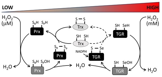
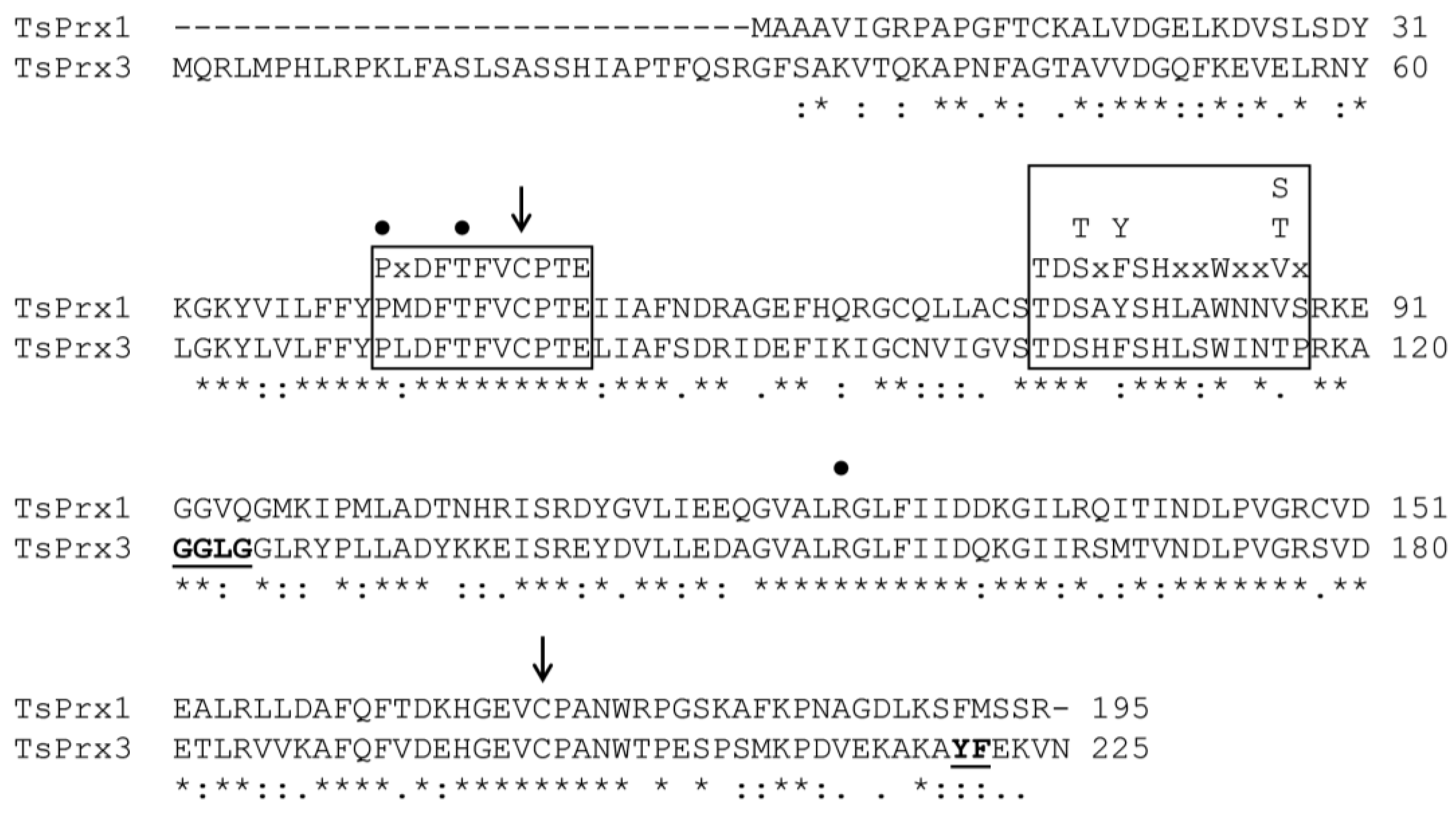
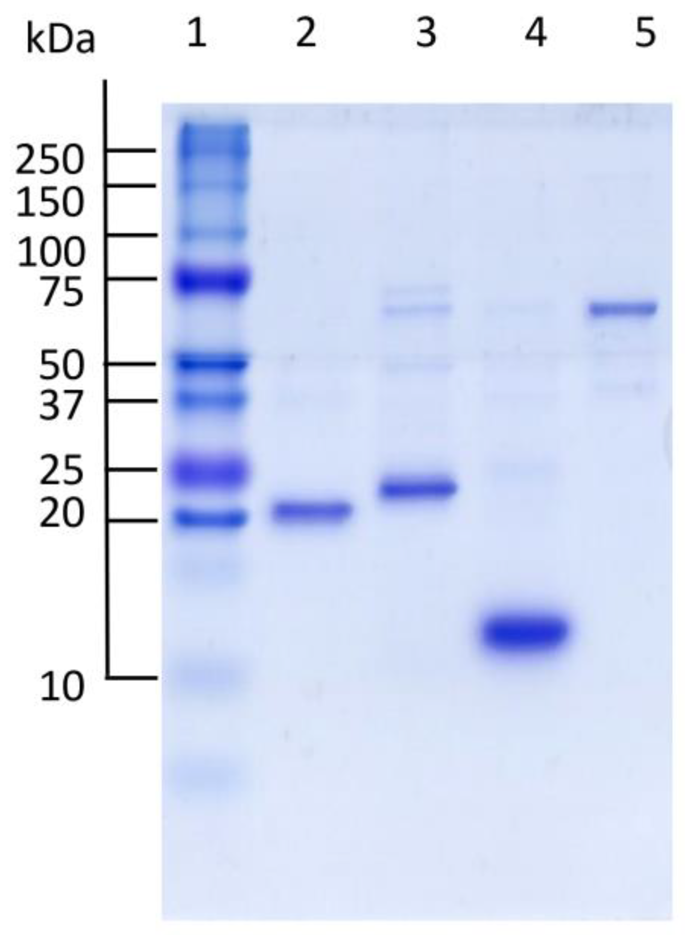
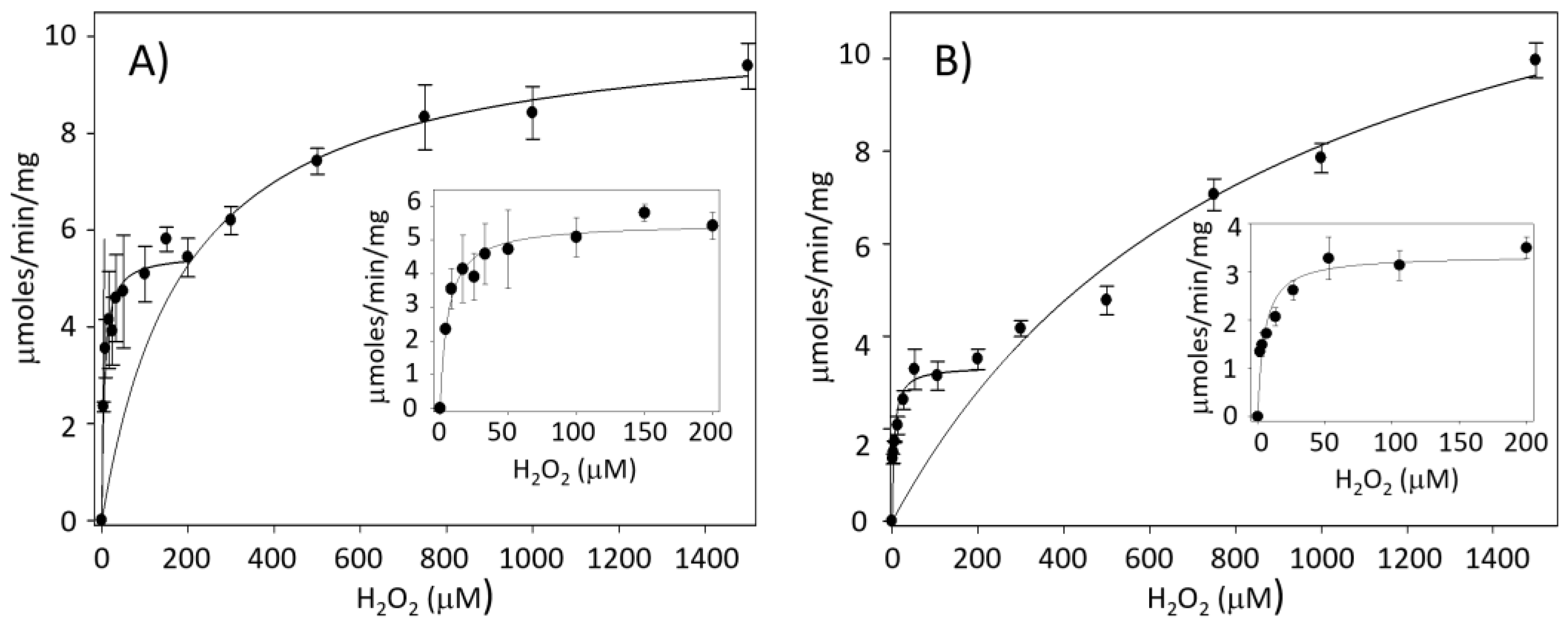
A Physiological Approach to Explore How Thioredoxin–Glutathione Reductase (TGR) and Peroxiredoxin (Prx) Eliminate H2O2 in Cysticerci of Taenia

by
Alberto Guevara-Flores
1,
Gabriela Nava-Balderas
2,*,
José de Jesús Martínez-González
1,
César Vásquez-Lima
1,
Juan Luis Rendón
1 and
Irene Patricia del Arenal Mena
1,*
1
Departamento de Bioquímica, Facultad de Medicina, Universidad Nacional Autónoma de México (UNAM), Apartado Postal 70-159, Mexico City 04510, Mexico
2
Departamento de Microbiología y Parasitología, Facultad de Medicina, Universidad Nacional Autónoma de México (UNAM), Apartado Postal 70-159, Mexico City 04510, Mexico
*
Authors to whom correspondence should be addressed.
Antioxidants 2024, 13(4), 444; https://doi.org/10.3390/antiox13040444
Submission received: 1 February 2024 / Revised: 2 April 2024 / Accepted: 4 April 2024 / Published: 10 April 2024
(This article belongs to the Section Antioxidant Enzyme Systems)

Abstract

:
Peroxiredoxins (Prxs) and glutathione peroxidases (GPxs) are the main enzymes of the thiol-dependent antioxidant systems responsible for reducing the H2O2 produced via aerobic metabolism or parasitic organisms by the host organism. These antioxidant systems maintain a proper redox state in cells. The cysticerci of Taenia crassiceps tolerate millimolar concentrations of this oxidant. To understand the role played by Prxs in this cestode, two genes for Prxs, identified in the genome of Taenia solium (TsPrx1 and TsPrx3), were cloned. The sequence of the proteins suggests that both isoforms belong to the class of typical Prxs 2-Cys. In addition, TsPrx3 harbors a mitochondrial localization signal peptide and two motifs (-GGLG- and -YP-) associated with overoxidation. Our kinetic characterization assigns them as thioredoxin peroxidases (TPxs). While TsPrx1 and TsPrx3 exhibit the same catalytic efficiency, thioredoxin–glutathione reductase from T. crassiceps (TcTGR) was five and eight times higher. Additionally, the latter demonstrated a lower affinity (>30-fold) for H2O2 in comparison with TsPrx1 and TsPrx3. The TcTGR contains a Sec residue in its C-terminal, which confers additional peroxidase activity. The aforementioned aspect implies that TsPrx1 and TsPrx3 are catalytically active at low H2O2 concentrations, and the TcTGR acts at high H2O2 concentrations. These results may explain why the T. crassiceps cysticerci can tolerate high H2O2 concentrations.

Graphical Abstract

1. Introduction

Reactive oxygen species (ROS) including the superoxide anion, the hydroxyl radical, the O2 singlet, and hydrogen peroxide (H2O2) are among the compounds resulting from aerobic metabolism. H2O2 possesses characteristics that reveal its relevance inside cells, such as the following: (I) it has no charge; (II) it is a very stable molecule compared to other ROS, and consequently, it has the longest half-life; (III) it has the highest diffusion rate, which allows it to diffuse in the whole cell; and (IV) at low concentrations, it acts as a second messenger in signaling pathways [1,2]. To avoid the deleterious accumulation of H2O2, organisms rely upon diverse metal-dependent peroxidases, including catalase (CAT) [3] and two thiol-dependent (-SH) antioxidant systems: (a) the glutathione system, composed of glutathione tripeptide (GSH), glutathione reductase (GR), and glutathione peroxidase (GPx), and (b) the thioredoxin system, composed of the small protein thioredoxin (Trx), thioredoxin reductase (TrxR), and peroxiredoxin (Prx). Both thiol-dependent antioxidant systems require NADPH [4]. Together, these systems regulate the H2O2 concentration, which maintains an adequate intracellular redox homeostasis in most organisms [5]. It is important to note that CAT is usually confined to peroxisomes [6], and in many endoparasitic organisms like cestodes, this enzyme is absent [7]. In contrast, GPxs and Prxs are present in most organisms, with different isoforms found in diverse cell compartments [8]. Prxs are characterized mainly by two points: (1) their catalytic efficiency (kcat/Km) for H2O2 is lower (104−5 M−1 s−1) [9,10,11], and this low catalytic efficiency is compensated by (2) their high intracellular concentration that ranges between 15 and 60 μM [12].
All Prxs depend on the presence of a catalytic cysteine around position 50 (Cys~50) that reacts with H2O2; identified as peroxidatic cysteine (CPSH). Based on this, Prxs are most often classified by the number of catalytic cysteine residues per subunit. Prxs with one cysteine (Prx 1-Cys) and two cysteines (Prx 2-Cys) exist. For Prx 2-Cys, the second cysteine (Cys~170) was identified as the resolving cysteine (CRSH) [13,14]. The reduction of H2O2 is performed through the oxidation of CPSH to sulfenic acid (CpSOH); subsequently, this sulfenic acid reacts with CRSH, generating a disulfide bond (CPS-SCR). When this disulfide bond is intermolecular, the Prxs are “typical”, and when it occurs in the same subunit, they are described as “atypical” [15]. In both cases, the disulfide bond is generated anew to its dithiol form by the reduced forms of thioredoxin (Trx-(SH)2) or glutathione (GSH) [16]. A shared feature by most Prxs is that they are sensitive to overoxidation, with micromolar concentrations of H2O2, and are known as “sensitive Prxs” [16,17,18]. In sensitive Prxs, two structural motifs (-GGLG- and -YP-) have been described that are predicted to confer sensitivity to H2O2. These sites are highly conserved among the Prxs of eukaryote cells; however, recently, “robust Prxs” (resistant to overoxidation) have been reported in bacteria including Escherichia coli and Salmonella, which lack these motifs and instead harbor two highly conserved motifs that have been associated with resistance to H2O2 [19]: (A and B: -D(N/G)H(G/S)- and -T(S/T)-, respectively).
The enzymatic activity of Prxs was determined with an assay coupled with TrxR and Trx using as reducer to NADPH. Generally, for this assay, the enzyme coupling of E. coli [20,21] and yeast [22] are the most used. These organisms’ reductases lack a Sec residue, so their TrxR is termed TrxR-Cys. Markedly, endogenous proteins were used to determine the activity of Prxs for a few organisms, like Plasmodium falciparum (PfTrxR-Cys and PfTrx) [23]. The eukaryotic TrxRs are selenocysteine-dependent (termed TrxR-Sec) and generally have the capacity to recognize Trxs of another origin as substrates [24], whereas the TrxR-Cys of prokaryotes are usually highly specific for their own endogenous Trx [24,25]. On the other hand, the specificity of Prxs for Trxs of other origins is not well documented. This information is relevant to establish which system is more appropriate to determine the activity of Prxs in a physiological context.
Parasite plathelminths of the cestode class must have a robust mechanism for the depuration of ROS that are either generated by the host’s immunological system [26] or from their own metabolism [26,27]. Studies performed in Taenia crassiceps have demonstrated significant amounts of H2O2 production under basal conditions [28], and the larval form can tolerate exposure to higher concentrations of H2O2 in culture conditions [29]. However, it is widely documented in diverse parasitic platyhelminths at both the genomic and proteomic levels that cestodes lack CAT, TrxR, and GR [7,30]; hence, their redox homeostasis relies on a bifunctional enzyme: the thioredoxin–glutathione reductase (TGR-Sec), which is the sole enzyme responsible for maintaining both thioredoxin and glutathione in their reduced state. Regarding the thiol-dependent peroxidases, a gene that encodes a GPx has been previously described in the Taenia solium genome, which is predicted to be associated with the plasma membrane [31], as well as two genes that encode 2-Cys Prxs isoforms [31].
This work aimed to identify the factors involved in the high tolerance of the Taenia genus to millimolar concentrations of H2O2. In this study, two Prxs of the T. solium cysticerci were cloned and expressed. Here, we characterize how they remove H2O2 using their endogenous thioredoxin system and the role of T. crassiceps cysticerci (Tc) TGR-Sec in this process.

2. Materials and Methods

2.1. Chemicals

NADPH, H2O2, ter-butyl hydroperoxide solution (Luperox), cumene hydroperoxide, Trizol®, bacto yeast, bacto tryptone, IPTG, ampicillin, and chloramphenicol, as well as Tris, EDTA, oxidized glutathione (GSSG), reduced glutathione (GSH), PMSF, manganese (II) chloride, L-glutamine, hydroxylamine, ADP, DEAE-cellulose, HA-Ultrogel, and Cibacron Blue 3G-A were obtained from Sigma-Aldrich, Merck KGaA, (Darmstadt, Germany). All other chemicals were purchased from JT Baker Chemical, Phillipsburg, NJ, USA.

2.2. Biological Material

T. solium cysticerci were obtained from the skeletal muscle of naturally infected pigs from City of Cuautla, State of Morelos, México. The cysticerci were washed with phosphate buffer (PBS), pH 7.4, and frozen until use. T. crassiceps cysticerci (HYG strain) were obtained from the peritoneum of experimentally infected BALB/c mice as described [32], washed with PBS, and frozen until use. All animal care and research protocols were carried out in accordance with the guidelines for the ethical care of experimental animals according to the guidelines of the Official Mexican Standards for the production, care, and use of laboratory animals (NOM-062-ZOO-1999). Further, the experimental protocols reported in the present work were approved by the Internal Committee for the Care and Use of Laboratory Animals (CICUAL) of the Facultad de Medicina, Universidad Nacional Autónoma de México (008-CIC-2023). All efforts were made to minimize animal suffering and to reduce the number of animals used.

2.3. Cloning and Overexpression of TsPrx1 and TsPrx3

Plasmid pET-23a (+) was obtained from Novagen®, Merck KGaA group (Darmstadt, Germany). E. coli strains TOP10 and BL-21 Codon Plus (DE3) were purchased from the Invitrogen corporation (Carlsbad, CA, USA). The plasmid purification kit was obtained from Thermo-Scientific (Waltham, MA, USA), as were the NdeI and Xho I restriction enzymes and the RevertAid First Strand synthesis kit used to obtain the cDNA. The amplified (TAQ DNA polymerase) was obtained from BioTecMol (Mexico City, Mexico). T4 DNA ligase was purchased from Promega Corporation (Madison, WI, USA), and the GelRed® was obtained from Biotium (Fremont, CA, USA). Trx from E. coli, Trx from humans, TrxR from rats, TrxR from E. coli, and GR from yeast, were obtained from Sigma-Aldrich.
Two Prxs coding sequences were identified in the WormBase Parasite (https://parasite.wormbase.org/Taenia_solium_prjna170813/Info/Index/, accessed on 2 August 2022): TsPrx1 (22 kDa) [33] and TsPrx3 (25 kDa). The total RNA from three T. solium cysticerci was extracted with TriZol®, and the cDNA synthesis was carried out using the RevertAid First Strand synthesis kit (Thermo Scientific, Waltham, MA, USA) with the supplier’s specifications and using oligo (dT)12 primer (5 μM final concentration). The synthesized cDNA (2 μL) was used as a template to amplify the Prxs genes by means of PCR reactions (50 μL total volume), using 100 ng/μL (0.2 μM) of each oligonucleotide 5′-ATTCATATGGCTGCTGCTGTCATCGGG-3′ and 3′-AAACTCGAGTCTTGAGCTCATGAACGAC-5′ for the TsPrx1 isoform; for TsPrx3, the oligonucleotides 5′AAGCATATGCAGCGTCTTATGCCTCATC-3′ and 3′TATCTCGAGGTTGACCTTCTCAAAGTACGC-5′ were used. The PCR reactions were carried out at an initial incubation temperature of 94 °C for 30 s; the alignment temperature was 61 °C for 35 s, and the extension temperature was 72 °C for 90 s; the final extension temperature was 72 °C for 10 min. PCR products were analyzed by electrophoresis on a 1.5% agarose gel with known molecular weight (MW) markers and visualized with GelRed® at λ = 312 nm; the products were purified and sequenced via the Sanger method [34] at the Sequencing Unit of the Institute for Biomedical Research (IIB, Cuernavaca, Morelos, Mexico). The resultant sequences were aligned and compared with the sequences of TsPrx1 and TsPrx3 identified in the GeneBank database, using the NCBI BLAST page (https://blast.ncbi.nlm.nih.gov), accessed on 29 September 2023.
The amplified TsPrx1 and TsPrx3 genes were cloned into the pET-23a(+) expression vector (Novegen, Dublin, Ireland), using the NdeI and XhoI cutting sites. The constructs were used to transform E. coli TOP 10 and Codon Plus bacteria. Positive clones were identified by PCR reactions with the specific oligos. Codon Plus positive bacterial clones were grown in LB culture (Luria–Bertani) with ampicillin (0.1 mg/mL) and chloramphenicol (34 μg/mL). The induction of the expression of clones TsPrx1 and TsPrx3, both with His tags in their amino terminal ends, was carried out by adding 1 mM IPTG at 37 °C and 300 rpm. After 4 h of incubation, bacteria were recovered by centrifugation and lysed by sonication at a frequency of 20 KHz. The expression of TsPrx was confirmed by SDS-PAGE according to Laemmli [35] and stained with Coomassie blue. The Prxs were purified from the soluble bacterial lysate by affinity chromatography on IMAC Sepharose (BioRad, Hercules, CA, USA). The protein concentration was determined with the extinction coefficient (ε) of each protein [36].

2.4. Purification of the TGR from T. crassiceps and Recombinant Trx from T. solium

The protocol followed in the purification of cytosolic TGR from T. crassiceps has been described elsewhere [37], using 20 infected mice (around 400 cysticerci per mouse). The recombinant Trx from T. solium was obtained following the protocol previously described [38].

2.5. Bioinformatics Analysis

The amino acid sequence alignment of TsPrx1 and TsPrx3 was performed using the Clustal Omega program (https://www.uniprot.org/align/, accessed on 19 October 2022). The prediction of the subcellular location of an N-terminal peptide corresponding to TsPrx3 was performed using DeepLoc-1.0 (https://services.healthtech.dtu.dk/services/DeepLoc-1.0/, accessed on 6 June 2023) and resulted in being mitochondrial-directed.

2.6. Electrophoresis

Polyacrylamide gel (4, 10, and 16%) electrophoresis under denaturing conditions was performed as described by Shägger [39]. Gels were stained by conventional procedures. The purity degree of the TsPrx1, TsPrx3, Trx proteins from T. solium and the TGR from T. crassiceps was established by analyzing the densitometry of each protein in the SDS-PAGE using ImageJ (https://imagej.nih.gov/ij/), accessed on 8 November 2023.

2.7. Protein Determination

The concentration of TsPrx1, TsPrx3, and TsTrx was determined by measuring their absorbance at 278 nm. The corresponding extinction coefficients (ε) were as follows: TsPrx1 = 20.6 mM−1 cm−1; TsPrx3 = 21.4 mM−1 cm−1; and TsTrx = 7.8 mM−1 cm−1. For TcTGR, its protein concentration was determined at 460 nm based on its FAD content (ε = 11.3 mM−1 cm−1). The protein concentration was corroborated by the densitometric method [40].

2.8. Enzyme Assays

This section pertains to the thioredoxin reductase activity of TcTGR. The reductase activity was determined by following the decrease in absorbance at 340 nm due to the oxidation of NADPH (150 μM) in the presence of recombinant TsTrx. Assays were performed at 25 °C in 100 mM Tris-HCl buffer (pH 7.8) containing 1 mM EDTA (TE buffer) in a final volume of 0.6 mL. The reaction was started by adding insulin (to recycle TsTrx) at a final concentration of 25 μM. An extinction coefficient of 6.22 mM−1 cm−1 for NADPH was used for the calculations of enzyme activity, as described previously [37].
Here. the activity of the peroxiredoxins from T. solium is discussed. This activity was determined by either of the two methods described below. The final volume of the reaction mixture was 0.3 mL. Unspecific rates were subtracted from the specific rates. All activity assays were carried out in a UV/Vis spectrophotometer DU-730 from Beckman Coulter.

2.8.1. Peroxidase Activity Assays

The reductase activity of the recombinant Prxs using either H2O2 or organic hydroperoxides (cumene hydroperoxide and t-butyl hydroperoxide) as oxidizing substrates was determined in TE buffer by following the oxidation of 150 μM NADPH at 340 nm and 25 °C in a coupled assay with TcTGR (11.2 nM), TsPrx1 or TsPrx3 (1.25 μM) and recombinant TsTrx (60 μM), and the latter was tested as a reductant substrate; under these conditions, when the maximum reduction was obtained (baseline), the specific reaction was started by adding the corresponding peroxide. One unit of Prx activity was defined as the amount of enzyme required to cause the oxidation of 1 nmol of NADPH per minute under the assay conditions described. Alternatively, the peroxidase activity with GSH as the reductant was assayed with GR from Saccharomyces cerevisiae or TcTGR (own reductase). The reaction mixture contained the following: (a) 0.1 unit/mL ScGR (Sigma) or (b) 11.2 nM TcTGR, 150 μM NADPH, 1.25 μM of TsPrx1 or TsPrx3, and 1 mM GSH in a buffer containing 100 mM sodium phosphates (pH 7.0), 1 mM EDTA. The reaction was initiated by adding the corresponding peroxide, and the consumption of NADPH was recorded by following the decrease in absorbance at 340 nm and 25 °C.

2.8.2. Thioredoxin Peroxidase Activity of TcTGR and EcTrxR

The comparison of a selenocysteine-dependent enzyme (TcTGR) with a Cys-dependent enzyme (EcTrxR), regarding its ability to catalyze the Trx-dependent reduction of H2O2, was evaluated by mixing 150 μM NADPH with either 60 μM TsTrx and 11.2 nM TcTGR or 6 μM EcTrx and 83 nM EcTrxR in TE buffer. The reaction was started by adding 1 mM H2O2, and the absorbance at 340 nm was measured. The final volume of the reaction mixture was 0.6 mL.
The kinetic constants Km and kcat of TsPrx1 and TsPrx3 for either H2O2, t-butyl hydroperoxide, or the cumene hydroperoxide substrates were determined by varying the concentration of the corresponding peroxide at a constant concentration of both NADPH (150 μM) and TsTrx (60 μM). To obtain the kinetic parameters for TsTrx, a constant concentration of 50 μM H2O2 was used at varying TsTrx concentrations. In all cases, fixed concentrations of TsPrxs (1.25 μM) and TcTGR (11.2 nM) were used (these last concentrations were previously determined to prevent them being limiting). The kinetic constants of TcTGR toward H2O2 was obtained by varying the concentration of the peroxide at a constant concentration of NADPH (150 μM) and TsTrx (60 μM). All initial velocity data were fitted to the Michaelis–Menten equation through non-linear regression analysis using Sigma-Plot Software version 12.

2.8.3. Glutamine Synthetase Protection Assay

The ability of cytosolic TcTGR, TsPrx1, and mitochondrial TsPrx3 to protect glutamine synthetase (GS) from oxidation was performed as previously described [41]. For both TsPrx1 and TsPrx3, the inactivation mixture contained 0.15 μM GS from E. coli, 3 μM FeCl3, and 10 mM DTT either in the presence or in the absence of 1.25 μM of the corresponding Prx in 50 mM HEPES buffer (pH 7). The final volume of the mixture was 50 μL. For TcTGR, the inactivation mixture additionally contained 160 μM NADPH and recombinant TsTrx either in the presence or in the absence of TcTGR. After 15 min of incubation at 30 °C, the residual activity of GS was determined by adding 1 mL of the assay mixture (0.4 mM ADP, 0.15 M glutamine, 10 mM Na2HAsO4, 20 mM NH2OH, and 0.4 mM MnCl2 in 100 mM HEPES buffer), pH 7.4. The resultant solution was incubated for 30 min at 30 °C; then, the reaction was terminated by adding 0.25 mL of stop mixture (0.3 M FeCl3 and 5.8 M HCl), and the formation of the γ-glutamylhydroxamine-Fe3+ complex was measured at 540 nm.

2.9. Data Presentation and Statistical Analysis

The data shown below represent the mean ± S.D. of three independent experiments. Data were evaluated for statistical significance using Student’s t-test and Statistical Software OriginPro (version 8, OriginLab Corporation, Northampton, MA, USA).

3. Results

3.1. Recombinant Peroxiredoxins

Two Prxs of T. solium cysticerci were cloned and expressed: TsPrx1 as described in [33] and TsPrx3. The Prx1 gene has been reported previously in the T. solium genome with the number TsM_001155200. This gene is 655 bp in length with two exons, the first 134 bp, and the second one 454 bp with a 67 bp intron. The two exons encode a protein sequence of 195 residues. The sequence reported for a second peroxiredoxin (TsPrx3) in the WormBase indicates a length of 2176 bp constituted by four exons of 357, 235, 30, and 86 bp and three introns with a length of 80, 1150, and 238 bp. The splicing of the two exons encodes a sequence of 235 amino acid residues. However, in the present work, a sequence of 224 amino acid residues was obtained because exon 3 is fused with introns 2 and 3 so that the gene for TsPrx3 is constituted by three exons of 357, 235, and 86 bp and two introns of 80 and 1418 bp.
Figure 1 depicts the sequences. TsPrx1, previously reported by Molina-López et al. [33], corresponds to a cytosolic Prx with a MW of ~22 kDa, whereas TsPrx3 has a MW of ~25 kDa. These two proteins are 56.4% identical. TsPrx3 has 29 additional residues in its N-terminal end. It was therefore analyzed with the DeepLoc-1.0 program to pinpoint its subcellular location.
This peptide resulted a mitochondrial recognition signal with a probability of ~0.44 (Figure S1A). A similar result (0.57) was obtained with the hierarchical tree method (Figure S1B). Both results suggest that it must correspond to a mitochondrial isoform (TsPrx3), comparable to that reported for the mitochondrial Prxs of Haemonchus contortus and Caenorhabditis elegans, which also have a signaling peptide [42]. Both Prxs can be classified as “typical” because of the presence of two essential resolving cysteines: the peroxidatic cysteine49 (CP) localized in the N-terminal end and the cysteine170 (CR) located in the C-terminal end. A relevant difference between the two Prxs is the additional presence of two motifs (-GGLG-) and (-YF-) in the TsPrx3 isoform (Figure 1), suggesting a possible higher sensitivity to H2O2.

3.2. Purity Degree of Recombinant Proteins

The degree of purity of TsPrx1 and TsPrx3 and TsTrx recombinant proteins, and that of TcTGR, was determined through electrophoresis in denaturing conditions (SDS-PAGE). Figure 2 shows that the four proteins had a significant degree of purity, which was confirmed through densitometry analysis of each band, revealing a purity greater than 75% for all proteins.

3.3. Peroxidase Activity of the Recombinant TsPrx1 and TsPrx3

Based on results (see below), 60 μM of TsTrx was used in the activity assays, one and a half times the Km for TcTGR. For GSH, 1 mM of GSH was used, which corresponds to the concentration reported in T. crassiceps cysticerci [27]. The activity of TsPrx1 and TsPrx3 was determined by changing the concentration of H2O2 (Table 1).

3.4. Kinetic Analysis of TcTGR

Kinetic constants of TcTGR were determined using TsTrx as a substrate with the following results: Km = 41.5 μM and kcat/Km = 1.2 × 10⁶ M−1 s−1 (Table S1); despite having different Km, the catalytic efficiency values were comparable to those reported previously [43] and those reported for TsTGR and the recombinant TsTrx [38]. Additionally, the comparison of the TsTGR gene (ID: TsM_000506200) of the T. solium genome submitted in WormBase Parisite database (GENOME ID: PRJNA170813) and the TcTGR gene (ID: JAKROA010000003.1) submitted in the GenBank database (GENOME ID: GCA_023375655.1.) showed an identity above 90%, and the genomic sequences of the TsPrx1 and TcPrx1 genes showed 94% identity [29]. These data suggest that independently of the origin of the proteins used in the activity assays, either of T. solium or T. crassiceps, the kinetic parameters were within the same range, and the high rates of identity of the sequences protein or genomics of the different components of the thioredoxin system (TS) enabled us to use TcTGR and the recombinant TsTrx with confidence in our assays.
Unexpectedly, in Figure S2, it is shown that when using GSH as substrate, it was not possible to detect the peroxidase activity in TsPrx1 and TsPrx3 in the presence of ScGR or TcTGR in the coupled assay (as mentioned under Materials and Methods Section 2). Table 1 and Table S2 show that the two Prxs depict a high affinity for different peroxides (Km < 8.4 μM), except for TsPrx1, whose affinity for the t-butyl hydroperoxide was significantly lower (Km = 18.1 μM). The catalytic efficiency for the different peroxides was about ~104 M−1 s−1; these kinetic parameters were within the same order of magnitude as other Prxs [10,11].
Peroxidase activity was not detected using GSH and with other organic peroxides as oxidizing substrates. Afterward, the kinetic constants for both Prxs toward the Trx were determined at a constant concentration of 50 μM of H2O2. The results obtained are shown in Table 2. It is interesting to point out that the affinity of TsPrx3 for TsTrx was significantly lower compared to that of TsPrx1. Again, GSH was not efficient as a reducer.

3.5. Dependence of the Peroxidase Activity of TsPrx1 and TsPrx3 on the H2O2 Concentration

As mentioned, only TsPrx3 has the two motifs that provide sensitivity to H2O2 in its sequence. To determine the susceptibility of both Prxs to H2O2, peroxidase activity was analyzed with a wide range of H2O2 concentrations. Figure 3 shows the saturation curves of both enzymes with a clear biphasic pattern, suggesting the presence of two components with peroxidase activity. A comparison of the two activity profiles reveals that the apparent maximal velocity of the component with the highest affinity is higher for the assays with TsPrx1.
However, in both cases, the total maximal velocity is essentially identical. A non-linear regression analysis yielded the corresponding kinetic parameters for both systems (Table 3). Because TcTGR is present as an auxiliary enzyme in the activity assays of both TsPrxs, it is possible that one of the components observed in the saturation graphs could be due to TcTGR. Consequently, the potential activity of the peroxidase of TcTGR was analyzed in the absence of TsPrxs (Figure S3). The results revealed that the peroxidase activity of TcTGR is significant (Km: 79.8 μM), overlapping with the activity observed in the assays performed in the presence of TsPrx1. Therefore, it can be concluded that the main contribution of the peroxidase activity is exerted by TcTGR, particularly at high H2O2 concentrations. Despite its significantly lower affinity for the peroxide, the catalytic efficiency of TcTGR is approximately five and eight times higher than that of TsPrx1 and TsPrx3, respectively (Table 3).

3.6. Peroxidase Activity of TrxR of E. coli

To determine whether the selenocysteine (Sec) residue plays a critical role in the high peroxidase activity of the TGR, its activity was compared to that of a TrxR lacking such residue, using the enzyme of E. coli. The results (Figure 4) revealed that the peroxidase activity of TcTGR was significantly higher (9.4 μmol min−1 mg−1) compared with that of EcTrxR (0.47 μmol min−1mg−1).

3.7. Protection of the Glutamine Synthetase

The peroxidase activity present in the TcTGR or TsPrxs and their consequent protective activity of the GS from ROS was compared. As shown in Figure 5A, in the presence of TS (NADPH + TsTrx + TcTGR), ~50% protection was obtained. The addition of TsPrx1 resulted in ~80% protection, whereas TsPrx3 did not protect and had the same magnitude regarding protection as TS (Figure 5B).

4. Discussion

Peroxiredoxins, enzymes that reduce H2O2, are widely represented among organisms [44]. A search in the T. solium genome revealed that this parasite possesses two peroxiredoxins: TsPrx1 and TsPrx3. The sequence analysis of both Prxs was performed, and the TsPrx3 sequence showed the presence of a signaling peptide, suggesting its localization to mitochondria (Figure S1). Both sequences indicated that they could be classified within the “typical Prx 2-Cys” group (Figure 1). The TsPrx3 isoform harbored the motifs (-GGLG-) and (-YP-) associated with the hyperoxidation produced by H2O2 [16,17]. Interestingly, the presence of these motifs in TsPrx3 did not confer a higher or lower kinetic behavior compared to TsPrx1. Both peroxiredoxins could recognize H2O2 with a catalytic efficiency of ~104 M−1 s−1 (Table 1), which is comparable to other organic peroxides (Table S2) used in the present work as substrates.
TsPrx’s affinity for H2O2 is clearly higher if compared with the Prxs of the Schistosoma mansoni trematode [20]. However, its catalytic efficiency is comparable to those reported for the Prxs of diverse organisms, such as the H. contortus nematode [42], Bacillus subtilis [45], and Helicobacter pylori [46], which reduce H2O2 using only Trx-(SH)2 and do not recognize GSH. In contrast, Prxs that can use both GSH and Trx-(SH)2 have been reported in P. falciparum [23,47], S. mansoni [20], and Clonorchis sinensis [21]. It is interesting to point out that among Prxs that use both reducing substrates, some, including P. falciparum and S. mansoni, use GSH more efficiently as a substrate. The results of this work indicate that GSH cannot serve as a reducing substrate and therefore is a marked preference for Trx-(SH)2; hence, we suggest that both TsPrx1 and TsPrx3 must be considered true thioredoxin peroxidases (TPx).
In Prx 2-Cys the CPSH thiol can reach different states of oxidation by reacting sequentially with one, two, or three H2O2 molecules, giving rise sequentially to sulfenic (CPSOH), sulfinic (CPSO2H), and sulfonic (CPSO3H) acids. The reaction needed to generate the CPSO2H is reversible through an ATP-dependent sulfiredoxin (Srx), whereas the reaction that generates the CPSO3H is irreversible [11,17]. The overoxidation of this thiol promotes the Prx to restructure and generate decamer-type oligomers (five homodimers, also known as “toroids”). At this point, the antioxidant activity of the Prx diminishes, favoring its transformation into a protein with a chaperone function. Only “typical” Prxs are believed to generate this type of oligomer [44] due to the presence of the motifs sensitive to H2O2, which are absent in TsPrx1 (Figure 1). This suggests that TsPrx1 could be a robust Prx similarly to that of the Prx (AhpC) of Salmonella typhimurium [19].
As previously mentioned, the low peroxidase activity of the Prxs could be related to the fact that the catalytic residues are cysteines [16,17,44], in contrast with those GPx selenium-dependent (GPx-Sec), which are generally more active [47]. The insertion of a Sec residue in a protein through site-directed mutagenesis enables enzymes to use a greater spectrum of substrates, including H2O2. In addition, the substitution of the essential serine residue by a Sec residue (Ser/Sec) in the subtilisin protease led to a loss of its original activity and the acquisition of a peroxidase activity [48]. A similar result was obtained for the GPx-Sec: the substitution of Sec residue with a Cys residue drastically reduced its activity and increased its sensitivity to overoxidation by H2O2 [49]. In our study, we found that TcTGR possesses an essential Sec residue that is likely responsible for its high peroxidase activity (Figure 4). Calculations of the initial velocity, using H2O2 as a substrate, revealed a 20 times higher activity compared to the activity of EcTrxR. These data support the important role of the Sec residue in the peroxidase activity of this enzyme.
The results shown in Figure 3 and Table 3 reveal that TcTGR contributes greatly to reduce H2O2. This suggests that when the assay system contains TGR and Prx, the peroxidase activity observed at low H2O2 concentrations is due mainly to TsPrx1 and TsPrx3, whereas at high concentration of the peroxide, where TsPrxs are already saturated, the reducing activity must be attributed to TcTGR. Additionally, in the intact organism, the TGR and its corresponding Prx coexist physiologically and are present in both cytosol and mitochondria; hence, their relative participation in peroxides depuration will depend not only on their kinetic parameters but also on the concentration in each organelle. In this sense, it is well known that the peroxiredoxins represent an important fraction of the total protein in a large variety of organisms, reaching up to 1% of the total soluble protein [44]. In this case, in the cestodes, it will be necessary to assess the concentration of these enzymes in intact organisms to obtain conclusive evidence about their relative importance in H2O2 depuration. We found no significant differences in the kinetic parameters for TsPrxs1 and TsPrx3, under the conditions used in this study. However, we found differences between the two Prxs in the GS protection assay (Figure 5). TsPrx3 does not protect GS from oxidative damage, possibly because this isoform harbors the motifs sensitive to overoxidation. On the other hand, TcTGR and TsPrx1 do protect GS from oxidative damage.
As mentioned in the Introduction, T. crassiceps cysticerci can tolerate high H2O2 concentrations in the millimolar range [28,29]. Although, under physiological conditions it is barely probable to reach such levels, the kinetic characteristics described for TcTGR (Km ~200 μM by H2O2 and Vmax ~10.36 μmol min−1 mg−1) seem to have evolved to work in the presence of moderately high H2O2 concentrations. TsPrxs1 and TsPrx3 have significantly higher affinities for the peroxide, compared with TcTGR (Table 3), which suggests that these enzymes constitute the first in vivo line of defense to avoid oxidative damage. Although using the TcTGR of the parasite in the present work as a coupling enzyme exceeded the activity of TsPrxs, its presence in the enzymatic assays reflects a situation closer to the physiological conditions of the parasite where the three enzymes act in the presence of the others. Hence, in these types of parasites, two very efficient systems have evolved for removing H2O2, one cytosolic represented by the cytosolic TGR and Prx1 and another mitochondrial that involves the mitochondrial TGR variant and Prx3.

5. Conclusions

The high peroxidase activity of TGR within TS could explain two relevant aspects in the physiology of the T. crassiceps cysticerci: (a) the tolerance of the parasite to millimolar H2O2 concentrations [29] and (b) the lack of the CAT gene in trematodes and cestodes [7]. The Prx/Trx/TGR system would compensate for the catalase activity, highlighting TGR’s role in redox homeostasis in these two groups of parasites.

Supplementary Materials

The following supporting information can be downloaded at: https://www.mdpi.com/article/10.3390/antiox13040444/s1, Figure S1: Prediction of TsPrx3 subcellular localization. Peptide (–MQRLMPHLRPKLFASLSASSHIAPTFQSR-) of TsPrx3 was analyzed using DeepLoc-1.0. (A) Table of predicted subcellular localization. (B) Hierarchical tree; Figure S2: Activity of TsPrx1 and TsPrx3 was determined with the glutathione system. Assays containing 1 mM GSH, 150 µM NADPH, 1.25 µM TsPrx1 (circles) or TsPrx3 (filled circles) were incubated with: (A) 8 nM ScGR (squares) or (B) 11.2 nM TcTGR (squares) in 100 mM Tris/HCl (pH 7.8) 1 mM EDTA for 5 min to allow the reaction to stabilize. H2O2 (50 µM) was added to initiate the reaction that was monitored for 6 min at 25 °C; Figure S3: Thioredoxin peroxidase activity of TcTGR. H2O2 reduction was measured in the absence of TsPrx1. Measurements obtained as described under Materials and Methods Section 2. Data are the means of three independent measurements; Table S1: Kinetic constants for TsTGR and TcTGR toward recombinant Trx from T. solium and Trx from T. crassiceps; Table S2: Kinetic constants for recombinant TsPrx1 and TsPrx3 toward hydroperoxides in the presence of TsTrx.

Author Contributions

Conceptualization, A.G.-F. and G.N.-B.; methodology, A.G.-F., G.N.-B. and C.V.-L.; software, A.G.-F.; validation, J.d.J.M.-G., C.V.-L. and J.L.R.; formal analysis, A.G.-F., J.d.J.M.-G., J.L.R. and I.P.d.A.M.; investigation, A.G.-F. and G.N.-B.; resources, I.P.d.A.M.; data curation, I.P.d.A.M., J.d.J.M.-G. and J.L.R.; writing—original draft preparation, A.G.-F. and G.N.-B.; writing—review and editing, A.G.-F., G.N.-B., J.d.J.M.-G., J.L.R., J.d.J.M.-G. and J.L.R.; visualization, A.G.-F. and J.d.J.M.-G.; supervision, I.P.d.A.M.; project administration, I.P.d.A.M.; funding acquisition, I.P.d.A.M. All authors have read and agreed to the published version of the manuscript.

Funding

This work was supported by the research grant IN215223 from Dirección General de Asuntos del Personal Académico (DGAPA) at Universidad Nacional Autónoma de México (UNAM).

Institutional Review Board Statement

All animal care and research protocols were carried out in accordance with the guidelines for the ethical care of experimental animals according to the guidelines of the Mexican Official Standard for the production, care, and use of laboratory animals (NOM-062-ZOO-1999). Further, the experimental protocols reported in the present work were approved by the Internal Committee for the Care and Use of Laboratory Animals (CICUAL) of the Facultad de Medicina, Universidad Nacional Autónoma de México (008-CIC-2023). All efforts were made to minimize animal suffering and to reduce the number of animals used.

Informed Consent Statement

Not applicable.

Data Availability Statement

The authors declare that all data supporting the findings of this study are available within the article and its Supplementary Materials.

Acknowledgments

The authors are grateful to Rachel Duffié (Zuckerman Mind Brain and Behavior Institute, Columbia University, New York, NY, USA) for proofreading the English of the manuscript and to Sandra D. Rodríguez Montaño for technical support.

Conflicts of Interest

The authors declare no conflicts of interest.

References

  1. Winterbourn, C.C. Hydrogen peroxide reactivity and specificity in thiol-based cell signalling. Biochem. Soc. Trans. 2020, 48, 745–754. [Google Scholar] [CrossRef] [PubMed]
  2. Cardoso, A.R.; Chausse, B.; da Cunha, F.; Luévano-Martínez, L.A.; Marazzi, T.B.M.; Pessoa, P.S.; Queliconi, B.B.; Kowaltowski, A.J. Mitochondrial compartmentalization of redox processes. Free Radic. Biol. Med. 2012, 52, 2201–2208. [Google Scholar] [CrossRef] [PubMed]
  3. Sepasi Tehrani, H.; Moosavi-Movahedi, A.A. Catalase and its mysteries. Prog. Biophys. Mol. Biol. 2018, 140, 5–12. [Google Scholar] [CrossRef] [PubMed]
  4. Das, K.C.; White, C.W. Redox systems of the cell: Possible links and implications. Proc. Natl. Acad. Sci. USA 2002, 99, 9617–9618. [Google Scholar] [CrossRef]
  5. Lennicke, C.; Cochemé, H.M. Redox metabolism: ROS as specific molecular regulators of cell signaling and function. Mol. Cell. 2021, 81, 3691–3707. [Google Scholar] [CrossRef] [PubMed]
  6. Sies, H. Role of Metabolic H2O2 Generation. Redox Signaling and Oxidative Stress. J. Biol. Chem. 2014, 289, 8735–8741. [Google Scholar] [CrossRef]
  7. Williams, D.L.; Bonilla, M.; Gladyshev, V.N.; Salinas, G. Thioredoxin Glutathione Reductase-Dependent Redox Networks in Platyhelminth Parasites. Antioxid. Redox Signal. 2013, 19, 735–745. [Google Scholar] [CrossRef] [PubMed]
  8. Rhee, S.G.; Woo, H.A.; Kil, I.S.; Bae, S.H. Peroxiredoxin functions as a peroxidase and a regulator and sensor of local peroxides. J. Biol. Chem. 2012, 287, 4403–4410. [Google Scholar] [CrossRef] [PubMed]
  9. Flohé, L.; Budde, H.; Hofmann, B. Peroxiredoxins in antioxidant defense and redox regulation. BioFactors 2003, 19, 3–10. [Google Scholar] [CrossRef] [PubMed]
  10. Gretes, M.C.; Poole, L.B.; Karplus, P.A. Peroxiredoxins in parasites. Antioxid. Redox Signal. 2012, 17, 608–633. [Google Scholar] [CrossRef] [PubMed]
  11. Guevara-Flores, A.; Martínez-González, J.J.; Rendón, J.L.; Del Arenal, I.P. The Architecture of The Antioxidants Systems among Invertebrate Parasites. Molecules 2017, 22, 259. [Google Scholar] [CrossRef] [PubMed]
  12. Andreyev, A.Y.; Kushnareva, Y.E.; Murphy, A.N.; Starkov, A.A. Mitochondrial ROS Metabolism: 10 Years Later. Biochemistry 2015, 80, 517–531. [Google Scholar] [CrossRef] [PubMed]
  13. Barranco-Medina, S.; Lázaro, J.J.; Dietz, K.J. The oligomeric conformation of peroxiredoxins links redox state to function. FEBS Lett. 2009, 583, 1809–1816. [Google Scholar] [CrossRef] [PubMed]
  14. Rhee, S.G. Overview on peroxiredoxin. Mol. Cells. 2016, 39, 1–5. [Google Scholar] [CrossRef] [PubMed]
  15. Nelson, K.J.; Knutson, S.T.; Soito, L.; Klomsiri, C.; Poole, L.B.; Fetrow, J.S. Analysis of the peroxiredoxin family: Using active site structure and sequence information for global classification and residue analysis. Proteins 2010, 79, 947–964. [Google Scholar] [CrossRef] [PubMed]
  16. Wood, Z.A.; Schröder, E.; Robin, H.J.; Poole, L.B. Structure, mechanism, and regulation of peroxiredoxins. Trends Biochem. Sci. 2003, 28, 32–40. [Google Scholar] [CrossRef] [PubMed]
  17. Wood, Z.A.; Poole, L.B.; Karplus, P.A. Peroxiredoxin Evolution and the Regulation of Hydrogen Peroxide Signaling. Science 2003, 300, 650–653. [Google Scholar] [CrossRef] [PubMed]
  18. Hall, A.; Karplus, P.A.; Poole, L.B. Typical 2-Cys peroxiredoxins: Structures, mechanisms, and functions. FEBS J. 2009, 276, 2469–2477. [Google Scholar] [CrossRef] [PubMed]
  19. Bolduc, J.A.; Nelson, K.J.; Haynes, A.C.; Lee, J.; Reisz, J.A.; Graff, A.H.; Clodfelter, J.E.; Parsonage, D.; Poole, L.B.; Furdui, C.M.; et al. Novel hyperoxidation resistance motifs in 2-Cys peroxiredoxins. J. Biol. Chem. 2019, 293, 11901–11912. [Google Scholar] [CrossRef]
  20. Sayed, A.A.; Williams, D.L. Biochemical characterization of 2-Cys peroxiredoxins from Schistosoma mansoni. J. Biol. Chem. 2004, 279, 26159–26166. [Google Scholar] [CrossRef]
  21. Bae, Y.A.; Kim, S.H.; Lee, E.G.; Sohn, W.M.; Kong, Y. Identification, and biochemical characterization of two novel peroxiredoxins in a liver fluke, Clonorchis sinensis. Parasitology 2011, 138, 1143–1153. [Google Scholar] [CrossRef] [PubMed]
  22. Kim, J.A.; Park, S.J.; Kim, K.; Rhee, S.G.; Kang, S.W. Activity assay of mammalian 2-Cys peroxiredoxins using yeast thioredoxin reductase system. Anal. Biochem. 2005, 338, 216–223. [Google Scholar] [CrossRef] [PubMed]
  23. Krnajski, Z.; Walter, R.D.; Müller, S. Isolation, and functional analysis of two thioredoxin peroxidases (peroxiredoxins) from Plasmodium falciparum. Mol. Biochem. Parasitol. 2001, 113, 303–308. [Google Scholar] [CrossRef]
  24. Kanzok, S.M.; Schirmer, R.H.; Turbachova, I.; Lozef, R.; Becker, K. The thioredoxin system of the malaria parasite Plasmodium falciparum. Glutathione reduction revisited. J. Biol. Chem. 2000, 275, 40180–40186. [Google Scholar] [CrossRef] [PubMed]
  25. Kanzok, S.M.; Fechner, A.; Bauer, H.; Ulschmid, J.K.; Müller, H.M.; Botella-Munoz, J.; Schneuwly, S.; Schirmer, R.H.; Becker, K. Substitution of the Thioredoxin System for Glutathione Reductase in Drosophila melanogaster. Science 2001, 291, 643–646. [Google Scholar] [CrossRef] [PubMed]
  26. Vaca-Paniagua, F.; Torres-Rivera, A.; Parra-Unda, R.; Landa, A. Taenia solium: Antioxidant metabolism enzymes as targets for cestocidal drugs and vaccines. Curr. Top. Med. Chem. 2008, 8, 393–399. [Google Scholar] [CrossRef] [PubMed]
  27. Martínez-González, J.J.; Guevara-Flores, A.; Rendón, J.L.; Del Arenal, I.P. Auranofin-induced oxidative stress causes redistribution of the glutathione pool in Taenia crassiceps cysticerci. Mol. Biochem. Parasitol. 2015, 201, 16–25. [Google Scholar] [CrossRef] [PubMed]
  28. del Arenal, I.P.; Rubio, M.E.; Ramírez, J.; Rendón, J.L.; Escamilla, J.E. Cyanide-resistant respiration in Taenia crassiceps metacestode (cysticerci) is explained by the H2O2-producing side-reaction of respiratory complex I with O2. Parasitol. Int. 2005, 54, 185–193. [Google Scholar] [CrossRef] [PubMed]
  29. Vaca-Paniagua, F.; Parra-Unda, R.; Landa, A. Characterization of one typical 2-Cys Peroxiredoxin gene of Taenia solium and Taenia crassiceps. Parasitol. Res. 2009, 105, 781–787. [Google Scholar] [CrossRef] [PubMed]
  30. Gupta, A.; Kesherwani, M.; Velmurugan, D.; Tripathi, T. Fasciola gigantica thioredoxin glutathione reductase: Biochemical properties and structural modeling. Int. J. Biol. Macromol. 2016, 89, 152–173. [Google Scholar] [CrossRef] [PubMed]
  31. Tsai, I.J.; Zarowiecki, M.; Holroyd, N.; Garciarrubio, A.; Sánchez-Flores, A.; Brooks, K.L.; Tracey, A.; Bobes, R.J.; Fragoso, G.; Sciutto, E.; et al. Taenia solium Genome Consortium. The genomes of four tapeworm species reveal adaptations to parasitism. Nature 2013, 496, 57–63. [Google Scholar] [CrossRef] [PubMed]
  32. Esquivel-Velázquez, M.; Hernández, R.; Larralde, C.; Ostoa-Saloma, P. Crosstalk among Taenia crassiceps (ORF Strain) cysts regulates their rates of budding by ways of soluble and contact signals exchanged between them. Biomed. Res. Int. 2014, 2014, 703693. [Google Scholar] [CrossRef]
  33. Molina-López, J.; Jiménez, L.; Ochoa-Sánchez, A.; Landa, A. Molecular cloning and characterization of a 2-Cys peroxiredoxin from Taenia solium. J. Parasitol. 2006, 92, 796–802. [Google Scholar] [CrossRef] [PubMed]
  34. Sanger, F.; Coulson, A.R. A rapid method for determining sequences in DNA by primed synthesis with DNA polymerase. J. Mol. Biol. 1975, 94, 441–448. [Google Scholar] [CrossRef] [PubMed]
  35. Laemmli, U.K. Cleavage of structural proteins during the assembly of the head of bacteriophage T4. Nature 1970, 227, 680–685. [Google Scholar] [CrossRef] [PubMed]
  36. Gill, S.C.; von Hippel, P.H. Calculation of Protein Extinction Coefficients from Amino Acids Sequence Data. Anal. Biochem. 1989, 182, 319–326. [Google Scholar] [CrossRef]
  37. Rendón, J.L.; del Arenal, I.P.; Guevara-Flores, A.; Uribe, A.; Plancarte, A.; Mendoza-Hernández, G. Purification, characterization, and kinetic properties of the multifunctional thioredoxin-glutathione reductase from Taenia crassiceps metacestode (cysticerci). Mol. Biochem. Parasitol. 2004, 133, 61–69. [Google Scholar] [CrossRef]
  38. Nava, G.; Maldonado, G.; Plancarte, A. Cloning, expression, purification, and kinetic characterization of mitochondrial thioredoxin (TsTrx2), cytosolic thioredoxin (TsTrx1), and glutaredoxin (TsGrx1) from Taenia solium. Parasitol. Res. 2019, 118, 1785–1797. [Google Scholar] [CrossRef] [PubMed]
  39. Shägger, H.; von Jagow, G. Tricine-sodium Dodecyl-sulphate- Polyacrylamide gel electrophoresis for the separation of proteins in the range from 1 to 100 kDa. Anal. Biochem. 1987, 166, 368–379. [Google Scholar] [CrossRef] [PubMed]
  40. Villela, S.M.A.; Kraïem, H.; Bouhaouala-Zahar, B.; Bideaux, C.; Aceves Lara, C.A.; Fillaudeau, L. A protocol for recombinant protein quantification by densitometry. MicrobiologyOpen 2020, 9, e1027. [Google Scholar] [CrossRef]
  41. Kim, K.; Kim, H.; Lee, K.Y.; Rhee, S.G.; Stadtman, E.R. The isolation and purification of a specific “protector” protein which inhibits enzyme inactivation by a thiol/Fe(III)/O2 mixed-function oxidation system. J. Biol. Chem. 1988, 263, 4704–4711. [Google Scholar] [CrossRef] [PubMed]
  42. Hudson, A.L.; Sotirchos, I.M.; Davey, M.W. The activity and hydrogen peroxide sensitivity of the peroxiredoxins from the parasitic nematode Haemonchus contortus. Mol. Biochem. Parasitol. 2011, 176, 17–24. [Google Scholar] [CrossRef] [PubMed]
  43. Martínez-González, J.J.; Guevara-Flores, A.; Rendón, J.L.; Sosa-Peinado, A.; Del Arenal Mena, I.P. Purification and characterization of Taenia crassiceps cysticerci thioredoxin: Insight into thioredoxin-glutathione-reductase (TGR) substrate recognition. Parasitol. Int. 2015, 64, 194–201. [Google Scholar] [CrossRef] [PubMed]
  44. Sharapov, M.G.; Ravin, V.K.; Novoselov, V.I. Peroxiredoxins as Multifunctional Enzymes. Mol. Biol. 2014, 48, 600–628. [Google Scholar] [CrossRef]
  45. Cha, M.K.; Bae, Y.J.; Kim, K.J.; Park, B.J.; Kim, I.H. Characterization of two alkyl hydroperoxide reductase C homologs alkyl hydroperoxide reductase C_H1 and alkyl hydroperoxide reductase C_H2 in Bacillus subtilis. World J. Biol. Chem. 2015, 6, 249–264. [Google Scholar] [CrossRef] [PubMed]
  46. Baker, L.M.S.; Raudonikiene, A.; Hoffman, P.S.; Poole, L.B. Essential Thioredoxin-Dependent Peroxiredoxin System from Helicobacter pylori: Genetic and Kinetic Characterization. J. Bacteriol. 2001, 183, 1961–1973. [Google Scholar] [CrossRef] [PubMed]
  47. Sztajer, H.; Gamain, B.; Aumann, K.-D.; Slomianny, C.; Becker, K.; Brigelius-Flohé, R.; Flohé, L. The putative glutathione peroxidase gene of Plasmodium falciparum codes for a thioredoxin peroxidase. J. Biol. Chem. 2001, 276, 7397–7403. [Google Scholar] [CrossRef] [PubMed]
  48. Mousa, R.; Notis Dardashti, R.; Metanis, N. Selenium and Selenocysteine in Protein Chemistry. Angew. Chem. Int. Ed. Engl. 2017, 56, 15818–15827. [Google Scholar] [CrossRef] [PubMed]
  49. Maroney, M.J.; Hondal, R.J. Selenium versus sulfur: Reversibility of chemical reactions and resistance to permanent oxidation in proteins and nucleic acids. Free Radic. Biol. Med. 2018, 127, 228–237. [Google Scholar] [CrossRef] [PubMed]
Figure 1. Protein sequence alignment of TsPrx1 and TsPrx3. The alignment was obtained using ClustalO. Symbols indicate (*) identical amino acids, (:) similar amino acids, (.) amino acids with different biochemical properties. Arrows are the cysteine residues (C49: peroxidatic cysteine and C170: resolving cysteine) involved in the catalytic reaction. In boxes, two highly conserved motifs in the “typical” 2-Cys Prx. Points are residues involved in the stabilization of C49. In bold and underlined, TsPrx3-containing residues involved in the hyperoxidation of H2O2.
Antioxidants 13 00444 g001
Figure 2. Electrophoretic patterns of thioredoxin system proteins from the Taenia genus. Proteins were obtained from the different purification protocols. Lanes are as follows: lane 1, MW markers; lane 2, TsPrx1 (8.0 μg); lane 3, TsPrx3 (10.6 μg); lane 4, TsTrx1 (5.3 μg); lane 5, TcTGR (4.4 μg). Purity grade determined by densitometry for TsPrx1 (87%), TsPrx3 (81%), TsTrx (85%), and TcTGR (75%).
Antioxidants 13 00444 g002
Figure 3. Two enzymes with peroxidative activity. (A) TsPrx1 and TcTGR, Michaelis–Menten plot; (insert) magnification of the lower concentrations <200 μM H2O2 and (B) TsPrx3 and TcTGR. The graphs were adjusted to protein concentration of TcTGR [11.2 nM] as well as TsPrx1 and TsPrx3 [1.25 μM]. Data are the means of three independent measurements.
Antioxidants 13 00444 g003
Figure 4. Influence of Sec or Cys residues in hydroperoxide reductase activity. The ability to reduce hydroperoxide of TcTGR-Sec (in black) and EcTrxR-Cys (in red) was determined. Measurements obtained as described under Materials and Methods Section 2. Black line: 11.2 nM TcTGR and 60 μM TsTrx, and red line: 83 nM EcTrxR and 6 μM EcTrx, and 1 mM H2O2 was added to start the reaction. The decrease in absorbance at 340 nm was recorded.
Antioxidants 13 00444 g004
Figure 5. Protection of glutamine synthetase by the different components of thioredoxin system and by the TsPrx1, TsPrx3, and TcTGR enzymes. The different components of TS: TcTGR (11.2 nM), TsTrx (60 μM), NADPH (100 μM), TsPrx1 (1.25 μM), or TsPrx3 (1.25 μM) were incubated with GS from E. coli (150 nM) in the presence of a mixed-function oxidation system (OS) in a final volume of 50 μL. After 15 min, 2 mL of the γ-glutamyl transferase assay mixture were added. Additional details are described under Materials and Methods Section 2. (A) TS bar 1, positive control; bar 2, negative control; bar 3, mixture without TsTrx and NADPH; bar 4, mixture without NADPH; bar 5, full mixture. (B) Enzymes TsPrx1, TsPrx3, and TcTGR. Bar 1, positive control; bar 2, negative control; bar 3, full mixture with TcTGR; bar 4, full mixture with TsPrx3; and bar 5, full mixture with TsPrx1. Statistical significance was considered at a p-value < 0.05, as indicated: ∗ = comparison between the different components of the TS vs. GS activity control; & = comparison between the different components of the TS vs. GS residual activity in the presence of the OS; # = comparison between TS vs. TsPrx1 or TsPrx3.
Antioxidants 13 00444 g005
Table 1. Kinetic constants of recombinant TsPrx1 and TsPrx3 toward H2O2 in the presence of Trx.
Hydrogen Peroxide
EnzymeReducing
Substrate
Km
(M)
kcat
(s−1)
kcat/Km
(M−1 s−1)
TsPrx1TsTrx1.8 ± 0.5 × 10−6160 ± 7.1 × 10−38.8 × 104
TsPrx3TsTrx1.3 ± 0.5 × 10⁻⁶ 90 ± 4.2 × 10−36.9 × 104
Data obtained using 150 μM NADPH, 11.2 nM TcTGR, 60 μM TsTrx, 1.25 μM TsPrx1, or TsPrx3, and increasing concentrations of H2O2 at 25 °C and pH 7.8; data are the means of three independent measurements.
Table 2. Kinetic constants for recombinant TsPrx1 and TsPrx3 for TsTrx.
EnzymeTsTrx
Km
(M)
kcat
(s−1)
kcat/Km
(M−1 s−1)
TsPrx138.6 ± 1.8 × 10−6160 ± 4.0 × 10−34.1 × 103
TsPrx3122.0 ± 14.5 × 10−6100 ± 7.3 × 10−30.8 × 103
Measurements obtained as described under Materials and Methods Section 2; data are the means of three independent measurements.
Table 3. Kinetic constants for H2O2 reduction by recombinant TsPrx1 and TsPrx3 and by TcTGR.
Hydrogen Peroxide
EnzymeThioredoxin
System
Km
(M)
kcat
(s−1)
kcat/Km
(M−1 s−1)
TsPrx1 *TcTGR + TsTrx5.8 ± 1.0 × 10−664 ± 2.1 × 10−31.0 × 104
TsPrx3 **TcTGR + TsTrx4.9 ± 1.2 × 10−635 ± 1.9 × 10−30.7 × 104
TcTGR *TsPrx1 + TsTrx192.0 ± 16.1 × 10−611,200 ± 230.0 × 10−35.8 × 104
Data obtained using 150 μM NADPH, 60 μM TsTrx, 11.2 nM TcTGR, 1.25 μM TsPrx1, and increasing concentrations of H2O2 (2–1500 μM) at 25 °C and pH 7.8; * data obtained from the Michaelis–Menten graph of two enzymes with peroxidase activity (Figure 3A and insert). Lines one and three (cytosolic TsPrx1 and TcTGR); ** data obtained from the Michaelis–Menten graph of two enzymes with peroxidase activity (Insert, Figure 3B). Line two (mitochondrial TsPrx3).
Disclaimer/Publisher’s Note: The statements, opinions and data contained in all publications are solely those of the individual author(s) and contributor(s) and not of MDPI and/or the editor(s). MDPI and/or the editor(s) disclaim responsibility for any injury to people or property resulting from any ideas, methods, instructions or products referred to in the content.

