

Actinidia chinensis Planch Ameliorates Photoaging in UVB-Irradiated NIH-3T3 Cells and SKH-1 Hairless Mice by Controlling the Reactive Oxygen Species/AKT Pathway


Abstract
:
1. Introduction
2. Materials and Methods
2.1. Materials
2.2. Preparation of EEACP
2.3. Cytotoxicity Assay of EEACP
2.4. Irradiation with UVB
2.5. ROS Measurement
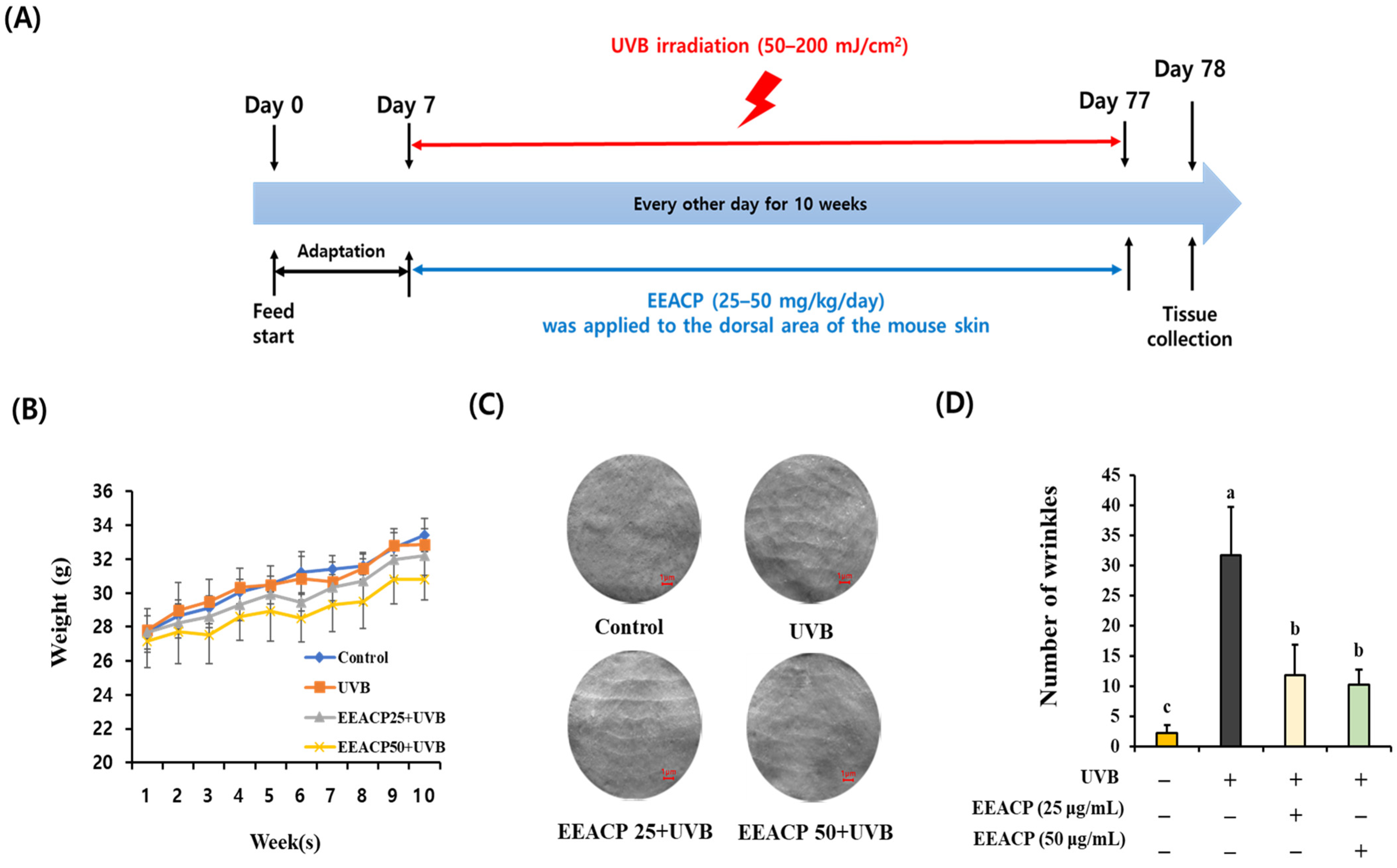
2.6. Animal Experiments
2.7. Skin Replica Assay and Tissue Staining
2.8. Quantitative Real-Time Polymerase Chain Reaction (qRT-PCR)
2.9. Western Blotting
2.10. Single Components Analysis
2.11. Statistical Analysis
3. Results
3.1. Administration of EEACP Attenuated UVB-Induced Wrinkle Formation
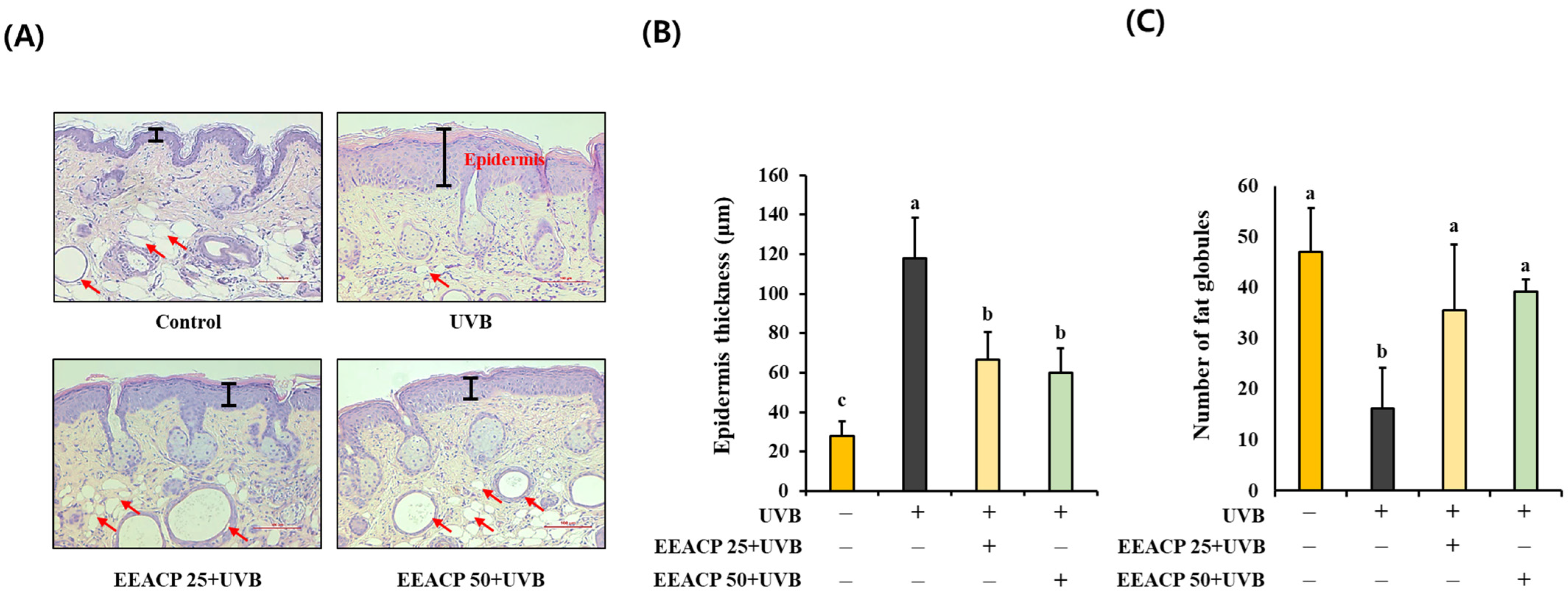
3.2. UVB-Induced Epidermal Thickening Was Restored by EEACP Treatments
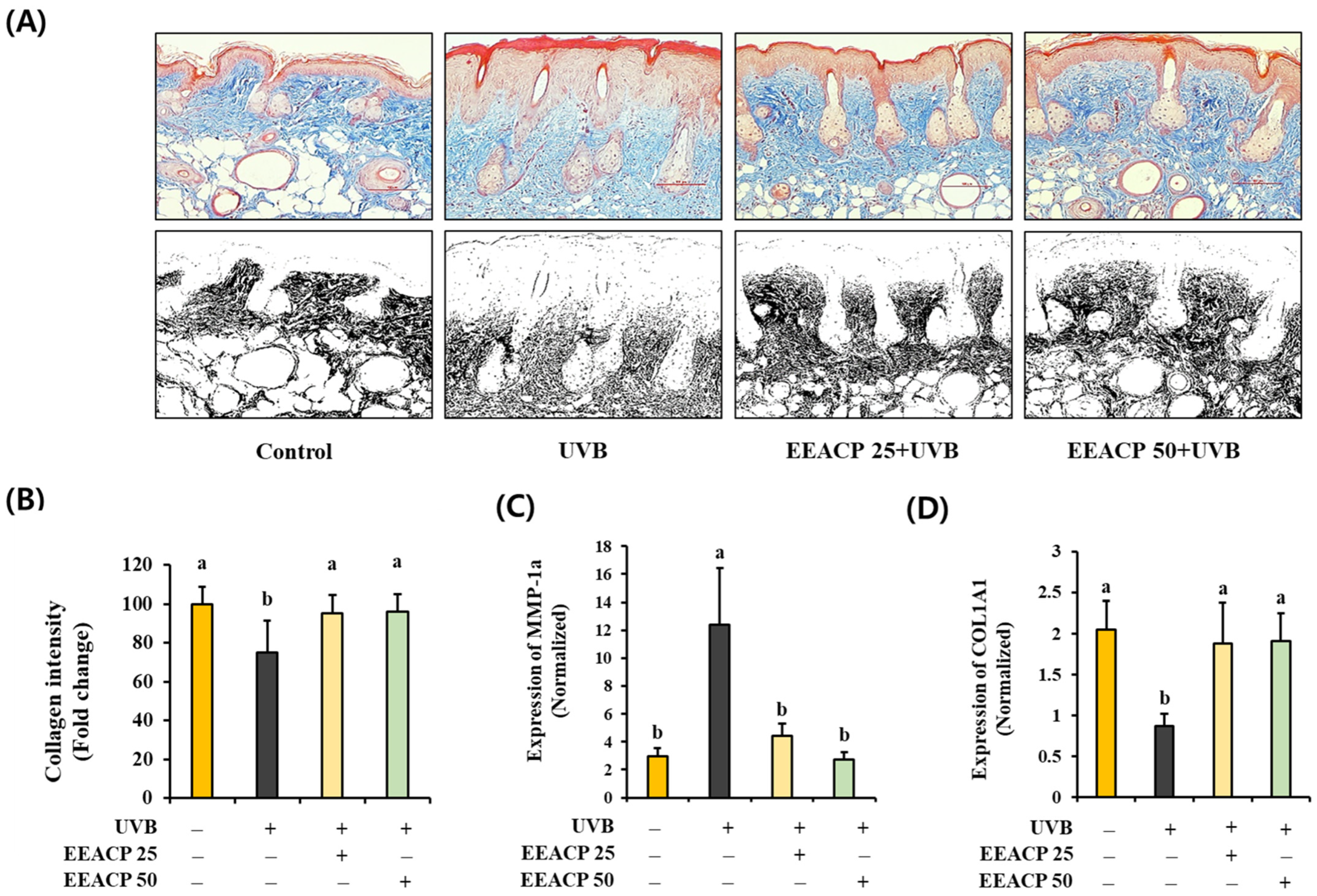
3.3. The Loss of Collagen Contents in UVB-Irradiated Skin Was Resolved by EEACP Treatments
3.4. EEACP Attenuated the Abnormal Expression of Photoaging-Related Genes in UVB-Irradiated NIH-3T3 Skin Fibroblast Cells
3.5. UVB-Induced ROS Production Was Attenuated by EEACP
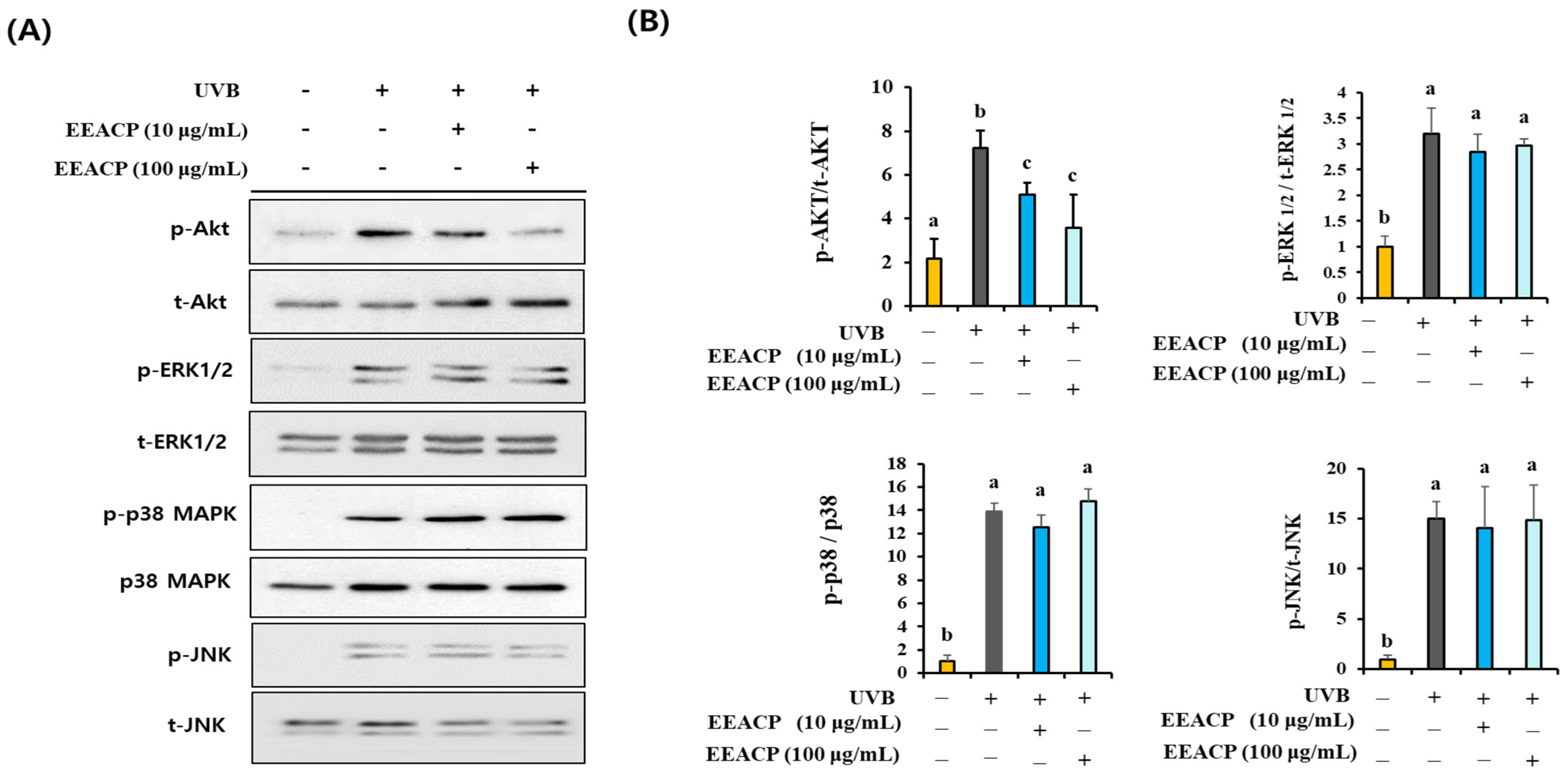
3.6. EEACP Prevented UVB-Induced Intracellular ROS-Mediated Signaling
3.7. EEACP and ROS Inhibitor Showed Equivalent Efficacy in Preventing Photoaging
3.8. Single Constituents of EEACP Identified by High-Performance Liquid Chromatography/Mass Spectrometry (HPLC/MS) Analysis
4. Discussion
5. Conclusions
Supplementary Materials
Author Contributions
Funding
Institutional Review Board Statement
Informed Consent Statement
Data Availability Statement
Conflicts of Interest
References
- Chen, X.; Yang, C.; Jiang, G. Research progress on skin photoaging and oxidative stress. Postep. Dermatol. Alergol. 2021, 38, 931–936. [Google Scholar] [CrossRef] [PubMed]
- Salminen, A.; Kaarniranta, K.; Kauppinen, A. Photoaging: UV radiation-induced inflammation and immunosuppression accelerate the aging process in the skin. Inflamm. Res. 2022, 71, 817–831. [Google Scholar] [CrossRef]
- Kim, E.J.; Jin, X.J.; Kim, Y.K.; Oh, I.K.; Kim, J.E.; Park, C.H.; Chung, J.H. UV decreases the synthesis of free fatty acids and triglycerides in the epidermis of human skin in vivo, contributing to development of skin photoaging. J. Dermatol. Sci. 2010, 57, 19–26. [Google Scholar] [CrossRef] [PubMed]
- Kwon, K.R.; Alam, M.B.; Park, J.H.; Kim, T.H.; Lee, S.H. Attenuation of UVB-Induced Photo-Aging by Polyphenolic-Rich Spatholobus Suberectus Stem Extract Via Modulation of MAPK/AP-1/MMPs Signaling in Human Keratinocytes. Nutrients 2019, 11, 1341. [Google Scholar] [CrossRef]
- Kang, W.; Choi, D.; Park, T. Dietary Suberic Acid Protects Against UVB-Induced Skin Photoaging in Hairless Mice. Nutrients 2019, 11, 2948. [Google Scholar] [CrossRef]
- Myung, D.B.; Han, H.S.; Shin, J.S.; Park, J.Y.; Hwang, H.J.; Kim, H.J.; Ahn, H.S.; Lee, S.H.; Lee, K.T. Hydrangenol Isolated from the Leaves of Hydrangea serrata Attenuates Wrinkle Formation and Repairs Skin Moisture in UVB-Irradiated Hairless Mice. Nutrients 2019, 11, 2354. [Google Scholar] [CrossRef]
- Jung, J.M.; Choi, J.K.; Kwon, O.Y.; Lee, S.H. Anti-Photoaging Activity of Scutellaria barbata D. Don (Family Lamiaceae) on Ultraviolet B-Irradiated NIH-3T3 Skin Fibroblast and SKH-1 Hairless Mouse. Molecules 2022, 27, 3803. [Google Scholar] [CrossRef] [PubMed]
- Kammeyer, A.; Luiten, R.M. Oxidation events and skin aging. Ageing Res. Rev. 2015, 21, 16–29. [Google Scholar] [CrossRef]
- Lee, J.E.; Oh, J.; Song, D.; Lee, M.; Hahn, D.; Boo, Y.C.; Kang, N.J. Acetylated Resveratrol and Oxyresveratrol Suppress UVB-Induced MMP-1 Expression in Human Dermal Fibroblasts. Antioxidants 2021, 10, 1252. [Google Scholar] [CrossRef]
- Hwang, Y.P.; Kim, H.G.; Han, E.H.; Choi, J.H.; Park, B.H.; Jung, K.H.; Shin, Y.C.; Jeong, H.G. N-Acetylglucosamine suppress collagenases activation in ultraviolet B-irradiated human dermal fibroblasts: Involvement of calcium ions and mitogen-activated protein kinases. J. Dermatol. Sci. 2011, 63, 93–103. [Google Scholar] [CrossRef]
- Choi, J.K.; Kwon, O.Y.; Lee, S.H. Kaempferide Prevents Photoaging of Ultraviolet-B Irradiated NIH-3T3 Cells and Mouse Skin via Regulating the Reactive Oxygen Species-Mediated Signalings. Antioxidants 2022, 12, 11. [Google Scholar] [CrossRef]
- He, X.; Fang, J.; Chen, X.; Zhao, Z.; Li, Y.; Meng, Y.; Huang, L. Actinidia chinensis Planch.: A Review of Chemistry and Pharmacology. Front. Pharmacol. 2019, 10, 1236. [Google Scholar] [CrossRef]
- Wei, L.B.; Ma, S.Y.; Liu, H.X.; Huang, C.S.; Liao, N. Cytotoxic Triterpenoids from Roots of Actinidia chinensis. Chem. Biodivers. 2018, 15, e1700454. [Google Scholar] [CrossRef]
- Deng, J.; Liu, Q.; Zhang, C.; Cao, W.; Fan, D.; Yang, H. Extraction Optimization of Polyphenols from Waste Kiwi Fruit Seeds (Actinidia chinensis Planch.) and Evaluation of Its Antioxidant and Anti-Inflammatory Properties. Molecules 2016, 21, 832. [Google Scholar] [CrossRef] [PubMed]
- Liao, J.C.; Huang, S.S.; Deng, J.S.; Lee, C.Y.; Lin, Y.C.; Huang, G.J. Chemical characterization and in vivo anti-inflammatory activities of Actinidia callosa var. ephippioides via suppression of proinflammatory cytokines. Am. J. Chin. Med. 2013, 41, 405–423. [Google Scholar] [CrossRef] [PubMed]
- Niu, H.; Song, D.; Sun, Y.; Zhang, W.; Mu, H.; Duan, J. Preparation and sulfation of an alpha-glucan from Actinidia chinensis roots and their potential activities. Int. J. Biol. Macromol. 2016, 92, 981–987. [Google Scholar] [CrossRef] [PubMed]
- Kwon, O.Y.; Lee, S.H. Ishige okamurae Attenuates Neuroinflammation and Cognitive Deficits in Mice Intracerebroventricularly Injected with LPS via Regulating TLR-4/MyD88-Dependent Pathways. Antioxidants 2022, 12, 78. [Google Scholar] [CrossRef]
- Kwon, O.Y.; Lee, S.H. Ameliorating Activity of Ishige okamurae on the Amyloid Beta-Induced Cognitive Deficits and Neurotoxicity through Regulating ERK, p38 MAPK, and JNK Signaling in Alzheimer’s Disease-Like Mice Model. Mol. Nutr. Food Res. 2020, 64, e1901220. [Google Scholar] [CrossRef] [PubMed]
- Choi, J.K.; Kwon, O.Y.; Lee, S.H. Oral Administration of Bifidobacterium lactis Ameliorates Cognitive Deficits in Mice Intracerebroventricularly Administered Amyloid Beta via Regulation the Activation of Mitogen-activated Protein Kinases. Food Sci. Anim. Resour. 2024, 44, 607–619. [Google Scholar] [CrossRef]
- Lee, S.H.; Yu, S.Y.; Nakayama, J.; Khoo, K.H.; Stone, E.L.; Fukuda, M.N.; Marth, J.D.; Fukuda, M. Core2 O-glycan structure isessential for the cell surface expression of sucrase isomaltase and dipeptidyl peptidase-IV during intestinal cell differentiation. J. Biol. Chem. 2010, 285, 37683–37692. [Google Scholar] [CrossRef]
- Kielkopf, C.L.; Bauer, W.; Urbatsch, I.L. Bradford assay for determining protein concentration. Cold Spring Harb Protoc. 2020, 2020, 102269. [Google Scholar] [CrossRef] [PubMed]
- Retinasamy, T.; Shaikh, M.F.; Kumari, Y.; Abidin, S.A.Z.; Othman, I. Orthosiphon stamineus Standardized Extract Reverses Streptozotocin-induced Alzheimer’s Disease-Like Condition in a Rat Model. Biomedicines 2020, 8, 104. [Google Scholar] [CrossRef] [PubMed]
- The Human Metabolome Database, Showing Metabocard for Phenethylamine Glucuronide (HMDB0010323). Available online: https://hmdb.ca/metabolites/HMDB0010323 (accessed on 25 June 2024).
- Telofski, L.S.; Morello, A.P., 3rd; Mack Correa, M.C.; Stamatas, G.N. The infant skin barrier: Can we preserve, protect, and enhance the barrier? Dermatol. Res. Pract. 2012, 2012, 198789. [Google Scholar] [CrossRef] [PubMed]
- Chambers, E.S.; Vukmanovic-Stejic, M. Skin barrier immunity and ageing. Immunology 2020, 160, 116–125. [Google Scholar] [CrossRef] [PubMed]
- Korkina, L. Metabolic and redox barriers in the skin exposed to drugs and xenobiotics. Expert Opin. Drug Metab. Toxicol. 2016, 12, 377–388. [Google Scholar] [CrossRef]
- Jung, J.M.; Kwon, O.Y.; Choi, J.K.; Lee, S.H. Alpinia officinarum Rhizome ameliorates the UVB induced photoaging through attenuating the phosphorylation of AKT and ERK. BMC Complement. Med. Ther. 2022, 22, 232. [Google Scholar] [CrossRef]
- Park, K.M.; Yoo, Y.J.; Ryu, S.; Lee, S.H. Nelumbo Nucifera leaf protects against UVB-induced wrinkle formation and loss of subcutaneous fat through suppression of MCP3, IL-6 and IL-8 expression. J. Photochem. Photobiol. B 2016, 161, 211–216. [Google Scholar] [CrossRef]
- Kim, E.J.; Kim, Y.K.; Kim, J.E.; Kim, S.; Kim, M.K.; Park, C.H.; Chung, J.H. UV modulation of subcutaneous fat metabolism. J. Investig. Dermatol. 2011, 131, 1720–1726. [Google Scholar] [CrossRef]
- Bosch, R.; Philips, N.; Suarez-Perez, J.A.; Juarranz, A.; Devmurari, A.; Chalensouk-Khaosaat, J.; Gonzalez, S. Mechanisms of Photoaging and Cutaneous Photocarcinogenesis, and Photoprotective Strategies with Phytochemicals. Antioxidants 2015, 4, 248–268. [Google Scholar] [CrossRef]
- Chen, Y.; Cai, X.; Li, G.; He, X.; Yu, X.; Yu, X.; Xiao, Q.; Xiang, Z.; Wang, C. Chemical constituents of radix Actinidia chinensis planch by UPLC-QTOF-MS. Biomed. Chromatogr. 2021, 35, e5103. [Google Scholar] [CrossRef]
- Zhao, T.; He, J.; Wang, X.; Ma, B.; Wang, X.; Zhang, L.; Li, P.; Liu, N.; Lu, J.; Zhang, X. Rapid detection and characterization of major phenolic compounds in Radix Actinidia chinensis Planch by ultra-performance liquid chromatography tandem mass spectrometry. J. Pharm. Biomed. Anal. 2014, 98, 311–320. [Google Scholar] [CrossRef] [PubMed]
- Nishiyama, T.; Suzuki, T.; Hashiguchi, Y.; Shiotsu, S.; Fujioka, M. Antioxidant activity of aromatic cyclic amine derivatives. Polym. Degrad. Stab. 2002, 75, 549–554. [Google Scholar] [CrossRef]
- Yu, J.H.; Shan, Y.; Li, S.; Zhang, L.F. Potential contribution of Amadori compounds to antioxidant and angiotensin I converting enzyme inhibitory activities of raw and black garlic. LWT-Food Sci. Technol. 2020, 129, 109553. [Google Scholar] [CrossRef]


| Genes | Sequences | Species | |
|---|---|---|---|
| Forward | Reverse | ||
| COL1A1 | 5′-CACTGCTGTTGGTCCACGT-3′ | 5′-AAAGCACAGCACTCGCCC-3′ | Mouse |
| MMP-1a | 5′-ACTTTCCAGCCAGGCCCA-3′ | 5′-CACTGCTGTTGGTCCACGT-3′ | Mouse |
| IL-6 | 5′-ACAACCACGGCCTTCCCT-3′ | 5′-AGCCTCCGACTTGTGAA-3′ | Mouse |
| IL-8 | 5′-TGTCCCATGCCACTCAGAGA-3′ | 5′-AGCAGGTGCTCCGGTTGTAT-3′ | Mouse |
| MCP-3 | 5′-ATAGCCGCTGCTTTCAGCAT-3′ | 5′-CTTCCCAGGGACACCGACTA-3′ | Mouse |
| GAPDH | 5′-AAGCTGTGGCGTGATGGC-3′ | 5′-AAGCTGTGGCGTGATGGC-3′ | Mouse |
| Natural Product | Peak | RT (min) | Observed Mass | Fragment ions | Single Compound | Molecular Structure | Formula | Molecular Mass (g/mol) | Ref. |
|---|---|---|---|---|---|---|---|---|---|
| Actinidia chinensis Planch | Peak 1 | 9.79 | 617.6337 | 365.3053, 297.2925, 279.2860 | N-(1-deoxy-1-fructosyl) valine | 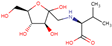 | C11H21NO7 | 279.13 | [22] |
| Phenethylamine glucuronide |  | C14H19NO6 | 297.12 | [23] |
Disclaimer/Publisher’s Note: The statements, opinions and data contained in all publications are solely those of the individual author(s) and contributor(s) and not of MDPI and/or the editor(s). MDPI and/or the editor(s) disclaim responsibility for any injury to people or property resulting from any ideas, methods, instructions or products referred to in the content. |
© 2024 by the authors. Licensee MDPI, Basel, Switzerland. This article is an open access article distributed under the terms and conditions of the Creative Commons Attribution (CC BY) license (https://creativecommons.org/licenses/by/4.0/).
Share and Cite
Jung, J.-M.; Kim, S.-Y.; Kwon, O.-Y.; Lee, S.-H. Actinidia chinensis Planch Ameliorates Photoaging in UVB-Irradiated NIH-3T3 Cells and SKH-1 Hairless Mice by Controlling the Reactive Oxygen Species/AKT Pathway. Antioxidants 2024, 13, 1091. https://doi.org/10.3390/antiox13091091
Jung J-M, Kim S-Y, Kwon O-Y, Lee S-H. Actinidia chinensis Planch Ameliorates Photoaging in UVB-Irradiated NIH-3T3 Cells and SKH-1 Hairless Mice by Controlling the Reactive Oxygen Species/AKT Pathway. Antioxidants. 2024; 13(9):1091. https://doi.org/10.3390/antiox13091091
Chicago/Turabian StyleJung, Jong-Min, Seo-Young Kim, Oh-Yun Kwon, and Seung-Ho Lee. 2024. "Actinidia chinensis Planch Ameliorates Photoaging in UVB-Irradiated NIH-3T3 Cells and SKH-1 Hairless Mice by Controlling the Reactive Oxygen Species/AKT Pathway" Antioxidants 13, no. 9: 1091. https://doi.org/10.3390/antiox13091091