Article

Flagellin Improves the Immune Response of an Infectious Bursal Disease Virus (IBDV) Subunit Vaccine

1
International Program in Animal Vaccine Technology, International College, National Pingtung University of Science and Technology, Pingtung 91201, Taiwan
2
Centre for Immunology & Infection, Hong Kong Science and Technology Park, Hong Kong, China
3
General Research Service Center, National Pingtung University of Science and Technology, Pingtung 91201, Taiwan
4
Graduate Institute of Animal Vaccine Technology, College of Veterinary Medicine, National Pingtung University of Science and Technology, Pingtung 91201, Taiwan
*
Authors to whom correspondence should be addressed.
Vaccines 2022, 10(11), 1780; https://doi.org/10.3390/vaccines10111780
Submission received: 14 September 2022 / Revised: 11 October 2022 / Accepted: 21 October 2022 / Published: 22 October 2022

Abstract

:
Flagellin activates the immune system through Toll-like receptor 5 (TLR5) and can work as an adjuvant for subunit vaccines. In this study, we tested the adjuvancy of two different N-terminal fragments of flagellin, (1) FliC99, residues 1–99, and (2) FliC176, residues 1–176, to incorporate larger areas of the hotspot region for potentially higher levels of TLR5 activation and immune response. A truncated version of the VP2 protein (name tVP2, residues 199–356) of the Infectious bursal disease virus (IBDV) was genetically linked to the flagellin constructs, and the immune response was evaluated in chickens. Results showed that both chimeric antigen–adjuvant constructs increased humoral (total IgG titers), cellular and cytokine immune response (IL-4, IFN-γ). The resulting antibody also successfully neutralized IBDV. We conclude that the N-terminus of flagellin can act as an immune activator to enhance vaccine efficacy.

1. Introduction

The flagellum is a whip-like organelle responsible for locomotion in bacteria [1]. Flagellin, the main protein component of the flagellum, is one of the pathogen-associated molecular patterns (PAMPs) that are recognized by the host immune system through receptors such as TLR5 [2] and NOD-like receptor protein four inflammasome receptor NAIP5/6 [3]. TLR5 detects the flagellin in the host and activates the MyD88-dependent signaling pathway, producing various proinflammatory cytokines and chemokines of the innate immune system. Flagellin has been used as an adjuvant for a number of antigens (bacterial, viral, and parasitic) with equal success [4,5].
Flagellin of Salmonella enterica serovar Typhimurium (S. Typhimurium) contains four domains, D0, D1, D2 and D3. D0 and D1, highly conserved and buried in the flagellum core filament, are responsible for innate immune activation through TLR5. On the other hand, D2 and D3, hypervariable and exposed on the surface of the filament, are accountable for antigenicity as well as undesirable toxicity [6,7,8].
Previous work has identified a crucial hotspot region (including conserved residues R89, L93 and E114) within D1 for binding to the leucine-rich repeat 9 (LRR9) region of TLR5 [6]. In our previous studies, we used an N-terminal portion of flagellin (residue 1–99, containing R89 and L93 of the hotspot region), and the results showed that it is sufficient for immune activation and vaccine efficacy enhancement [9,10,11]. In this study, we designed and tested two different N-terminal fragments of flagellin, (1) FliC99, residue 1–99, and (2) FliC176, residue 1–176, to incorporate more significant areas of the hotspot region, including E114, for potentially higher levels of TLR5 activation and immune response.
To evaluate the above flagellin constructs as adjuvants in vivo, a truncated version of the VP2 protein (name tVP2, residue 199–356) of the Infectious bursal disease virus (IBDV) was employed as the vaccine antigen [12]. IBDV depletes B lymphocytes in the bursa of Fabricius, causing immunosuppression [13]. There are many vaccines available against IBD on the market. Still, due to the RNA genome of the virus, it is continuously evolving, making it challenging to control the disease with available vaccines [14]. In this study, flagellin constructs were genetically linked to tVP2 via a flexible glycine-serine (GS) linker. Chickens were immunized with antigen-adjuvant constructs to evaluate humoral and cellular immune responses.

2. Materials and Methods

2.1. Bacteria Strain and Virus

S. Typhimurium (ATCC 14028) was obtained and cultured in Tryptic Soy Broth at 37 °C. The VP2 gene of Infectious bursal disease virus strain 3529/92 was obtained as a stable plasmid (VP2-pET32a, gift from Dr. Guanming Ke) and later confirmed by primers.

2.2. Epitope and Antigencity Prediction in the Antigen (IBDV)

To predict the B cell and antigenicity in the VP2 protein of the IBDV, BepiPred-2.0: Sequential B-Cell Epitope Predictor and Hydropath/Kyte & Doolittle methods were used, respectively. Using a Random Forest algorithm trained on epitopes and non-epitope amino acids with a threshold value set above 0.5 for all servers, the BepiPred-2.0 Sequential B-Cell Epitope Predictor server forecasts B-cell epitopes from a protein sequence. Briefly, an amino acid sequence was fed to IEDB tools for B-cell epitope prediction, and those which reside in a yellow color above the threshold (0.5 value) are the predicted epitopes, whereas the Y-axes depict residue scores and X-axes residue positions in the sequence. For predicting antigenicity, the sequence was fed to IEDB and Hydropath/Kyte & Doolittle methods using a threshold value of 0.5. The Y-axes represent the residue score and X-axes residue positions in the sequence.

2.3. Cloning of the Desired Flagellin and IBDV Antigenic Segments and Expression of Antigen-Adjuvant Recombinant Proteins

To evaluate the adjuvancy of flagellin, three recombinant proteins were expressed as subunit vaccines: (1) FliC99-tVP2, the truncated N terminus of the flagellin (1–99 residues) fused with the truncated VP2 (199–356 residues) of the IBD virus, (2) FliC176-tVP2, the N terminus (1–176 residues) of the flagellin fused with the truncated VP2 (199–356 residues) of the IBD virus, and (3) tVP2, truncated VP2 only. First, tVP2 was cloned from full-length VP2 using the primers listed in Table 1 and ligated into the vector pET32a (Novagen, Darmstadt, Germany). For the cloning of the FliC99 and FLiC176, full-length flagellin or FliC clone (DNA of S. Typhimurium and inserted into pET32a) was used, and subcloning of the FliC99 (residues 1–99) and FliC176 (residues 1–176) was done using primers for FliC99 and FLiC176 (Table 1, Figures S1 and S2). To construct the FliC99-tVP2 and FliC176-tVP2, a chimeric polymerase reaction (PCR) was carried out using the tVP2, FliC99, and FliC176 PCR products containing glycine-serine linker as a template. Primers for the chimeric reaction are listed in Table 1. pET32a was used as a vector to insert the final construct, and plasmid was sequenced for reconfirmation of the construct.
For the expression of the (1) FliC99-tVP2, (2) FliC176-tVP2 and (3) tVP2, the plasmids containing these constructs were transformed to Escherichia coli BL21 (DE3) (Yeastern Biotech, Taipei, Taiwan) according to the manufacturer’s instructions. 1-mM isopropyl-b-D-galactopyranoside (IPTG; Sigma, Darmstadt, Germany) was used to induce the protein expression at room temperature overnight. Cells were harvested, lysed in native lysis buffer (300-mM KCl, 50-mM KH2PO4 and 5-mM Imidazole) and sonicated. His-tag Bio-scale Mini Profinity IMAC cartridges (1 mL) (Bio-Rad, Hercules, CA, USA) were used to purify the recombinant protein from the insoluble fraction. Twelve percent sodium dodecyl sulfate-polyacrylamide gel electrophoresis (SDS-PAGE) analysis using BSA protein as standards was used to determine the expression levels of the recombinant proteins. Image Lab software predicted the concentration of the recombinant proteins. A Western blot assay was performed to confirm the identity of the recombinant proteins. In brief, after gel electrophoresis, proteins were transferred onto polyvinylidene difluoride (PVDF) membranes (Merck, Darmstadt, Germany). 6X-His Tag antibody solution (Gentex, Hsinchu, Taiwan) at 1:5000 dilution was used as the primary antibody, and rabbit anti-mouse antibody conjugated to HRP (Gentex, Taiwan) was used as the secondary antibody at 1:5000 dilution. Western Lightning PLUS (PerkinElmer, Waltham, MA, USA) was used for color development. PyMOL 2.5 (Schrodinger, New York, NY, USA) was used to predict the crystal structure of the FliC176, FliC99 and tVP2. Briefly, PDB ID (Protein Data Bank identifier) of flagellin Salmonella enterica serovar Typhimurium (S. Typhimurium) and Infectious Bursal Disease Virus (IBDV) were fetched on the PyMOL 2.5 and structure was drawn according to the amino acids sequence.

2.4. Vaccine Preparation and Immunization of Chickens

Four vaccine formulations were prepared: (1) FliC176-tVP2, (2) FliC99-tVP2, (3) tVP2 and (4) PBS as the control. Each purified recombinant protein dose was 50 mg. Water-in-oil adjuvant Summit-P101 (Country Best Biotech, Taipei, Taiwan) was used for the formulation at the ratio of 2:1 (adjuvant: antigen) to make up the final injection volume of 0.2 mL per chicken.
For immunization, 16 two-week-old Brown Leghorns chickens were obtained from a local farm and randomly assigned to four groups of three chickens for the four different vaccine formulations. Chickens were immunized subcutaneously twice, two weeks apart. Whole blood was collected from three chickens per vaccine group on days 0, 7, 14, 21 (Table 2) and 28 post vaccinations to analyze the immune response. All experimental animal protocols (NPUST-106-055) were approved by the Animal Care and Use Committee, National Pingtung University of Science and Technology (NPUST). The Ethical Rules and Laws of NPUST were followed during the experiments.

2.5. Analysis of Antibody Immune Response

An indirect enzyme-linked immunosorbent assay (ELISA) was done to determine the antibody response in the chicken. Whole blood was allowed to coagulate and then centrifuged at 700× g for 5 min to separate the serum from erythrocytes. The ELISA plates were coated with 50-ng/well-purified tVP2 overnight at 4 °C. The next day, washing and blocking were done, and the primary antibody (serum) was used at 1:10,000 dilution. For the secondary antibody, horseradish peroxidase (HRP)-conjugated anti-chicken IgG (Sigma, Carlsbad, CA, USA) was used at a 1:5000 dilution. The Peroxidase Kit (KPL, Gaithersburg, MD, USA) was used for color development. In the end, the optical density of the plates was read at 450 nm on the MultiskanTM FC microplate photometer (Thermo Fisher Scientific, Vantaa, Finland).

2.6. Analysis of Cell-Mediated Immune Response

To check the T-cell proliferation activity, an MTT ((3-(4,5-dimethylthiazol-2-yl)-2,5-diphenyltetrazolium bromide) assay was done. Blood was collected from chickens (n = 3, two weeks-old Brown Leghorns) on days 2, 14 and 28 to separate the peripheral blood mononuclear cells (PBMCs) and stimulated with tVP2. Briefly, Blood was collected in BD Vacutainer™ EDTA Blood Collection Tubes (BD Biosciences, Franklin Lakes, NJ, USA), and an equal volume of PBS was added to the blood. The PBS–blood mixture was slowly added to another tube’s equal volume of Ficoll-Paque (Amersham Biosciences, Piscataway, NJ, USA). The mixture was centrifuged at 252× g for 40 min to collect the buffy coat layer containing PBMCs fraction. Cells were rinsed with PBS twice and resuspended in RPMI-1640 (Gibco Invitrogen, Carlsbad, CA, USA) supplemented with 5% fetal bovine serum (Gibco Invitrogen, Carlsbad, CA, USA) at 2 × 10⁶ cells/mL. For antigen stimulation, PBMCs (4 × 10⁵ cells/well) were added to 96-well plates incubated with 10 μg/mL of the purified recombinant tVP2 for 48 h at 37 °C, 5% CO2. Concanavalin A (Thermo Fisher Scientific, Ward Hill, MA, USA) at 5 µg/mL was used as the positive control for cell stimulation, with cell-only as the negative control and medium-only as the background. The cell proliferation was measured using EZcountTM MTT Cell Assay Kit (Himedia, India). The results were read at 550 nm on the MultiskanTM FC microplate photometer (Thermo Fisher Scientific, Vantaa, Finland). Stimulation index (SI) = (OD of treatment − OD of background)/(OD of the negative control − OD of background).

2.7. Th1 and Th2-Type Cytokine mRNA Expression Level

PBMCs were collected from the vaccinated group on Day 28, as described in Section 2.5. Briefly, the cells (2 × 10⁶ cells/well) were added to the 6-wells plate containing 10 μg/mL of the tVP2 for the stimulation and incubated for 2 h incubation at 37 °C, 5% CO2. A Miniprep system (Viogene, Taipei, Taiwan) was used for the extraction of total RNA, and a Reverse Transcriptase Kit (Applied Biosystems, Foster, CA, USA) was used to convert the RNA into complementary DNA (cDNA). Chicken cytokines primers listed in Table 3 were used for the Real-time PCR in the SmartCycler I (Cepheid, Sunnyvale, CA, USA) for TH1 (IFN-γ) and TH2-type cytokines (IL-4) and the housekeeping gene glyceraldehyde-3-phosphate dehydrogenase (GAPDH). GAPDH was used as the reference gene to normalize the cytokine expression levels, and changes were expressed as n-fold higher or lower in levels relative to the PBS control.

2.8. IBD Virus Neutralization Assay

Serum samples from Day 28 were used for the neutralizing antibody assay. The titers of the virus-neutralizing antibodies were examined using IBD virus and Vero cell lines. Briefly, the serum was heat-inactivated, and a 2-fold dilution of the serum was done on a 96-well plate. Then, serum samples of each dilution were mixed with an equal volume of the virus (100 TCID50) and incubated for 1 h at 37 °C. Vero cell lines were added and again incubated for 2 h at 37 °C. After that, the supernatant was removed, and the virus was allowed to grow on the cell lines for 5 to 7 days. Cells were monitored for cytopathic effects, and the virus-neutralizing antibody titer of the serum was calculated according to the method of Reed and Munch (1938).

2.9. Statistical Analysis

For statistical analysis, the IBM SPSS Statistics software was used. One-way analysis of variance (ANOVA) was used to compare data on antibody response, lymphocyte proliferation assay and cytokine mRNA levels. For the comparison of neutralization assay data, the t-test was used. The significance level (p) was set at 0.05 for all the experiments. All the data are expressed as mean ± standard error of the mean (SEM).

3. Results

3.1. Antigen-Adjuvant Chimeric Proteins Were Formulated as Subunit Vaccines

B-cell epitope prediction and hydrophobicity plot showed that the region 199–356 (Figure 1) of IBDV VP2 contains several epitopes and can be highly antigenic. Therefore, this region was selected as the antigen for vaccine construction.
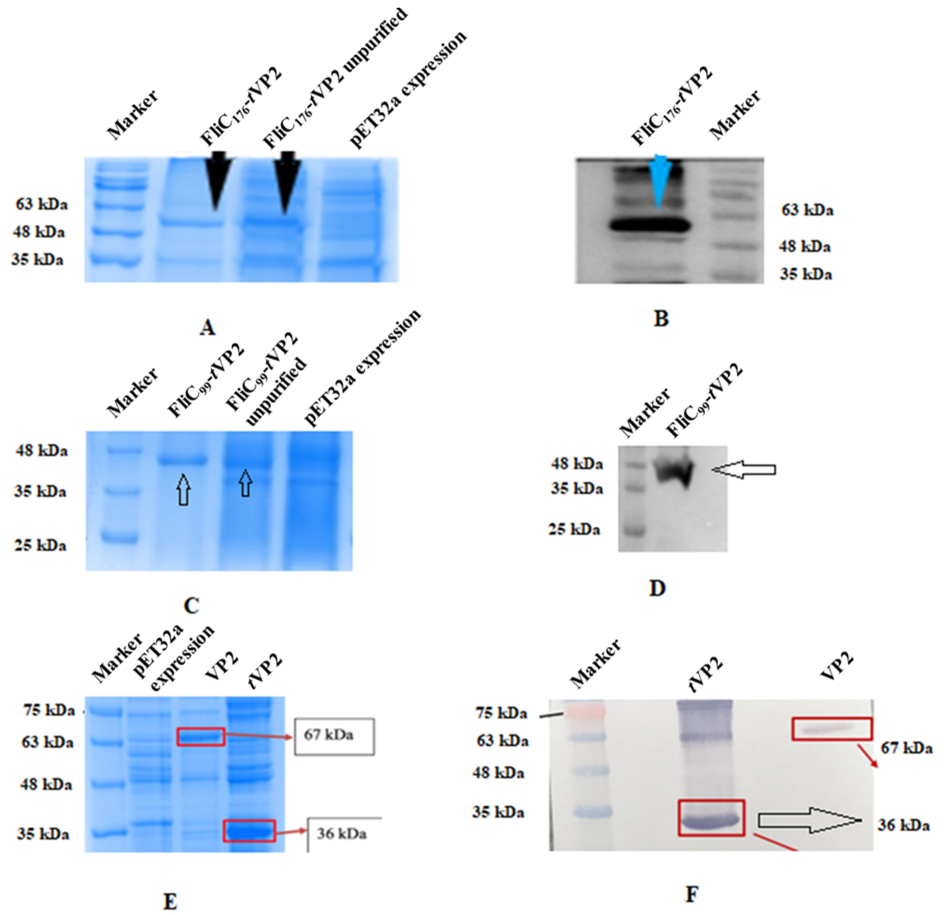
Two separate chimeric proteins were constructed to evaluate the adjuvant effect of flagellin N terminus 1–99 and 1–176: the N flagellin 1–99 linked to tVP2 (199–356) of IBD virus and N flagellin 1–176 also linked to tVP2. Protein expression was confirmed by SDS-PAGE (Figure 2C) and Western blot (Figure 2D) with a size of 48kDa (FliC99-tVP2). The size of the FliC176-tVP2 was 55kDa (Figure 2A,B). Full-length VP2 and tVP2 were also expressed, and the size was confirmed by running SDS-PAGE (Figure 2E) and Western blot (Figure 2F). 20-kDa was an additional size protein carried with it when we cloned in pET32a, because the pET32a vector inserts a 20-kDa Trx-His-S-enterokinase tag. The concentration value was up to 140 µg/mL for FliC99-tVP2 and 150 µg/mL for the FliC176-tVP2 variably in different purification attempts. Four vaccine formulations were prepared for the immunization of chickens: (1) FliC99-tVP2, (2) FliC176-tVP2, (3) tVP2 and (4) PBS as the negative control.
Molecular structure of FliC176, FliC99 and tVP2 were predicted using PyMOL 2.5 (Figure 3A–C).

3.2. Both FliC176 and FliC99 Resulted in an Increase in Antibody Levels

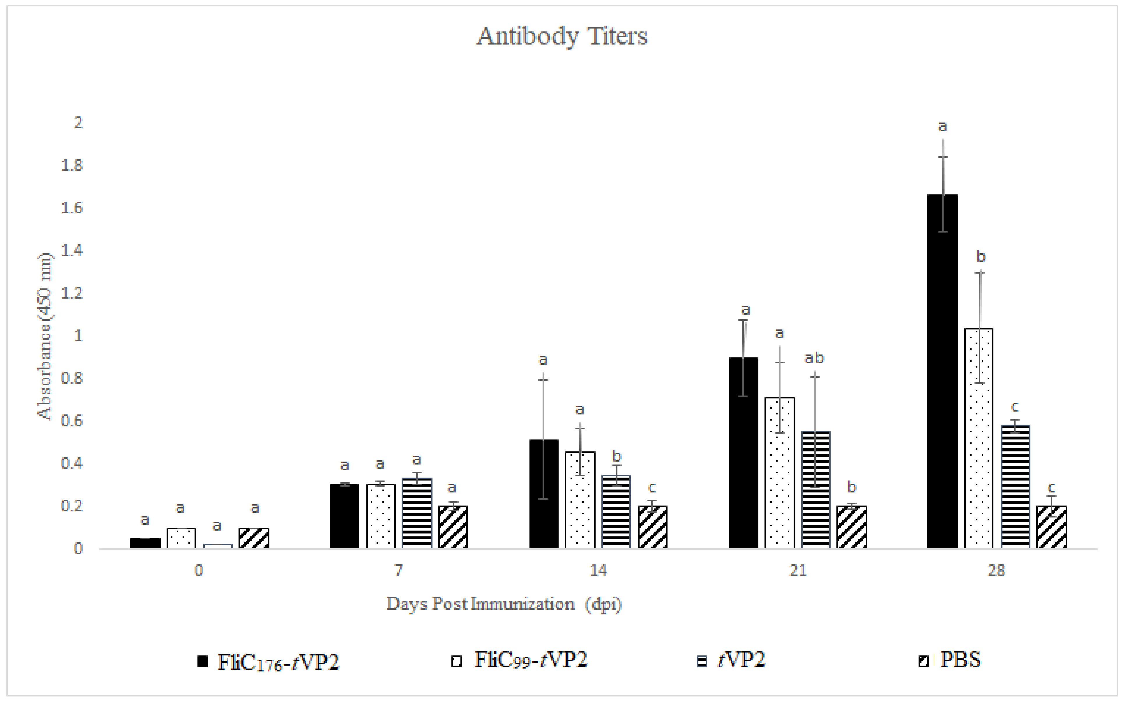
Indirect ELISA was used to determine the levels of antibody response, with tVP2 as the coating antigen. On Day 21 and 28, the levels of the antibodies went up in the FliC176-tVP2 and FliC99-tVP2 groups as compared to the tVP2-only group (Figure 4), indicating that the flagellin enhanced antibody production.

3.3. Both FliC176 and FliC99 Increased the Proliferation of the Lymphocytes

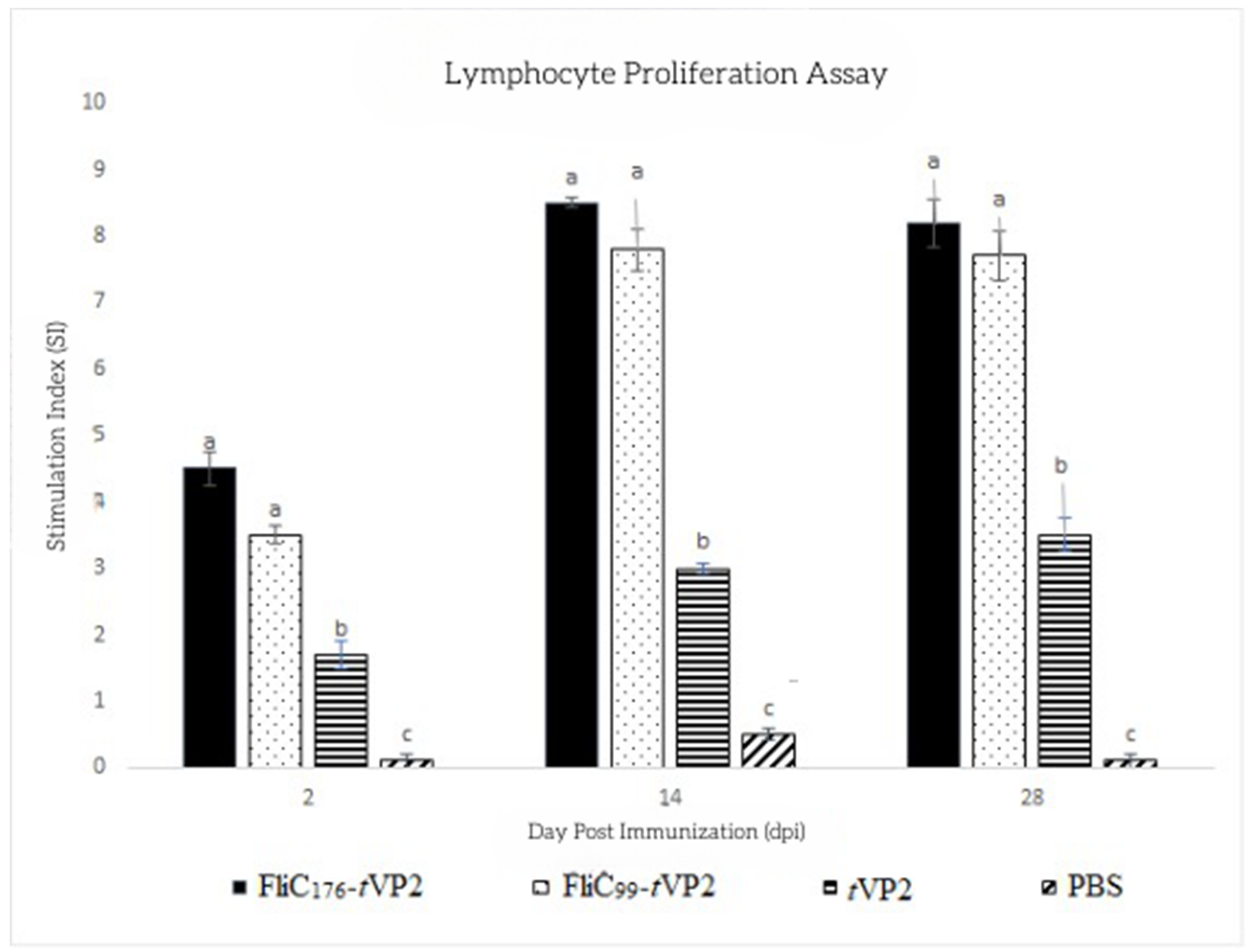
To measure lymphocytes’ metabolic activity, the MTT assay was performed using PBMCs. Cells were stimulated with tVP2, and the proliferation activity was measured. After the 2nd dose of the vaccine, on the 14th and 28th days, the proliferation activity of both groups, FliC176-tVP2 and FliC99-tVP2, rose considerably compared to the tVP2 and PBS groups (Figure 5). This indicates that the flagellin was essential in increasing the number of lymphocytes in the vaccinated chickens.

3.4. Both FliC176 and FliC99 Raised the Gene Expression of the Cytokines

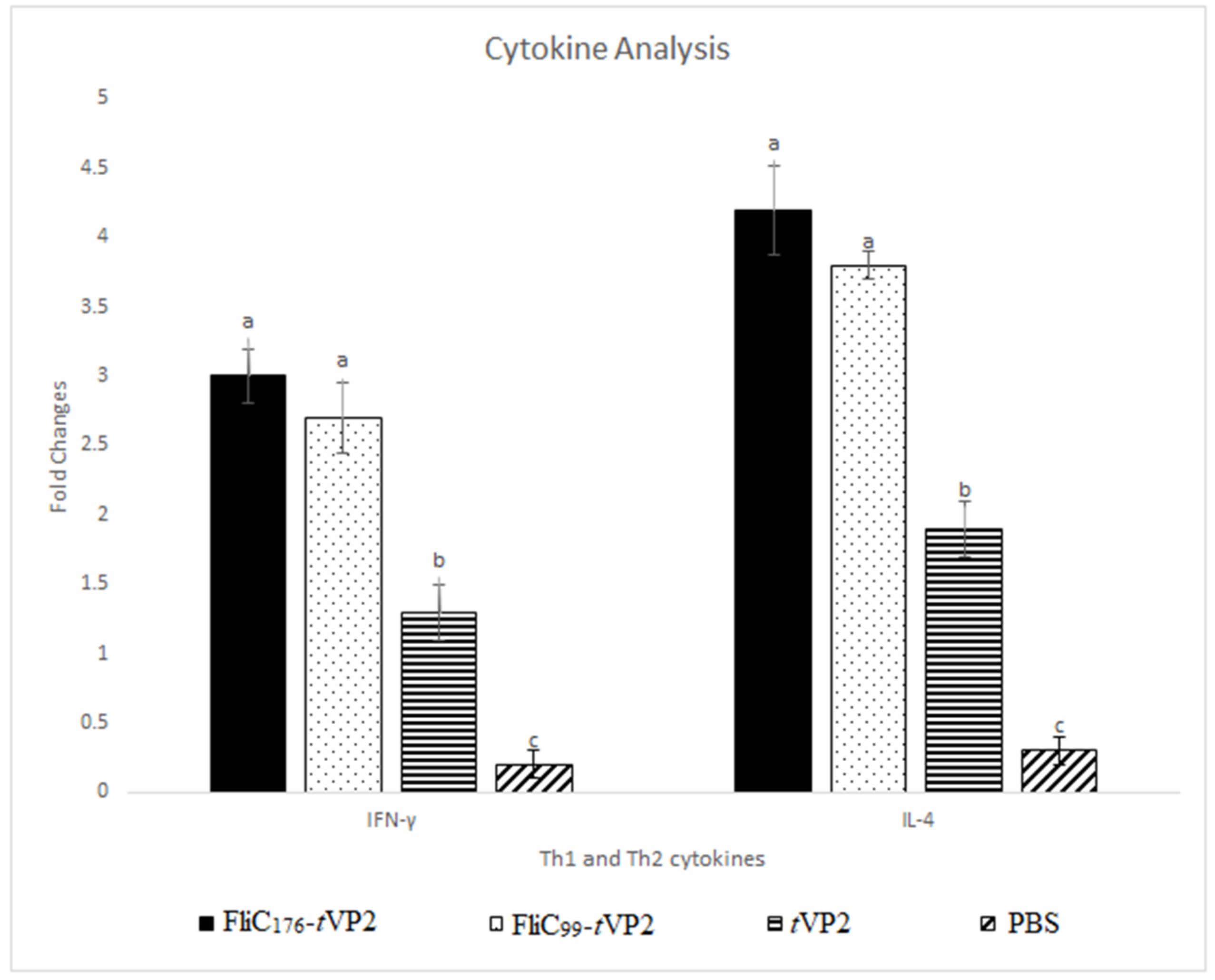
Two weeks after the 2nd dose, on the 28th day, the PBMCs were collected and stimulated with tVP2, and cytokine mRNA levels were quantified. Th1 (IFN-γ) levels and Th2 (IL-4) cytokines increased significantly in the vaccinated chickens (Figure 6).

3.5. Neutralization Assay

On day 28, after vaccinations, the serum samples were used for the neutralization assay with 100 TCID50 of the Infectious Bursal Disease Virus. The serum sample of the FliC176-tVP2 group neutralized the virus up to the 5th dilution of the log2, while on the other hand, serum of FliC99-tVP2 neutralized the virus up to the 4th dilution of the log2. In the tVP2 serum, only the 1st dilution of the log2 neutralized the virus, whereas neutralization was not noticed in the PBS-only group (Figure 7 and Figure S3).

4. Discussion

This study demonstrated that N-terminal fragments of flagellin encompassing the hotspot region can enhance immune response when linked to an antigen. FliC176 and FliC99 both boosted antigen-specific antibody levels, virus-neutralizing activity, cellular immunity and cytokine gene expression.
Our experimental data revealed that there is not a statistically significant difference between FliC176 and FliC99 in terms of immune modulation (antibody titers, T-cell stimulation and cytokine expression). We hypothesize that the hotspot is the main contributor to the TLR5 activation. Liu et al., showed that residues 85 to 111 of flagellin encompassing the hotspot region resulted in significantly higher titers of neutralizing antibodies and cytokines against PCV2 than antigens alone [15]. A similar trend of immune activation was measured in the anti-tumor vaccine (B16-OVA melanoma, P815 tumor model) [16,17]. Therefore, any length of flagellin encompassing the hot-spot region of flagellin may be sufficient for adjuvancy. Both of our truncated designs of flagellin-VP2 elicit statistically insignificant antibody and cytokine titers. However, neutralizing antibody titers did show significant difference among the vaccine groups.
Previous work using flagellin as a therapeutic agent pointed out that the development of anti-flagellin antibody upon repeated exposure can hamper effectiveness. A case in point is Entolimod, an anti-radiation drug developed from flagellin D0, D1. GP532 (modified flagellin) was developed as an anti-radiation drug with minimal anti-flagellin antibodies to circumvent this issue. Deimmunization was accomplished after deleting B-cell, T-cell and inflammasome activation epitopes. In light of this, future truncated flagellin designs should be screened for anti-flagellin antibodies [18].
Truncation of flagellin can be carried out in many other ways, and the best possible truncation design could be narrowed down for therapeutic use. The N-terminal domain of D0 was removed in the GP532 design without impacting TLR5 binding and activation. Hot-spot is pivotal to flagellin-TLR5 adjuvancy. Thus, the binding domain of flagellin (D1) could be linked to either the N or the C-terminus of D0. In short, various truncations can be performed in D0, D1 with respect to the goal of the experiment by retaining the hot-spot region in truncation. In addition to truncation designs, adjuvancy of naturally conserved (D0, D1) flagellin from Bacillus cereus and Bacillus subtilis should be considered for future studies. These short flagellins offer adjuvancy comparable to FliC. Moreover, reduced proinflammatory properties were observed in mice compared to flagellin from S. Typhimurium [19]. Further studies are required to determine the de novo antigenicity of these flagellins.

5. Conclusions

This study finds that the N-terminus of flagellin, both FliC99 and FliC176, can act as an immune activator to enhance vaccine efficacy for an IBDV VP2 subunit vaccine.

Supplementary Materials

The following supporting information can be downloaded at: https://www.mdpi.com/article/10.3390/vaccines10111780/s1, (i) Supplementary material Figure S1: (A) Gel photo of the SDS-PAGE of the recombinant FliC99, (B) Western Blot membrane photo of the recombinant FliC99; (ii) Supplementary material. Figure S2: Gel photo of the SDS-PAGE of the recombinant FliC176; (iii) Supplementary material. Figure S3: (A) Neutralization by FliC176-tVP2 (B) Neutralization by FliC99tVP2 (C) Virus Cytopathic effect (D) Positive Control (E) Negative Control.

Author Contributions

A.M. designed the study, carried out the laboratory work and drafted the manuscript. H.A. carried out the laboratory work and drafted the manuscript. T.-D.D. contributed to data analysis. G.-M.K. contributed to study design and data interpretation. L.-T.C. acquired funding, supervised the study and wrote the manuscript. All authors have read and approved the final manuscript.

Funding

This work was supported in part by an internal grant from NPUST, Pingtung, Taiwan (No. 1085702-63).

Institutional Review Board Statement

All animal experimental protocols (NPUST-106-055) were approved by the Animal Care and Use Committee, National Pingtung University of Science and Technology (NPUST). The experiments were conducted based on the Ethical Rules and Law of NPUST.

Informed Consent Statement

Not applicable.

Data Availability Statement

Not applicable.

Conflicts of Interest

All authors have no financial or personal relationships with other people or organizations that could inappropriately influence or bias their work.

References

  1. Mitchell, D.R. The Evolution of Eukaryotic Cilia and Flagella as Motile and Sensory Organelles. Adv. Exp. Med. Biol. 2007, 607, 130–140. [Google Scholar] [CrossRef] [PubMed] [Green Version]
  2. Muñoz, N.; Van Maele, L.; Marqués, J.M.; Rial, A.; Sirard, J.C.; Chabalgoity, J.A. Mucosal Administration of Flagellin Protects Mice from Streptococcus Pneumoniae Lung Infection. Infect. Immun. 2010, 78, 4226–4233. [Google Scholar] [CrossRef] [PubMed] [Green Version]
  3. Li, W.; Yang, J.; Zhang, E.; Zhong, M.; Xiao, Y.; Yu, J.; Zhou, D.; Cao, Y.; Yang, Y.; Li, Y.; et al. Activation of NLRC4 Downregulates TLR5-Mediated Antibody Immune Responses against Flagellin. Cell. Mol. Immunol. 2016, 13, 514–523. [Google Scholar] [CrossRef] [PubMed] [Green Version]
  4. Bargieri, D.Y.; Rosa, D.S.; Braga, C.J.M.; Carvalho, B.O.; Costa, F.T.M.; Maria, N.; José, A.; Soares, I.S.; Ferreira, L.C.S.; Rodrigues, M.M. New Malaria Vaccine Candidates Based on the Plasmodium Vivax Merozoite Surface Protein-1 and the TLR-5 Agonist Salmonella Typhimurium FliC Flagellin. Vaccine 2008, 26, 6132–6142. [Google Scholar] [CrossRef] [PubMed]
  5. Nguyen, C.T.; Hong, S.H.; Sin, J.I.; Vu, H.V.D.; Jeong, K.; Cho, K.O.; Uematsu, S.; Akira, S.; Lee, S.E.; Rhee, J.H. Flagellin Enhances Tumor-Specific CD8+ T Cell Immune Responses Through TLR5 Stimulation in a Therapeutic Cancer Vaccine Model. Vaccine 2013, 31, 3879–3887. [Google Scholar] [CrossRef] [PubMed]
  6. Song, W.S.; Jeon, Y.J.; Namgung, B.; Hong, M.; Yoon, S. Il A Conserved TLR5 Binding and Activation Hot Spot on Flagellin. Sci. Rep. 2017, 7, 40878. [Google Scholar] [CrossRef] [PubMed] [Green Version]
  7. López-Yglesias, A.H.; Lu, C.-C.; Zhao, X.; Chou, T.; VandenBos, T.; Strong, R.K.; Smith, K.D. FliC’s Hypervariable D3 Domain Is Required for Robust Anti-Flagellin Primary Antibody Responses. ImmunoHorizons 2019, 3, 422–432. [Google Scholar] [CrossRef] [PubMed] [Green Version]
  8. Nempont, C.; Cayet, D.; Rumbo, M.; Villeret, V.; Sirard, J.; Cayet, D.; Rumbo, M.; Bompard, C.; Villeret, V.; Sirard, J. Deletion of Flagellin’s Hypervariable Region Abrogates Antibody-Mediated Neutralization and Systemic Activation of TLR5-Dependent Immunity. J. Immunol. 2008, 181, 2036–2043. [Google Scholar] [CrossRef] [PubMed] [Green Version]
  9. Doan, T.D.; Wang, H.Y.; Ke, G.M.; Cheng, L.T. N-Terminus of Flagellin Fused to an Antigen Improves Vaccine Efficacy against Pasteurella Multocida Infection in Chickens. Vaccines 2020, 8, 283. [Google Scholar] [CrossRef] [PubMed]
  10. Truong, T.-N.; Cheng, L.-T. Development of a Subunit Vaccine against Duck Hepatitis A Virus Serotype 3. Vaccines 2022, 10, 523. [Google Scholar] [CrossRef] [PubMed]
  11. Chuekwon, K.; Chu, C.-Y.; Cheng, L.-T. N-Terminus of Flagellin Enhances Vaccine Efficacy against Actinobacillus Pleuropneumoniae. BMC Vet. Res. 2022, 18, 279. [Google Scholar] [CrossRef] [PubMed]
  12. Dey, S.; Pathak, D.; Ramamurthy, N.; Maity, H.K.; Chellappa, M.M. Infectious Bursal Disease Virus in Chickens: Prevalence, Impact, and Management Strategies. Vet. Med. Res. Rep. 2019, 10, 85–97. [Google Scholar] [CrossRef] [PubMed] [Green Version]
  13. Sharma, J.M.; Kim, I.J.; Rautenschlein, S.; Yeh, H.Y. Infectious Bursal Disease Virus of Chickens: Pathogenesis and Immunosuppression. Dev. Comp. Immunol. 2000, 24, 223–235. [Google Scholar] [CrossRef]
  14. Müller, H.; Mundt, E.; Eterradossi, N.; Islam, M.R. Current Status of Vaccines against Infectious Bursal Disease. Avian Pathol. 2012, 41, 133–139. [Google Scholar] [CrossRef] [PubMed]
  15. Liu, X.; Liu, Y.; Zhang, Y.; Zhang, F.; Du, E. Incorporation of a Truncated Form of Flagellin (TFlg) into Porcine Circovirus Type 2 Virus-like Particles Enhances Immune Responses in Mice. BMC Vet. Res. 2020, 16, 45. [Google Scholar] [CrossRef] [PubMed] [Green Version]
  16. Faham, A.; Altin, J.G. Antigen-Containing Liposomes Engrafted with Flagellin-Related Peptides Are Effective Vaccines That Can Induce Potent Antitumor Immunity and Immunotherapeutic Effect. J. Immunol. 2010, 185, 1744–1754. [Google Scholar] [CrossRef] [PubMed] [Green Version]
  17. Faham, A.; Herringson, T.; Parish, C.; Suhrbier, A.; Khromykh, A.A.; Altin, J.G. PDNA-Lipoplexes Engrafted with Flagellin-Related Peptide Induce Potent Immunity and Anti-Tumour Effects. Vaccine 2011, 29, 6911–6919. [Google Scholar] [CrossRef] [PubMed]
  18. Mett, V.; Kurnasov, O.V.; Bespalov, I.A.; Molodtsov, I.; Brackett, C.M.; Burdelya, L.G.; Purmal, A.A.; Gleiberman, A.S.; Toshkov, I.A.; Burkhart, C.A.; et al. A Deimmunized and Pharmacologically Optimized Toll-like Receptor 5 Agonist for Therapeutic Applications. Commun. Biol. 2021, 4, 466. [Google Scholar] [CrossRef] [PubMed]
  19. Côté-Cyr, M.; Gauthier, L.; Zottig, X.; Bourgault, S.; Archambault, D. Recombinant Bacillus Subtilis Flagellin Hag Is a Potent Immunostimulant with Reduced Proinflammatory Properties Compared to Salmonella Enterica Serovar Typhimurium FljB. Vaccine 2022, 40, 11–17. [Google Scholar] [CrossRef] [PubMed]
Figure 1. (A) The figure represents the B−cell epitope prediction in the hypervariable region of VP2 by BepiPred−2.0: Sequential B−cell Epitope Predictor. Amino acids sequence was fed to IEDB tools for B−cell epitope prediction. The yellow color represents the epitopes above the threshold while green ones are below the threshold value. Epitopes were selected from the hypervariable region having values above 0.5 thresholds. (B) The figure represents the hydrophobicity index of full−length VP2 by Hydropath/Kyte & Doolittle method. Amino acids in the hypervariable region represent values above the threshold of 0.5.
Vaccines 10 01780 g001
Figure 2. (A) SDS-PAGE of FliC176-tVP2. (B) Western blot analysis of FliC176-tVP2. (C) SDS-PAGE of FliC99-tVP2. (D) Western blot analysis of FliC99-tVP2. (E) SDS-PAGE of VP2; and tVP2 (F) Western blot analysis of VP2 and tVP2.
Vaccines 10 01780 g002
Figure 3. Predicted structure of (A) FliC176 and (B) FliC99 based on PDB ID: 1UCU. (C) Predicted structure of tVP2 199–356 based on PDB ID: 2DF7 using PyMOL2.5.
Vaccines 10 01780 g003
Figure 4. Three chickens from each group (n = 3) were immunized with the FliC176-tVP2, FliC99-tVP2 tVP2 and PBS. Sera were analyzed by indirect ELISA using tVP2 as coating antigen (10 µg). The bar chart shows the antigen-specific antibodies of the immunized chicken. Data are presented as mean ± SEM. Different letters indicate significant differences (p < 0.05) between treatment groups at the same time point.
Vaccines 10 01780 g004
Figure 5. Each group’s chickens (n = 3) were immunized with the FlIC176-tVP2, FliC99-tVP2, tVP2 and PBS. PMBCs were collected, and after stimulation with tVP2, lymphocyte proliferation was analyzed using an MTT assay. Data are presented as mean ± SEM. Different letters indicate significant differences (p < 0.05) between treatment groups at the same time point.
Vaccines 10 01780 g005
Figure 6. Peripheral blood mononuclear cells (PBMCs) were taken from chickens (number of chickens in each group = 3) and treated with 10 μg/mL of the tVP2. Expression levels of the cytokines, such as IFN-γ and IL-4, were determined. Data are presented as mean ± SEM. Different letters indicate significant differences (p < 0.05) between treatment groups at the same time point.
Vaccines 10 01780 g006
Figure 7. Serum samples were taken from the chicken (number of chickens in each group = 3), and 100 TCID50 of the virus was used to check the neutralization activity of the antibody. Data are presented as mean ± SEM, and the t-test showed a significant difference (p < 0.05) between different groups. *. Indicate the significant differences (p < 0.05) between treatment groups at the same time point.
Vaccines 10 01780 g007
Table 1. Primers for gene cloning and recombinant protein construction.
TargetSequence (5′-3′)RE Gene-DNA
Gene SiteLength (bp)Templates
FliCF     1 ggatccatgaaaaagacaatcgtagcBamHI1500ATCC 14028
R     gtcgacttagaagtgtacgcgtaaacSalI
tVP2F     2 ggcgggggcggcagcaccata
actgcagccgatg
-489ATCC 5848
R     ctcgagcgtaacgacagaccctgtXhol
FliC176F      ggatccaacccgctgcagaaaattgBamHI543FliC
R     gctgccgcccccgccacgcag
taaagagaggac
-
FliC176-tVP2F    ggatccaacccgctgcagaaaattgBamHI1017tVP2 and FliC176
R     ctcgagcgtaacgacagaccctgtXhol
FliC99F     gaattcatggcacaagtcattaatacaaacEcoRI312FliC
R     gctgccgcccccgccagactgaaccgccagttc-
FliC99-tVP2F     gaattcatggcacaagtcattaatacaaacEcoRI786tVP2 and FliC99
R     ctcgagcgtaacgacagaccctgtXhol
1 Italics in the primers represent restriction enzyme (RE) sites. 2 Bold fonts in the primers represent glycine–serine linkers.
Table 2. Design of chicken immunization.
Group1st Dose2nd DoseAdjuvantImmunization DoseRoute
FliC176-tVP20 Day14 DaysSummit-P10150 μgS/C
FliC99-tVP20 Day14 DaysSummit-P10150 μgS/C
tVP20 Day14 DaysSummit-P10150 μgS/C
PBS0 Day14 DaysSummit-P10150 μgS/C
Table 3. Primers for cytokine genes.
TargetSequence (5′-3′)LengthAnnealing Temp.GenBank
Gene (bp)(°C)
R cttcca agggatcttcattt
IFN-γF gacggtggacctattatt25550HQ739082
R ggctttgcgctggattc
IL-4F tgtgcccacgctgtgcttaca19361AJ621249.1
R cttgtggcagtgctggctctcc
GAPDHF tgctgcccagaacatcatcc14255NM_204305
R acggcaggtcaggtcaacaa
GAPDH: glyceraldehyde 3 phosphate dehydrogenase; IL: interleukin; IFN-γ: interferon gamma.
Publisher’s Note: MDPI stays neutral with regard to jurisdictional claims in published maps and institutional affiliations.