Article

Codon Usage and Context Analysis of Genes Modulated during SARS-CoV-2 Infection and Dental Inflammation

by
Rekha Khandia
1,
Megha Katare Pandey
2,
Azmat Ali Khan
3,
Igor Vladimirovich Rzhepakovsky
4,
Pankaj Gurjar
5 and
Mohmed Isaqali Karobari
6,7,8,*
1
Department of Biochemistry and Genetics, Barkatullah University, Bhopal 462026, India
2
Department of Translational Medicine, All India Institute of Medical Sciences, Bhopal 462020, India
3
Pharmaceutical Biotechnology Laboratory, Department of Pharmaceutical Chemistry, College of Pharmacy, King Saud University, Riyadh 11451, Saudi Arabia
4
Medical and Biological Faculty, North Caucasus Federal University, 355009 Stavropol, Russia
5
Department of Science and Engineering, Novel Global Community Educational Foundation, Hebersham, NSW 2770, Australia
6
Conservative Dentistry Unit, School of Dental Sciences, Universiti Sains Malaysia, Kubang Kerian 16150, Malaysia
7
Department of Conservative Dentistry & Endodontics, Saveetha Dental College & Hospitals, Saveetha Institute of Medical and Technical Sciences University, Chennai 600077, India
8
Department of Restorative Dentistry & Endodontics, University of Puthisastra, Phnom Penh 12211, Cambodia
*
Author to whom correspondence should be addressed.
Vaccines 2022, 10(11), 1874; https://doi.org/10.3390/vaccines10111874
Submission received: 15 September 2022 / Revised: 30 October 2022 / Accepted: 31 October 2022 / Published: 6 November 2022
(This article belongs to the Section Epidemiology)

Abstract

:
The overexpression of SARS-CoV-2 primary receptors and co-receptors (ACE2, TMPRSS2, FURIN, and CD147) enhance the likeliness of SARS-CoV-2 infection. The genes for same receptors are overexpressed in the periodontal tissues of periodontitis patients. On the other hand, BMAL1 is recognized to play a crucial role in regulating pulmonary inflammation and enhancing susceptibility to viral infection. Silenced BMAL1 disrupts circadian transcriptional regulations, enhances vulnerability to SARS-CoV-2 infections, and may trigger the further production of TNF-α and other pro-inflammatory cytokines that propagate the cytokine storm and exacerbate periodontal inflammation. Therefore ACE2, TMPRSS2, FURIN, CD147, and BMAL1 are the crossroads between SARS-CoV-2 and Periodontitis genes. The enhanced expression of ACE2, TMPRSS2, FURIN, and CD147 and the diminished expression of BMAL1 may be a strategy to check both ailments simultaneously. In gene manipulation techniques, oligos are introduced, which contain all the necessary information to manipulate gene expression. The data are derived from the studies on genes’ molecular patterns, including nucleotide composition, dinucleotide patterns, relative synonymous codon usage, codon usage bias, codon context, and rare and abundant codons. Such information may be used to manipulate the overexpression and underexpression of the genes at the time of SARS-CoV-2 infection and periodontitis to mitigate both ailments simultaneously; it can be explored to uncover possible future treatments.

1. Introduction

COVID-19 was caused by the SARS-CoV-2 virus and had a catastrophic effect on the world’s demographics leading to more than 6 million deaths worldwide up to early 2022 and is emerging as the most critical health crisis. The symptoms of COVID-19 include fever, malaise, dry cough, sore throat, dyspnea, and respiratory complications that can eventually lead to multiple health problems and death [1]. Nasal and oral cavities play a significant role in preventing some pathogens. COVID-19 patients have a higher viral load in saliva and nasopharyngeal secretions [2], and long-term SARS-CoV-2 RNA shedding has also been reported [3]. The presence of the virus in saliva and nasopharyngeal secretions indicates the possibility of the virus spreading through the oral and nasal routes. For entry into host cells, the SARS-CoV-2 virus uses the ACE2 receptor [4] and spike protein priming through serine protease TMPRSS2 [5]. TMPRSS2 is also required for viral spread and pathogenesis [6]. Another protein that helps in spike priming is FURIN, and priming is an essential step of virus binding to the ACE2 receptor. During the biosynthesis of the virus, the S protein is cleaved by FURIN protease into S1 and S2 units which remain associated [7]. S1 subunits bind to the receptor, and S2 anchors the virion, mediating fusion between the virus and the host cells [8]. There are emerging reports pointing towards the role of CD147 as a receptor for SARS-CoV-2 [9]. Therefore ACE2, TMPRSS2, CD147, and FURIN may be considered receptors for SARS-CoV-2 entry [10,11]. It is obvious that if the protein expression level of receptors for SARS-CoV-2 entry is modulated, it will also affect the severity of infection. Experimentally the notion is supported by the work of Dobrindt and colleagues [12], who reported that host gene manipulation using the shRNA strategy to knockdown the expression of ACE2, CD147, FURIN, and TMPRSS2 in neurons of SARS-CoV-2-infected cells resulted in the reduction in viral RNA expression to 26.6%, 61.1%, 37.7%, and 38.7% for ACE2, CD147, FURIN, and TMPRSS2 genes, respectively [12]. In various bacterial and viral infection models, the loss of the circadian gene BMAL1 resulted in increased viral burden and disease severity [13].
ACE2, TMPRSS2, FURIN, and CD147, which are SARS-CoV-2 primary receptors and co-receptors, are overexpressed in the periodontal tissues of periodontitis patients, presenting with inflammation, periodontal pathogens, and damage-induced pyroptosis [14]. Immunohistochemistry results also supported that SARS-CoV-2 invasion-related molecules are abundant in the oral cavity. Based on the National Electronic Health Records of the State of Qatar between February and July 2020, an association between periodontitis and severe COVID-19 complications, such as death, ICU admissions, and/or assisted ventilation, has been observed [15], and periodontitis can be a risk factor for COVID-19 severity.
The BMAL1 gene regulates the innate immune system, and its disruption mediates the release of pro-inflammatory cytokines, which are involved in periodontitis, an inflammatory disorder [16]. Diminished BMAL1 may trigger TNF-α and other pro-inflammatory cytokines production leading to cytokine storm and exacerbating periodontal inflammation [17].
Other than ACE2, BMAL1, CD147, FURIN, and TMPRSS2, more genes, including CD26(DPP4), IFITM3, HLA, ABO, SLC6A20, GSTT1-M1, and DBP [18], have also been implicated in SARS-CoV-2 and periodontitis. CD26 has a role in expressing the essential regulatory genes participating in SARS-CoV-2 internalization [19]; however, at periodontitis location, the expression of CD26 is not significantly altered [20]. Periodontitis induced an 8-fold higher expression of the IFITM3 gene [21], while the artificial overexpression of IFITMs blocks SARS-CoV-2 infection [22]. Among the ABO blood group system, a higher percentage of “O” group patients were found in the periodontitis group, while group “O” of the same presented a lower risk of SARS-CoV-2 infection leading to severe disease [23]. The aggressive forms were statistically associated with the double null GSTM1 [24].
Vitamin D binding protein (DBP) showed a positive correlation (p < 0.05) between COVID-19 prevalence, mortality rates, and GT genotype among all populations. On the other hand, it showed a significant negative correlation (p < 0.05) between prevalence, mortality rates, and TT genotype at the rs7041 locus among all populations [25]. Significantly high levels (p < 0.05) of serum DBP were observed in periodontitis patients, with a significant linear relationship (p = 0.005) between the levels of DBP and the severity of periodontitis [26]. Contrarily low plasma DBP has been documented in patients with acute respiratory distress syndrome (ARDS) associated with SARS-CoV-2 infection. Since the frequency of GSTM1−/−, GSTT1−/−, and GSTM1−/−/GSTT1−/− is higher in severe COVID-19 patients [27] and the aggressive forms of periodontitis were associated with the double null GSTM1 and GSTT1 combination [24].
In attempts at gene manipulation, gene therapy has been employed. To enhance the gene expression, a codon-optimized copy of the gene may be inserted into the engineered virus as a delivery vector where the engineered virus contains a minimal amount of viral DNA, just sufficient for genetic material packing [28]. Adenovirus, adeno-associated virus, lentivirus, and herpesvirus are a few popular viral vectors. For example, in Parkinson’s patients, dopamine levels are decreased, and by increasing the L-DOPA levels, dopamine levels may be augmented. For augmentation, an aromatic L-amino acid decarboxylase (AADC) gene is delivered that converts L-DOPA to dopamine [29]. To suppress gene expression, siRNA or antisense RNAs are used [30]. Information, such as nucleotide composition, dinucleotide patterns, relative synonymous codon usage, codon usage bias, codon context, and rare and abundant codons, may be utilized in constructing the oligos that may further be used in gene therapy techniques.

2. Materials and Methods

2.1. Sequence Retrieval

The transcripts for each gene sequence were retrieved from National Center for Biotechnology Information. For each gene, the transcripts resulting in a protein product were studied. All the sequences had a base number in multiples of three and did not contain stop codons in between the sequence. In the present study, 06, 51, 06, 07, and 03 transcripts qualified for ACE2, BMAL1, CD147, FURIN, and TMPRSS2 genes, respectively (accession ID, nucleotide composition, and RSCU (defined in Section 2.4) are given in Supplementary Materials).

2.2. Compositional Analysis

Base composition determines many of the molecular features of any genome, including codon usage [31,32]. Subsequently, it influences other factors, such as the fine-tuning of gene expression [33], protein translation efficacy [34], the tuning of the protein co-translational folding process [35], stress regulation during the starvation condition [36] or oscillations in gene expression during the cell cycle [37]. The overall percent composition of nucleotides A, T, C, and G at the third codon position was determined (%A3, %T3, %C3, %G3, and %GC3).

2.3. Dinucleotide Odds Ratio Analysis

Dinucleotide bias affects the overall codon usage bias in several organisms. Moreover, their differential representation is observed in viruses. The expected occurrence of dinucleotide is called the odds ratio, and a value above 1.23 and below 0.78 is conserved as overrepresented and underrepresented dinucleotides, respectively [38]. The odds ratio was calculated using the formula:
P x y = f x y f x f y
where fx, fy, and fxy represent the frequency of nucleotide x, y, and dinucleotide xy, respectively.

2.4. Relative Synonymous Codon Usage (RSCU)

Except for Trp and Met and stop codons, all other codons are encoded by two or more than two codons. RSCU refers to the ratio of the observed frequency to the expected frequency of a specific codon out of many synonymous codons present for a single amino acid and is used to standardize the codon usage bias between different sequences [39]. An RSCU value of 1 indicates no bias in codon usage and all codons are used equally. An RSCU value of less than 0.6 and above 1.6 represents underrepresentation and overrepresentation, respectively. The RSCU value of each codon was calculated using the program CodonW 1.4.2.

2.5. Rare Codon Analysis

Rare codon ramps are often used as a rate-limiting steps for translating protein so that the translated protein may be folded appropriately [40]. Moreover, recoding viral genes using rare codons leads to virus attenuation [41]. The number of rare codons was calculated using the Anaconda 2.0 software [42]. Furthermore, the number of rare codons was normalized for all the transcripts.

2.6. Codon Context Analysis

Codon pair context is the possibility of the occurrence of two codons together. Few codon pairs are reported to be greater in number than other codon pairs. The frequency tables were prepared through Anconda2.0 software, which permits the contingency table’s statistical analysis and indicates whether the context is significantly biased. The chi-square (χ2) test of independence identifies the preferred and rejected pairs of codons in any transcript [43].

3. Results

3.1. Compositional Analysis

The composition of all five genes is given in Figure 1. Maximum variation within transcripts was observed for the CD147 gene. However, in the BMAL1 gene, despite having the maximum number of transcripts, the variation in nucleotide composition was significantly less. The analysis revealed that %T and %A are low in CD147, FURIN, and TMPRSS2 genes while high in ACE2 and BMAL1, and the opposite is true for %C and %G.

3.2. Odds Ratio Revealed Two Distinct Odds Ratio Pattern Sets

Odds ratios show the underrepresentation or overrepresentation of certain dinucleotides. For example, in Human Rhinoviruses A, B, and C, significantly different (p < 0.001; Dunns Repeated Measures test) occurrence of CpG, CpA, and TpG dinucleotides has been observed along with a small borderline underrepresentation of TpA and CpG. The composition also influences the odds ratio for a dinucleotide pair, and Megremis and colleagues [44] found the occurrence of TpA dinucleotide in an inversely correlated manner with overall %CG and %TA composition (p < 0.0001).
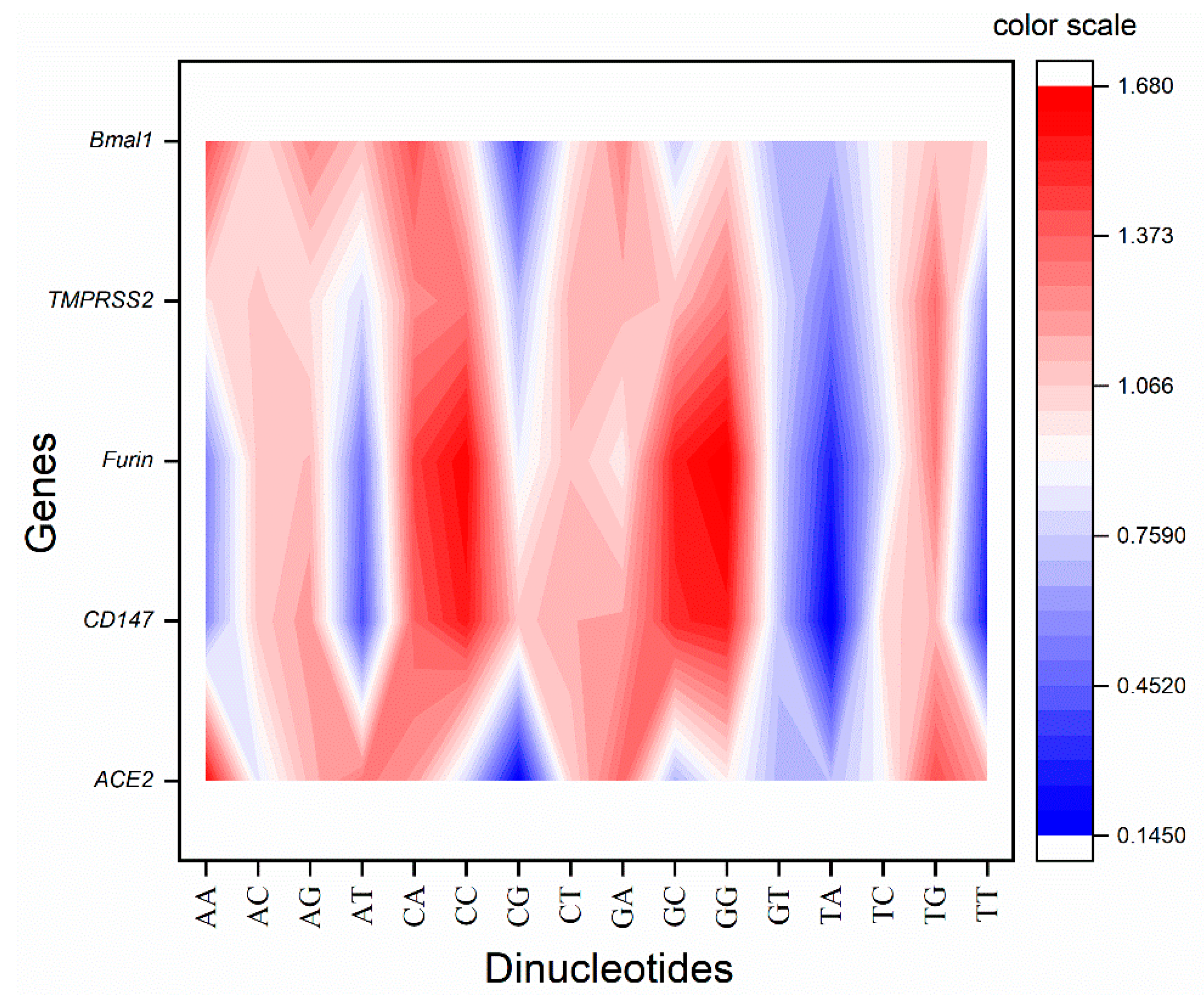
In the present study, five genes were envisaged where BMAL1 is found to be downregulated, and ACE2, CD147, FURIN, and TMPRSS1 are upregulated in periodontitis conditions and the SARS-CoV-2 infection. CpT dinucleotide is present nearly unbiasedly in the transcripts of all the genes. CD147 and FURIN genes showed an almost similar pattern of dinucleotide preference for ApA, ApC, ApG, ApT, CpA, CpC, CpT, GpC, and GpG dinucleotides. On the other hand, ACE2 and BMAL1 showed similar dinucleotide patterns in dinucleotide ApA, ApC, CpC, CpG, CpT, GpA, GpC, and GpG. CpA, CpC, and GpG are overrepresented, while TpT dinucleotide is underrepresented in the CD147, FURIN and TMPRSS2 genes’ set. TpA is underrepresented in all the genes envisaged. Similarly, GpT is also underrepresented in all the genes but TMPRSS2. Based on the odds ratio, the envisaged genes may be divided into two groups, one comprising CD147, FURIN and TMPRSS2 and the other comprising ACE2 and BMAL1 (Figure 2).

3.3. RSCU Analysis Revealed G/C Ending Codons Preference in CD147, FURIN, and TMPRSS2 Gene Set While A/T Ending Codons in ACE2 and BMAL1

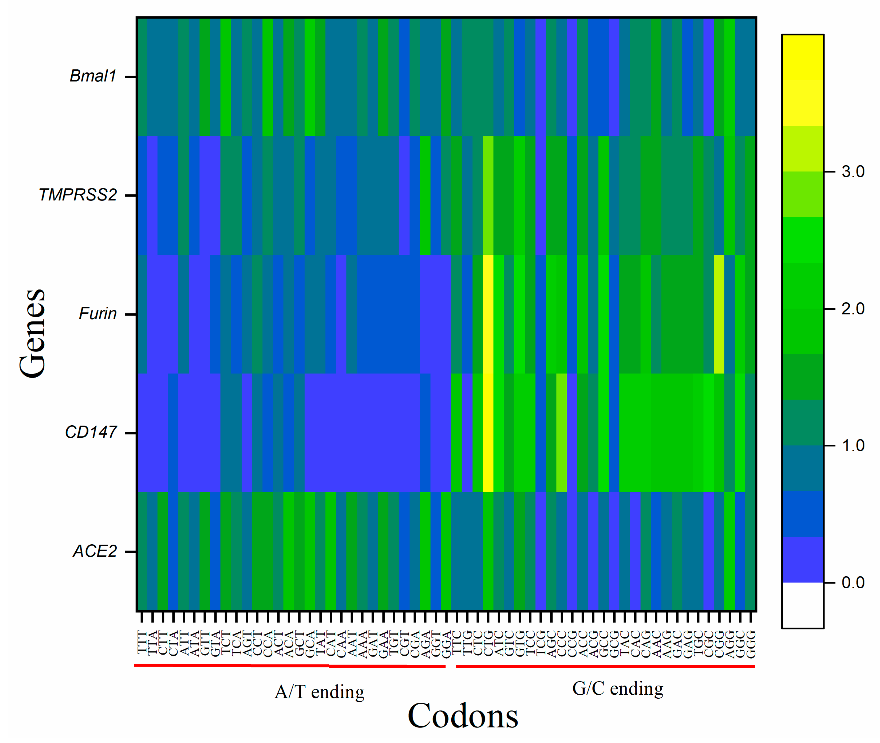
In the studied five genes, two patterns in RSCU were observed. Genes CD147, and FURIN, and TMPRSS2 showed the similar overrepresentation of C/G ending codons, while BMAL1 and ACE2 genes set showed a similar pattern and a preference for A/T ending codons. Three codons, TCG, CCG, and GCG, were underrepresented in all the genes, while CTG and GTG codons were overrepresented in all the genes but Bmal (Figure 3). CD147 was the gene that bypassed eight codons and did not use TTA, GTT, TGT, CGT, CGA, TAT, CAT, and CAA even for a single time in the transcripts. The ACE2 gene bypassed TCG, CCG, and GCG codons. BMAL1, FURIN, and TMPRSS2 genes used all available codons. The RSCU values of individual genes corresponding to their amino acid are given in Table 1.

3.4. Codons CGT, TCG and CTA Are Rarely Used in All the Genes

Codon usage bias is ruled by several mechanisms, resulting in various consequences. Optimal and unbiased codons are used in mRNA to be robustly translated [44]. On the other hand, the abundance of rare codons results in slower translation rates and deleteriously affects gene expression [45] and, in a few instances, reduces translational accuracy [46]. In the case of SARS-CoV-2, codon bias influences the host–pathogen interaction too. The presence of a specific rare codon is position-specific, and the presence at the 5′ end increases translational efficiency [47]. In the present study, we investigated six-fold, four-fold, two-fold, and three-fold degenerate codons separately (Figure 4). Among six-fold degenerated codons, CGT, TCG, and CTA encoding for Arg, Ser, and Leu, respectively, are rarely used (frequency less than 10 in 1000 bp) in all the five genes envisaged that are involved both in the periodontitis and the SARS-CoV-2 infection. Furthermore, AGA and CGA are the codons that were rare in four genes (codon AGA is not rare in ACE2 and CGA is not rare in BMAL1). For four-fold degenerate codons, GTA (Val), GGT (Gly), and CCG (Pro) are rare in four out of five genes (GTA, GGT, and CCG are not rare in BMAL1 and TMPRSS2 genes, respectively).

3.5. Codon Context Analysis Revealed Val (GTG) Initiated Codon Pair Abundance in CD147, FURIN, and TMPRSS2

There is the presence of a specific bias for a certain combinations of codons [48]. The impact of codon context is further strengthened by pieces of evidence produced by codon pair deoptimization [49,50] and codon pair optimization [51]. Table 2 presents the top 30 codon pairs in the genes.
The analysis revealed that CAA-GAA, GTG-CTG, GTG-GCC, GTG-TGC, and GAA-TAT are the most abundantly found codon pair in ACE2, CD147, FURIN, TMPRSS2, and BMAL1 genes, respectively. In CD147, FURIN, and TMPRSS2 genes Val (GTG)-initiated codon pairs were most abundant, while in ACE2 and BMAL1 genes, Gln (CAA)- and Glu (GAA)-initiated codons were most abundant. Among all genes, TMPRSS2 showed preference for Asn (AAC-CCC, AAC-AAU)-, Phe (UUG-AAC, UUC-AUG)-, Tyr (UAU-GAC, UAC-GGG, UAC-CAA)-, Val (GUC, GUG)-, Gly (GUG-UAC, GUC-GAU)-, Gly (GGG-GCC, GGA-UAC)-, Ala (GCG-CUG, GCC-UGC, GCC-GGC)-, Asp (GAC-UGG, GAC-UCC)-, Glu (GAA-AAC, GAA-AAA)-, Pro (CCU-CUG, CCC-ACU)-, and Gln (CAG-UAC, CAG-CCC)-initiated codons. CD147 gene showed 25 codon pairs (GAG-GAC, GGC-UCC, CUG-AAG, UCA-GAG, GUG-AAG, GCG-CUG, GCC-CUC, CUG-GUC, CCC-GGC, CCC-GAG, AUC-AUC, ACU-GAC, ACG-GCC, AAC-GGC, UUC-GUG, UCC-GAC, UCC-AAG, GGC-CAG, GGC-ACC, GCC-GGC, GAC-GAC, GAC-CAG, CUG-GGC, AAU-GAC) to be highly used, followed by FURIN (three codon pairs, GUG-GCC, GCC-AAC, CUG-GGC) and BMAL1 (two codon pairs, GAA-UAU, AAA-GAU).
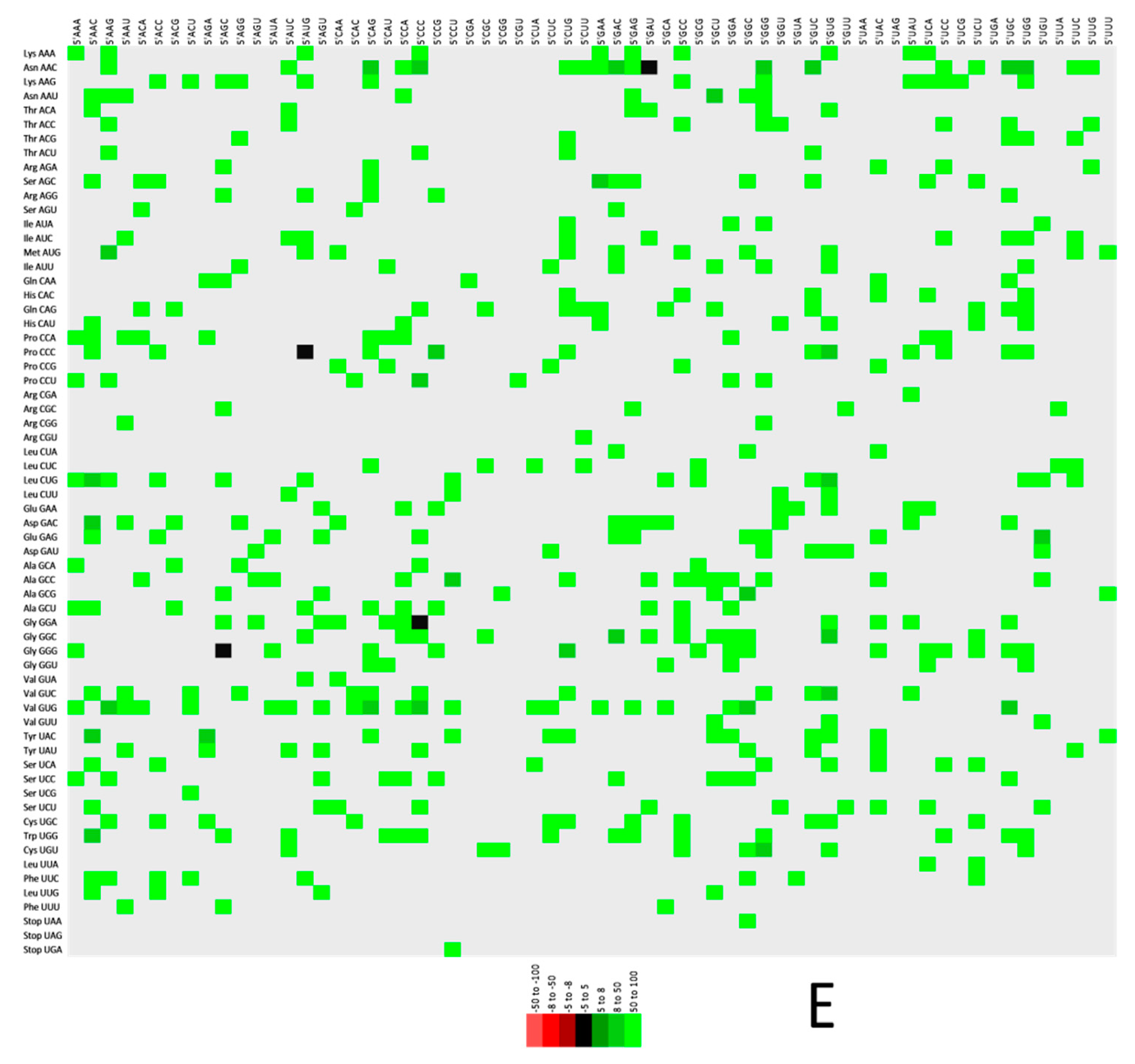
Figure 5A–E demonstrates the codon context in two color-coded maps. Strongly preferred codon context bias is depicted as green, while firmly rejected codon context is shown as red. In the case where the 3′ context is not strongly biased, it is defined as black, revealing that in TMPRSS2 gene, the rejected codon pairs are absent.

4. Discussion

COVID-19, caused by the SARS-CoV-2 virus, emerged as a severe health problem leading to the death of more than 6 million worldwide up to early 2022 [1]. ACE2, TMPRSS2, CD147, and FURIN act as receptors and coreceptors for SARS-CoV-2 entry. Their overexpression led to the enhanced severity and knocking down the expression of these genes by shRNA strategy, showed a reduction in viral protein expression [12]. Similar genes are overexpressed in the periodontal tissues of periodontitis patients [14]. Contrary to the four genes mentioned above, the loss of circadian gene BMAL1 shows enhanced viral burden and disease severity [13]. Diminished BMAL1 may trigger TNF-α and other pro-inflammatory cytokines production leading to cytokine storm and exacerbating periodontal inflammation [17].
Therefore, based on the above evidence, we studied five genes at the crossroads of periodontitis and SARS-CoV-2. Gene manipulations that alter the gene expression level of these genes might be helpful in simultaneously controlling both the SARS-CoV-2 infection and periodontitis. Here the upregulation of ACE2, TMPRSS2, FURIN, CD147, and downregulation of BMAL1 genes predispose towards the ailment. Therefore, if a strategy can be planned to modulate the gene expression of these genes in the future, it might help to prevent and cure the COVID-19 infection and periodontitis.
Codon optimization and codon pair optimization are the strategies to increase gene expression [52]. There are various approaches for codon optimization. Some approaches use the most optimal codons for all instances of a particular amino acid [53], while others adjust codon distribution proportionally to its natural distribution [54]. These approaches incorporate codon harmonization techniques where slow translation regions are identified and maintained to preserve the proper folding of protein to maintain its antigenicity and immunogenicity [55]. Alternatively, rare codons are replaced with abundant ones or codon pairs which are responsible for slow translation are avoided [56].
Therefore, we analyzed the different codon patterns in the present study and the knowledge will help modulate the expression of five genes associated with periodontitis and SARS-CoV-2 pathology. Based on the nucleotide composition of the genome, the codon usage of any organism is decided [57]. Organisms with GC-rich genomes use G/C ending codons, while organisms with AT-rich genomes use A/T ending codons [58]. In our study, CD147, FURIN, and TMPRSS2 genes are found to be GC rich and vice versa for ACE2 and BMAL1 genes. Based on RSCU, it is evident that G/C ending codons preference in CD147, FURIN, and TMPRSS2 gene set, while A/T-ending codons in ACE2 and BMAL1 and our results are in concordance with the results of human albumin superfamily members, which are AT-rich genes and preference for A/T-ending codons [59].
Rare codons slow down the translation rate due to the rarity of their cognate tRNAs. A linear relationship has been reported between the gene expression level and the number of rare codons. Wang and colleagues [60] engineered a rare codon device to finely tune the expression level of four genes responsible for the fatty acid synthesis II (FASII) pathway in E. Coli that not only switches the expression of reporter genes between the On and Off state but also helped in precisely regulating the protein expression at intermediate levels. A proof-of-concept experiment shows that altering the rare codon number might modulate protein expression levels. Similarly, the antibiotic pyoluteorin biosynthetic gene pltL encompasses six AGA rare codons, substituting four with CGC and two with CGG significantly enhanced antibiotic production [61].
In the present study CGT (Arg), AGA (Arg), CGA (Arg), TCG (Ser), CTA (Leu), GTA (Val), GGT (Gly) and CCG (Pro) codons were rare in at least four out of five genes. AGA and AGG codons are the rarest codons in E.coli [62], and the same is true for CD147, FURIN, and TMPRSS2 genes. To attenuate the virus, viral protein production needs to be reduced; therefore, the strategy is to increase the number of rare codons in recoded virus sequences. As a result, particular codons pair with specific codons at a frequency higher than expected, while some codon combinations are avoided. This phenomenon is called codon pair bias.
Various researchers have used codon pair deoptimization to construct the attenuated virus for vaccine candidate development. Examples are poliovirus [63], avian influenza virus [63], HIV-1 [64], human respiratory syncytial [65], and porcine reproductive and respiratory syndrome virus [66], where segments of viruses have been modified. Similarly, using the information presented here regarding rare codon pairs, the genes ACE2, CD147, FURIN, and TMPRSS2 may be deoptimized since, apart from participation in pathogenesis, these genes also contribute to physiological roles, so we cannot wholly knock down the genes. Instead, if using the CRISPR-Cas technique if we can recode the genes, the strategy might be used to ameliorate the ailment. Likewise, we will need to enhance the expression of the BMAL1 gene, which may be carried out using optimized codons in the recoded sequence to be inserted through CRISPR.
In the present study, amongst the genes envisaged the highest utilization of Val (GTG)–initiated codon pairs in CD147, FURIN, and TMPRSS2 genes was observed, while in ACE2 and BMAL1 genes, Gln (CAA)- and Glu (GAA)-initiated codons were most abundant. On the other hand, in the SARS-CoV-2 isolates, a bias towards valine-initiated (GUU-UUA, GUU-GAA, GUU-UAU, GUU-GUA) and glycine-initiated codon pairs (GGU-GUU, GGU-GAU, GGU-AAA, GGU-GGU) was observed [67]. Consistently there are under- and overrepresentations of codon pairs in all the organisms that appear to influence the translation machinery [68]. The first example of gene expression reduction through codon pair deoptimization was presented by Coleman and colleagues [63]. They generated synthetic poliovirus capsid gene sequences that contained codon pairs specifically rejected in human coding sequences. Such a codon pair deoptimized sequence was inserted in the infectious cDNA clone of poliovirus and a remarkably attenuated viral phenotype owing to the reduced viral protein expression was observed. However, codon pair-deoptimized sequences are predisposed to mistranslation, decreased mRNA stability, and reduced translation efficiency [69]. While attenuating a highly virulent Marek’s disease herpesvirus by codon pair bias deoptimization, the UL30 gene, encoding for the catalytic subunit of the viral DNA polymerase, was codon pair deoptimized. First, the gene was divided into three segments of equal length, and then the segments were codon pair deoptimized. A minimum of 224 codon pairs deoptimized in one segment resulted in reduced viral plaque size. In comparison, the introduction of changes in 770 codon pairs appeared highly lethal for the virus and no viable progeny was generated [70]. There is no gold standard for the number of deoptimized codon pairs for an effective reduction in protein production. However, some scientists may argue that codon pair deoptimization has such a minuscule effect on gene expression that one needs to introduce hundreds of changes in codon pairs [71]. In the experiment of Coleman and colleagues [63] in poliovirus capsid protein (PV), codon pairs were deoptimized. PV-Min construct contained 631 deoptimized codon pairs, while its segments PV-MinXY and PV-MinZ contained 407 and 224 deoptimized pairs. The F-Luc reporter was used to seeing the translatability of PV’s capsid region. The attenuation order was PV-Min > PV-MinZ > PV-MinXY, corresponding to 631, 224, and 407 deoptimized codon pairs. So, it appears that the reduction in translation is more sequence-specific and less dependent on the number of deoptimized codon pairs.
Contrarily, while deoptimizing HIV, mutant HIV-P minA showed significantly reduced replication only with 15 substitutions [72]. Hence there is broad scope in research related to optimizing the number required for codon pairs to modulate gene expression effectively. The present study provided valuable knowledge regarding the codon usage patterns, codon bias, and information regarding codon context, where, using this knowledge, gene expression patterns may be modulated as per the requirement under the conditions of periodontitis and SARS-CoV-2 infection. After extensively reviewing the literature, we found only five simultaneously upregulated and downregulated genes in both states that were utilized in the study.
Expression studies have been carried out in mice lungs where IL-1β is transiently expressed using adenoviral gene transfer via intratracheal administration. As a result, the high expression of IL-1β accompanied by a local increase of the pro-inflammatory cytokines IL-6 and TNF-α was observed with evidence of lung injury [73]. The experiment is a proof of concept experiment showing therapeutic interventions using gene transfer techniques. Hydrodynamic gene delivery has been proven effective in a rodent model to transiently express apolipoprotein L-I (APOL-I) and haptoglobin-related protein [74]. The gene copies present in human cells can be modified. The proof of concept is the FDA-approved treatment for spinal muscular atrophy (SMA) where the drug Spinraza® modifies the existing SMN2 gene through the intrathecal administration of four loading doses of oligonucleotides in such a way that the SMN2 gene is produced in higher amounts. Risdiplam® is another SMA therapy where the SMN2 gene is modified; however, the drug molecule is orally active. Zolgensma® is a more traditional gene therapy where a single intravenous dose is delivered to correct the missing or non-functional copy of the SMN1 gene [75]. The gendicine gene therapy drug, developed by Shenzhen SiBionoGeneTech and approved by China Food and Drug Administration [76], harbors a copy of the Tp53 gene embedded in the E1 region of human serotype 5 adenovirus (Ad5). Tumor suppressor genes, including TP53, NOTCH1, CDKN2A, PIK3CA, and FBXW7, are found inactivated in head and neck squamous cell carcinoma (HNSCC). The expression of Tp53 in cancer cells via gene therapy initiates the apoptotic pathways and has antitumor effects.
In the present case, out of the five genes we envisaged, four genes, ACE2, CD147, FURIN, and TMPRSS2, which serve as the receptors and co-receptors for SARS-CoV-2 and are overexpressed during COVID-19 infection and in the periodontal tissues of periodontitis patients too, presenting with inflammation, periodontal pathogens, and damage-induced pyroptosis [14], are required to be reduced to an average level. Elevated IL-1β activity is a feature of COVID-19 [77] and periodontitis [78]. BMAL1, an immune system regulator, limits the production of the pro-inflammatory cytokine IL-1β [77]. Therefore, the downregulation of ACE2, CD147, FURIN, and TMPRSS2 genes and the upregulation of BMAL1 may offer a therapeutic strategy against COVID-19 and periodontitis.
In light of knowledge from gene therapy studies, it is evident now that the desired upregulation or downregulation may be achieved in human tissue using codon usage information. For gene expression reduction, however, many codon changes are required to achieve the desired result, and this is the limitation inherently associated with codon pair deoptimization. The addition of rare codons might help in this context. With that, a uniform strategy cannot be made based on our studies for all the ailments and every disease model; it has to be studied and optimized separately.
Synonymous mutation influence phenotypic effects, owing to control over mRNA stability and translational regulation. However, our technique has the limitation that it is challenging to pre-judge the changes in which part of the gene will be most effective [79].
Presently the gene therapy technique is limited to treating genetic disorders and life-threatening diseases, such as cancer; however, in the future, with the advancement of technology, it will be possible to apply this to other conditions as well, such as in the case of COVID-19 and dental infections, and the codon usage and context analysis of genes may help reach this target.

5. Conclusions

We believe that our study about codon usage, codon bias, availability of rare codons, and codon context will help modulate the gene expression pattern within the organism and, therefore, simultaneously, the COVID-19 and periodontitis problem may be addressed. However, more study is required to obtain an optimal number of codon pairs or rare codons to be replaced in order to obtain an optimal level of gene expression, which is required to maintain the normal physiological functions of the ACE2, BMAL1, CD147, FURIN, and TMPRSS2 genes so that the pathological consequences of SARS-CoV-2 infection and periodontitis may be avoided.

Supplementary Materials

The following supporting information can be downloaded at: https://www.mdpi.com/article/10.3390/vaccines10111874/s1.

Author Contributions

Conceptualization, R.K. and M.K.P.; methodology, R.K.; software, P.G. and M.I.K.; validation, I.V.R., M.K.P. and M.I.K.; formal analysis, R.K.; investigation, M.I.K.; resources, M.K.P.; data curation, M.K.P. and P.G.; writing—original draft preparation, R.K.; writing—review and editing, R.K.; visualization, A.A.K.; supervision, R.K. and M.I.K.; project administration, R.K. and M.I.K.; funding acquisition, A.A.K. All authors have read and agreed to the published version of the manuscript.

Funding

This work was funded by the Researchers Supporting Project Number (RSP-2021/339), King Saud University, Riyadh, Saudi Arabia.

Institutional Review Board Statement

Not applicable.

Informed Consent Statement

Not applicable.

Data Availability Statement

Available upon request.

Acknowledgments

The authors are thankful for their respective Universities and Institutes for providing the research opportunity.

Conflicts of Interest

The authors declare no conflict of interest.

References

  1. Wiersinga, W.J.; Rhodes, A.; Cheng, A.C.; Peacock, S.J.; Prescott, H.C. Pathophysiology, Transmission, Diagnosis, and Treatment of Coronavirus Disease 2019 (COVID-19): A Review. JAMA 2020, 324, 782–793. [Google Scholar] [CrossRef] [PubMed]
  2. Azzi, L.; Carcano, G.; Gianfagna, F.; Grossi, P.; Gasperina, D.D.; Genoni, A.; Fasano, M.; Sessa, F.; Tettamanti, L.; Carinci, F.; et al. Saliva is a reliable tool to detect SARS-CoV-2. J. Infect. 2020, 81, e45–e50. [Google Scholar] [CrossRef] [PubMed]
  3. Zhu, J.; Guo, J.; Xu, Y.; Chen, X. Viral dynamics of SARS-CoV-2 in saliva from infected patients. J. Infect. 2020, 81, e48–e50. [Google Scholar] [CrossRef]
  4. Zhou, P.; Yang, X.-L.; Wang, X.-G.; Hu, B.; Zhang, L.; Zhang, W.; Si, H.-R.; Zhu, Y.; Li, B.; Huang, C.-L.; et al. A pneumonia outbreak associated with a new coronavirus of probable bat origin. Nature 2020, 579, 270–273. [Google Scholar] [CrossRef] [Green Version]
  5. Glowacka, I.; Bertram, S.; Müller, M.A.; Allen, P.; Soilleux, E.; Pfefferle, S.; Steffen, I.; Tsegaye, T.S.; He, Y.; Gnirss, K.; et al. Evidence that TMPRSS2 activates the severe acute respiratory syndrome coronavirus spike protein for membrane fusion and reduces viral control by the humoral immune response. J. Virol. 2011, 85, 4122–4134. [Google Scholar] [CrossRef] [PubMed] [Green Version]
  6. Iwata-Yoshikawa, N.; Okamura, T.; Shimizu, Y.; Hasegawa, H.; Takeda, M.; Nagata, N. TMPRSS2 Contributes to Virus Spread and Immunopathology in the Airways of Murine Models after Coronavirus Infection. J. Virol. 2019, 93, e01815–e01818. [Google Scholar] [CrossRef] [PubMed] [Green Version]
  7. Shang, J.; Ye, G.; Shi, K.; Wan, Y.; Luo, C.; Aihara, H.; Geng, Q.; Auerbach, A.; Li, F. Structural basis of receptor recognition by SARS-CoV-2. Nature 2020, 581, 221–224. [Google Scholar] [CrossRef] [Green Version]
  8. Jackson, C.B.; Farzan, M.; Chen, B.; Choe, H. Mechanisms of SARS-CoV-2 entry into cells. Nat. Rev. Mol. Cell Biol. 2022, 23, 3–20. [Google Scholar] [CrossRef]
  9. Wang, K.; Chen, W.; Zhang, Z.; Deng, Y.; Lian, J.-Q.; Du, P.; Wei, D.; Zhang, Y.; Sun, X.-X.; Gong, L.; et al. CD147-spike protein is a novel route for SARS-CoV-2 infection to host cells. Signal Transduct. Target. Ther. 2020, 5, 283. [Google Scholar] [CrossRef]
  10. Peng, R.; Wu, L.-A.; Wang, Q.; Qi, J.; Gao, G.F. Cell entry by SARS-CoV-2. Trends Biochem. Sci. 2021, 46, 848–860. [Google Scholar] [CrossRef]
  11. Qiao, J.; Li, W.; Bao, J.; Peng, Q.; Wen, D.; Wang, J.; Sun, B. The expression of SARS-CoV-2 receptor ACE2 and CD147, and protease TMPRSS2 in human and mouse brain cells and mouse brain tissues. Biochem. Biophys. Res. Commun. 2020, 533, 867–871. [Google Scholar] [CrossRef] [PubMed]
  12. Dobrindt, K.; Hoagland, D.A.; Seah, C.; Kassim, B.; O’Shea, C.P.; Iskhakova, M.; Fernando, M.B.; Deans, P.J.M.; Powell, S.K.; Javidfar, B.; et al. Common genetic variation in humans impacts in vitro susceptibility to SARS-CoV-2 infection. Biorxiv 2021, 16, 505–518. [Google Scholar] [CrossRef] [PubMed]
  13. Early, J.O.; Menon, D.; Wyse, C.A.; Cervantes-Silva, M.P.; Zaslona, Z.; Carroll, R.G.; Palsson-McDermott, E.M.; Angiari, S.; Ryan, D.G.; Corcoran, S.E.; et al. Circadian clock protein BMAL1 regulates IL-1β in macrophages via NRF2. Proc. Natl. Acad. Sci. USA 2018, 115, E8460–E8468. [Google Scholar] [CrossRef] [PubMed] [Green Version]
  14. Baima, G.; Marruganti, C.; Sanz, M.; Aimetti, M.; Romandini, M. Periodontitis and COVID-19: Biological Mechanisms and Meta-analyses of Epidemiological Evidence. J. Dent. Res. 2022, 11, 1430–1440. [Google Scholar] [CrossRef]
  15. Marouf, N.; Cai, W.; Said, K.N.; Daas, H.; Diab, H.; Chinta, V.R.; Hssain, A.A.; Nicolau, B.; Sanz, M.; Tamimi, F. Association between periodontitis and severity of COVID-19 infection: A case-control study. J. Clin. Periodontol. 2021, 48, 483–491. [Google Scholar] [CrossRef] [PubMed]
  16. Sehirli, A.Ö.; Chukwunyere, U.; Aksoy, U.; Sayiner, S.; Abacioglu, N. The circadian clock gene BMAL1: Role in COVID-19 and periodontitis. Chronobiol. Int. 2021, 38, 779–784. [Google Scholar] [CrossRef] [PubMed]
  17. Li, J.; Li, Y.; Pan, S.; Zhang, L.; He, L.; Niu, Y. Paeonol attenuates ligation-induced periodontitis in rats by inhibiting osteoclastogenesis via regulating Nrf2/NF-κB/NFATc1 signaling pathway. Biochimie 2019, 156, 129–137. [Google Scholar] [CrossRef]
  18. Cai, W.; Marouf, N.; Said, K.N.; Tamimi, F. Nature of the Interplay Between Periodontal Diseases and COVID-19. Front. Dent. Med. 2021, 2, 735126. [Google Scholar] [CrossRef]
  19. Senapati, S.; Kumar, S.; Singh, A.K.; Banerjee, P.; Bhagavatula, S. Assessment of risk conferred by coding and regulatory variations of TMPRSS2 and CD26 in susceptibility to SARS-CoV-2 infection in human. J. Genet. 2020, 99, 53. [Google Scholar] [CrossRef]
  20. Manna, P.M.L.; Costa, J.E.; Gomez, R.S. CD26 Immuno-Expression and Periodontal Disease Progression. J. Biomed. Biotechnol. 2001, 1, 91–94. [Google Scholar] [CrossRef]
  21. Bakthavatchalu, V.; Meka, A.; Mans, J.J.; Sathishkumar, S.; Lopez, M.C.; Bhattacharyya, I.; Boyce, B.F.; Baker, H.V.; Lamont, R.J.; Ebersole, J.L.; et al. Polymicrobial periodontal pathogen transcriptomes in calvarial bone and soft tissue. Mol. Oral Microbiol. 2011, 26, 303–320. [Google Scholar] [CrossRef] [PubMed]
  22. Prelli Bozzo, C.; Nchioua, R.; Volcic, M.; Koepke, L.; Krüger, J.; Schütz, D.; Heller, S.; Stürzel, C.M.; Kmiec, D.; Conzelmann, C.; et al. IFITM proteins promote SARS-CoV-2 infection and are targets for virus inhibition in vitro. Nat. Commun. 2021, 12, 4584. [Google Scholar] [CrossRef] [PubMed]
  23. Goel, R.; Bloch, E.M.; Pirenne, F.; Al-Riyami, A.Z.; Crowe, E.; Dau, L.; Land, K.; Townsend, M.; Jecko, T.; Rahimi-Levene, N.; et al. ABO blood group and COVID-19: A review on behalf of the ISBT COVID-19 Working Group. Vox Sang. 2021, 116, 849–861. [Google Scholar] [CrossRef] [PubMed]
  24. Concolino, P.; Cecchetti, F.; D’Autilia, C.; Santonocito, C.; Di Stasio, E.; Zuppi, C.; Arcuri, C.; Deli, G.; Giardina, B.; Capoluongo, E.; et al. Association of periodontitis with GSTM1/GSTT1-null variants—A pilot study. Clin. Biochem. 2007, 40, 939–945. [Google Scholar] [CrossRef] [PubMed]
  25. Karcioglu Batur, L.; Hekim, N. The role of DBP gene polymorphisms in the prevalence of new coronavirus disease 2019 infection and mortality rate. J. Med. Virol. 2021, 93, 1409–1413. [Google Scholar] [CrossRef] [PubMed]
  26. Rafique, S.; Hingorjo, M.R.; Mumtaz, M.; Qureshi, M.A. The relationship of 1,25-dihydroxyvitamin D and Vitamin D binding protein in periodontitis. Pak. J. Med. Sci. 2019, 35, 847–851. [Google Scholar] [CrossRef] [Green Version]
  27. Abbas, M.; Verma, S.; Verma, S.; Siddiqui, S.; Khan, F.H.; Raza, S.T.; Siddiqi, Z.; Eba, A.; Mahdi, F. Association of GSTM1 and GSTT1 gene polymorphisms with COVID-19 susceptibility and its outcome. J. Med. Virol. 2021, 93, 5446–5451. [Google Scholar] [CrossRef]
  28. Hitti, F.L.; Yang, A.I.; Gonzalez-Alegre, P.; Baltuch, G.H. Human gene therapy approaches for the treatment of Parkinson’s disease: An overview of current and completed clinical trials. Park. Relat. Disord. 2019, 66, 16–24. [Google Scholar] [CrossRef]
  29. Muramatsu, S.; Fujimoto, K.; Kato, S.; Mizukami, H.; Asari, S.; Ikeguchi, K.; Kawakami, T.; Urabe, M.; Kume, A.; Sato, T.; et al. A phase I study of aromatic L-amino acid decarboxylase gene therapy for Parkinson’s disease. Mol. Ther. 2010, 18, 1731–1735. [Google Scholar] [CrossRef] [Green Version]
  30. Watts, J.K.; Corey, D.R. Silencing disease genes in the laboratory and the clinic. J. Pathol. 2012, 226, 365–379. [Google Scholar] [CrossRef]
  31. Chakraborty, S.; Deb, B.; Barbhuiya, P.A.; Uddin, A. Analysis of codon usage patterns and influencing factors in Nipah virus. Virus Res. 2019, 263, 129–138. [Google Scholar] [CrossRef] [PubMed]
  32. Blitvich, B.J.; Firth, A.E. Insect-specific flaviviruses: A systematic review of their discovery, host range, mode of transmission, superinfection exclusion potential and genomic organization. Viruses 2015, 7, 1927–1959. [Google Scholar] [CrossRef] [PubMed] [Green Version]
  33. Quax, T.E.F.; Claassens, N.J.; Söll, D.; van der Oost, J. Codon Bias as a Means to Fine-Tune Gene Expression. Mol. Cell 2015, 59, 149–161. [Google Scholar] [CrossRef] [PubMed] [Green Version]
  34. Frumkin, I.; Lajoie, M.J.; Gregg, C.J.; Hornung, G.; Church, G.M.; Pilpel, Y. Codon usage of highly expressed genes affects proteome-wide translation efficiency. Proc. Natl. Acad. Sci. USA 2018, 115, E4940–E4949. [Google Scholar] [CrossRef] [Green Version]
  35. Zhang, G.; Hubalewska, M.; Ignatova, Z. Transient ribosomal attenuation coordinates protein synthesis and co-translational folding. Nat. Struct. Mol. Biol. 2009, 16, 274–280. [Google Scholar] [CrossRef] [PubMed]
  36. Xu, Y.; Ma, P.; Shah, P.; Rokas, A.; Liu, Y.; Johnson, C.H. Non-optimal codon usage is a mechanism to achieve circadian clock conditionality. Nature 2013, 495, 116–120. [Google Scholar] [CrossRef] [Green Version]
  37. Frenkel-Morgenstern, M.; Danon, T.; Christian, T.; Igarashi, T.; Cohen, L.; Hou, Y.-M.; Jensen, L.J. Genes adopt non-optimal codon usage to generate cell cycle-dependent oscillations in protein levels. Mol. Syst. Biol. 2012, 8, 572. [Google Scholar] [CrossRef]
  38. Di Giallonardo, F.; Schlub, T.E.; Shi, M.; Holmes, E.C. Dinucleotide Composition in Animal RNA Viruses Is Shaped More by Virus Family than by Host Species. J. Virol. 2017, 91, e02381-16. [Google Scholar] [CrossRef] [Green Version]
  39. Chen, Y.; Shi, Y.; Deng, H.; Gu, T.; Xu, J.; Ou, J.; Jiang, Z.; Jiao, Y.; Zou, T.; Wang, C. Characterization of the porcine epidemic diarrhea virus codon usage bias. Infect. Genet. Evol. 2014, 28, 95–100. [Google Scholar] [CrossRef]
  40. Clarke, T.F.; Clark, P.L. Rare codons cluster. PLoS ONE 2008, 3, e3412. [Google Scholar] [CrossRef]
  41. Wang, W.; Cheng, X.; Buske, P.J.; Suzich, J.A.; Jin, H. Attenuate Newcastle disease virus by codon modification of the glycoproteins and phosphoprotein genes. Virology 2019, 528, 144–151. [Google Scholar] [CrossRef] [PubMed]
  42. Moura, G.; Pinheiro, M.; Arrais, J.; Gomes, A.C.; Carreto, L.; Freitas, A.; Oliveira, J.L.; Santos, M.A.S. Large scale comparative codon-pair context analysis unveils general rules that fine-tune evolution of mRNA primary structure. PLoS ONE 2007, 2, e847. [Google Scholar] [CrossRef] [PubMed] [Green Version]
  43. Moura, G.; Pinheiro, M.; Silva, R.; Miranda, I.; Afreixo, V.; Dias, G.; Freitas, A.; Oliveira, J.L.; Santos, M.A. Comparative context analysis of codon pairs on an ORFeome scale. Genome Biol. 2005, 6, R28. [Google Scholar] [CrossRef] [Green Version]
  44. Presnyak, V.; Alhusaini, N.; Chen, Y.-H.; Martin, S.; Morris, N.; Kline, N.; Olson, S.; Weinberg, D.; Baker, K.E.; Graveley, B.R.; et al. Codon optimality is a major determinant of mRNA stability. Cell 2015, 160, 1111–1124. [Google Scholar] [CrossRef] [PubMed] [Green Version]
  45. Sharp, P.M.; Li, W.H. The codon Adaptation Index--a measure of directional synonymous codon usage bias, and its potential applications. Nucleic Acids Res. 1987, 15, 1281–1295. [Google Scholar] [CrossRef] [Green Version]
  46. Kramer, E.B.; Farabaugh, P.J. The frequency of translational misreading errors in E. coli is largely determined by tRNA competition. RNA 2007, 13, 87–96. [Google Scholar] [CrossRef] [PubMed] [Green Version]
  47. Chaney, J.L.; Steele, A.; Carmichael, R.; Rodriguez, A.; Specht, A.T.; Ngo, K.; Li, J.; Emrich, S.; Clark, P.L. Widespread position-specific conservation of synonymous rare codons within coding sequences. PLoS Comput. Biol. 2017, 13, e1005531. [Google Scholar] [CrossRef] [Green Version]
  48. Gutman, G.A.; Hatfield, G.W. Nonrandom utilization of codon pairs in Escherichia coli. Proc. Natl. Acad. Sci. USA 1989, 86, 3699–3703. [Google Scholar] [CrossRef] [Green Version]
  49. Groenke, N.; Trimpert, J.; Merz, S.; Conradie, A.M.; Wyler, E.; Zhang, H.; Hazapis, O.-G.; Rausch, S.; Landthaler, M.; Osterrieder, N.; et al. Mechanism of Virus Attenuation by Codon Pair Deoptimization. Cell Rep. 2020, 31, 107586. [Google Scholar] [CrossRef]
  50. Tulloch, F.; Atkinson, N.J.; Evans, D.J.; Ryan, M.D.; Simmonds, P. RNA virus attenuation by codon pair deoptimisation is an artefact of increases in CpG/UpA dinucleotide frequencies. Elife 2014, 3, e04531. [Google Scholar] [CrossRef]
  51. Le Nouën, C.; Luongo, C.L.; Yang, L.; Mueller, S.; Wimmer, E.; DiNapoli, J.M.; Collins, P.L.; Buchholz, U.J. Optimization of the Codon Pair Usage of Human Respiratory Syncytial Virus Paradoxically Resulted in Reduced Viral Replication In Vivo and Reduced Immunogenicity. J. Virol. 2020, 94, e01296-19. [Google Scholar] [CrossRef] [PubMed] [Green Version]
  52. Mauro, V.P.; Chappell, S.A. A critical analysis of codon optimization in human therapeutics. Trends Mol. Med. 2014, 20, 604–613. [Google Scholar] [CrossRef] [PubMed] [Green Version]
  53. Richardson, S.M.; Wheelan, S.J.; Yarrington, R.M.; Boeke, J.D. GeneDesign: Rapid, automated design of multikilobase synthetic genes. Genome Res. 2006, 16, 550–556. [Google Scholar] [CrossRef] [PubMed] [Green Version]
  54. Villalobos, A.; Ness, J.E.; Gustafsson, C.; Minshull, J.; Govindarajan, S. Gene Designer: A synthetic biology tool for constructing artificial DNA segments. BMC Bioinform. 2006, 7, 285. [Google Scholar] [CrossRef] [PubMed] [Green Version]
  55. Punde, N.; Kooken, J.; Leary, D.; Legler, P.M.; Angov, E. Codon harmonization reduces amino acid misincorporation in bacterially expressed P. falciparum proteins and improves their immunogenicity. AMB Express 2019, 9, 1–14. [Google Scholar] [CrossRef]
  56. Hatfield, G.W.; Roth, D.A. Optimizing scaleup yield for protein production: Computationally Optimized DNA Assembly (CODA) and Translation EngineeringTM. In Biotechnology Annual Review; El-Gewely, M.R., Ed.; Elsevier: Amsterdam, The Netherlands, 2007; Volume 13, pp. 27–42. [Google Scholar]
  57. Jenkins, G.M.; Holmes, E.C. The extent of codon usage bias in human RNA viruses and its evolutionary origin. Virus Res. 2003, 92, 1–7. [Google Scholar] [CrossRef]
  58. Yang, X.; Luo, X.; Cai, X. Analysis of codon usage pattern in Taenia saginata based on a transcriptome dataset. Parasit Vectors 2014, 7, 527. [Google Scholar] [CrossRef]
  59. Mirsafian, H.; Mat Ripen, A.; Singh, A.; Teo, P.H.; Merican, A.F.; Mohamad, S.B. A comparative analysis of synonymous codon usage bias pattern in human albumin superfamily. Sci. World J. 2014, 2014, 639682. [Google Scholar] [CrossRef] [Green Version]
  60. Wang, Y.; Li, C.; Khan, M.R.I.; Wang, Y.; Ruan, Y.; Zhao, B.; Zhang, B.; Ma, X.; Zhang, K.; Zhao, X.; et al. An Engineered Rare Codon Device for Optimization of Metabolic Pathways. Sci. Rep. 2016, 6, 20608. [Google Scholar] [CrossRef] [Green Version]
  61. Yan, Q.; Philmus, B.; Hesse, C.; Kohen, M.; Chang, J.H.; Loper, J.E. The Rare Codon AGA Is Involved in Regulation of Pyoluteorin Biosynthesis in Pseudomonas protegens Pf-5. Front. Microbiol. 2016, 7, 497. [Google Scholar] [CrossRef]
  62. Chen, G.T.; Inouye, M. Role of the AGA/AGG codons, the rarest codons in global gene expression in Escherichia coli. Genes Dev. 1994, 8, 2641–2652. [Google Scholar] [CrossRef] [PubMed] [Green Version]
  63. Coleman, J.R.; Papamichail, D.; Skiena, S.; Futcher, B.; Wimmer, E.; Mueller, S. Virus attenuation by genome-scale changes in codon pair bias. Science 2008, 320, 1784–1787. [Google Scholar] [CrossRef] [PubMed] [Green Version]
  64. Martrus, G.; Nevot, M.; Andres, C.; Clotet, B.; Martinez, M.A. Changes in codon-pair bias of human immunodeficiency virus type 1 have profound effects on virus replication in cell culture. Retrovirology 2013, 10, 78. [Google Scholar] [CrossRef] [PubMed] [Green Version]
  65. Le Nouën, C.; Brock, L.G.; Luongo, C.; McCarty, T.; Yang, L.; Mehedi, M.; Wimmer, E.; Mueller, S.; Collins, P.L.; Buchholz, U.J.; et al. Attenuation of human respiratory syncytial virus by genome-scale codon-pair deoptimization. Proc. Natl. Acad. Sci. USA 2014, 111, 13169–13174. [Google Scholar] [CrossRef] [Green Version]
  66. Ni, Y.-Y.; Zhao, Z.; Opriessnig, T.; Subramaniam, S.; Zhou, L.; Cao, D.; Cao, Q.; Yang, H.; Meng, X.-J. Computer-aided codon-pairs deoptimization of the major envelope GP5 gene attenuates porcine reproductive and respiratory syndrome virus. Virology 2014, 450, 132–139. [Google Scholar] [CrossRef] [Green Version]
  67. Saha, J.; Bhattacharjee, S.; Pal Sarkar, M.; Saha, B.K.; Basak, H.K.; Adhikary, S.; Roy, V.; Mandal, P.; Chatterjee, A.; Pal, A. A comparative genomics-based study of positive strand RNA viruses emphasizing on SARS-CoV-2 utilizing dinucleotide signature, codon usage and codon context analyses. Gene Rep. 2021, 23, 101055. [Google Scholar] [CrossRef]
  68. Tats, A.; Tenson, T.; Remm, M. Preferred and avoided codon pairs in three domains of life. BMC Genom. 2008, 9, 463. [Google Scholar] [CrossRef] [Green Version]
  69. Broadbent, A.J.; Santos, C.P.; Anafu, A.; Wimmer, E.; Mueller, S.; Subbarao, K. Evaluation of the attenuation, immunogenicity, and efficacy of a live virus vaccine generated by codon-pair bias de-optimization of the 2009 pandemic H1N1 influenza virus, in ferrets. Vaccine 2016, 34, 563–570. [Google Scholar] [CrossRef] [Green Version]
  70. Eschke, K.; Trimpert, J.; Osterrieder, N.; Kunec, D. Attenuation of a very virulent Marek’s disease herpesvirus (MDV) by codon pair bias deoptimization. PLoS Pathog. 2018, 14, e1006857. [Google Scholar] [CrossRef] [Green Version]
  71. Mueller, S.; Coleman, J.R.; Papamichail, D.; Ward, C.B.; Nimnual, A.; Futcher, B.; Skiena, S.; Wimmer, E. Live attenuated influenza virus vaccines by computer-aided rational design. Nat. Biotechnol. 2010, 28, 723–726. [Google Scholar] [CrossRef]
  72. Fu, J.; Sha, B.E.; Thomas, L.L. HIV-1-infected peripheral blood mononuclear cells enhance neutrophil survival and HLA-DR expression via increased production of GM-CSF: Implications for HIV-1 infection. J. Acquir. Immune Defic. Syndr. 2011, 56, 16–25. [Google Scholar] [CrossRef] [PubMed] [Green Version]
  73. Kolb, M.; Margetts, P.J.; Anthony, D.C.; Pitossi, F.; Gauldie, J. Transient expression of IL-1beta induces acute lung injury and chronic repair leading to pulmonary fibrosis. J. Clin. Investig. 2001, 107, 1529–1536. [Google Scholar] [CrossRef] [PubMed] [Green Version]
  74. Sendra, L.; Herrero, M.J.; Aliño, S.F. Translational Advances of Hydrofection by Hydrodynamic Injection. Genes 2018, 9, 136. [Google Scholar] [CrossRef] [PubMed] [Green Version]
  75. National Academies of Sciences, Engineering, and Medicine; Health and Medicine Division; Board on Health Sciences Policy; Forum on Regenerative Medicine. Exploring Novel Clinical Trial Designs for Gene-Based Therapies: Proceedings of a Workshop; Beachy, S.H., Alper, J., Hackmann, M., Addie, S., Eds.; The National Academies Collection: Reports funded by National Institutes of Health; National Academies Press (US): Washington, DC, USA, 2020; ISBN 978-0-309-67298-6. [Google Scholar]
  76. Peng, Z. Current status of gendicine in China: Recombinant human Ad-p53 agent for treatment of cancers. Hum. Gene Ther. 2005, 16, 1016–1027. [Google Scholar] [CrossRef] [PubMed]
  77. Vieira, E.; Mirizio, G.G.; Barin, G.R.; de Andrade, R.V.; Nimer, N.F.S.; La Sala, L. Clock Genes, Inflammation and the Immune System-Implications for Diabetes, Obesity and Neurodegenerative Diseases. Int. J. Mol. Sci. 2020, 21, 9743. [Google Scholar] [CrossRef]
  78. Cheng, R.; Wu, Z.; Li, M.; Shao, M.; Hu, T. Interleukin-1β is a potential therapeutic target for periodontitis: A narrative review. Int. J. Oral. Sci. 2020, 12, 2. [Google Scholar] [CrossRef] [Green Version]
  79. Van Leuven, J.T.; Ederer, M.M.; Burleigh, K.; Scott, L.; Hughes, R.A.; Codrea, V.; Ellington, A.D.; Wichman, H.A.; Miller, C.R. ΦX174 Attenuation by Whole-Genome Codon Deoptimization. Genome Biol. Evol. 2021, 13, evaa214. [Google Scholar] [CrossRef]
Figure 1. Average per cent composition of the genes.
Vaccines 10 01874 g001
Figure 2. Contour plot for the dinucleotide odds ratio. The blue color shows underrepresentation, while the red shows an overrepresentation of dinucleotides.
Vaccines 10 01874 g002
Figure 3. Heat map analysis for RSCU values of A/T and G/C ending codons of ACE2, CD147, FURIN, TMPRSS2, and BMAL1. Analysis revealed the dominance of G/C ending codons in CD147, FURIN, and TMPRSS2 genes while the dominance of A/T ending codons in ACE2 and BMAL1.
Vaccines 10 01874 g003
Figure 4. Rare codons in (A) Six-fold, (B) four-fold, (C) two-fold, and (D) three-fold degenerate codons in ACE2, BMAL1, TMPRSS2, FURIN, and CD147 genes. Codons less than 10 in number in a standard transcript of 1000 bp (normalized transcript length) were considered rare. Codons that are altogether absent in the transcript of a gene are boxed with red color.
Vaccines 10 01874 g004aVaccines 10 01874 g004b
Figure 5. Codon context analysis for (A) ACE2, (B) BMAL1, (C) CD147, (D) FURIN, and (E) TMPRSS2 genes. The green color shows the preferred codon pair, while the red color represents the rejected codon pairs. Statistically insignificant values are expressed as black. The grey color indicates unrepresented codon pairs.
Vaccines 10 01874 g005aVaccines 10 01874 g005bVaccines 10 01874 g005c
Table 1. Average RSCU values of ACE2, CD147, FURIN, TMPRSS2, and BMAL1 genes.
CODONSSingle Letter Amino AcidACE2CD147FURINTMPRSS2BMAL1
TTTF1.1040.0610.7630.5451.178
TTC0.8961.9391.2371.4550.822
TTAL0.7430.0000.0950.2300.965
TTG0.7780.1360.5550.7001.233
CTT1.4740.1360.0950.5840.778
CTC0.6911.6831.4571.1061.205
CTA0.5340.3490.2860.4060.730
CTG1.7803.6973.5112.9751.090
ATTI1.1580.2580.6071.1191.267
ATC1.1782.5852.3551.3440.793
ATA0.6640.1580.0370.5370.940
GTTV1.4740.0000.0720.2431.482
GTC0.6371.5861.0651.3561.084
GTA0.4730.1040.4350.2090.833
GTG1.4162.3102.4282.1920.600
TCTS1.5770.7120.6131.3261.790
TCC1.2872.2941.4961.5000.837
TCA1.2290.7120.4571.2350.814
TCG0.0000.4750.5470.1320.182
AGT0.7260.2980.8000.3981.241
AGC1.1821.5072.0871.4091.136
CCTP1.5000.7241.1540.6290.942
CCC0.9722.6921.6081.5170.928
CCA1.5280.3620.6831.2621.975
CCG0.0000.2220.5550.5920.156
ACTT1.1620.8900.5150.7900.959
ACC0.8261.3711.7311.5791.107
ACA1.8040.4480.9330.9481.579
ACG0.2081.2910.8200.6830.355
GCTA1.5030.6480.3641.0111.143
GCC0.8392.5362.4051.7890.511
GCA1.6580.2600.7510.5802.228
GCG0.0000.5550.4800.6200.119
TATY1.2330.0000.6350.7721.362
TAC0.7682.0001.3651.2280.638
CATH1.7550.0000.4210.9030.699
CAC0.2452.0001.5791.0971.301
CAAQ0.8160.0000.2300.5450.754
CAG1.1852.0001.7701.4551.246
AATN1.4120.3100.7210.5710.638
AAC0.5891.6901.2791.4291.362
AAAK1.1480.2220.4090.9051.236
AAG0.8521.7781.5911.0950.764
GATD0.9560.2830.5970.6870.941
GAC1.0441.7171.4031.3131.059
GAAE1.3600.2320.4230.7671.487
GAG0.6401.7681.5771.2330.513
TGTC1.0830.0000.4850.6561.157
TGC0.9172.0001.5151.3440.843
CGTR0.4410.0000.3750.1250.350
CGC0.1752.5511.1861.2460.114
CGA0.8040.0000.5070.3761.220
CGG0.6151.7773.1740.7521.343
AGA1.9420.4870.0801.6220.709
AGG2.0241.1850.6781.8802.263
GGTG0.4360.0430.3150.3560.782
GGC0.5682.6292.0871.1640.932
GGA1.7380.3030.2441.0151.369
GGG1.2581.0251.3541.4640.917
Table 2. Table presenting codon numbers and normalized codon numbers in transcripts of genes. Normalized codon pair numbers above a value of 5 (five codon pairs per thousand codon pairs) are indicated in bold.
ACE2 (4249 Codons)CD147 (1666 Codons)FURIN (5425 Codons)TMPRSS2 (1522 Codons)BMAL1 (30,873 Codons)
S. No.Codon PairCodon NumbersNormalized Codon NumberCodon PairCodon NumbersNormalized Codon NumberCodon PairCodon NumbersNormalized Codon NumberCodon PairCodon NumbersNormalized Codon NumberCodon PairCodon NumbersNormalized Codon Number
1CAA-GAA225.18GUG-CUG2012.0GUG-GCC417.56GUG-UGC95.91GAA-UAU1966.35
2AAU-GAA214.94GAG-GAC1810.8GCC-AAC285.16CUG-CAG95.91AAA-GAU1655.34
3AAA-AAU204.71GGC-UCC169.60CUG-GGC285.16AAC-CCC95.91GAU-GAA1484.79
4UAU-GAA194.47CUG-AAG148.40GGC-GAG274.98AAC-AAU95.91AUA-GAU1484.79
5GAA-AAU194.47UCA-GAG127.20AAU-GAC274.98UUG-AAC65.91GAA-AUC1464.73
6CUG-UUC194.47GUG-AAG127.20GGG-CUG264.79UUC-AUG65.91AGC-AUG1464.73
7GUU-GGG184.24GCG-CUG127.20GCC-CCC264.79UGU-GCC65.91GCA-GAU1304.21
8GAG-AUG174.00GCC-CUC127.20GAG-GUG264.79UGG-AUU65.91AUG-GAC1304.21
9GGA-UUC163.77CUG-GUC127.20CGG-CUG264.79UGC-AUC65.91AUG-AUU1153.72
10GAA-GAC163.77CCC-GGC127.20CAG-CAG264.79UCC-GGG65.91AUG-AAC1103.56
11CAG-AAA163.77CCC-GAG127.20GUG-GAG254.61UCC-AAC65.91GAA-UUG1013.27
12AUG-GCA163.77AUC-AUC127.20CUG-CCC254.61UAU-GAC65.91UUU-GUC1003.24
13AAA-CCA163.77ACU-GAC127.20UGG-GCC213.87UAC-GGG65.91UUG-UUU1003.24
14GAC-CAG153.53ACG-GCC127.20GGC-UAC213.87UAC-CAA65.91GUC-UCA1003.24
15GAA-GAG153.53AAC-GGC127.20GGC-CGG213.87GUG-UAC65.91GAU-AAA1003.24
16GCA-UAU143.29UUC-GUG106.00GGC-ACC213.87GUC-GAU65.91ACA-GAA1003.24
17CUU-GGA143.29UCC-GAC106.00GAG-GCC213.87GGG-GCC65.91AAC-UAC1003.24
18AUG-AAU143.29UCC-AAG106.00GAG-CCC213.87GGA-UAC65.91GUU-UUA993.21
19AAA-GCA143.29GGC-CAG106.00CUG-GCC213.87GCG-CUG65.91GCA-GCA993.21
20UUU-CUG122.82GGC-ACC106.00CUC-ACC213.87GCC-UGC65.91GCA-AUG993.21
21UUU-CAA122.82GCC-GGC106.00CGG-GAC213.87GCC-GGC65.91GAA-GCA993.21
22UUG-AAA122.82GAC-GAC106.00CGG-AAG213.87GAC-UGG65.91CUA-UCA993.21
23UUC-CUG122.82GAC-CAG106.00CAG-GGC213.87GAC-UCC65.91CCC-UCU993.21
24UUC-CAU122.82CUG-GGC106.00CAC-AUC213.87GAA-AAC65.91CAG-CUC993.21
25UGU-GAC122.82AAU-GAC106.00ACC-CUG213.87GAA-AAA65.91CAA-GGA993.21
26UGG-AUG122.82UAC-GAG84.80AAC-CAC213.87CUG-AAC65.91AUG-GCU993.21
27GGG-GAA122.82GUC-UUC84.80GCC-UUC203.69CCU-CUG65.91AGG-AUG993.21
28GCU-AAU122.82GUC-CUG84.80CAG-AAG203.69CCC-ACU65.91AGG-AUA993.21
29GAA-GCU122.82GUC-CGC84.80CAC-CUG203.69CAG-UAC65.91ACU-GUU993.21
30GAA-ACA122.82GGG-CAG84.80GUC-UUC193.50CAG-CCC65.91AAU-GAU993.21
Publisher’s Note: MDPI stays neutral with regard to jurisdictional claims in published maps and institutional affiliations.