Article

B-Cell Epitope Mapping of the Plasmodium falciparum Malaria Vaccine Candidate GMZ2.6c in a Naturally Exposed Population of the Brazilian Amazon

by
Barbara de Oliveira Baptista
1,2,
Ana Beatriz Lopes de Souza
1,2,
Luana Santos de Oliveira
1,2,
Hugo Amorim dos Santos de Souza
1,2,
Jenifer Peixoto de Barros
1,2,
Lucas Tavares de Queiroz
1,2,
Rodrigo Medeiros de Souza
3,
Linda Eva Amoah
4,
Susheel Kumar Singh
5,6,
Michael Theisen
5,6,
Rodrigo Nunes Rodrigues-da-Silva
7,
Evelyn Kety Pratt Riccio
1,2,
Paulo Renato Rivas Totino
1,2,
Josué da Costa Lima-Junior
8,
Cláudio Tadeu Daniel-Ribeiro
1,2 and
Lilian Rose Pratt-Riccio
1,2,*
1
Laboratório de Pesquisa em Malária, Instituto Oswaldo Cruz (IOC), Fundação Oswaldo Cruz (Fiocruz), Rio de Janeiro 21040-900, RJ, Brazil
2
Centro de Pesquisa, Diagnóstico e Treinamento em Malária (CPD-Mal), Fiocruz e Secretaria de Vigilância em Saúde, Ministério da Saúde, Rio de Janeiro 21040-900, RJ, Brazil
3
Centro de Pesquisa em Doenças Infecciosas, Universidade Federal do Acre–Campus Floresta (UFAC), Cruzeiro do Sul 69895-000, AC, Brazil
4
Immunology Department, Noguchi Memorial Institute for Medical Research, University of Ghana, Accra P.O. Box LG 25, Ghana
5
Centre for Medical Parasitology, Department of International Health, Immunology and Microbiology, University of Copenhagen, DK-2200 Copenhagen, Denmark
6
Department of Infectious Disease, Copenhagen University Hospital, DK-2200 Copenhagen, Denmark
7
Laboratório de Tecnologia Imunológica, Instituto de Tecnologia em Imunobiológicos (Bio-Manguinhos), Fiocruz, Rio de Janeiro 21040-900, RJ, Brazil
8
Laboratório de Imunoparasitologia, IOC, Fiocruz, Rio de Janeiro 21040-900, RJ, Brazil
*
Author to whom correspondence should be addressed.
Vaccines 2023, 11(2), 446; https://doi.org/10.3390/vaccines11020446
Submission received: 9 January 2023 / Revised: 9 February 2023 / Accepted: 13 February 2023 / Published: 15 February 2023

Abstract

:
The GMZ2.6c malaria vaccine candidate is a multi-stage P. falciparum chimeric protein that contains a fragment of the sexual-stage Pfs48/45-6C protein genetically fused to GMZ2, an asexual-stage vaccine construction consisting of the N-terminal region of the glutamate-rich protein (GLURP) and the C-terminal region of the merozoite surface protein-3 (MSP-3). Previous studies showed that GMZ2.6c is widely recognized by antibodies from Brazilian exposed individuals and that its components are immunogenic in natural infection by P. falciparum. In addition, anti-GMZ2.6c antibodies increase with exposure to infection and may contribute to parasite immunity. Therefore, identifying epitopes of proteins recognized by antibodies may be an important tool for understanding protective immunity. Herein, we identify and validate the B-cell epitopes of GMZ2.6c as immunogenic and immunodominant in individuals exposed to malaria living in endemic areas of the Brazilian Amazon. Specific IgG antibodies and subclasses against MSP-3, GLURP, and Pfs48/45 epitopes were detected by ELISA using synthetic peptides corresponding to B-cell epitopes previously described for MSP-3 and GLURP or identified by BepiPred for Pfs48/45. The results showed that the immunodominant epitopes were P11 from GLURP and MSP-3c and DG210 from MSP-3. The IgG1 and IgG3 subclasses were preferentially induced against these epitopes, supporting previous studies that these proteins are targets for cytophilic antibodies, important for the acquisition of protective immunity. Most individuals presented detectable IgG antibodies against Pfs48/45a and/or Pfs48/45b, validating the prediction of linear B-cell epitopes. The higher frequency and antibody levels against different epitopes from GLURP, MSP-3, and Pfs48/45 provide additional information that may suggest the relevance of GMZ2.6c as a multi-stage malaria vaccine candidate.

1. Introduction

Malaria, an infectious disease caused by Plasmodium parasites, remains a major public health problem worldwide. In 2021, 247 million cases and 619.000 deaths due to malaria were estimated globally, most of them caused by P. falciparum [1]. Despite the remarkable progress in control over the last few years, the emergence of resistant parasites to artemisinin-based combination therapies (ACTs), the first line of treatment for uncomplicated P. falciparum malaria in all endemic countries [2,3,4,5,6], and resistance of Anopheles to available insecticides [7] highlights the need for an effective vaccine to be implemented, complementing the existing tools. Recently, the World Health Organization (WHO) recommended the widespread use of the RTS,S/AS01 vaccine among children in regions with moderate to high P. falciparum transmission [1]. Despite its partial and low efficacy, this vaccine might prevent about 30% of child deaths [8]. However, vaccine candidates with a higher protective efficacy should be developed. Considering the complex life cycle, an ideal malaria vaccine should be directed against antigens expressed in different parasite development stages.
The GMZ2.6c malaria vaccine candidate is a multi-stage P. falciparum chimeric protein that contains a fragment of the sexual-stage Pfs48/45-6C protein genetically fused to GMZ2, an asexual-stage vaccine construct consisting of conserved domains of glutamate-rich protein (GLURP) and merozoite surface protein-3 (MSP-3) [9]. GLURP is expressed in all parasite life cycle stages in the vertebrate host and may contribute to merozoite invasion and formation of the parasitophorous vacuole [10,11], MSP-3 is expressed in the erythrocytic stage and is involved in the invasion of red blood cells, as well as in parasite protection against heme released from unprocessed hemoglobin released after schizont egress [12,13], and Pfs48/45 is expressed on the surface of gametocytes and gametes, playing a central role in fertilization [14]. Both GLURP and MSP-3 are targets of cytophilic IgG subclasses (IgG1 and IgG3) able to control P. falciparum growth in vitro by opsonic phagocytosis [15] and antibody-dependent cellular inhibition (ADCI) [16,17,18] correlated to protection against high parasitemia and clinical disease in individuals living in endemic areas, while naturally acquired antibodies against Pfs48/45 can recognize extra-erythrocytic gametes, reducing transmission by preventing fertilization and sporogonic development in the mosquito midgut [19].
Previous studies showed that GMZ2.6c protein was widely recognized by naturally acquired antibodies from individuals of Brazilian endemic areas and that its components (MSP-3, GLURP, and Pfs48/45) are immunogenic in natural infection by P. falciparum. Moreover, anti-GMZ2.6c antibodies seem to increase with exposure to malaria infection and may contribute to parasite immunity [20]. Considering that antibodies recognize several antigenic determinants of the protein, identifying the contribution of different immunodominant B-cell epitopes of antimalarial vaccine candidates that induce specific immune responses may be an important tool for understanding protective immunity. Therefore, the present work aimed to identify and validate immunodominant B-cell epitopes of the P. falciparum chimeric protein GMZ2.6c in individuals exposed to malaria living in the Brazilian Amazon.

2. Materials and Methods

2.1. Study Area and Volunteers

A cross-sectional cohort study was carried out from June to August 2016 and 2018 in three malaria-endemic areas of the Brazilian Amazon: Cruzeiro do Sul (07°37′50″ S/72°40′13″ W) and Mâncio Lima (07°36′49″ S/72°53′47″ W), both high-risk areas situated in Juruá Valley, Acre State, the main P. falciparum malaria hotspot in Brazil, and Guajará (02°58′18″ S/57°40′38″ W), a medium-risk area situated in Amazonas State. Cruzeiro do Sul, Mâncio Lima, and Guajará registered 5.447, 1.432, and 674 cases in 2016, accounting for 52.6% of all P. falciparum infections in Brazil. Due to the increase in P. falciparum cases in other municipalities, this number decreased to 24.1% in 2018, with 2.915, 1.719, and 452 P. falciparum infections in Cruzeiro do Sul, Mâncio Lima, and Guajará, respectively [21].
Serum samples from 303 malaria-exposed individuals identified as responders to GLURP27−500 (n = 258), MSP-3155–249 (n = 170), and/or Pfs48/45291–428 (n = 108) recombinants (Table S1), as previously described [20], were used to investigate specific naturally acquired antibody response to GLURP, MSP-3, and Pfs48/45 epitopes. In addition, serum samples from 5 individuals of the laboratory staff (Rio de Janeiro, Brazil) who had neither history of malaria nor contact with malaria transmission areas were included in our study (Rio de Janeiro controls).

2.2. Epidemiological Survey

Donors providing informed consent answered an epidemiological survey. To evaluate the degree of malaria exposure, subjects responded to questions related to personal data such as age, time of residence in the endemic area, number of previous malaria episodes, time elapsed from the last infection, use of malaria prophylaxis, and presence of symptoms.

2.3. Blood Sampling and Malaria Diagnosis

Venous peripheral blood (20 mL) was collected into Heparin or EDTA tubes for antibodies analysis or molecular diagnosis, respectively. The plasma was stored at −20 °C, and the pellets, containing peripheral blood cells collected into EDTA tubes, were mixed with equal volumes of a cryopreservation solution (0.9% NaCl/4.2% sorbitol/20% glycerol) and were stored at −70 °C until use.
Thin and thick blood smears were examined for identification of malaria parasites by a technician experienced in malaria diagnosis from the Laboratory of Malaria Research (Fiocruz), which is the headquarters of the CEMART (Centre for Malaria Research and Training), a reference center for malaria diagnosis in the Extra-Amazonian Region for the Brazilian Ministry of Health. Thick blood smears from all the subjects were stained with Giemsa, and a total of 200 microscopic fields were examined under a 1000-fold magnification. Thin blood smears of the positive samples were examined for species identification. The parasite density was determined by counting the parasites in a predetermined number of white blood cells in the thick blood films, and the number of blood parasites per milliliter was calculated [22]. To increase the sensitivity of the parasite detection, molecular analysis using specific primers for genus (Plasmodium sp.) and species (P. falciparum and P. vivax) was performed in all samples, as previously described [23]. Positive donors for P. vivax and/or P. falciparum at the time of blood collection were subsequently treated by the chemotherapeutic regimen recommended by the Brazilian Ministry of Health [24].

2.4. B-Cell Epitope Prediction of Pfs48/45

The prediction of linear B-cell epitopes of Pfs48/45291–428 (UniProt: Q8I6T1) was carried out using the program BepiPred 1.0 [25]. This method is based on the combination of hidden Markov and propensity scale models, which consider hydrophilicity and secondary structure prediction. The server outputs a prediction score for each amino acid for each input FASTA sequence. To identify potential linear B-cell epitopes was used the cut-off value of 0.35, ensuring a sensibility of 49% and specificity of 75% to this approach. Linear B-cell epitopes are predicted to be located at the residues with the highest scores. Sequences with a BepiPred score above 0.35 were considered potential linear B-cell epitopes in regions that could be accessed by naturally acquired antibodies.

2.5. Synthetic Peptides

B-cell epitope mapping was performed using four synthetic peptides of MSP-3155–249 (MSP-3a, MSP-3b, MSP-3c, and DG210) and 13 synthetic peptides of GLURP27–500 (P1, P2, P3, P4, P5, P6, P7, P8, P9, P10, P11, S2, and S3), previously described [26,27]. Moreover, two synthetic peptides of Pfs48/45291–428 (Pfs48/45a and Pfs48/45b) identified by prediction of linear B-cell epitopes were used (Figure 1). The selected sequences were synthesized by fluorenylmethoxycarbonyl (F-moc) solid-phase chemistry (GenOne Biotechnologies, Brazil). Analytical chromatography of the peptide demonstrated a purity of >95%.

2.6. Enzyme-Linked Immunosorbent Assay

Microtiter 96-well plates (Maxisorp, NUNC, Roskilde, Denmark) were coated with 5 µg/mL of each synthetic peptide in carbonate-bicarbonate buffer, pH 9.6, at 100 µL/well overnight at 4 °C. The plates were washed with phosphate-buffered saline 0.05% Tween20 (PBST), and the uncoated sites were blocked with 5% powdered milk containing PBST for 1 h at 37 °C. Serum samples diluted 1:100 in dilution buffer (1% powdered milk containing PBST) were added in duplicate wells, and plates were incubated for 1 h at 37 °C. The plates were washed, 100 µL of peroxidase-conjugated mouse anti-human IgG (Sigma, St. Louis, MO, USA) 1:1000 in dilution buffer was added, and the plates were incubated for 1 h at 37 °C. To detect specific IgG subclass, plates were incubated with peroxidase-conjugated mouse anti-human IgG1, IgG2, IgG3, and IgG4 (Clones: 4E3, 31-7-4, HP6050, and HP6025 for IgG1, IgG2, IgG3, and IgG4, respectively; SouthernBiotech, Birmingham, AL, USA) 1:1000 in dilution buffer for 1 h at 37 °C. After washing, 100 µL of a solution of 0.4 mg/mL orthophenylenediamine (OPD, Sigma) and H2O2 30% (Sigma) in citrate-phosphate buffer (24 mM citric acid, Sigma, and 51 mM dibasic sodium phosphate, Sigma), pH 5.0, were added to each well, the plates were incubated for 5 min at room temperature in the dark, and then 50 µL/well of 2N H2SO4 (Sigma) were added. Optical density was identified at 492 nm using a SpectraMax 250 ELISA reader (Molecular Devices, Sunnyvale, CA, USA). Samples of non-endemic controls obtained from 5 individuals of the laboratory staff (Rio de Janeiro controls) were used to establish the normal range for each assay. The cut-off values were determined as the mean optical density (OD) plus 3 standard deviations of the Rio de Janeiro controls. The results were expressed as the reactivity index (RI), which was calculated by the OD mean of each tested sample divided by the cut-off value. Subjects were scored as responders if the RI against each epitope was higher than 1.0.

2.7. Statistical Analysis

Data were stored in the Epi-Info version 6 data bank system (Centers for Disease Control and Prevention, Atlanta, GA, USA) and analyzed using Epi-Info version 6 and GraphPad Prism (GraphPad Sftware, Inc., San Diego, CA, USA) software programs. Normality tests were performed in all variables using the one-sample Kolmogorov–Smirnov test. The chi-square analysis was applied to compare the prevalence among groups. Kruskal–Wallis, followed by uncorrected Dunn’s test (for multiples comparisons) or Mann–Whitney test (comparisons between two groups), was used to analyze differences in distributions of continuous numerical variables. The Spearman rank coefficient test was used to analyze the correlation between variables. A two-sided p-value of ≤0.05 was considered statistically significant.

3. Results

3.1. Population Characteristics

The studied population was composed of 303 individuals living in three malaria-endemic areas of the Brazilian Amazon (Table 1). The population age ranged from 12 to 88 years old (median: 32 years; interquartile range: 28–34) and presented a similar frequency of female (47.5%) and male (52.5%) individuals. Most participants (99%) were naturally exposed to malaria infection throughout the years (median: 31 years; interquartile range: 28–34), and 295 (97.4%) reported one or more previous malaria episodes. Among those who remembered the Plasmodium species, previous episodes were attributed only to P. falciparum (12 cases), to P. vivax (50 cases), or both parasites (199 cases). The number of reported past malaria episodes varied greatly among participants, ranging from 0 to 100 (median: 8; interquartile range: 6–10). The time elapsed since the last malaria infection ranged from 0 to 684 months (median: 12 months; interquartile range: 6–12). At the time of blood sampling, 145 individuals (47.8%) presented symptoms that started 1 to 30 days earlier (median: 4 days; interquartile range: 2–4), with headache, fever, and chills the most frequent ones. A total of 135 individuals (44.5%) had detectable parasites, 53 (17.5%) were infected with P. falciparum (median parasitemia: 8000 parasites/µL of blood; interquartile range: 4000–12,000), and 82 (27%) were infected with P. vivax (median parasitemia: 20,000 parasites/µL of blood; interquartile range: 7000–32,000).

3.2. Frequencies and IgG Levels of Pre-Identified Linear B-Cell Epitopes of GLURP27–500 and MSP-3155–249

Firstly, amino acid sequences from GLURP27–500 (P1, P2, P3, P4, P5, P6, P7, P8, P9, P10, P11, S2, and S3) and MSP-3155–249 (MSP-3a, MSP-3b, MSP-3c, and DG210) previously identified as B-cell epitopes to validate their immunodominance in the studied population were selected. The frequencies of individuals with IgG antibodies that recognized at least one of the peptides were 70.9% and 64.7% for GLURP and MSP-3, respectively. The frequency of IgG antibody response varied depending on the peptide, showing that P11 from GLURP27–500 and MSP-3c and DG210 from MSP-3155–249 were preferentially recognized by antibodies from exposed individuals (Figure 2). Although lower, the frequencies of individuals with IgG antibodies against epitopes P3, P4, P5, P6, P7, and S3 were higher than the frequencies of individuals with IgG antibodies against epitopes P1, P2, P9, and S2 (Figure 2).
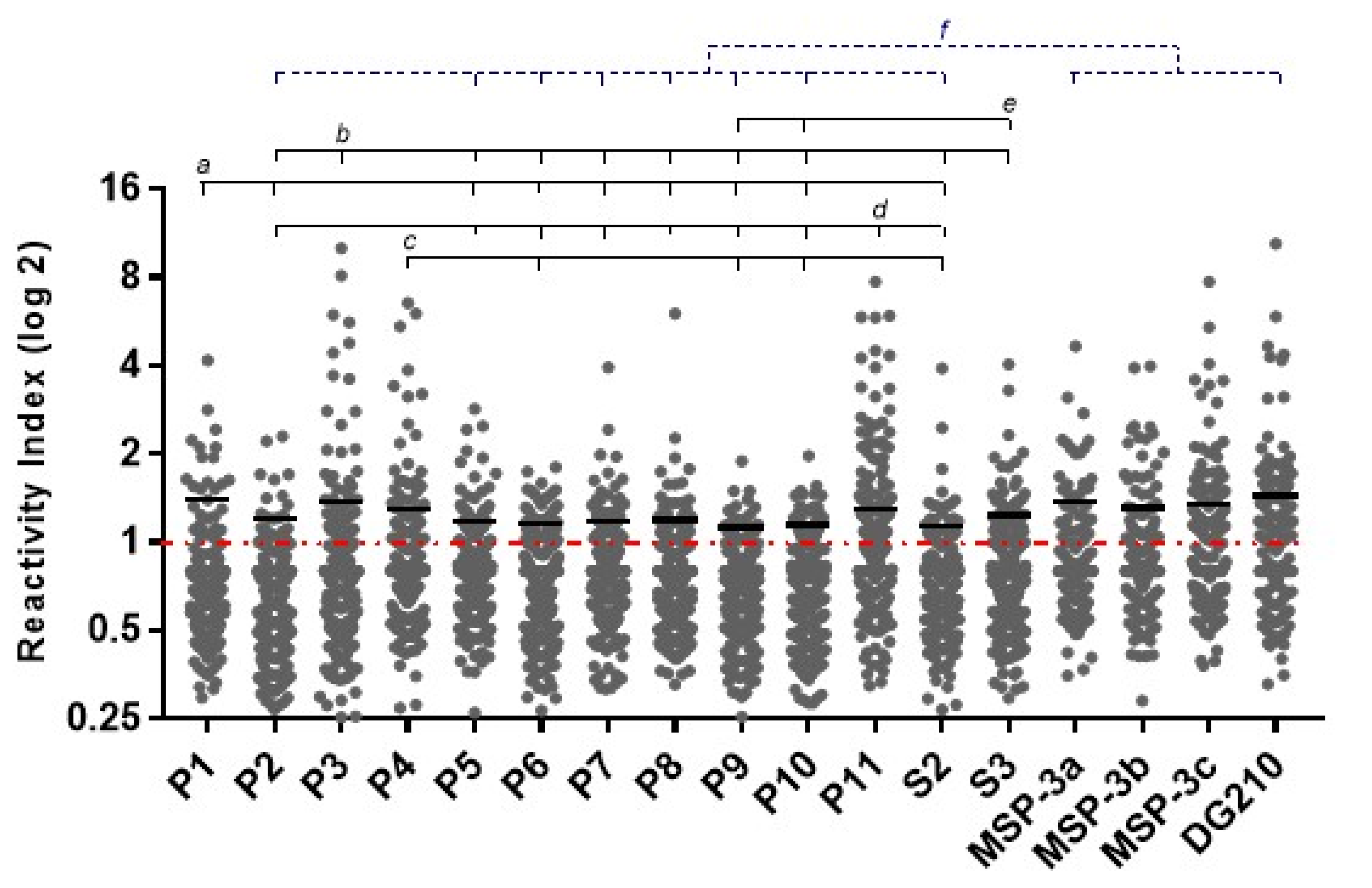
Regarding the magnitude of response to epitopes among responders, higher levels of IgG antibodies against epitopes P1, P3, P4, P11, and S3 were observed. No difference was observed in IgG antibody levels between MSP-3155–249 epitopes. However, MSP-3 epitopes MSP-3a, MSP-3c, and DG210 induced higher IgG antibody levels than GLURP epitopes P2, P5, P6, P7, P8, P9, P10, and S2 (Figure 3).
No association was observed among frequency of responders or IgG antibody levels and age, sex, time of exposure, presence of symptoms, number of reported previous malaria episodes, the time elapsed since the last malaria episode, and current or last infecting plasmodial species.

3.3. IgG Subclasses Distribution against the Immunodominant Epitopes of GLURP27–500 and MSP-3155–249

To evaluate the balance between cytophilic and non-cytophilic antibodies, immunodominant epitopes derived from GLURP27–500 (P1, P3, P4, P11, and S3) and MSP-3155–249 (MSP-3a, MSP-3b, MSP-3c, and DG210) were selected. The results showed that epitopes P1, P3, P11, and S3 of GLURP and MSP-3b, MSP-3c, and DG210 of MSP-3 were preferentially recognized by IgG1 and IgG3 cytophilic antibodies, while epitopes P4 of GLURP and MSP-3a of MSP-3 were preferentially recognized by antibodies of the IgG1 subclass. No difference was observed between IgG1, IgG2, IgG3, and IgG4 antibody levels in responders to P3, P4, MSP-3a, MSP-3b, and MSP-3c. However, higher IgG1 than IgG3 antibody levels against P1 and IgG1 than IgG2, IgG3, and IgG4 antibody levels against P11 were detected. In addition, IgG1 and IgG3 were higher than IgG4 antibody levels against S3 and IgG2 antibody levels against DG210 (Figure 4).
IgG1 antibody levels against P1 and P11 were positively correlated with age and time of residence in malaria-endemic area (P1: p = 0.002; r = 0.530; for both; P11: p = 0.02; r = 0.207; p = 0.03; r = 0.201; respectively). In addition, IgG3 antibody levels against DG210 were associated with the number of previous malaria episodes (p = 0.034; r = 0.240), while IgG1 antibody levels against P3 were negatively correlated with time elapsed since the last malaria episode (p = 0.024; r = −0.316).

3.4. Experimental Validation of Predicted Linear B-Cell Epitopes of Pfs48/45291–428

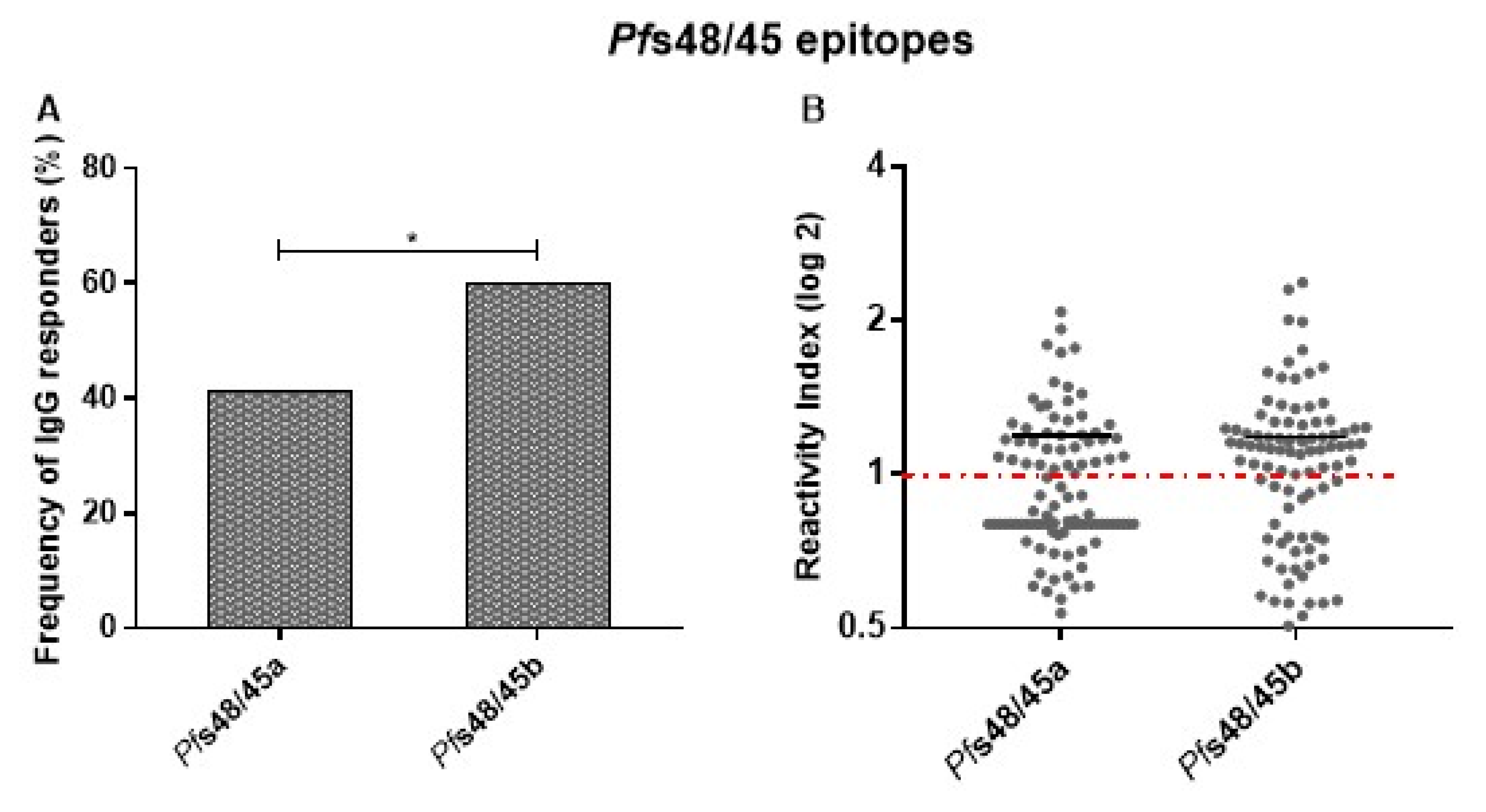
Two sequences were predicted as potential linear B-cell epitopes on Pfs48/45291–428 (Pfs48/45a and Pfs48/45b) and selected for experimental validation. Most responders to Pfs48/45291–428 recombinant protein presented detectable IgG antibodies against epitopes Pfs48/45a and/or Pfs48/45b. A higher frequency of individuals presenting IgG antibodies against Pfs48/45b than Pfs48/45a was observed. However, low IgG antibody levels were detected in epitopes, without differences between Pfs48/45a and Pfs48/45b (Figure 5).
The analyses of IgG subclasses were performed in all samples with detectable IgG antibodies against Pfs48/45a or Pfs48/45b. The frequencies of IgG1, IgG2, IgG3, and IgG4 against Pfs48/45a were 21%, 0%, 37% and 9%, respectively, and against Pfs48/45b were 43%, 11%, 26%, and 21%, respectively. The data showed a higher frequency of individuals presenting IgG3 than IgG4 antibodies against Pfs48/45a (p = 0.002) and IgG1 than IgG2, IgG3, and IgG4 antibodies against Pfs48/45b (p < 0.0001, p = 0.03 and p = 0.007 versus IgG2, IgG3, and IgG4, respectively). No difference was observed in IgG1, IgG2, IgG3, and IgG4 antibody levels against Pfs48/45 epitopes.

3.5. Frequencies and IgG Antibody Levels in Non-Infected and Infected by P. vivax and P. falciparum Individuals

Frequencies and IgG antibody levels to GLURP27–500, MSP-3155–249, and Pfs48/45291–428 epitopes were compared between non-infected and infected by P. vivax and P. falciparum individuals. The results showed higher frequencies of individuals responders to P7 and P8 infected by P. falciparum when compared with P. vivax-infected individuals. Responders to S3 epitope infected by P. vivax were less frequent than non-infected and P.falciparum-infected individuals. Individuals infected by P. falciparum presented a higher frequency of response to MSP-3a, MSP-3b, and MSP-3c epitopes than non-infected and P.vivax-infected individuals, while the frequency of responders to DG210 infected by P. falciparum was higher than non-infected individuals (Table 2). No difference was observed in frequencies of responders to P1, P2, P3, P4, P5, P6, P9, P10, P11, S2, Pfs48/45a, and Pfs48/45b between individuals non-infected and infected by P. vivax or P. falciparum.
IgG antibody levels against P1 were higher in P. vivax and P. falciparum-infected than in non-infected individuals. In addition, P. falciparum-infected individuals presented higher IgG antibody levels than P. vivax-infected and non-infected individuals against DG210 than non-infected individuals against Pfs48/45b (Figure 6). No difference was observed in antibody levels against P2, P3, P4, P5, P6, P7, P8, P9, P10, P11, S2, S3, MSP-3a, MSP-3b, MSP-3c, and Pfs48/45a epitopes between non-infected and infected by P. vivax or P. falciparum individuals.

4. Discussion

The GMZ2.6c malaria vaccine candidate is a multi-stage P. falciparum chimeric protein that contains the sexual-stage Pfs48/45291–428 fragment genetically fused to GMZ2, an asexual-stage vaccine construction consisting of GLURP27–500 and MSP-3155–249 antigens [9]. The GMZ2.6c and its components were widely recognized by naturally acquired antibodies from Brazilian exposed individuals [20]. Antibodies recognize and bind their target protein antigens by surface-accessible sites, known as antigenic determinants or epitopes [28]. The identification of B-cell epitopes of proteins is an important tool for the development of epitope-based vaccines [29], diagnostic tests [30], immunotherapy [31], and understanding specific immune responses against pathogens. This work identified and validated the immunodominant B-cell epitopes of GLURP27–500, MSP-3155–249, and Pfs48/45291–428 antigens, components of GMZ2.6c malaria vaccine candidate in individuals exposed to malaria in the Brazilian Amazon.
The studied population was composed of individuals living in three malaria-endemic areas of the Brazilian Amazon, Cruzeiro do Sul and Mâncio Lima, Acre State, and Guajará, Amazonas State. The highly variable range of age, time of residence in the endemic area, reported number of previous malaria episodes, and the time elapsed from the last infection indicated different degrees of exposure among the studied individuals, an important determinant of protection against clinical malaria [32,33]. Most of the individuals reported previous infections by both P. falciparum and P. vivax, but P. vivax was the most prevalent infecting plasmodial species, reflecting the current malaria scenario in Brazil, where 89% of the recorded cases are due to P. vivax infection [34].
GLURP and MSP-3 were identified as targets of naturally acquired antibodies capable of mediating parasite killing in cooperation with monocytes [35,36] associated with malaria protection in several epidemiological statuses [37,38,39,40,41,42]. Considering the protective role of antibodies against GLURP and MSP-3, the antigenicity of these proteins has been evaluated in epitope mapping studies. In the present work, 13 peptides derived from GLURP27–500 previously predicted as potential B-cell epitopes [27], four peptides derived from MSP-3155–249, a polypeptide recognized by antibodies from clinical immune individuals utilizing clones from a DNA library, and its three overlapping peptides [26,35] were used.
Epitope P11 was identified as the immunodominant of the GLURP with both higher frequency of responders and IgG antibody levels in the studied population. Likewise, P11 was identified as the immunodominant B-cell epitope of the GLURP in individuals living in Rondônia State, also in the Brazilian Amazon [43], and Saimiri sciureus monkeys immunized with a hybrid protein containing the R0 region of GLURP and the C-terminal region of MSP-3 [44]. In contrast, only 29% of clinical immune Liberian adults present antibodies against P11, while the immunodominant epitopes were P1, P3, P4, and S3 [27]. The differences in the immunodominance of GLURP B-cell epitopes may be due to polymorphisms in the R0 region of GLURP when isolates from different geographic areas were compared [45], genetic restriction of the antibody response against GLURP-R0 epitopes [43] or differences transmission intensities [46] between endemic areas.
Although with low frequency, the responding individuals displayed high levels of IgG antibodies against the P1, P3, P4, and S3 GLURP epitopes. Interestingly, P1, P3, and P4 contain a common motif (EPFPXQXHK) that appears to be targeted by cross-reactive antibodies [27]. Similarly, affinity-purified antibodies to S3 can recognize the S4 peptide derived from the C-terminal R2 region of GLURP [27]. This finding may suggest that the high antibody levels against these peptides could be the result of cross-reactivity.
The linear peptide containing the amino acid sequence of DG210 from MSP-3 was also shown to be widely recognized by high levels of naturally acquired IgG antibodies. Interestingly, similar frequency and antibody levels to peptide MSP-3c were found, suggesting that antibody response against DG210 may primarily target this epitope in the studied population. Many seroepidemiological studies demonstrated distinct profiles of antibody response to peptides derived from MSP-3CT. Peptide MSP-3b is the major B-cell epitope in hyperimmune individuals [26,35], while subjects with diverse degrees of exposure showed no differences between prevalence and antibody levels against MSP-3b, MSP-3c, and MSP-3d [47]. Antibodies induced after immunization with MSP-3CT LPS were directed against peptides MSP-3c and MSP-3d [48,49], but similar antibody levels against all peptides were found.
It is widely accepted that cytophilic IgG1 and IgG3 isotypes are the main mediators of the protective humoral response against P. falciparum blood-stage antigens, promoting Fc-mediated effector functions as antibody-dependent cellular inhibition (ADCI) [50,51], opsonic phagocytosis [52,53], antibody-dependent respiratory bursty (ADRB) [54] and complement activation [55,56]. In contrast, non-cytophilic IgG subclasses (IgG2 and IgG4) with the same specificity could block these effector mechanisms [57]. In our cohort, all the immunodominant epitopes of GLURP and MSP-3 are primarily targeted by IgG1 and/or IgG3 antibodies—predominantly IgG3 isotype against epitope S3 and IgG1 isotype against other epitopes. The prevalence of cytophilic antibodies against MSP-3 and GLURP epitopes was also observed in exposed individuals from Africa [47,58] and immunized European volunteers [48] associated with protection. It is known that genetic background, degrees of exposure of the population, and malaria transmission levels may influence the distribution of IgG subclasses against plasmodial antigens [59]. However, these data suggest that intrinsic characteristics of these antigens might be the major factor that determines antibody cytophilic IgG subclasses response, driving to B-cell activation by the binding of CD40 to CD40L expressed on the surface of activated T cells providing the costimulatory signal and cytokines release, influencing the IgG class switching [60,61].
Correlation analysis of the immune response and epidemiological data showed that IgG1 antibody levels against GLURP epitopes P1 and P11 were positively correlated with age and time of residence in malaria-endemic area, while IgG3 antibody levels against MSP-3 epitope DG210 were associated with the number of previous malaria episodes. In addition, IgG1 antibody levels against GLURP epitope P3 were negatively correlated with time elapsed since the last malaria episode. These may reflect the cumulative exposure of these individuals to P. falciparum infections and possibly maturation of the immune system over time [17] but did not provide evidence of a possible protective role of these antibodies since no relationship was verified between the prevalence or levels of specific antibodies and the presence or absence of parasites in the blood, the presence or absence of symptoms at the time of collection, and parasitemia. A detailed evaluation of the functional activity of specific antibodies against these peptides is currently being conducted in our laboratory and may generate further evidence of their possible protective role.
The Pfs48/45 antigen is one of the most well-characterized antibody targets of P. falciparum gametocytes. Several studies showed that antibodies against Pfs48/45 displayed a transmission reduction activity in the standard membrane-feeding assay (SMFA) [62,63], and antibodies against the conformational epitope I in the 6c domain of Pfs48/45 have strong transmission block potential in genetically diverse parasite strains and multiclonal infections [64,65]. Transmission-blocking antibodies are known to often target conformational-dependent epitopes [66], and no linear B-cell epitopes against Pfs48/45 have been described. Here, we identified two potential linear B-cell epitopes (Pfs48/45a: Pfs48/45358–366 and Pfs48/45b: Pfs48/45382–391) of Pfs48/45-6c domain using in silico BepiPred algorithm [25]. Most of the responders to Pfs48/45291–428 recombinant protein (68.5%) presented detectable IgG antibodies against Pfs48/45a and/or Pfs48/45b peptides, validating the prediction of linear B-cell epitopes. These frequencies are comparable to those found for GLURP27–500 (70.9%) and MSP-3155–249 (64.7%) peptides in this study, as well as other P. falciparum antigens [67,68,69]. A higher frequency of responders to Pfs48/45b was observed, despite no difference between the antibody levels against both epitopes. Interestingly, antibody levels against Pfs48/45a and Pfs48/45b were similar to those against Pfs48/45291–428 recombinant protein [20]. These findings indicate that although the minority of B-cell epitopes are linear (~10%) [70], they might be an interesting antibody target. However, the most potent transmission-blocking epitope in Pfs48/45-6c is conformational, and the production of a non-properly folded protein did not elicit functional antibodies in mice [71]. Further, the immunization with full-length Pfs48/45 has revealed that a larger number of antibodies with no transmission-blocking potential recognize the 6c domain of Pfs48/45 [72]. Additional studies are needed to better understand the humoral immune response against these epitopes and their possible transmission-blocking activity.
Studies have revealed that the high transmission-blocking efficacy of the monoclonal antibody 85RF45.1 is due to its ability to block the normal function of Pfs48/45 or its binding partners [72], and several IgG subclasses may contribute to the transmission-blocking activity. Curiously, the high frequency of IgG1 and IgG3 against Pfs48/45b and Pfs48/45a, respectively, and the less proportion of IgG2 and IgG4 antibodies to both epitopes suggest a possible role of the classical complement activation pathway. Although there is no evidence of the relevance of the anti-Pfs48/45 antibody–complement system interaction in transmission-blocking immunity, antibodies against the sexual-stage antigen Pfs230 showed an increase in transmission-blocking activity in the presence of human complement system proteins [73].
Specific antibodies against sexual-stage antigens are also related to age and serve as markers of exposure, increasing according to gametocyte densities [62,74,75], but no correlation with epidemiological parameters was found in this study. This finding is not surprising since, in malaria-endemic areas, infections are determined by microscopic parasite counts, and gametocytes normally occur at low densities. In addition, no other higher-sensitive method to detect gametocytes was used.
In conclusion, the data presented here showed that P11 from GLURP, MSP-3c, and DG210 from MSP-3, and the predicted epitopes Pfs48/45a and Pfs48/45b from Pfs48/45291 were widely recognized by antibodies from individuals living in endemic areas of Brazil. Combined immunoinformatic and experimental approach strategies identified several linear B-cell epitopes from GLURP27–500, MSP-3155–249, and Pfs48/45291–428 fragments that composed GMZ2.6c, providing additional information that may suggest its relevance as a multi-stage malaria vaccine candidate. Further studies are needed to assess the potential of specific antibodies against GMZ2.6c epitopes in the inhibition of P. falciparum growth and transmission-blocking activity.

Supplementary Materials

The following supporting information can be downloaded at: https://www.mdpi.com/article/10.3390/vaccines11020446/s1, Table S1: IgG reactivity index for GLURP, MSP-3, and Pfs48/45 recombinant proteins.

Author Contributions

Conceptualization, L.R.P.-R., C.T.D.-R., M.T., S.K.S. and L.E.A.; Data curation, L.R.P.-R. and B.d.O.B.; Formal analysis, B.d.O.B.; Funding acquisition, L.R.P.-R. and C.T.D.-R.; Investigation, B.d.O.B., A.B.L.d.S., L.T.d.Q., L.S.d.O. and R.N.R.-d.-S.; Methodology, L.R.P.-R. and B.d.O.B.; Project administration, L.R.P.-R.; Resources, L.R.P.-R., B.d.O.B., A.B.L.d.S., E.K.P.R., P.R.R.T., R.M.d.S., J.d.C.L.-J., H.A.d.S.d.S. and J.P.d.B.; Supervision, L.R.P.-R.; Writing—Original draft, B.d.O.B.; Writing—Review and editing, L.R.P.-R., E.K.P.R., P.R.R.T., J.d.C.L.-J. and C.T.D.-R. All authors have read and agreed to the published version of the manuscript.

Funding

This work was supported by Fundação Oswaldo Cruz (Fiocruz/INOVA) project VPPCB-007-FIO-18-2-46, Conselho Nacional de Desenvolvimento Científico e Tecnológico (CNPq) project 425.422/2016-0, Programa de Apoio a Núcleos de Excelência (Pronex), Fundação de Amparo à Pesquisa do Estado do Rio de Janeiro (FAPERJ) project E-26/010.001268/2016. This study was also supported in part by the Coordenação de Aperfeiçoamento de Pessoal de Nível Superior—Brazil (CAPES)—Finance Code 0001. C.T.D.-R. is the recipient of a Productivity Research Fellowship from CNPq, Brazil, and is a “Cientista do Nosso Estado”, recognized by the FAPERJ, Rio de Janeiro, Brazil. The Laboratório de Pesquisa em Malária is an Associate Laboratory of the Instituto Nacional de Ciência e Tecnologia em Neuroimunomodulação (INCT-NIM/CNPq) project 465489/2014-1 and of the Rede de Neuroinflamação (Redes/FAPERJ) project 26010.002418/2019.

Institutional Review Board Statement

The study protocol was reviewed and approved by the Oswaldo Cruz Foundation Ethical Committee (CEP-FIOCRUZ CAAE 46084015.1.0000.5248).

Informed Consent Statement

Written informed consent has been obtained from all adult donors or donors’ parents in the cases of children. All the procedures adopted in this study fully complied with specific federal permits issued by the Brazilian Ministry of Health.

Data Availability Statement

The datasets supporting the conclusions of this article are included within the article and its Supplementary Materials.

Acknowledgments

We are grateful to all individuals who agreed to participate in this study for their cooperation and generous donation of blood samples, which made this study possible, and to the Endemic Diseases Coordination of the cities of Cruzeiro do Sul, Mâncio Lima, and Guajará for the support during the fieldwork. We are also thankful to Heloisa Maria Nogueira Diniz for helping with figures and to Marcelo Urbano Ferreira for providing facilities in the malaria-endemic areas.

Conflicts of Interest

The authors declare that they have no conflict of interest.

References

  1. World Health Organization World Malaria Report. Available online: https://www.malariaworld.org/blogs/world-malaria-report-2022 (accessed on 26 December 2022).
  2. Dimbu, P.R.; Horth, R.; Cândido, A.L.M.; Ferreira, C.M.; Caquece, F.; Garcia, L.E.A.; André, K.; Pembele, G.; Jandondo, D.; Bondo, B.J.; et al. Continued Low Efficacy of Artemether-Lumefantrine in Angola in 2019. Antimicrob. Agents Chemother. 2021, 65, e01949-20. [Google Scholar] [CrossRef] [PubMed]
  3. Mathieu, L.C.; Cox, H.; Early, A.M.; Mok, S.; Lazrek, Y.; Paquet, J.C.; Ade, M.P.; Lucchi, N.W.; Grant, Q.; Udhayakumar, V.; et al. Local Emergence in Amazonia of Plasmodium falciparum K13 C580Y Mutants Associated with in Vitro Artemisinin Resistance. Elife 2020, 9, e51015. [Google Scholar] [CrossRef]
  4. Gansané, A.; Moriarty, L.F.; Ménard, D.; Yerbanga, I.; Ouedraogo, E.; Sondo, P.; Kinda, R.; Tarama, C.; Soulama, E.; Tapsoba, M.; et al. Anti-Malarial Efficacy and Resistance Monitoring of Artemether-Lumefantrine and Dihydroartemisinin-Piperaquine Shows Inadequate Efficacy in Children in Burkina Faso, 2017–2018. Malar. J. 2021, 20, 48. [Google Scholar] [CrossRef] [PubMed]
  5. Uwimana, A.; Umulisa, N.; Venkatesan, M.; Svigel, S.S.; Zhou, Z.; Munyaneza, T.; Habimana, R.M.; Rucogoza, A.; Moriarty, L.F.; Sandford, R.; et al. Association of Plasmodium falciparum Kelch13 R561H Genotypes with Delayed Parasite Clearance in Rwanda: An Open-Label, Single-Arm, Multicentre, Therapeutic Efficacy Study. Lancet Infect. Dis. 2021, 21, 1120–1128. [Google Scholar] [CrossRef]
  6. Das, S.; Kar, A.; Manna, S.; Mandal, S.; Mandal, S.; Das, S.; Saha, B.; Hati, A.K. Artemisinin Combination Therapy Fails Even in the Absence of Plasmodium falciparum Kelch13 Gene Polymorphism in Central India. Sci. Rep. 2021, 11, 9946. [Google Scholar] [CrossRef]
  7. Hancock, P.A.; Hendriks, C.J.M.; Tangena, J.A.; Gibson, H.; Hemingway, J.; Coleman, M.; Gething, P.W.; Cameron, E.; Bhatt, S.; Moyes, C.L. Mapping Trends in Insecticide Resistance Phenotypes in African Malaria Vectors. PLoS Biol. 2020, 18, e3000633. [Google Scholar] [CrossRef]
  8. Mugo, R.M.; Mwai, K.; Mwacharo, J.; Shee, F.M.; Musyoki, J.N.; Wambua, J.; Otieno, E.; Bejon, P.; Ndungu, F.M. Seven-Year Kinetics of RTS, S/AS01-Induced Anti-CSP Antibodies in Young Kenyan Children. Malar. J. 2021, 20, 452. [Google Scholar] [CrossRef]
  9. Baldwin, S.L.; Roeffen, W.; Singh, S.K.; Tiendrebeogo, R.W.; Christiansen, M.; Beebe, E.; Carter, D.; Fox, C.B.; Howard, R.F.; Reed, S.G.; et al. Synthetic TLR4 Agonists Enhance Functional Antibodies and CD4+ T-Cell Responses against the Plasmodium falciparum GMZ2.6C Multi-Stage Vaccine Antigen. Vaccine 2016, 34, 2207–2215. [Google Scholar] [CrossRef] [Green Version]
  10. Borre, M.B.; Dziegiel, M.; Høgh, B.; Petersen, E.; Rieneck, K.; Riley, E.; Meis, J.F.; Aikawa, M.; Nakamura, K.; Harada, M.; et al. Primary Structure and Localization of a Conserved Immunogenic Plasmodium falciparum Glutamate Rich Protein (GLURP) Expressed in Both the Preerythrocytic and Erythrocytic Stages of the Vertebrate Life Cycle. Mol. Biochem. Parasitol. 1991, 49, 119–131. [Google Scholar] [CrossRef] [PubMed]
  11. del Quintana, M.P.; Ch’ng, J.H.; Zandian, A.; Imam, M.; Hultenby, K.; Theisen, M.; Nilsson, P.; Qundos, U.; Moll, K.; Chan, S.; et al. SURGE Complex of Plasmodium falciparum in the Rhoptry-Neck (SURFIN4.2-RON4-GLURP) Contributes to Merozoite Invasion. PLoS ONE 2018, 13, e0201669. [Google Scholar] [CrossRef] [PubMed]
  12. McColl, D.J.; Anders, R.F. Conservation of Structural Motifs and Antigenic Diversity in the Plasmodium falciparum Merozoite Surface Protein-3 (MSP-3). Mol. Biochem. Parasitol. 1997, 90, 21–31. [Google Scholar] [CrossRef] [PubMed]
  13. Imam, M.; Singh, S.; Kaushik, N.K.; Chauhan, V.S. Plasmodium falciparum Merozoite Surface Protein 3. J. Biol. Chem. 2014, 289, 3856–3868. [Google Scholar] [CrossRef] [Green Version]
  14. van Dijk, M.R.; Janse, C.J.; Thompson, J.; Waters, A.P.; Braks, J.A.M.; Dodemont, H.J.; Stunnenberg, H.G.; van Gemert, G.J.; Sauerwein, R.W.; Eling, W. A Central Role for P48/45 in Malaria Parasite Male Gamete Fertility. Cell 2001, 104, 153–164. [Google Scholar] [CrossRef] [Green Version]
  15. Kana, I.H.; Adu, B.; Tiendrebeogo, R.W.; Singh, S.K.; Dodoo, D.; Theisen, M. Naturally Acquired Antibodies Target the Glutamate-Rich Protein on Intact Merozoites and Predict Protection Against Febrile Malaria. J. Infect. Dis. 2017, 215, 623–630. [Google Scholar] [CrossRef] [Green Version]
  16. Dodoo, D.; Theisen, M.; Kurtzhals, J.A.L.; Akanmori, B.D.; Koram, K.A.; Jepsen, S.; Nkrumah, F.K.; Theander, T.G.; Hviid, L. Naturally Acquired Antibodies to the Glutamate-Rich Protein Are Associated with Protection against Plasmodium falciparum Malaria. J. Infect. Dis. 2000, 181, 1202–1205. [Google Scholar] [CrossRef] [Green Version]
  17. Soe, S.; Theisen, M.; Roussilhon, C.; Aye, K.S.; Druilhe, P. Association between Protection against Clinical Malaria and Antibodies to Merozoite Surface Antigens in an Area of Hyperendemicity in Myanmar: Complementarity between Responses to Merozoite Surface Protein 3 and the 220-Kilodalton Glutamate-Rich Protein. Infect. Immun. 2004, 72, 247–252. [Google Scholar] [CrossRef] [Green Version]
  18. Lusingu, J.P.A.; Vestergaard, L.S.; Alifrangis, M.; Mmbando, B.P.; Theisen, M.; Kitua, A.Y.; Lemnge, M.M.; Theander, T.G. Cytophilic Antibodies to Plasmodium falciparum Glutamate Rich Protein Are Associated with Malaria Protection in an Area of Holoendemic Transmission. Malar. J. 2005, 4, 48. [Google Scholar] [CrossRef] [Green Version]
  19. Bousema, J.T.; Drakeley, C.J.; Kihonda, J.; Hendriks, J.C.M.; Akim, N.I.J.; Roeffen, W.; Sauerwein, R.W. A Longitudinal Study of Immune Responses to Plasmodium falciparum Sexual Stage Antigens in Tanzanian Adults. Parasite Immunol. 2007, 29, 309–317. [Google Scholar] [CrossRef] [PubMed]
  20. Baptista, B.O.; de Souza, A.B.L.; Riccio, E.K.P.; Bianco-Junior, C.; Totino, P.R.R.; Martins da Silva, J.H.; Theisen, M.; Singh, S.K.; Amoah, L.E.; Ribeiro-Alves, M.; et al. Naturally Acquired Antibody Response to a Plasmodium falciparum Chimeric Vaccine Candidate GMZ2.6c and Its Components (MSP-3, GLURP, and Pfs48/45) in Individuals Living in Brazilian Malaria-Endemic Areas. Malar. J. 2022, 21, 6. [Google Scholar] [CrossRef] [PubMed]
  21. Ministry of Health of Brazil. Epidemiological Situation of Malaria in Brazil. Available online: https://portalarquivos.saude.gov.br/images/pdf/2019/dezembro/03/3.%20a%20-%20Situa%C3%A7%C3%A3o%20Epidemiol%C3%B3gica%20da%20Mal%C3%A1ria_Brasil_CIT_28.11.2018.pdf (accessed on 15 December 2020).
  22. Shute, G.T. The Microscpopic Diagnosis of Malaria. In Malaria: Principles and Practice of Malariology; Wernsdorfer, W., McGregor, S., Eds.; Churchill Livingstone: New York, NY, USA, 1988; pp. 781–814. [Google Scholar]
  23. Snounou, G. Detection and Identification of the Four Malaria Parasite Species Infecting Humans by PCR Amplification. Methods Mol. Biol. 1996, 50, 263–291. [Google Scholar] [CrossRef]
  24. Ministry of Health of Brazil. Guide to Malaria Treatment in Brazil. Available online: http://bvsms.saude.gov.br/bvs/publicacoes/guia_tratamento_malaria_brasil.pdf (accessed on 19 November 2020).
  25. Larsen, J.E.P.; Lund, O.; Nielsen, M. Improved Method for Predicting Linear B-Cell Epitopes. Immunome Res. 2006, 2, 2. [Google Scholar] [CrossRef] [PubMed] [Green Version]
  26. Oeuvray, C.; Bouharoun-Tayoun, H.; Gras-Masse, H.; Bottius, E.; Kaidoh, T.; Aikawa, M.; Filgueira, M.; Tartar, A.; Druilhe, P. Merozoite Surface Protein-3: A Malaria Protein Inducing Antibodies That Promote Plasmodium falciparum Killing by Cooperation with Blood Monocytes. Blood 1994, 84, 1594–1602. [Google Scholar] [CrossRef] [Green Version]
  27. Theisen, M.; Soe, S.; Jessing, S.G.; Okkels, L.M.; Danielsen, S.; Oeuvray, C.; Druilhe, P.; Jepsen, S. Identification of a Major B-Cell Epitope of the Plasmodium falciparum Glutamate-Rich Protein (GLURP), Targeted by Human Antibodies Mediating Parasite Killing. Vaccine 2000, 19, 204–212. [Google Scholar] [CrossRef]
  28. Beutling, U.; Frank, R. Epitope Analysis Using Synthetic Peptide Repertoires Prepared by SPOT Synthesis Technology. In Antibody Engineering; Springer: Berlin/Heidelberg, Germany, 2010; Volume 1, pp. 537–571. [Google Scholar]
  29. Ghattas, M.; Dwivedi, G.; Lavertu, M.; Alameh, M.G. Vaccine Technologies and Platforms for Infectious Diseases: Current Progress, Challenges, and Opportunities. Vaccines 2021, 9, 1490. [Google Scholar] [CrossRef] [PubMed]
  30. Noya, O.; Patarroyo, M.; Guzman, F.; de Noya, B. Immunodiagnosis of Parasitic Diseases with Synthetic Peptides. Curr. Protein Pept. Sci. 2003, 4, 299–308. [Google Scholar] [CrossRef] [PubMed]
  31. Shirai, H.; Prades, C.; Vita, R.; Marcatili, P.; Popovic, B.; Xu, J.; Overington, J.P.; Hirayama, K.; Soga, S.; Tsunoyama, K.; et al. Antibody Informatics for Drug Discovery. Biochim. Biophys. Acta 2014, 1844, 2002–2015. [Google Scholar] [CrossRef]
  32. Doolan, D.L.; Dobaño, C.; Baird, J.K. Acquired Immunity to Malaria. Clin. Microbiol. Rev. 2009, 22, 13–36, Table of Contents. [Google Scholar] [CrossRef] [Green Version]
  33. Gonzales, S.J.; Reyes, R.A.; Braddom, A.E.; Batugedara, G.; Bol, S.; Bunnik, E.M. Naturally Acquired Humoral Immunity Against Plasmodium falciparum Malaria. Front. Immunol. 2020, 11, 594653. [Google Scholar] [CrossRef]
  34. Ministry of Health of Brazil. Boletim Epidemiológico. Available online: http://portalsaude.saude.gov.br (accessed on 11 November 2021).
  35. Oeuvray, C.; Bouharoun-Tayoun, H.; Grass-Masse, H.; Iepers, J.P.; Ralamboranto, L.; Tartar, A.; Druilhe, P. A Novel Merozoite Surface Antigen of Plasmodium falciparum (MSP-3) Identified by Cellular-Antibody Cooperative Mechanism Antigenicity and Biological Activity of Antibodies. Mem. Inst. Oswaldo Cruz 1994, 89, 77–80. [Google Scholar] [CrossRef] [Green Version]
  36. Theisen, M.; Soe, S.; Oeuvray, C.; Thomas, A.W.; Vuust, J.; Danielsen, S.; Jepsen, S.; Druilhe, P. The Glutamate-Rich Protein (GLURP) of Plasmodium falciparum Is a Target for Antibody-Dependent Monocyte-Mediated Inhibition of Parasite Growth In Vitro. Infect. Immun. 1998, 66, 11–17. [Google Scholar] [CrossRef]
  37. Nebie, I.; Diarra, A.; Ouedraogo, A.; Soulama, I.; Bougouma, E.C.; Tiono, A.B.; Konate, A.T.; Chilengi, R.; Theisen, M.; Dodoo, D.; et al. Humoral Responses to Plasmodium falciparum Blood-Stage Antigens and Association with Incidence of Clinical Malaria in Children Living in an Area of Seasonal Malaria Transmission in Burkina Faso, West Africa. Infect. Immun. 2008, 76, 759–766. [Google Scholar] [CrossRef] [Green Version]
  38. Osier, F.H.A.; Fegan, G.; Polley, S.D.; Murungi, L.; Verra, F.; Tetteh, K.K.A.; Lowe, B.; Mwangi, T.; Bull, P.C.; Thomas, A.W.; et al. Breadth and Magnitude of Antibody Responses to Multiple Plasmodium falciparum Merozoite Antigens Are Associated with Protection from Clinical Malaria. Infect. Immun. 2008, 76, 2240–2248. [Google Scholar] [CrossRef] [PubMed] [Green Version]
  39. Courtin, D.; Oesterholt, M.; Huismans, H.; Kusi, K.; Milet, J.; Badaut, C.; Gaye, O.; Roeffen, W.; Remarque, E.J.; Sauerwein, R.; et al. The Quantity and Quality of African Children’s IgG Responses to Merozoite Surface Antigens Reflect Protection against Plasmodium falciparum Malaria. PLoS ONE 2009, 4, e7590. [Google Scholar] [CrossRef] [PubMed] [Green Version]
  40. Iriemenam, N.C.; Khirelsied, A.H.; Nasr, A.; ElGhazali, G.; Giha, H.A.; Elhassan A-Elgadir, T.M.; Agab-Aldour, A.A.; Montgomery, S.M.; Anders, R.F.; Theisen, M.; et al. Antibody Responses to a Panel of Plasmodium falciparum Malaria Blood-Stage Antigens in Relation to Clinical Disease Outcome in Sudan. Vaccine 2009, 27, 62–71. [Google Scholar] [CrossRef] [PubMed]
  41. Dodoo, D.; Atuguba, F.; Bosomprah, S.; Ansah, N.A.; Ansah, P.; Lamptey, H.; Egyir, B.; Oduro, A.R.; Gyan, B.; Hodgson, A.; et al. Antibody Levels to Multiple Malaria Vaccine Candidate Antigens in Relation to Clinical Malaria Episodes in Children in the Kasena-Nankana District of Northern Ghana. Malar. J. 2011, 10, 108. [Google Scholar] [CrossRef]
  42. Guiyedi, V.; Bécavin, C.; Herbert, F.; Gray, J.; Cazenave, P.A.; Kombila, M.; Crisanti, A.; Fesel, C.; Pied, S. Asymptomatic Plasmodium falciparum Infection in Children Is Associated with Increased Auto-Antibody Production, High IL-10 Plasma Levels and Antibodies to Merozoite Surface Protein 3. Malar. J. 2015, 14, 162. [Google Scholar] [CrossRef] [Green Version]
  43. Pratt-Riccio, L.R.; Lima-Junior, J.C.; Carvalho, L.J.M.; Theisen, M.; Espíndola-Mendes, E.C.; Santos, F.; Oliveira-Ferreira, J.; Goldberg, A.C.; Daniel-Ribeiro, C.T.; Banic, D.M. Antibody Response Profiles Induced by Plasmodium falciparum Glutamate-Rich Protein in Naturally Exposed Individuals from a Brazilian Area Endemic for Malaria. Am. J. Trop. Med. Hyg. 2005, 73, 1096–1103. [Google Scholar] [CrossRef]
  44. Carvalho, L.J.M.; Alves, F.A.; Bianco, C.; Oliveira, S.G.; Zanini, G.M.; Soe, S.; Druilhe, P.; Theisen, M.; Muniz, J.A.P.C.; Daniel-Ribeiro, C.T. Immunization of Saimiri sciureus Monkeys with a Recombinant Hybrid Protein Derived from the Plasmodium falciparum Antigen Glutamate-Rich Protein and Merozoite Surface Protein 3 Can Induce Partial Protection with Freund and Montanide ISA720 Adjuvants. Clin. Diagn. Lab. Immunol. 2005, 12, 242–248. [Google Scholar] [CrossRef] [Green Version]
  45. Pratt-Riccio, L.R.; de Perce-da-Silva, D.S.; Lima-Junior, J.d.C.; Theisen, M.; Santos, F.; Daniel-Ribeiro, C.T.; de Oliveira-Ferreira, J.; Banic, D.M. Genetic Polymorphisms in the Glutamate-Rich Protein of Plasmodium falciparum Field Isolates from a Malaria-Endemic Area of Brazil. Mem. Inst. Oswaldo Cruz 2013, 108, 523–528. [Google Scholar] [CrossRef]
  46. Ambrosino, E.; Dumoulin, C.; Orlandi-Pradines, E.; Remoue, F.; Toure-Baldé, A.; Tall, A.; Sarr, J.B.; Poinsignon, A.; Sokhna, C.; Puget, K.; et al. A Multiplex Assay for the Simultaneous Detection of Antibodies against 15 Plasmodium falciparum and Anopheles gambiae Saliva Antigens. Malar. J. 2010, 9, 317. [Google Scholar] [CrossRef]
  47. Singh, S.; Soe, S.; Mejia, J.P.; Roussilhon, C.; Theisen, M.; Corradin, G.; Druilhe, P. Identification of a Conserved Region of Plasmodium falciparum MSP3 Targeted by Biologically Active Antibodies to Improve Vaccine Design. J. Infect. Dis. 2004, 190, 1010–1018. [Google Scholar] [CrossRef] [PubMed] [Green Version]
  48. Druilhe, P.; Spertini, F.; Soesoe, D.; Corradin, G.; Mejia, P.; Singh, S.; Audran, R.; Bouzidi, A.; Oeuvray, C.; Roussilhon, C. A Malaria Vaccine That Elicits in Humans Antibodies Able to Kill Plasmodium falciparum. PLoS Med. 2005, 2, e344. [Google Scholar] [CrossRef] [PubMed]
  49. Nebie, I.; Diarra, A.; Ouedraogo, A.; Tiono, A.B.; Konate, A.T.; Gansane, A.; Soulama, I.; Cousens, S.; Leroy, O.; Sirima, S.B. Humoral and Cell-Mediated Immunity to MSP3 Peptides in Adults Immunized with MSP3 in Malaria Endemic Area, Burkina Faso. Parasite Immunol. 2009, 31, 474–480. [Google Scholar] [CrossRef] [PubMed]
  50. Yagi, M.; Bang, G.; Tougan, T.; Palacpac, N.M.Q.; Arisue, N.; Aoshi, T.; Matsumoto, Y.; Ishii, K.J.; Egwang, T.G.; Druilhe, P.; et al. Protective Epitopes of the Plasmodium falciparum SERA5 Malaria Vaccine Reside in Intrinsically Unstructured N-Terminal Repetitive Sequences. PLoS ONE 2014, 9, e98460. [Google Scholar] [CrossRef] [Green Version]
  51. Tohmoto, T.; Takashima, E.; Takeo, S.; Morita, M.; Nagaoka, H.; Udomsangpetch, R.; Sattabongkot, J.; Ishino, T.; Torii, M.; Tsuboi, T. Anti-MSP11 IgG Inhibits Plasmodium falciparum Merozoite Invasion into Erythrocytes in Vitro. Parasitol. Int. 2019, 69, 25–29. [Google Scholar] [CrossRef]
  52. Hill, D.L.; Wilson, D.W.; Sampaio, N.G.; Eriksson, E.M.; Ryg-Cornejo, V.; Harrison, G.L.A.; Uboldi, A.D.; Robinson, L.J.; Beeson, J.G.; Siba, P.; et al. Merozoite Antigens of Plasmodium falciparum Elicit Strain-Transcending Opsonizing Immunity. Infect. Immun. 2016, 84, 2175–2184. [Google Scholar] [CrossRef] [Green Version]
  53. Kana, I.H.; Singh, S.K.; Garcia-Senosiain, A.; Dodoo, D.; Singh, S.; Adu, B.; Theisen, M. Breadth of Functional Antibodies Is Associated with Plasmodium falciparum Merozoite Phagocytosis and Protection Against Febrile Malaria. J. Infect. Dis. 2019, 220, 275–284. [Google Scholar] [CrossRef]
  54. Mbengue, B.; Fall, M.M.; Varela, M.L.; Loucoubar, C.; Joos, C.; Fall, B.; Niang, M.S.; Niang, B.; Mbow, M.; Dieye, A.; et al. Analysis of Antibody Responses to Selected Plasmodium falciparum Merozoite Surface Antigens in Mild and Cerebral Malaria and Associations with Clinical Outcomes. Clin. Exp. Immunol. 2019, 196, 86–96. [Google Scholar] [CrossRef]
  55. Feng, G.; Boyle, M.J.; Cross, N.; Chan, J.-A.; Reiling, L.; Osier, F.; Stanisic, D.I.; Mueller, I.; Anders, R.F.; McCarthy, J.S.; et al. Human Immunization with a Polymorphic Malaria Vaccine Candidate Induced Antibodies to Conserved Epitopes That Promote Functional Antibodies to Multiple Parasite Strains. J. Infect. Dis. 2018, 218, 35–43. [Google Scholar] [CrossRef] [Green Version]
  56. Reiling, L.; Boyle, M.J.; White, M.T.; Wilson, D.W.; Feng, G.; Weaver, R.; Opi, D.H.; Persson, K.E.M.; Richards, J.S.; Siba, P.M.; et al. Targets of Complement-Fixing Antibodies in Protective Immunity against Malaria in Children. Nat. Commun. 2019, 10, 610. [Google Scholar] [CrossRef]
  57. Dobaño, C.; Santano, R.; Vidal, M.; Jiménez, A.; Jairoce, C.; Ubillos, I.; Dosoo, D.; Aguilar, R.; Williams, N.A.; Díez-Padrisa, N.; et al. Differential Patterns of IgG Subclass Responses to Plasmodium falciparum Antigens in Relation to Malaria Protection and RTS,S Vaccination. Front. Immunol. 2019, 10, 439. [Google Scholar] [CrossRef] [Green Version]
  58. Theisen, M.; Dodoo, D.; Toure-Balde, A.; Soe, S.; Corradin, G.; Koram, K.K.; Kurtzhals, J.A.L.; Hviid, L.; Theander, T.; Akanmori, B.; et al. Selection of Glutamate-Rich Protein Long Synthetic Peptides for Vaccine Development: Antigenicity and Relationship with Clinical Protection and Immunogenicity. Infect. Immun. 2001, 69, 5223–5229. [Google Scholar] [CrossRef] [PubMed] [Green Version]
  59. Scopel, K.K.G.; Fontes, C.J.F.; Ferreira, M.U.; Braga, E.M. Factors Associated with Immunoglobulin G Subclass Polarization in Naturally Acquired Antibodies to Plasmodium falciparum Merozoite Surface Proteins: A Cross-Sectional Survey in Brazilian Amazonia. Clin. Vaccine Immunol. 2006, 13, 810–813. [Google Scholar] [CrossRef] [PubMed] [Green Version]
  60. Tangye, S.G.; Ferguson, A.; Avery, D.T.; Ma, C.S.; Hodgkin, P.D. Isotype Switching by Human B Cells Is Division-Associated and Regulated by Cytokines. J. Immunol. 2002, 169, 4298–4306. [Google Scholar] [CrossRef] [Green Version]
  61. Cassiano, G.C.; Furini, A.A.C.; Capobianco, M.P.; Storti-Melo, L.M.; Cunha, M.G.; Kano, F.S.; Carvalho, L.H.; Soares, I.S.; Santos, S.E.; Póvoa, M.M.; et al. Polymorphisms in B Cell Co-Stimulatory Genes Are Associated with IgG Antibody Responses against Blood–Stage Proteins of Plasmodium vivax. PLoS ONE 2016, 11, e0149581. [Google Scholar] [CrossRef] [PubMed]
  62. Stone, W.J.R.; Campo, J.J.; Ouédraogo, A.L.; Meerstein-Kessel, L.; Morlais, I.; Da, D.; Cohuet, A.; Nsango, S.; Sutherland, C.J.; van de Vegte-Bolmer, M.; et al. Unravelling the Immune Signature of Plasmodium falciparum Transmission-Reducing Immunity. Nat. Commun. 2018, 9, 558. [Google Scholar] [CrossRef] [Green Version]
  63. Stone, W.J.R.; Dantzler, K.W.; Nilsson, S.K.; Drakeley, C.J.; Marti, M.; Bousema, T.; Rijpma, S.R. Naturally Acquired Immunity to Sexual Stage P. falciparum Parasites. Parasitology 2016, 143, 187–198. [Google Scholar] [CrossRef] [PubMed]
  64. Roeffen, W.; Teelen, K.; van As, J.; vd Vegte-Bolmer, M.; Eling, W.; Sauerwein, R. Plasmodium falciparum: Production and Characterization of Rat Monoclonal Antibodies Specific for the Sexual-Stage Pfs48/45 Antigen. Exp. Parasitol. 2001, 97, 45–49. [Google Scholar] [CrossRef]
  65. de Jong, R.M.; Meerstein-Kessel, L.; Da, D.F.; Nsango, S.; Challenger, J.D.; van de Vegte-Bolmer, M.; van Gemert, G.J.; Duarte, E.; Teyssier, N.; Sauerwein, R.W.; et al. Monoclonal Antibodies Block Transmission of Genetically Diverse Plasmodium falciparum Strains to Mosquitoes. NPJ Vaccines 2021, 6, 101. [Google Scholar] [CrossRef]
  66. Singh, S.K.; Plieskatt, J.; Chourasia, B.K.; Fabra-García, A.; Garcia-Senosiain, A.; Singh, V.; Bengtsson, K.L.; Reimer, J.M.; Sauerwein, R.; Jore, M.M.; et al. A Reproducible and Scalable Process for Manufacturing a Pfs48/45 Based Plasmodium falciparum Transmission-Blocking Vaccine. Front. Immunol. 2021, 11, 606266. [Google Scholar] [CrossRef]
  67. Biswas, S.; Seth, R.K.; Tyagi, P.K.; Sharma, S.K.; Dash, A.P. Naturally Acquired Immunity and Reduced Susceptibility to falciparum Malaria in Two Subpopulations of Endemic Eastern India. Scand. J. Immunol. 2008, 67, 177–184. [Google Scholar] [CrossRef]
  68. Balam, S.; Olugbile, S.; Servis, C.; Diakité, M.; D’Alessandro, A.; Frank, G.; Moret, R.; Nebie, I.; Tanner, M.; Felger, I.; et al. Plasmodium falciparum Merozoite Surface Protein 2: Epitope Mapping and Fine Specificity of Human Antibody Response against Non-Polymorphic Domains. Malar. J. 2014, 13, 510. [Google Scholar] [CrossRef] [PubMed] [Green Version]
  69. Nixon, C.E.; Park, S.; Pond-Tor, S.; Raj, D.; Lambert, L.E.; Orr-Gonzalez, S.; Barnafo, E.K.; Rausch, K.M.; Friedman, J.F.; Fried, M.; et al. Identification of Protective B-Cell Epitopes within the Novel Malaria Vaccine Candidate Plasmodium falciparum Schizont Egress Antigen 1. Clin. Vaccine Immunol. 2017, 24, e00068-17. [Google Scholar] [CrossRef] [Green Version]
  70. Sanchez-Trincado, J.L.; Gomez-Perosanz, M.; Reche, P.A. Fundamentals and Methods for T- and B-Cell Epitope Prediction. J Immunol. Res. 2017, 2017, 2680160. [Google Scholar] [CrossRef] [PubMed] [Green Version]
  71. Lee, S.M.; Hickey, J.M.; Miura, K.; Joshi, S.B.; Volkin, D.B.; King, C.R.; Plieskatt, J.L. A C-Terminal Pfs48/45 Malaria Transmission-Blocking Vaccine Candidate Produced in the Baculovirus Expression System. Sci. Rep. 2020, 10, 395. [Google Scholar] [CrossRef] [Green Version]
  72. Lennartz, F.; Brod, F.; Dabbs, R.; Miura, K.; Mekhaiel, D.; Marini, A.; Jore, M.M.; Søgaard, M.M.; Jørgensen, T.; de Jongh, W.A.; et al. Structural Basis for Recognition of the Malaria Vaccine Candidate Pfs48/45 by a Transmission Blocking Antibody. Nat. Commun. 2018, 9, 3822. [Google Scholar] [CrossRef] [PubMed] [Green Version]
  73. Kurtovic, L.; Boyle, M.J.; Opi, D.H.; Kennedy, A.T.; Tham, W.; Reiling, L.; Chan, J.; Beeson, J.G. Complement in Malaria Immunity and Vaccines. Immunol. Rev. 2020, 293, 38–56. [Google Scholar] [CrossRef]
  74. Ouédraogo, A.L.; Eckhoff, P.A.; Luty, A.J.F.; Roeffen, W.; Sauerwein, R.W.; Bousema, T.; Wenger, E.A. Modeling the Impact of Plasmodium falciparum Sexual Stage Immunity on the Composition and Dynamics of the Human Infectious Reservoir for Malaria in Natural Settings. PLoS Pathog. 2018, 14, e1007034. [Google Scholar] [CrossRef] [Green Version]
  75. Ayanful-Torgby, R.; Sarpong, E.; Abagna, H.B.; Donu, D.; Obboh, E.; Mensah, B.A.; Adjah, J.; Williamson, K.C.; Amoah, L.E. Persistent Plasmodium falciparum Infections Enhance Transmission-Reducing Immunity Development. Sci. Rep. 2021, 11, 21380. [Google Scholar] [CrossRef]
Figure 1. Amino acid sequence and linear epitopes of GMZ2.6c hybrid protein. Amino acid sequence of PfGLURP (orange), PfMSP-3 (green), and Pfs48/45 (blue). Bold letters represent linear epitopes. Orange, green, and blue lines represent PfGLURP, PfMSP-3, and Pfs48/45 epitopes, respectively.
Vaccines 11 00446 g001
Figure 2. Frequency of individuals presenting IgG antibodies against epitopes derived from GLURP27–500, MSP-3155–249. Frequency of IgG responders to PfGLURP and PfMSP-3 epitopes. a: p < 0.0001 P11 versus all PfGLURP epitopes; b: p < 0.05 P3 versus S2, p < 0.005 P3 versus P1 and P9, p < 0.0005 P3 versus P2; c: p < 0.005 P4 versus S2, p < 0.0005 P4 versus P1, P2, and P9; d: p < 0.05 P5 versus S2, p < 0.0005 P5 versus P1, P2, and P9; e: p < 0.05 P6 versus P9 and S2, p < 0.005 P6 versus P1, p < 0.0005 P6 versus P2; f: p < 0.05 P7 versus S2, p < 0.0005 P7 versus P1, P2, and P9; g: p < 0.05 S3 versus P9 and S2, p < 0.005 S3 versus P1, p < 0.0005 S3 versus P2; h: p < 0.0001 MSP-3c versus MSP-3a, p = 0.0004 MSP-3c versus MSP-3b; i: p < 0.0001 DG210 versus MSP-3a, p = 0.0001 DG210 versus MSP-3b.
Vaccines 11 00446 g002
Figure 3. Levels of IgG antibodies (reactivity index) against GLURP27–500 and MSP-3155–249 epitopes. Reactivity indices are individual values. The dashed red line represents the positivity limit. Lines represent the median. a: p < 0.05 P1 versus P2, P5, P7, and P8; p < 0.005 P1 versus P6, P9, P10, and S2; b: p < 0.05 P3 versus P2 and S3; p < 0.005 P3 versus P5, P7, and P8; p < 0.0005 P3 versus P6, P9, P10, and S2; c: p < 0.05 P4 versus P6, P9, and S2; p < 0.005 P4 versus P10; d: p < 0.05 P11 versus P2, P5, P7, and P8; p < 0.005 P11 versus S2; p < 0.0005 P11 versus P6, P9, and P10; e: p < 0.05 S3 versus P9 and P10; f: p < 0.05 MSP-3a versus P2, P5, P7, and P8; MSP-3b versus P5 and S2; MSP-3c and DG210 versus P2; p < 0.005 MSP-3b versus P6, P9, and P10; MSP-3c and DG210 versus P5, P7, and P8; p < 0.0005 MSP-3a versus P6, P9, and P10; MSP-3c and DG210 versus P6, P9, P10, and S2.
Vaccines 11 00446 g003
Figure 4. Frequency and levels of IgG subclasses against the immunodominant epitopes from GLURP27–500 and MSP-3155–249. The bars represent the frequency of responders, and the circles with broken red lines represent IgG subclasses levels (median). Significant differences among subclass frequencies were indicated by * and significant differences among subclasses levels were indicated by +; (*; +) p < 0.05; (**; ++) p < 0.005; (***; +++) p < 0.0005.
Vaccines 11 00446 g004
Figure 5. Frequency and levels of IgG antibodies against predicted Pfs48/45291–428 epitopes. (A) Frequencies of IgG responders to Pfs48/45291–428 epitopes. * p = 0.0097. (B) Levels of IgG antibodies to Pfs48/45291–428 epitopes. Reactivity indices are individual values. The dashed red line represents the positivity limit. Lines represent the median.
Vaccines 11 00446 g005
Figure 6. Levels of IgG antibodies (reactivity index) against P1, DG210, and Pfs48/45b epitopes in non-infected and infected by P. vivax and P. falciparum individuals. Reactivity indices are individual values. The dashed red line represents the positivity limit. Lines represent the median. NI: non-infected individuals; PV: P. vivax-infected individuals; PF: P. falciparum-infected individuals. P1: * p = 0.02 NI versus PV, ** p = 0.002 NI versus PF; DG210: * p = 0.01 NI versus PF, ** p = 0.001 PV versus PF; Pfs48/45b: ** p = 0.002 NI versus PF.
Vaccines 11 00446 g006
Table 1. The studied population’s personal, clinical, and epidemiological characteristics.
Personal Data n = 303
SexMale159/303 (52.5%)
Female144/303 (47.5%)
Age (years) 32 (28–34)
Time of residence in malaria-endemic area (years) 31 (28–34)
Clinical And Epidemiological Data
Number of past malaria episodes 8 (6–10)
Time elapsed since the last malaria episode (months) 12 (6–12)
Time of symptoms (days) 4 (2–4)
DiagnosisP. falciparum53 (17.5%)
P. vivax82 (27%)
Parasitemia (parasites/µL of blood)P. falciparum8000 (4000–12,000)
P. vivax20,000 (7000–32,000)
Age, time of residence in malaria-endemic area (years), number of past malaria episodes, time elapsed since the last malaria episode (months), time of symptoms (days), and parasitemia (parasites/µL of blood) are represented by median (interquartile range). n: number; %: percentage.
Table 2. IgG antibody response in non-infected and P. vivax- or P. falciparum-infected individuals.
NIPVPF
P7Responders37/148 (25%)10/65 (15.4%)15/45 (33.3%) a
Non-responders111/148 (75%)55/65 (84.6%)30/45 (66.7%)
P8Responders30/152 (19.7%)6/63 (9.5%)11/43 (25.6%) b
Non-responders122/152 (80.3%)57/63 (90.5%)32/43 (74.4%)
S3Responders41/161 (25.5%)3/61 (4.9%) c11/36 (30.6%)
Non-responders120/161 (74.5%)58/61 (95.1%)25/36 (69.4%)
MSP-3aResponders15/91 (16.5%)6/43 (14%)20/36 (55.6%) d
Non-responders76/91 (83.5%)37/43 (86%)16/36 (44.4%)
MSP-3bResponders20/90 (22.2%)11/44 (25%)19/36 (52.8%) e
Non-responders70/90 (77.8%)33/44 (75%)17/36 (47.2%)
MSP-3cResponders42/94 (44.7%)16/42 (38.1%)24/34 (70.6%) f
Non-responders52/94 (55.3%)26/42 (61.9%)10/34 (29.4%)
DG210Responders38/93 (40.9%)23/42 (54.8%)24/35 (68.6%) g
Non-responders55/93 (59.1%)19/42 (45.2%)11/35 (31.4%)
NI: non-infected individuals; PV: P. vivax-infected individuals; PF: P. falciparum-infected individuals. a p = 0.02 PF versus PV; b p = 0.02 PF versus PV; c p = 0.0006 NI versus PV, p = 0.0005 PF versus PV; d p < 0.0001 NI versus PF, p = 0.01 PV versus PF; e p = 0.0008 NI versus PF, p = 0.01 PV versus PF; f p = 0.009 NI versus PF, p = 0.004 PV versus PF; g p = 0.005 NI versus PF.
Disclaimer/Publisher’s Note: The statements, opinions and data contained in all publications are solely those of the individual author(s) and contributor(s) and not of MDPI and/or the editor(s). MDPI and/or the editor(s) disclaim responsibility for any injury to people or property resulting from any ideas, methods, instructions or products referred to in the content.