Article

Rotavirus A Infection Prevalence and Spatio-Temporal Genotype Shift among Under-Five Children in Amhara National Regional State, Ethiopia: A Multi-Center Cross-Sectional Study

1
Department of Medical Microbiology, School of Biomedical and Laboratory Sciences, College of Medicine and Health Sciences, University of Gondar, Gondar, Ethiopia
2
Department of Immunology and Molecular Biology, School of Biomedical and Laboratory Sciences, College of Medicine and Health Sciences, University of Gondar, Gondar, Ethiopia
3
Ohio State University Global One Health Initiative LLC, Eastern Africa Regional Office, Bole Road, Noah Plaza, 2nd Floor, Addis Ababa, Ethiopia
4
Center for Food Animal Health, Department of Animal Sciences, College of Food Agricultural and Environmental Sciences, The Ohio State University, Wooster, OH 44691, USA
5
Institute of Clinical Immunology, Faculty of Medicine, University of Leipzig, 04103 Leipzig, Germany
6
Department of Veterinary Preventive Medicine, College of Veterinary Medicine, The Ohio State University, Columbus, OH 43210, USA
7
Department of Pediatrics and Child Health, School of Medicine, College of Medicine and Health Sciences, University of Gondar, Gondar, Ethiopia
8
Global One Health initiative (GOHi), The Ohio State University, Columbus, OH 43210, USA
*
Authors to whom correspondence should be addressed.
These authors contributed equally to this work.
Vaccines 2024, 12(8), 866; https://doi.org/10.3390/vaccines12080866
Submission received: 16 July 2024 / Revised: 24 July 2024 / Accepted: 28 July 2024 / Published: 1 August 2024

Abstract

:
Background: Globally, rotavirus (RV) A (RVA) is the most common cause of severe and sometimes fatal diarrhea in young children. It is also the major cause of acute gastroenteritis among children in Ethiopia. Currently, the WHO has prequalified four RVA vaccines for universal childhood immunization. Ethiopia introduced the monovalent Rotarix vaccine into its national immunization program in 2013. Since then, only a few studies on the burden and genotype distribution of RVA infection post-vaccine introduction have been conducted (mostly at sentinel surveillance sites). Therefore, this study aimed to assess RVA prevalence and genotype distribution among children under five years in Ethiopia (February 2021–December 2022). Methods: This multi-center hospital-based cross-sectional study involved 537 diarrheic children under-five years old. Rotavirus A detection was conducted using a one-step reverse-transcriptase polymerase chain reaction (RT-PCR). Genotyping was conducted by Sanger sequencing of the VP7 (complete) and VP4 (partial) genes. Descriptive analysis and Pearson’s chi-squared test were carried out using SPSS version 29. Phylogenetic analysis with 1000 bootstrap replicates was performed using MEGA version 11 software. Statistical significance was set at p < 0.05 for all analyses. Results: The prevalence of RVA infection among diarrheic children was 17.5%. The most prevalent G-types identified were G3 (37%), the previously uncommon G12 (28%), and G1 (20%). The predominant P-types were P[8] (51%), P[6] (29%), and P[4] (14%). The three major G/P combinations observed were G3P[8] (32.8%), G12P[6] (28.4%), and G1P[8] (19.4%). Phylogenetic analysis revealed clustering of Ethiopian strains with the globally reported strains. Many strains exhibited amino acid differences in the VP4 (VP8* domain) and VP7 proteins compared to vaccine strains, potentially affecting virus neutralization. Conclusions: Despite the high RVA vaccination rate, the prevalence of RVA infection remains significant among diarrheic children in Ethiopia. There is an observable shift in circulating RVA genotypes from G1 to G3, alongside the emergence of unusual G/P genotype combinations such as G9P[4]. Many of these circulating RVA strains have shown amino acid substitutions that may allow for neutralization escape. Therefore, further studies are warranted to comprehend the emergence of these unusual RVA strains and the diverse factors influencing the vaccine’s diminished effectiveness in developing countries.

1. Introduction

Rotavirus A (RVA) is the predominant cause of severe acute gastroenteritis among infants and young children globally. In 2016 alone, RVA infections were responsible for over 128,500 deaths in children under the age of 5 with a significant proportion occurring in sub-Saharan Africa [1]. This virus exerts a disproportionate burden on resource-limited regions, with more than 80% of all RVA-related fatalities concentrated in South Asia and sub-Saharan Africa [2]. Remarkably, nearly every child worldwide contracts RVA by the age of 5, regardless of their geographic or economic circumstances [3].
Rotaviruses have a genome consisting of 11 double-stranded RNA segments surrounded by a triple-layered icosahedral protein capsid that belongs to the Sedoreoviridae family [4]. The viral genome encodes six structural proteins (VP1–VP4, VP6, and VP7) and six non-structural proteins (NSP1–NSP6) [5]. Viral protein 2 (VP2) makes the first layer of the virus, with each vertex having VP1 and VP3 protein copies. Viral protein 6 (VP6) forms the second layer. The structural glycoprotein VP7 and the spike protein VP4 form the outermost protein layer. Based on antigenic relationships of their VP6, rotaviruses (RVs) are classified into nine groups (A–D and F–J) [4,6,7,8]. The combination of VP7 and VP4 genes, each of which encodes surface proteins capable of eliciting neutralizing antibodies independently, are used to classify RV species A (RVA) into different G (glycoprotein) and P (protease-sensitive) genotypes, respectively [9,10]. Currently, there are 36 G and 51 P genotypes of RVA described in both human and animals [10,11,12].
Although four groups of RVs (A, B, C, and H) are known to cause acute gastroenteritis in humans, RVA accounts for more than 90% human disease [13]. The predominant RVA genotypes are G1, G2, G3, G4, and G9 in conjunction with P[8], P[6], and P[4] worldwide [13]. Nonetheless, studies have shown wide geographical variation in G- and P-type prevalence across continents, global temporal changes in the frequency of dominant strains and emergence of unusual P and G types and their combinations were reported [13,14,15,16,17].
Two effective RVA vaccines, a single-strain attenuated human RVA vaccine (Rotarix, GlaxoSmithKline Biologicals, Rixensart, Belgium) and a multi-strain bovine-human reassortant vaccine (RotaTeq, Merck Sharp & Dohme LLC, Rahway, NJ, USA), have been recommended by the World Health Organization (WHO) for routine immunization of infants since 2009 [18]. Ethiopia has introduced RVA vaccine 4 years after WHO’s recommendation in 2013 to its Expanded Program on Immunization (EPI). The purpose of the RVA vaccine introduction in Ethiopia was primarily to prevent childhood deaths and hospitalizations related to RVA infection. Two doses of a single-strain attenuated RVA vaccine (Rotarix) is orally administered at 6 and 10 weeks of age [19]. A national level study reported 56% uptake of a complete RVA vaccine schedule among children aged 12–23 months [20]. A recent study also reported 52.3% and 68.86% RVA full immunization coverage at national level and Amhara National Regional State, respectively [21].
Some hospital-based studies in Ethiopia showed RVA as a major cause of nonbacterial acute gastroenteritis in infants and young children accounting for 18–28% of acute gastroenteritis cases [22,23,24,25,26]. However, evidence about the magnitude and genetic diversity of the circulating RVA strains in the study area is limited. Moreover, the impact of the RVA vaccine introduction on the magnitude of RVA infection among under-five children and the emergence of vaccine breakthrough genotypes is not well understood. Therefore, this study aimed to assess the magnitude and vaccine breakthrough genotypes of RVA infection among diarrheic children in Amhara National Regional State, Ethiopia.

2. Materials and Methods

2.1. Study Design and Settings

The study was conducted in Amhara National Regional State, Ethiopia. A multi-center hospital based cross-sectional study design was employed involving three hospitals (University of Gondar Comprehensive Specialized Hospital, Felege Hiwot Comprehensive Specialized Hospital, and Debre Markos Comprehensive Specialized Hospital) in Amhara National Regional State. A total of 537 under-five children with diarrhea visiting the outpatient and inpatient departments of the three hospitals from February 2021 to December 2022 were enrolled in this study. More than 50% of samples were collected from Gondar between February 2021 and December 2021, while another 50% of samples were collected from Bahir Dar and Debre Markos between May 2021 and December 2022.

2.2. Sociodemographic and Clinical Data Collection

Socio-demographic and clinical data of the study participants were collected by trained nurses using semi-structured questionnaires. Moreover, the immunization status, clinical presentation, and nutritional status of the children were determined by accessing immunization cards, conducting physical evaluation, and recording anthropometric measurements, respectively.

2.3. Nutritional Assessment and Clinical Severity Scorning of Diarrhea

Nutritional status of children was determined by using the World Health Organization (WHO) child growth standards (available at: https://www.who.int/tools/child-growth-standards/standards (accessed on 5 September 2023)). Malnutrition was defined as z-score < −SD as assessed by height for age, weight for age, and weight for height for stunting, underweight, and wasting, respectively [27]. Clinical severity of diarrhea was assessed using the Vesikari clinical severity scoring system that considers diarrhea episodes/24 h, diarrhea duration in days, vomiting episodes/24 h, vomiting duration in days, body temperature, dehydration status, and type of treatment received into account. The Vesikari clinical severity scoring system has a maximum of 20 points. Based on the Vesikari clinical severity score, clinical severity of diarrhea was categorized as mild (<7), moderate (7–10), and severe (≥11) [28,29].

2.4. Sample Collection, Transport, and Storage

Approximately 2 g of formed stool (or 2 mL for diarrheic stool) was collected from each under-five diarrheic child in a sterile stool cup. The collected stool samples were transferred into 2 mL cryovials and stored at −20 °C onsite. Samples were then transported in cold boxes to the Immunology and Molecular Biology Laboratory at the School of Biomedical and Laboratory Sciences, University of Gondar for further laboratory analyses. Subsequently, the samples were stored at −80 °C until tested.

2.5. RNA Extraction and RVA Detection

Viral RNA extraction was performed using QIAamp Mini spin viral RNA extraction kit (Qiagen, Hilden, Germany). One-step RT-PCR kit (Bio-Rad, Hercules, CA, USA) was employed to amplify non-structural protein 3 (NSP3) gene of RVA using NSP3F-5′-ACCATCTACACATGACCCTC-3′ and NSP3R-5′-GGTCACATAACGCCCC-3′ primers. Eighteen microliters of One-step RT-PCR master mix was prepared per sample from iTaq Universal SYBR green reaction mix (2x) (10 µL), nuclease free water (4.75 µL), iScriptRT enzyme (0.25 µL), NSP3F and NSP3R primers (10 µM) (1.5 µL) each. The master mix was added to the respective RNA sample, positive control, no template control and negative extraction control wells in a PCR plate. Two microliters of sample and respective controls were added to the respective wells to make up a total reaction volume of 20 µL. A reaction condition involving reverse transcription at 50 °C for 30 min followed initial denaturation at 95 °C for 10 min and 40 cycles of denaturation at 94 °C, annealing at 56 °C and extension at 72 °C for 30 s each was set for amplifying the target gene. Melting curve analysis was added to the program (denaturation at 95 °C, annealing 56 °C, and denaturation at 95 °C for 15 s each) to check for non-specific amplificon and primer-dimer formation.

2.6. RNA Shipment for Genotyping and Sequencing

The rotavirus-positive RNA samples were shipped to the Ohio State University, USA for genotyping and sequencing. The sample preparation for shipment is described briefly as follows. The RNA samples were first dried and stored in GENTegra RNA tubes (GenTegra LLC, Pleasanton, CA, USA) before shipment. According to the RNArchive protocol, a volume of 40 µL of RNA was added to the GENTegra tubes. The tubes containing the RNA were incubated for 5 min at room temperature (21–25 °C). The RNA was then mixed by pipetting up and down 10 times to solubilize and mix in the RNArchive matrix. The RNA was dried by letting the tubes open in a biosafety cabinet for 24 h. After shipment to the OSU, the dried RNA in the tubes were reconstituted with equal volume (40 µL) of molecular biology grade water to recover the RNA. Prior to the utilization of the kit, it was evaluated for its performance by comparing the initial concentration of the RNA in the sample and RNA concentration after four weeks of storage at room temperature using the kit. The kit has shown excellent performance in terms of maintaining the RNA concentration after four weeks of storage at room/ambient temperature. Upon arrival, the samples were tested for RVA using TaqMan qRT-PCR to confirm the integrity of the sample during shipping and almost all samples tested positive with satisfactory Ct values (16.4 ± 7.4 SD).

2.7. VP7 (G) and VP4 (P) Based Genotyping PCR

To determine the predominantly circulating RVA genotypes, one-step RT-PCR was conducted for all RVA-positive samples using genotyping primers targeting the outer capsid VP7 and VP4 genes. Beg 9-5′-GGC TTT AAA AGA GAG AAT TTC CGTCTGG-3′ and End 9-5′-GGT CAC ATC ATA CAA TTC TAA TCTAAG-3′ primer pairs targeting 1062 bp of the VP7 gene were used. VP4F-5′-ATGGCTTCGCTCATTTATAGACA-3′ and con2R-5′-ATT TCG GAC CAT TTA TAA CC-3′ primer pairs amplifying 877 bp fragment of the VP4 gene were used to amplify RVA VP4 genes. SuperScript IV One-Step RT-PCR kit (Thermo Fisher Scientific, Waltham, MA, USA) was used to amplify the target genes. For a large volume of PCR reactions, 37.5 µL of 2x Platinum™ SuperFi™ RT-PCR Master Mix, 4.5 µL of the respective forward and reverse primers each, 0.75 µL of SuperScript™ IV RT Mix, 20.25 µL of nuclease free water, and 6 µL of template RNA treated with 1.5 µL of DMSO were used to obtain 75 µL of the amplification products. The reaction condition involves 50 °C for 10 min for reverse transcription, 98 °C for 2 min of initial denaturation followed by 40 cycles of denaturation at 98 °C for 10 s, annealing at 50 °C for 10 s, and extension at 72 °C for 30 s. Final extension at 72 °C for 5 min followed by a hold at 4 °C was included in the PCR program. The resulting amplicons were gel-purified and subjected to Sanger sequencing following the methods indicated in previously published works [26,30,31,32].

2.8. Gel Electrophoresis and Sequencing

The amplicons from the one step RT-PCR reaction were run on 1.5% agarose gel (Figure 1). The PCR amplicons derived from VP7 (1062 bp) (Figure 1A) and VP4 gene (877 bp) (Figure 1B) were extracted from the gel using QIAquick gel extraction kit (Qiagen, Hilden, Germany). The amplicon + primer mixes were shipped to the Ohio State University James Comprehensive Cancer Center (CCC) Genomic Shared Resources (GSR) Laboratory for sequencing. Sequencing was performed using Sanger dideoxy method.

2.9. Genotyping and Phylogenetic Analysis

The VP7 and VP4 sequence data were verified by Basic Local Alignment Search Tool for Nucleotides (BLASTN) analysis on the National Center for Biotechnology Information (NCBI) database and a web-based RVA Genotyping Tool Version 0.1 (available at: https://www.rivm.nl/mpf/typingtool/rotavirusa/job/462661131/ (accessed on 30 May 2024)) to determine the G-types and P-types of the sequenced RVA VP7 and VP4 genes, respectively [33]. Multiple sequence alignments were conducted using the CLUSTAL Omega tool integrated into the Molecular Evolutionary Genetics Analysis (MEGA) version 11 software. Subsequently, phylogenetic trees were constructed using maximum likelihood method and General Time Reversible model validated by 1000 bootstrap replicates as previously reported [34]. Sequence data generated in this study were deposited at GenBank and assigned accession numbers PQ001370–PQ001437 and PQ001438–PQ001502 for VP7 and VP4 gene sequences, respectively.

2.10. VP7 and VP4 Protein Modeling

The three-dimensional VP7 and VP4 protein structures of the circulating as well as the vaccine strains were predicted by SWISS-MODEL (available at: https://swissmodel.expasy.org/interactive (accessed on 2 June 2024)) [35]. Subsequently, molecular graphics images were produced using the UCSF Chimera package version 1.5.3 (available at: http://www.cgl.ucsf.edu/chimera (accessed on 2 June 2024)) from the Resource for Biocomputing, Visualization, and Informatics at the University of California, San Francisco (supported by NIH P41 RR-01081) [36]. Comparative structural analysis was conducted between the circulating and vaccine strains to understand the ability of the former to escape vaccine induced neutralization immunity in the study area.

2.11. Statistical Analysis

The sociodemographic, clinical, and laboratory data were analyzed by SPSS version 29 statistical software. The frequency and cross-tabulations were performed to summarize descriptive data. Pearson’s chi-squared test was used to investigate the association between outcome and explanatory variables. Significance was set at p < 0.05 for all statistical tests.

3. Results

3.1. Sociodemographic and Clinical Characteristics

A total of 537 children with acute gastroenteritis were enrolled into the study. The mean age of the study participants was 26.4 + 15.2 months ranging from 2 to 59 months old. The majority of children involved in this study were (i) immunized against RVA 524/537 (97.6%), (ii) from urban settings 496/537 (92%), and (iii) from outpatient department 496/537 (92.4%) (Table 1).

3.2. Rotavirus A Prevalence

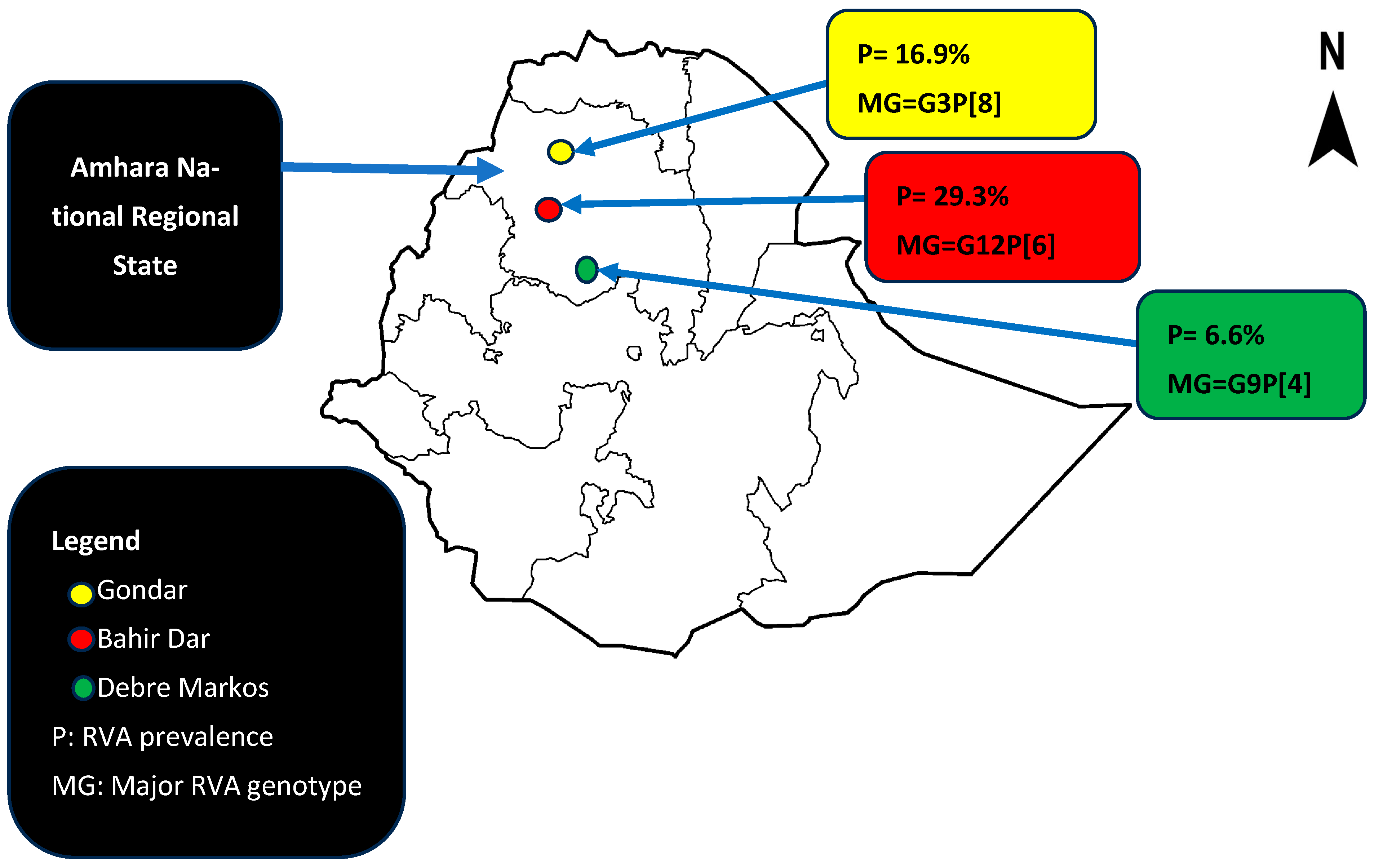
The prevalence of RVA infection among diarrheic children was 94/537 (17.5%, 95%CI = 14.3–21%). The prevalence was the highest in Bahir Dar city 41/140 (29.3%), followed by Gondar city 44/261 (16.9%) and Debre Markos 9/136 (6.6%) (Figure 2).
Younger age (<24 months old) children were more affected 63/279 (22.58%) compared to older (24–59 months old) children 31/258 (12%). Nearly all the study participants were immunized 7/537 (1.3%) and 517/537 (96.3%) for incomplete and complete series, respectively. Only 13/537 (2.4%) of the study participants have never received RVA vaccine. Participant location (p = 0.001), age of the child (p = 0.04), vomiting (p = 0.002), sunken eyes (p = 0.019), being on intravenous fluid therapy (p = 0.026), Vesikari clinical severity (p = 0.006), being underweight (p = 0.005), and wasting (p = 0.007) were found to be significantly associated with RVA infection (Table 2).
The overall diarrheic cases were higher during summer (June, July, and August), which is considered rainy season in Ethiopia. However, the prevalence of RVA-associated diarrhea was relatively higher during spring (February, March, and April), ranging from 29.6 to 46.2% (Figure 3).

3.3. Genotypic Distribution of RVAs

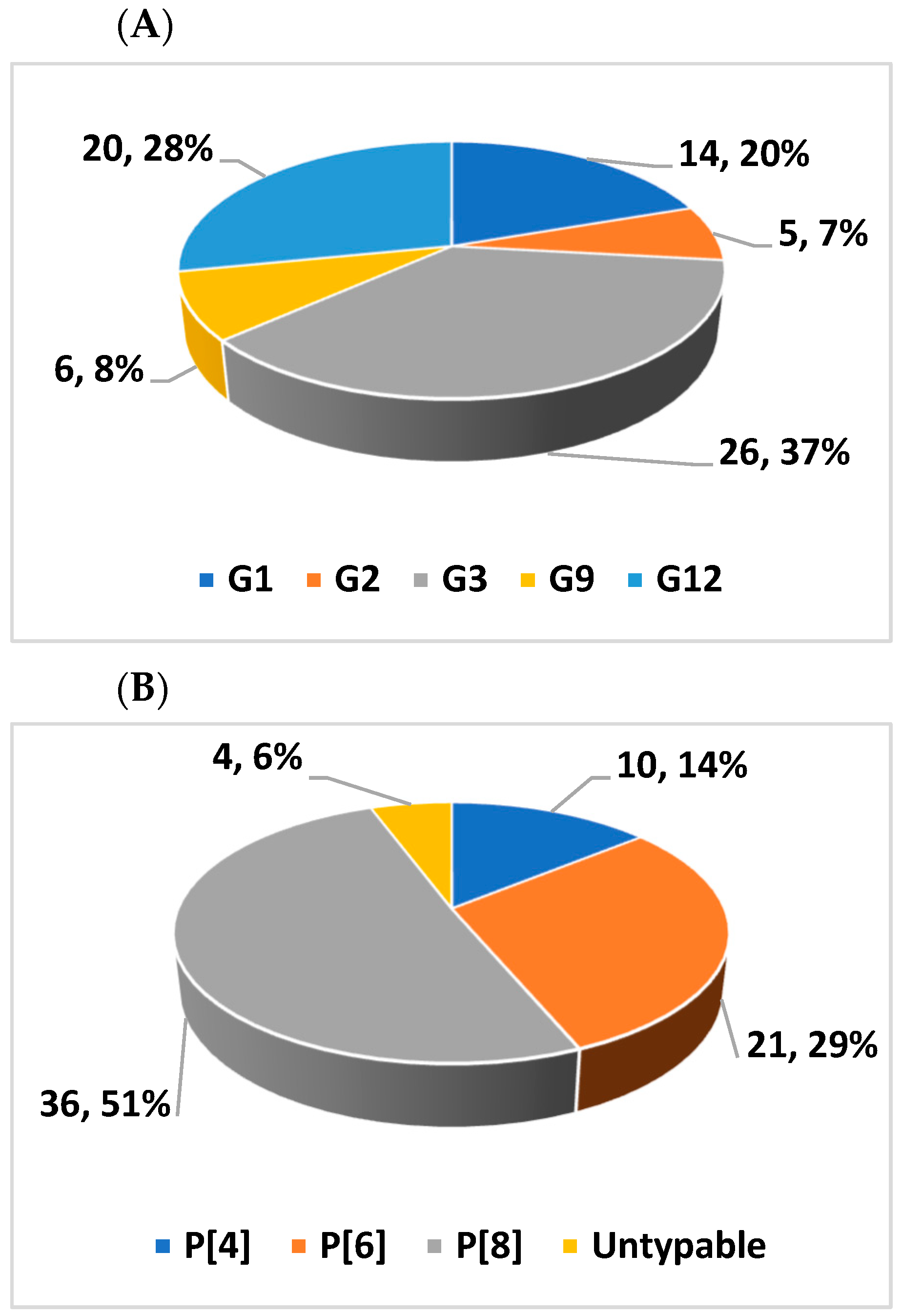
Based on the VP7 and VP4 gene sequences of the circulating RVAs, their G and P-types, respectively, were determined. Of the 94 positive samples, 71 (75.5%) for the VP7 and 67 (71.3%) for the VP4 were successfully genotyped. The circulating G-types identified were G1, G2, G3, G9, and G12 while the P-types included P[4], P[6], and P[8]. G3 was the most dominant G-type, detected in 26 (37%) followed by G12 in 20 (28%) and G1 in 14 (20%) of the samples (Figure 4A). The proportions of circulating P-types were P[8] 36 (51%), P[6] 21 (29%), and P[4] 10 (14%) (Figure 4B).
Eight different G/P combinations were identified. Of these, G3P[8] 22 (32.8%), G12P[6] 19 (28.4%), and G1P[8] 13 (19.4%) were relatively more frequent (Table 3). These three G/P combinations account for 80.6% of the circulating strains.
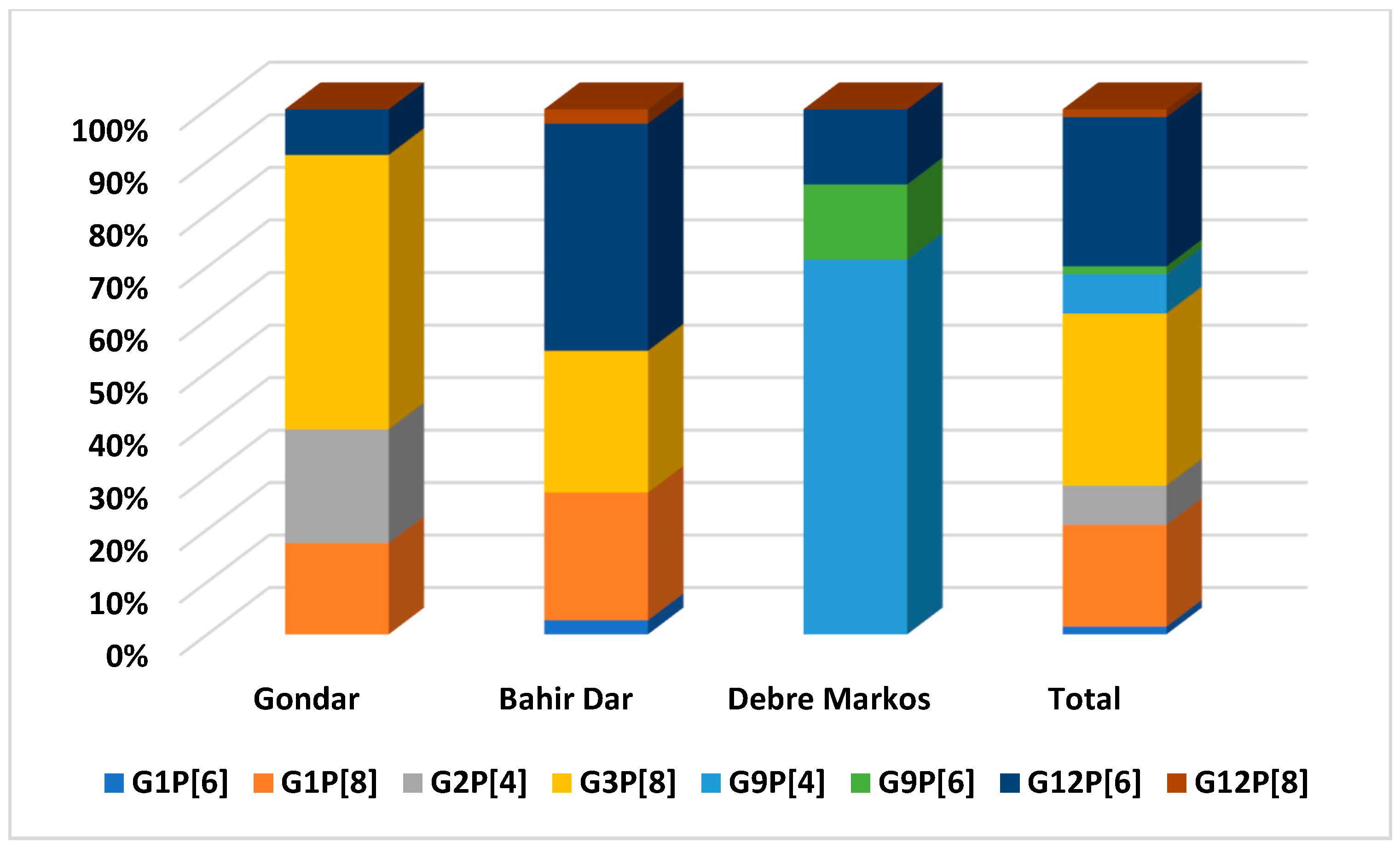
The diversity and distribution of RVA genotypes were different across the three sampling locations. G3, G12, and G9 were the dominantly circulating RVA genotypes in Gondar, Bahir Dar, and Debre Markos, respectively (p = 0.001). G2 and G9 were only detected in Gondar and Debre Markos, respectively. All the three P-types were detected in samples from Gondar and P[6] was predominantly detected in Bahir Dar. G3P[8], G12P[6], and G9P[4] are the predominantly circulating G/P combinations in Gondar, Bahir Dar, and Debre Markos, respectively (Figure 5).

3.4. Phylogenetic Analysis of VP7 Gene of the Circulating RVA Strains

Phylogenetic trees were constructed for 68 complete VP7 gene sequences of the circulating RVA strains. All the 13 G1 strains clustered together and were associated with P[8] except one, which was associated with the P[6]. There was high nucleotide identity among the circulating G1 strains (98.88–100%). Additionally, the circulating strains have shown high nucleotide identity with the Rotarix G1 strain (96.22–96.63%). These strains were also closely related to the wild-type human G1 strains reported from India, Pakistan, Russia, Iran, and the Rotarix G1 strain (Figure 6A).
All G2 strains were associated with P[4] and were identified in Gondar only. The circulating G2 strains were 100% identical to each other and clustered closely with the wild-type human G2P[4] strains from USA, Brazil, Kenya, Bangladesh, and a previous Ethiopian strain isolated in 2016 (Figure 6B).
Twenty one G3 genotypes were associated with P[8], while three G3 genotypes were associated with untypeable P-types. The nucleotide identity among the circulating G3 strains ranged from 91.23 to 100%. These G3 strains were grouped into two sub-clusters. The first sub-cluster included the strains identified in children from Gondar that were closely related to the previously reported G3P[8] Ethiopian strain, while the other half of the G3 strains predominantly from Bahir Dar clustered with the wild-type human RVA G3 strains from USA and Pakistan (Figure 6C).
There were six G9 strains in this study, and all of them were associated with P[4] except one that was associated with P[6]. The circulating G9 strains were shown to share high sequence identity among each other (99.69–100%). All the G9 strains were identified from Debre Markos. The G9 strains were shown to form a tight cluster with the wild-type G9P[4] strains of USA and Ghana. However, these strains seemed to be only distantly related to the previously reported wild-type G9P[8] strain from Bahir Dar, Ethiopia in 2016 (Figure 6D).
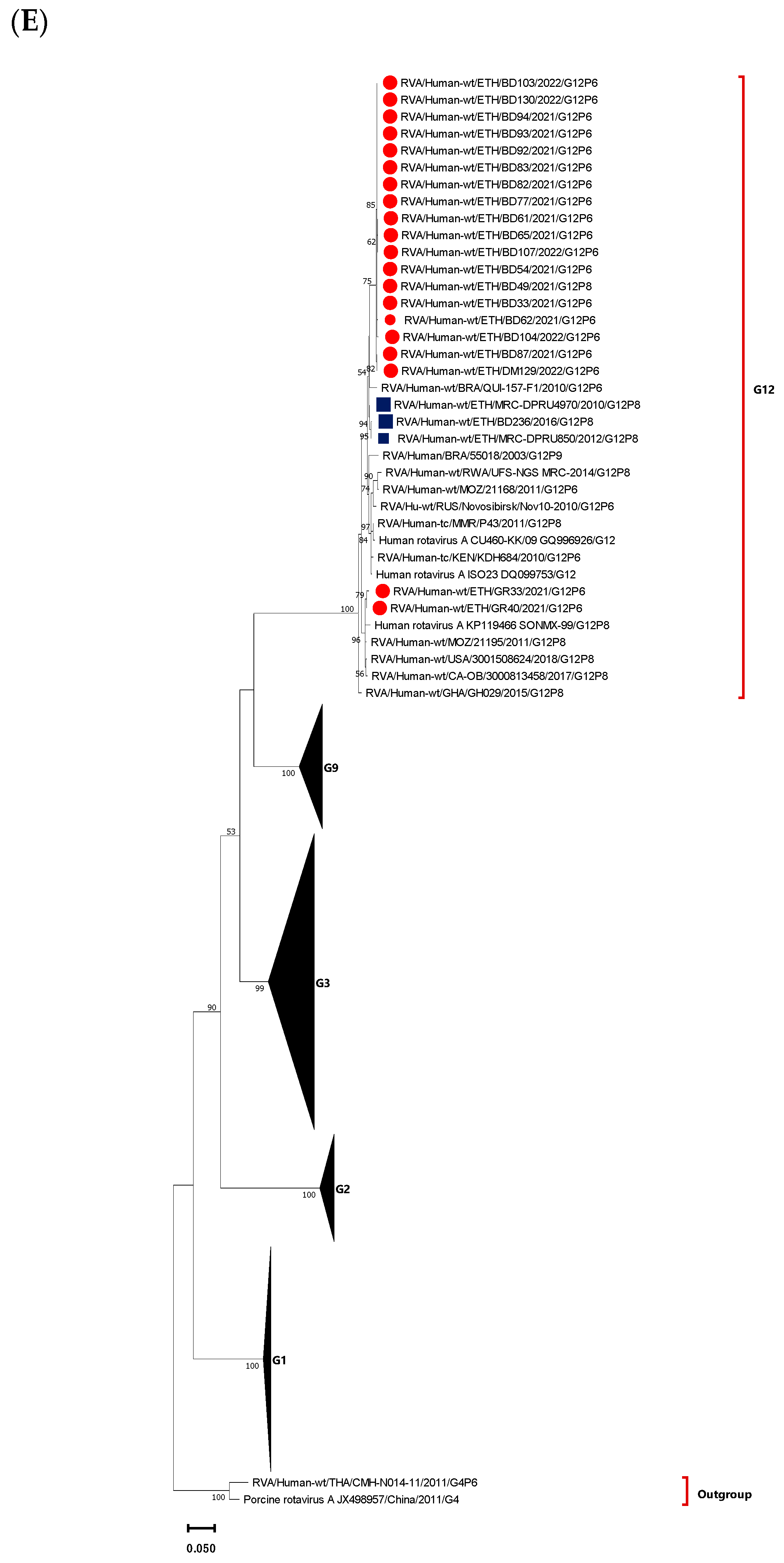
Twenty G12 RVA strains were identified in this study. The sequence identity among the circulating G12 strains ranged from 96.02 to 100%. The majority were from Bahir Dar (17/20) and were associated with P[6] except one associated with P[8]. These G12 strains were clustered into two distinct sub-clusters: the first sub-cluster comprised eighteen strains (seventeen from Bahir Dar and one from Debre Markos), and the second sub-cluster was a cluster of two strains both from Gondar. Unlike the first cluster, the second cluster was shown to be closely related to the human wild-type RVA strains previously reported from USA, Mozambique, and Mexico. However, none of the current G12 strains were closely related to G12 strains previously reported in 2010, 2012, and 2016 from Ethiopia (Figure 6E).

3.5. Phylogenetic Analysis of VP4 Gene of the Circulating RVA Strains

Phylogenetic analysis was performed for the VP4 sequences of the 65 RVA strains circulating in Amhara National Regional State, Ethiopia. The analysis grouped the P[4] strains into two closely related clusters. Interestingly, the P[4] sequences detected in association with G2 were clustered together and similarly, the sequences associated with G9 also clustered together, suggestive of sequence-specific inter-segment interactions between RVA genomic segments. The sequence identity between the circulating P[4] strains ranged from 96.79 to 100% among each other. The G9-associated P[4] strains are closely related to the wild-type G9P[4] human RVA strains reported from India and Kenya. On the other hand, G2-associated P[4] strains were shown to be closely related to the wild-type G2P[4] human RVA strains reported from South Korea and Belarus (Figure 7A).
Of the twenty-two P[6] genotypes, twenty were associated with G12 while the rest of the two P[6] genotypes were associated with G1 and G9, one each. The sequences of the circulating P[6] strains shared high nucleotide identity amongst each other, ranging from 98.15 to 100%. These P[6] strains formed a distinct cluster and were not related to previously reported strains in Ethiopia. A P[6] strain from Debre Markos clustered with the porcine-human reassortant G4P[6] and wild-type human G4P[6] strains isolated from children in China and Thailand, respectively (Figure 7B).
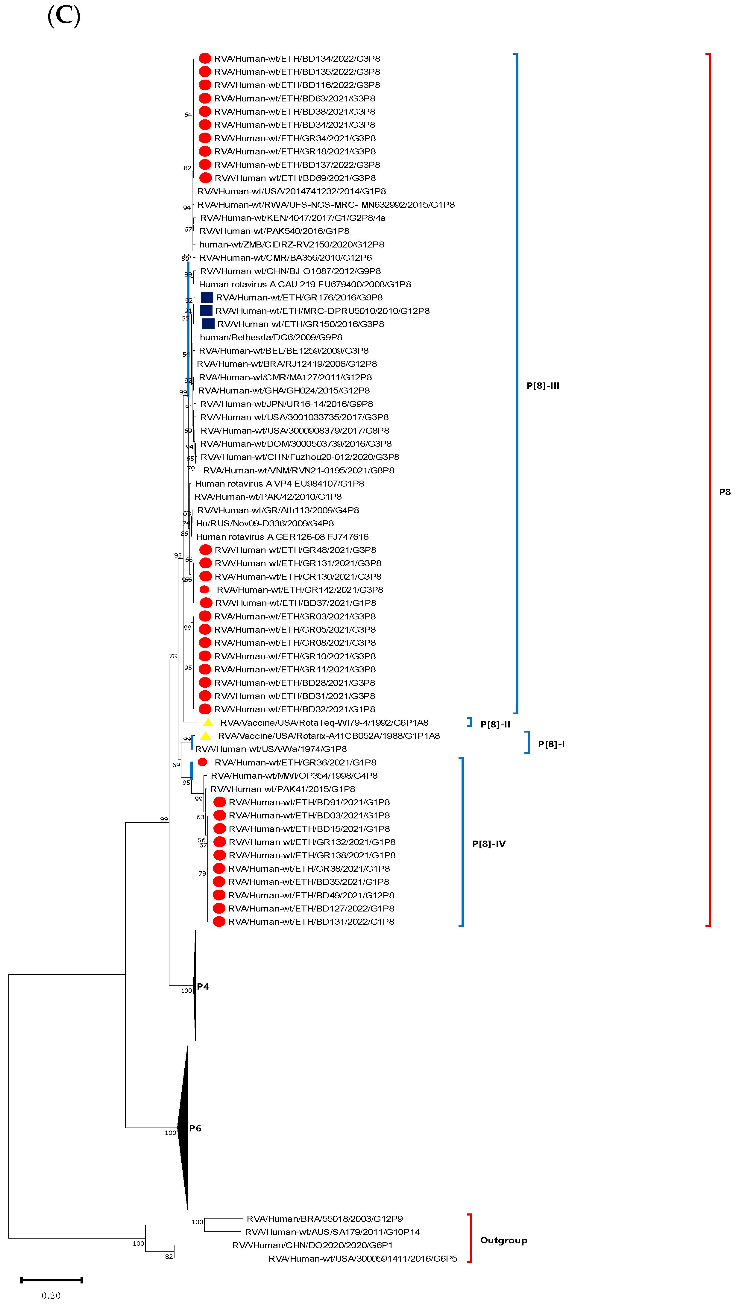
The majority of the P-genotypes were P[8] mostly associated with G3 and G1. The sequence identity among the circulating P[8] strains ranged from 88.23 to 100%, while with the Rotarix P[8] strain ranged from 88.59 to 91.26%. The P[8] strains were grouped into three clusters. The strains in the first cluster were associated with G1 and closely related to the Rotarix P[8] strain and wild-type human RVA G1P[8] strain reported from the USA. The remaining two clusters were associated with G3 genotype, but clustered based on the study location. One of the clusters of G3-associated P[8] strains was dominated by strains from Bahir Dar, while the other was dominated by strains from Gondar. The G3-associated P[8] strains were not closely related to the Rotarix vaccine P[8] strain. However, the dominant strains from Gondar were closely related to strains reported from Greece, Pakistan, and Russia, while the dominant G3-associated P[8] strains from Bahir Dar were closely related to strains reported from Kenya, Rwanda, Cameroon, USA, Zimbabwe, and Ethiopian P[8] strains associated with G3, G9, and G12 reported in 2010 and 2016 (Figure 7C).

3.6. Comparison of the VP7 Antigenic Epitopes with Vaccine Strains

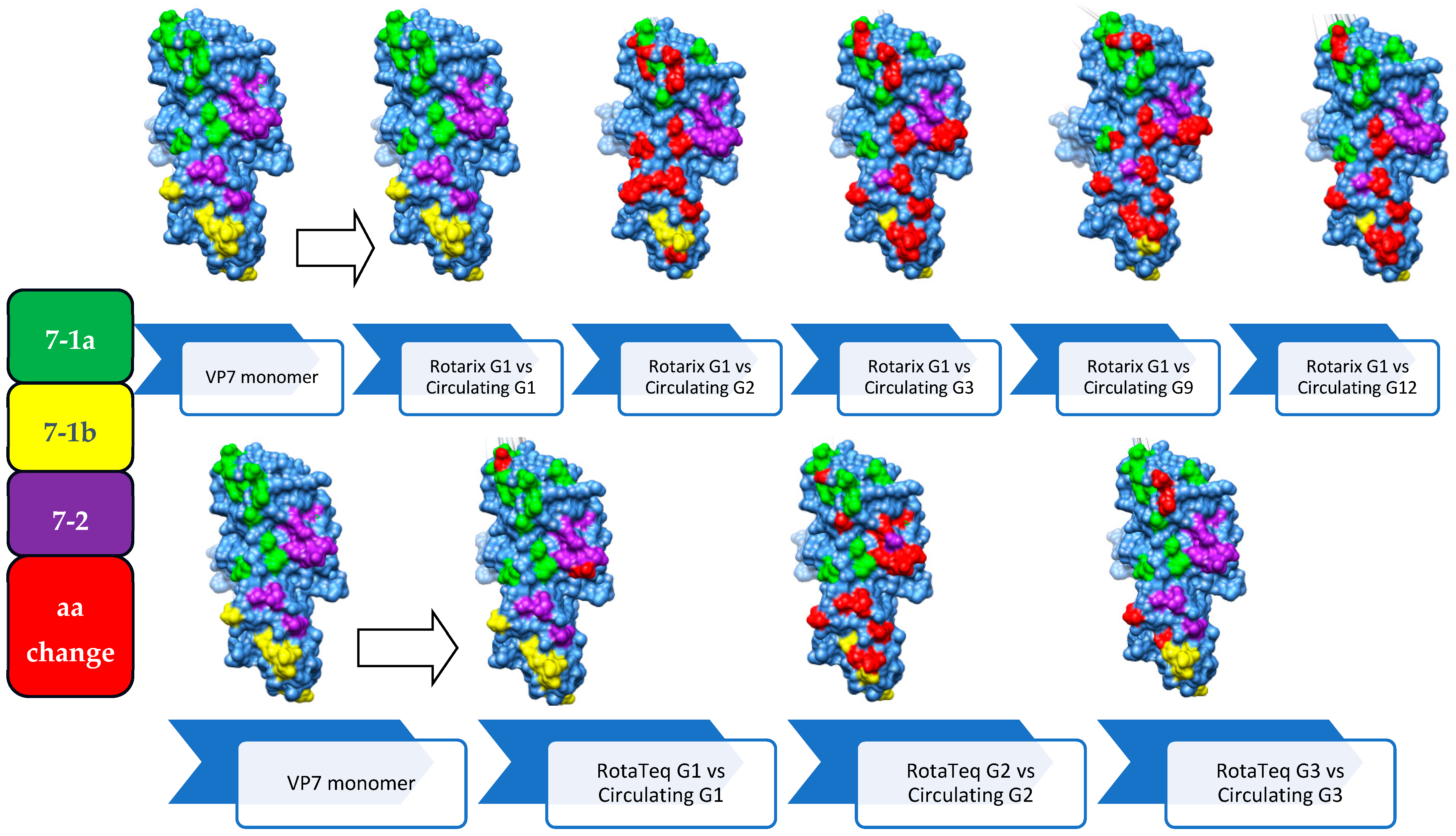
Amino acid sequences spanning the three major antigenic epitopes (7-1a, 7-1b, and 7-2) of the VP7 protein were compared between the identified RVA strains and Rotarix and RotaTeq vaccine strains, revealing substantial amino acid sequence heterogeneity.
All circulating G1 strains (N = 13) showed 100% identity with the Rotarix G1 vaccine strain in the epitopes 7-1a, 7-1b, and 7-2. The circulating G1P[8] strains maintained intact VP7 protein antigenic epitopes similar to the Rotarix vaccine G1 strain resulting in their clustering as a G1-II lineage. For the current Ethiopian G2 strains (N = 5), there were 18 amino acid substitutions out of 29 residues compared to the Rotarix G1 vaccine strain and 13 substitutions compared to the RotaTeq G2 strain. A comparison of the circulating G3 strains revealed amino acid differences ranging from 12 to 14 out of 29 amino acids distributed across all the three antigenic epitopes compared to the Rotarix G1 strain. All circulating G9 strains (n = 6) showed 12/29 and 13/29 amino acid differences compared to RotaTeq G3 and Rotarix G1 vaccine strains, respectively, distributed throughout the three antigenic epitopes. However, compared to the RotaTeq G1 vaccine strain (G1-III), two substitution mutations (D97E and S147N) were observed in the epitopes 7-1a and 7-2 of the circulating G1 strains, respectively. Among the twenty-four circulating G3 strains, twelve showed 2/29 amino acid substitutions (K238N and D242N), eleven showed 3/29 (T91N, K238N, D242N), and one showed 4/29 (T91N, N94D, K238N, D242N) across 7-1a and 7-1b epitopes compared to RotaTeq G3.
Thus, the highest antigenic epitope variation was observed among circulating G12 strains compared to the G4 strain of RotaTeq (19/29) and G1 strain of Rotarix (17/29) consistent with the increased phylogenetic distance between the circulating G12 and the vaccine strains. Almost all strains with amino acid substitutions involved at least one substitution associated with antibody neutralization escape (Table 4 and Figure 8).

3.7. Comparison of VP4 Antigenic Epitopes with Vaccine Strains

Comparative amino acid sequence analyses of the four neutralizing antigenic epitopes (epitope 8-1, epitope 8-2, epitope 8-3, and epitope 8-4) of the VP8* component of VP4 in the circulating RVA strains and the two vaccine strains exhibited significant amino acid substitutions associated with antibody neutralization escape.
Of the eleven circulating lineage IV P[8] strains associated with G1, ten showed four amino acid substitutions (N113D, N192D, N194T, and N195S) out of twenty-eight residues at epitopes 8-1, 8-2, and 8-3, while one strain exhibited only two amino acid substitutions (N194T and N195S) at epitope 8-2 compared to Rotarix P[8] (P[8]-I). P[8] strains associated with G3 showed more amino acid substitutions to the VP8* region of the Rotarix P[8] strain than compared to the P[8] strains associated with G1. Thirteen out of the twenty-two lineage III P[8] strains associated with G3 had six amino acid substitutions (E150D, N194D, N195G, S125N, S131N, and N135D), while the remaining nine had five amino acid substitutions (E150D, N195G, S125N, S131N/R, and N135D) were distributed across the three antigenic epitopes except epitope 8-4 of the Rotarix P[8] strain. Conversely, more amino acid substitutions were observed in the circulating lineage IV P[8] strains associated with G1 than lineage III P[8] strains associated with G3 compared to the RotaTeq P[8] (P[8]-II) vaccine strain. The circulating P[8] strains associated with G1 have shown a total of 7/28 amino acid substitutions (N113D, N192D, N194T, D195S, N125S, R131S, and D135N) across the three neutralizing antigenic epitopes (8-1, 8-2, and 8-3). However, relatively fewer amino acid variabilities were observed between circulating P[8] strains associated with G3 and the RotaTeq P[8] strain. Thirteen strains showed variability in four out of twenty-eight amino acids (E150D, N194D, D195G, and R131N), nine strains exhibited variability in three out of twenty-eight amino acids (E150D, D195G, and R131N), and one strain displayed variability in two out of twenty-eight amino acids (E150D and D195G).
Comparative analysis of the amino acid sequences in circulating P[4] and P[6] strains showed 10/28 and 18/28 amino acid variabilities, respectively, compared to the Rotarix P[8] strain. These strains have shown the highest heterogeneity (25/28 and 24/28 amino acid variabilities) compared to the RotaTeq P[8] strain (Table 5 and Figure 9).

4. Discussion

In this study, the prevalence of RVA infection among diarrheic children was 93/537 (17.5%). This prevalence is lower than in previous studies from Ethiopia before [17,37,38] and after the introduction of RVA vaccine (Rotarix) [26]. Nonetheless, at least one post vaccine era sentinel surveillance study in Ethiopia have reported comparable findings [37]. Reduction in RVA prevalence post vaccine introduction has been reported elsewhere [39,40,41], signifying the role of the vaccine in the reduction in RVA-associated acute gastroenteritis among under-five children.
This study identified significant geographic variability in RVA prevalence and genetic diversity among children, with Bahir Dar showing the highest prevalence at 28.6% (with G12P[6] strains being dominant), followed by Gondar at 16.9% (with G3P[8] strains being dominant), and Debre Markos at 6.6% (with G9P[4] strains being dominant). This striking variability may result from differences in sampling timing, variable climatic conditions, behavioral, and other location-specific factors. Additionally, the prevalence of symptomatic RVA infections was higher among younger children (22.58%) compared to older children (12%), possibly due to their less developed immune systems [42] and limited previous exposure to RVA [43], aligning with findings from Nigeria and Brazil where RVA is more prevalent among children under 24 months old [44,45,46]. In this study, though, there was a relative increase in the prevalence of RVA in spring (February, March, and April). RVA has circulated year-round in the study area, emphasizing the endemicity of RVA in Ethiopia. This observation is supported by a study which reported a year-round disease pattern in low- and low-middle income countries [47]. However, studies also reported the seasonal nature of RVA infection in which the highest peaks of RVA prevalence happened during the dry and cooler seasons [48,49].
In this study, RVA infection correlated with severe clinical signs such as vomiting, sunken eyes, intravenous fluid treatment, and high Vesikari clinical severity scores. These findings are consistent with other studies linking the severity of diarrhea to RVA infection [50,51,52]. Additionally, RVA infection was associated with acute malnutrition, as evidenced by higher rates of wasting and underweight status. Similar associations between diarrhea and malnutrition have been reported in studies from Bangladesh [53] and Sudan [54].
Based on the VP7 gene sequence analysis of 71 RVA-positive samples, five distinct G-types (G1, G2, G3, G9, and G12) were identified, with G3 being the most prevalent (37%), followed by G12 (28%) and G1 (20%). This trend aligns with a previous meta-analyses study in Ethiopia indicating an increase in the prevalence of G3 strains post-Rotarix vaccine introduction, possibly due to the vaccine-driven selective pressure on G1 strains of the same lineage (G1-II) [55]. Similar observations were noted in Australia and Colombia, where an increase in G3P[8] strain prevalence was observed following Rotarix vaccine introduction [56,57]. Despite its global distribution, G4 genotype was not identified in this study, consistent with previous Ethiopian studies [17,26,37,38,55,58]. The circulating RVA strains also included P[4], P[6], P[8], and four untypable P-types, with P[8] being the most common (51%). This finding is consistent with several previous studies in Ethiopia [17,26,37,38,58] and elsewhere including Cameroon [59], Kenya [60], and China [61]. Major G/P combinations including G3P[8] (32.8%), G12P[6] (28.4%), and G1P[8] (19.4%) reflected a shift towards G3P[8], G12P[8], G9P[8], and other unusual combinations observed in our previous meta-analysis study [55]. Similar trends were reported in Mozambique [62] and China [63] post-Rotarix vaccine introduction, indicating the vaccine’s influence on RVA genotype distribution and emergence of new genotypes. Thus, the vaccine-induced immunity continues to impact the prevalence of dominant and emerging genotypes highlighting that ongoing surveillance and vaccine efficacy monitoring are crucial for RVA control strategies.
Our observations of the variable predominant G/P combinations circulating in Gondar (G3P[8]), Bahir Dar (G12P[6]), and Debre Markos (G9P[4]) were similar to those reported in proximate study locations previously [26].
Phylogenetic analysis of the currently circulating Ethiopian strains revealed that most strains are closely related to the previously reported global and local RVA variants. However, the dominant circulating RVA strains seem to be changing over time. G3P[8] and the unusual G12P[6] and G9P[4] tend to rise in the post-vaccine introduction era. In general, the sequences from Ethiopia during the post-vaccine period formed distinct clusters as well as clusters with other globally reported strains on the global phylogeny. This indicates that distinct virus populations as well as imported strains were circulating during this period. Some Ethiopian strains such as G12 P[6] clustered together in a separate branch, distinct from other global strains, suggesting that local strains observed were likely persisting within the study area or neighboring regions, accumulating genetic changes over time. Moreover, some circulating Ethiopian strains such as G1P[8] and G12P[6] are not related to the previously reported Ethiopian strains. This might be either due to importation of new strains from other countries or due to significant mutations through random point mutations and intra-genotype reassortment in previously circulating strains, resulting in distinct clustering patterns of the circulating strains [64].
In this study, a comparative protein sequence analysis of the antigenic epitopes of VP7 and VP4 proteins revealed significant amino acid residue variability between the circulating Ethiopian RVA strains and the monovalent Rotarix and the pentavalent RotaTeq vaccine strains. Similar observation of high antigenic epitope variability between the circulating RVA strains and the vaccine strains were reported from China [65], Qatar [66], Belgium [67], and Gabone [68]. This might be one of the reasons to have significant prevalence of RVA infection despite high RVA immunization rate in the study area.
Similar to a previous study from Zambia [69], the circulating G1P[8] strains in our study belonged to the same lineage (G1-II) as the Rotarix vaccine G1 strain, and their VP7 antigenic epitopes were indistinguishable. Expectedly, G2, G3, G9, and G12 strains showed a significantly higher amino acid variability in the VP7 antigenic epitopes compared to Rotarix, suggesting that the vaccine-induced immunity could be inadequate to control heterotypic (non-G1) strains [69,70]. Although the multivalent RotaTeq vaccine (that was not a part of the EPI in Ethiopia) could provide a broader [homotypic (against G1, G2, and G3 strains) and heterotypic (against G9 and G12 strains)] immunity, the circulating Ethiopian strains belonged to different lineages (G1-II, G2-IV, and G3-I) than those in RotaTeq vaccine (G1-III, G2-II, and G3-II). This was associated with multiple amino acid mismatches, including substitutions linked to neutralization escape, as seen in studies from the USA [71] and elsewhere [65,66,67,68]. Thus, it remains unclear if RotaTeq vaccine would provide a superior protection against the strains circulating in Ethiopia compared to that induced by the Rotarix vaccine.
The circulating strains belonged to different lineages (P[8]-III and P[8]-IV) than Rotarix (P[8]-I) or RotaTeq (P[8]-II), displaying numerous amino acid substitutions in the VP8* component of VP4 [containing four neutralizing antigenic epitopes (8-1, 8-2, 8-3, and 8-4)] of the circulating RVA strains, which can enable neutralization escape. However, lineage III P[8] strains exhibited fewer substitutions than lineage IV strains relative to Rotarix, while the opposite trend was observed for RotaTeq vaccine, consistent with findings from Gabon [68] and Qatar [66]. These substitutions in the VP8* region may compromise neutralizing immunity, posing a risk of immune evasion [72]. The continuous dominance of the P[8] strains observed in our and other studies despite the vaccine-associated homotypic immunity suggests its increased genetic plasticity compared to the common G types. Comparative analyses with heterotypic strains (P[4] and P[6]) also revealed substantial amino acid variations across VP8* epitopes, suggesting a high likelihood of neutralization escape from both homotypic and less effective heterotypic immunity against VP4 proteins by P[8] and non-P[8] strains, respectively [66,67,68].

5. Conclusions and Recommendation

Despite the high rate of RVA immunization in the study area, RVA infections among diarrheic children remain considerably prevalent. The predominantly circulating RVA genotypes in the study area appear to change over time post-vaccine introduction. Phylogenetic analysis revealed that different RVA strains cluster with strains reported elsewhere globally. However, strong spatial genotype variations were also confirmed in this study. The circulating RVA strains demonstrate significant variability in amino acid residues, including those involved in neutralization escape within the VP7 and VP4 antigenic epitopes, compared to the two widely RVA vaccine strains licensed for global use. This finding underscores the importance of continued surveillance, vaccine updating and development, and potentially adapting immunization strategies to better match the evolving diversity of RVA strains globally.

Author Contributions

Conceptualization, D.D., A.N.V. and B.T.; data curation, D.D.; formal analysis, D.D. and A.N.V.; funding acquisition, A.G., Y.W., Y.A., Z.T., A.N.V. and B.T.; investigation, D.D., Y.A. and M.K.K.; methodology, D.D., A.G., Y.W., Z.T., A.N.V. and B.T.; project administration, D.D.; resources, A.G., Y.W., U.S., Z.H.M., A.N.V. and B.T.; software, Z.H.M. and A.N.V.; supervision, A.G., Y.W., Z.T., U.S., Z.H.M., A.N.V. and B.T.; validation, D.D.; visualization, D.D.; writing—original draft, D.D. and Z.H.M.; writing—review and editing, D.D., A.G., Y.W., Y.A., M.K.K., U.S., A.N.V. and B.T. All authors have read and agreed to the published version of the manuscript.

Funding

This work was supported by the University of Gondar internal competitive grant awarded in 2020 (R. No. R/T/T/C/E/C/D/03/2013). D.D. was supported by Sustainable One Health Research Training Capacity (OHEART): Molecular epidemiology of zoonotic foodborne and waterborne pathogens in Eastern Africa, funded by the NIH Fogarty International Center (D43TW008650), through the Global One Health initiative (GOHi) and Dr. Vlasova startup and discretionary funds from The Ohio State University. The funders had no role in study design, data collection and analysis, decision to publish, or preparation of the manuscript.

Institutional Review Board Statement

The protocol was approved by the University of Gondar Institutional Review Board (IRB) (R. No. V/P/RCS/05/538/2020) dated 15 December 2020. Moreover, health facilities involved in this study were communicated through written support letters obtained from the University of Gondar research and publication office (RPO). Written informed consent was obtained from the parents/guardians of the children before enrolment in the study. Data taken from study subjects were numerically coded, and the test results were used only for the study purpose and kept securely stored throughout the study period and thereafter.

Informed Consent Statement

Written informed consent was obtained from the parents/guardians of the children before enrolment in the study.

Data Availability Statement

Data supporting this manuscript are available in the manuscript or online (GenBank).

Acknowledgments

We would like to acknowledge the University of Gondar, the Ohio State University, and the NIH Fogarty International Center for their support. We are also grateful to the data and sample collectors, as well as the study participants.

Conflicts of Interest

The authors declare that they have no competing interests. Ohio state University Global Health Initiative LLC in Ethiopia is not a commercial entity. It is OSU Global One Health initiative (GOHi) affiliate office of the Eastern Africa region based in Ethiopia. The Office aims to improve community health, and provide learning opportunities for students, faculty and staff around the world as part of the OSU’s commitment to share knowledge and find solutions to global issues. It appears as LLC is not because it is a commercial entity, it is rather because it cannot have financial activities for the research and community engagement projects in Eastern African region (In Ethiopia) without establishing office as LLC as it is required by the Ethiopian law.

References

  1. Troeger, C.; Khalil, I.A.; Rao, P.C.; Cao, S.; Blacker, B.F.; Ahmed, T.; Armah, G.; Bines, J.E.; Brewer, T.G.; Colombara, D.V.; et al. Rotavirus Vaccination and the Global Burden of Rotavirus Diarrhea among Children Younger than 5 Years. JAMA Pediatr. 2018, 172, 958–965. [Google Scholar] [CrossRef] [PubMed]
  2. Parashar, U.D.; Burton, A.; Lanata, C.; Boschi-Pinto, C.; Shibuya, K.; Steele, D.; Birmingham, M.; Glass, R.I. Global mortality associated with rotavirus disease among children in 2004. J. Infect. Dis. 2009, 200 (Suppl. S1), S9–S15. [Google Scholar] [CrossRef] [PubMed]
  3. Bilcke, J.; Van Damme, P.; Van Ranst, M.; Hens, N.; Aerts, M.; Beutels, P. Estimating the incidence of symptomatic rotavirus infections: A systematic review and meta-analysis. PLoS ONE 2009, 4, e6060. [Google Scholar] [CrossRef] [PubMed]
  4. Matthijnssens, J.; Attoui, H.; Bányai, K.; Brussaard, C.P.; Danthi, P.; Del Vas, M.; Dermody, T.S.; Duncan, R.; Fāng, Q.; Johne, R. ICTV virus taxonomy profile: Sedoreoviridae 2022. J. Gen. Virol. 2022, 103, 001782. [Google Scholar] [CrossRef] [PubMed]
  5. Estes, M.K.; Greenberg, H.B. Rotaviruses. In Fields Virology, 6th ed.; Howley, P.M., Knipe, D.M., Griffin, D.E., Lamb, R.A., Martin, M.A., Roizman, B., Straus, S.E., Eds.; Wolters Kluwer Health/Lippincott Williams & Wilkins: Philadelphia, PA, USA, 2013; pp. 1347–1401. [Google Scholar]
  6. Matthijnssens, J.; Otto, P.H.; Ciarlet, M.; Desselberger, U.; Van Ranst, M.; Johne, R. VP6-sequence-based cutoff values as a criterion for rotavirus species demarcation. Arch. Virol. 2012, 157, 1177–1182. [Google Scholar] [CrossRef]
  7. Mihalov-Kovács, E.; Gellért, Á.; Marton, S.; Farkas, S.L.; Fehér, E.; Oldal, M.; Jakab, F.; Martella, V.; Bányai, K. Candidate new rotavirus species in sheltered dogs, Hungary. Emerg. Infect. Dis. 2015, 21, 660. [Google Scholar] [CrossRef]
  8. Bányai, K.; Kemenesi, G.; Budinski, I.; Földes, F.; Zana, B.; Marton, S.; Varga-Kugler, R.; Oldal, M.; Kurucz, K.; Jakab, F. Candidate new rotavirus species in Schreiber’s bats, Serbia. Infect. Genet. Evol. 2017, 48, 19–26. [Google Scholar] [CrossRef] [PubMed]
  9. Collins, P.J.; Martella, V.; Buonavoglia, C.; O’Shea, H. Identification of a G2-like porcine rotavirus bearing a novel VP4 type, P[32]. Vet. Res. 2010, 41, 73. [Google Scholar] [CrossRef] [PubMed]
  10. Esona, M.D.; Mijatovic-Rustempasic, S.; Conrardy, C.; Tong, S.; Kuzmin, I.V.; Agwanda, B.; Breiman, R.F.; Banyai, K.; Niezgoda, M.; Rupprecht, C.E. Reassortant group A rotavirus from straw-colored fruit bat (Eidolon helvum). Emerg. Infect. Dis. 2010, 16, 1844. [Google Scholar] [CrossRef]
  11. Abe, M.; Ito, N.; Masatani, T.; Nakagawa, K.; Yamaoka, S.; Kanamaru, Y.; Suzuki, H.; Shibano, K.-i.; Arashi, Y.; Sugiyama, M. Whole genome characterization of new bovine rotavirus G21P[29] and G24P[33] strains provides evidence for interspecies transmission. J. Gen. Virol. 2011, 92, 952–960. [Google Scholar] [CrossRef]
  12. Hayashi, M.; Murakami, T.; Kuroda, Y.; Takai, H.; Ide, H.; Awang, A.; Suzuki, T.; Miyazaki, A.; Nagai, M.; Tsunemitsu, H. Reinfection of adult cattle with rotavirus B during repeated outbreaks of epidemic diarrhea. Can. J. Vet. Res. = Rev. Can. Rech. Vet. 2016, 80, 189–196. [Google Scholar]
  13. Santos, N.; Hoshino, Y. Global distribution of rotavirus serotypes/genotypes and its implication for the development and implementation of an effective rotavirus vaccine. Rev. Med. Virol. 2005, 15, 29–56. [Google Scholar] [CrossRef] [PubMed]
  14. Khamrin, P.; Maneekarn, N.; Peerakome, S.; Chan-it, W.; Yagyu, F.; Okitsu, S.; Ushijima, H. Novel porcine rotavirus of genotype P[27] shares new phylogenetic lineage with G2 porcine rotavirus strain. Virology 2007, 361, 243–252. [Google Scholar] [CrossRef]
  15. Matthijnssens, J.; Ciarlet, M.; Rahman, M.; Attoui, H.; Bányai, K.; Estes, M.K.; Gentsch, J.R.; Iturriza-Gómara, M.; Kirkwood, C.D.; Martella, V. Recommendations for the classification of group A rotaviruses using all 11 genomic RNA segments. Arch. Virol. 2008, 153, 1621–1629. [Google Scholar] [CrossRef] [PubMed]
  16. Matthijnssens, J.; Ciarlet, M.; McDonald, S.M.; Attoui, H.; Bányai, K.; Brister, J.R.; Buesa, J.; Esona, M.D.; Estes, M.K.; Gentsch, J.R. Uniformity of rotavirus strain nomenclature proposed by the Rotavirus Classification Working Group (RCWG). Arch. Virol. 2011, 156, 1397–1413. [Google Scholar] [CrossRef] [PubMed]
  17. Mwenda, J.M.; Ntoto, K.M.; Abebe, A.; Enweronu-Laryea, C.; Amina, I.; Mchomvu, J.; Kisakye, A.; Mpabalwani, E.M.; Pazvakavambwa, I.; Armah, G.E. Burden and epidemiology of rotavirus diarrhea in selected African countries: Preliminary results from the African Rotavirus Surveillance Network. J. Infect. Dis. 2010, 202 (Suppl. S1), S5–S11. [Google Scholar] [CrossRef] [PubMed]
  18. WHO. Meeting of the Strategic Advisory Group of Experts on immunization, October 2009—Conclusions and recommendations. Wkly. Epidemiol. Rec. = Relev. Épidémiol. Hebd. 2009, 84, 517–532. [Google Scholar]
  19. FMoH. Ethiopian National Expanded Programme on Immunization: Comprhencive Munlti-Year Plan 2016–2020 EPI; Federal Ministry of Health of Ethiopia: Addis Ababa, Ethiopia, 2015; p. 28.
  20. Wondimu, A.; Cao, Q.; Wilschut, J.C.; Postma, M.J. Factors associated with the uptake of newly introduced childhood vaccinations in Ethiopia: The cases of rotavirus and pneumococcal conjugate vaccines. BMC Public Health 2019, 19, 1656. [Google Scholar] [CrossRef] [PubMed]
  21. Atalell, K.A.; Liyew, A.M.; Alene, K.A. Spatial distribution of rotavirus immunization coverage in Ethiopia: A geospatial analysis using the Bayesian approach. BMC Infect. Dis. 2022, 22, 830. [Google Scholar] [CrossRef]
  22. Muhe, L.; Fredrikzon, B.; Habte, D. Clinical profile of rotavirus enteritis in Ethiopian children. Ethiop. Med. J. 1986, 24, 1–6. [Google Scholar]
  23. Abebe, A.; Abebe, S.; Giday, M.; Taffesse, B. Rotavirus infection in under-five children in Yekatit 12 Hospital. Ethiop. J. Health Dev. (EJHD) 1995, 9, 71–75. [Google Scholar]
  24. Stintzing, G.; Bäck, E.; Tufvesson, B.; Johnsson, T.; Wadström, T.; Habte, D. Seasonal fluctuations in the occurrence of enterotoxigenic bacteria and rotavirus in paediatric diarrhoea in Addis Ababa. Bull. World Health Organ. 1981, 59, 67. [Google Scholar] [PubMed]
  25. Bizuneh, T.; Abebe, A.; Lema, E. Rotavirus infection in under-five children in Jimma Hospital, Southwest Ethiopia. Ethiop. J. Health Dev. (EJHD) 2004, 18, 1–66. [Google Scholar] [CrossRef]
  26. Gelaw, A.; Pietsch, C.; Liebert, U.G. Molecular epidemiology of rotaviruses in Northwest Ethiopia after national vaccine introduction. Infect. Genet. Evol. 2018, 65, 300–307. [Google Scholar] [CrossRef] [PubMed]
  27. Group, W.M.G.R.S.; de Onis, M. WHO Child Growth Standards based on length/height, weight and age. Acta Paediatr. 2006, 95, 76–85. [Google Scholar]
  28. Lewis, K.D.; Dallas, M.J.; Victor, J.C.; Ciarlet, M.; Mast, T.C.; Ji, M.; Armah, G.; Zaman, K.; Ferraro, A.; Neuzil, K.M. Comparison of two clinical severity scoring systems in two multi-center, developing country rotavirus vaccine trials in Africa and Asia. Vaccine 2012, 30, A159–A166. [Google Scholar] [CrossRef] [PubMed]
  29. Ruuska, T.; Vesikari, T. Rotavirus disease in Finnish children: Use of numerical scores for clinical severity of diarrhoeal episodes. Scand. J. Infect. Dis. 1990, 22, 259–267. [Google Scholar] [CrossRef]
  30. Gouvea, V.; Glass, R.I.; Woods, P.; Taniguchi, K.; Clark, H.F.; Forrester, B.; Fang, Z.-Y. Polymerase chain reaction amplification and typing of rotavirus nucleic acid from stool specimens. J. Clin. Microbiol. 1990, 28, 276–282. [Google Scholar] [CrossRef]
  31. Gault, E.; Chikhi-Brachet, R.; Delon, S.; Schnepf, N.; Albiges, L.; Grimprel, E.; Girardet, J.-P.; Begue, P.; Garbarg-Chenon, A. Distribution of human rotavirus G types circulating in Paris, France, during the 1997–1998 epidemic: High prevalence of type G4. J. Clin. Microbiol. 1999, 37, 2373–2375. [Google Scholar] [CrossRef]
  32. Gentsch, J.R.; Glass, R.; Woods, P.; Gouvea, V.; Gorziglia, M.; Flores, J.; Das, B.; Bhan, M. Identification of group A rotavirus gene 4 types by polymerase chain reaction. J. Clin. Microbiol. 1992, 30, 1365–1373. [Google Scholar] [CrossRef]
  33. Maes, P.; Matthijnssens, J.; Rahman, M.; Van Ranst, M. RotaC: A web-based tool for the complete genome classification of group A rotaviruses. BMC Microbiol. 2009, 9, 238. [Google Scholar] [CrossRef] [PubMed]
  34. Tamura, K.; Peterson, D.; Peterson, N.; Stecher, G.; Nei, M.; Kumar, S. MEGA5: Molecular evolutionary genetics analysis using maximum likelihood, evolutionary distance, and maximum parsimony methods. Mol. Biol. Evol. 2011, 28, 2731–2739. [Google Scholar] [CrossRef] [PubMed]
  35. Waterhouse, A.; Bertoni, M.; Bienert, S.; Studer, G.; Tauriello, G.; Gumienny, R.; Heer, F.T.; de Beer, T.A.P.; Rempfer, C.; Bordoli, L. SWISS-MODEL: Homology modelling of protein structures and complexes. Nucleic Acids Res. 2018, 46, W296–W303. [Google Scholar] [CrossRef] [PubMed]
  36. Pettersen, E.F.; Goddard, T.D.; Huang, C.C.; Couch, G.S.; Greenblatt, D.M.; Meng, E.C.; Ferrin, T.E. UCSF Chimera—A visualization system for exploratory research and analysis. J. Comput. Chem. 2004, 25, 1605–1612. [Google Scholar] [CrossRef] [PubMed]
  37. Abebe, A.; Getahun, M.; Mapaseka, S.L.; Beyene, B.; Assefa, E.; Teshome, B.; Tefera, M.; Kebede, F.; Habtamu, A.; Haile-Mariam, T. Impact of rotavirus vaccine introduction and genotypic characteristics of rotavirus strains in children less than 5 years of age with gastroenteritis in Ethiopia: 2011–2016. Vaccine 2018, 36, 7043–7047. [Google Scholar] [CrossRef]
  38. Yassin, M.A.; Kirby, A.; Mengistu, A.A.; Arbide, I.; Dove, W.; Beyer, M.; Cunliffe, N.A.; Cuevas, L.E. Unusual norovirus and rotavirus genotypes in Ethiopia. Paediatr. Int. Child Health 2012, 32, 51–55. [Google Scholar] [CrossRef] [PubMed]
  39. Assis, A.S.F.; Valle, D.A.; Antunes, G.R.; Tibiriça, S.H.C.; De Assis, R.M.S.; Leite, J.P.G.; De Carvalho, I.P.; da Rosa e Silva, M.L. Rotavirus epidemiology before and after vaccine introduction. J. Pediatr. 2013, 89, 470–476. [Google Scholar] [CrossRef] [PubMed]
  40. Hallowell, B.D.; Parashar, U.D.; Curns, A.; DeGroote, N.P.; Tate, J.E. Trends in the laboratory detection of rotavirus before and after implementation of routine rotavirus vaccination—United States, 2000–2018. Morb. Mortal. Wkly. Rep. 2019, 68, 539. [Google Scholar] [CrossRef] [PubMed]
  41. Zeller, M.; Rahman, M.; Heylen, E.; De Coster, S.; De Vos, S.; Arijs, I.; Novo, L.; Verstappen, N.; Van Ranst, M.; Matthijnssens, J. Rotavirus incidence and genotype distribution before and after national rotavirus vaccine introduction in Belgium. Vaccine 2010, 28, 7507–7513. [Google Scholar] [CrossRef]
  42. Saso, A.; Kampmann, B. (Eds.) Vaccine responses in newborns. In Seminars in Immunopathology; Springer: Berlin/Heidelberg, Germany, 2017. [Google Scholar]
  43. Lewnard, J.A.; Lopman, B.A.; Parashar, U.D.; Bar-Zeev, N.; Samuel, P.; Guerrero, M.L.; Ruiz-Palacios, G.M.; Kang, G.; Pitzer, V.E. Naturally acquired immunity against rotavirus infection and gastroenteritis in children: Paired reanalyses of birth cohort studies. J. Infect. Dis. 2017, 216, 317–326. [Google Scholar] [CrossRef]
  44. Ojobor, C.; Olovo, C.; Onah, L.; Ike, A. Prevalence and associated factors to rotavirus infection in children less than 5 years in Enugu State, Nigeria. Virusdisease 2020, 31, 316–322. [Google Scholar] [CrossRef] [PubMed]
  45. Junaid, S.A.; Umeh, C.; Olabode, A.O.; Banda, J.M. Incidence of rotavirus infection in children with gastroenteritis attending Jos university teaching hospital, Nigeria. Virol. J. 2011, 8, 233. [Google Scholar] [CrossRef] [PubMed]
  46. Gutierrez, M.B.; Fialho, A.M.; Maranhão, A.G.; Malta, F.C.; Andrade, J.d.S.R.d.; Assis, R.M.S.d.; Mouta, S.d.S.e.; Miagostovich, M.P.; Leite, J.P.G.; Machado Fumian, T. Rotavirus A in Brazil: Molecular epidemiology and surveillance during 2018–2019. Pathogens 2020, 9, 515. [Google Scholar] [CrossRef] [PubMed]
  47. Patel, M.M.; Pitzer, V.E.; Alonso, W.J.; Vera, D.; Lopman, B.; Tate, J.; Viboud, C.; Parashar, U.D. Global seasonality of rotavirus disease. Pediatr. Infect. Dis. J. 2013, 32, e134–e147. [Google Scholar] [CrossRef] [PubMed]
  48. Ureña-Castro, K.; Ávila, S.; Gutierrez, M.; Naumova, E.N.; Ulloa-Gutierrez, R.; Mora-Guevara, A. Seasonality of rotavirus hospitalizations at Costa Rica’s National Children’s Hospital in 2010–2015. Int. J. Environ. Res. Public Health 2019, 16, 2321. [Google Scholar] [CrossRef]
  49. Levy, K.; Hubbard, A.E.; Eisenberg, J.N. Seasonality of rotavirus disease in the tropics: A systematic review and meta-analysis. Int. J. Epidemiol. 2009, 38, 1487–1496. [Google Scholar] [CrossRef] [PubMed]
  50. Staat, M.A.; Azimi, P.H.; Berke, T.; Roberts, N.; Bernstein, D.I.; Ward, R.L.; Pickering, L.K.; Matson, D.O. Clinical presentations of rotavirus infection among hospitalized children. Pediatr. Infect. Dis. J. 2002, 21, 221–227. [Google Scholar] [CrossRef] [PubMed]
  51. Intusoma, U.; Sornsrivichai, V.; Jiraphongsa, C.; Varavithaya, W. Epidemiology, clinical presentations and burden of rotavirus diarrhea in children under five seen at Ramathibodi Hospital, Thailand. J. Med. Assoc. Thail. 2008, 91, 1350–1355. [Google Scholar]
  52. Rerksuppaphol, S.; Rerksuppaphol, L. Prevalence and clinical manifestations of rotavirus diarrhea in children of rural area of Thailand. Int. J. Collab. Res. Intern. Med. Public Health 2011, 3, 695–702. [Google Scholar]
  53. Ferdous, F.; Das, S.K.; Ahmed, S.; Farzana, F.D.; Latham, J.R.; Chisti, M.J.; Ud-Din, A.I.; Azmi, I.J.; Talukder, K.A.; Faruque, A.S. Severity of diarrhea and malnutrition among under five-year-old children in rural Bangladesh. Am. J. Trop. Med. Hyg. 2013, 89, 223. [Google Scholar] [CrossRef]
  54. Samani, E.F.Z.E.; Willett, W.C.; Ware, J.H. Association of malnutrition and diarrhea in children aged under five years: A prospective follow-up study in a rural Sudanese community. Am. J. Epidemiol. 1988, 128, 93–105. [Google Scholar] [CrossRef]
  55. Damtie, D.; Melku, M.; Tessema, B.; Vlasova, A.N. Prevalence and genetic diversity of rotaviruses among under-five children in Ethiopia: A systematic review and meta-analysis. Viruses 2020, 12, 62. [Google Scholar] [CrossRef] [PubMed]
  56. Roczo-Farkas, S.; Kirkwood, C.D.; Cowley, D.; Barnes, G.L.; Bishop, R.F.; Bogdanovic-Sakran, N.; Boniface, K.; Donato, C.M.; Bines, J.E. The impact of rotavirus vaccines on genotype diversity: A comprehensive analysis of 2 decades of Australian surveillance data. J. Infect. Dis. 2018, 218, 546–554. [Google Scholar] [CrossRef]
  57. Martinez-Gutierrez, M.; Arcila-Quiceno, V.; Trejos-Suarez, J.; Ruiz-Saenz, J. Prevalence and molecular typing of rotavirus in children with acute diarrhoea in Northeastern Colombia. Rev. Inst. Med. Trop. Sao Paulo 2019, 61, e34. [Google Scholar] [CrossRef]
  58. Abebe, A.; Teka, T.; Kassa, T.; Seheri, M.; Beyene, B.; Teshome, B.; Kebede, F.; Habtamu, A.; Maake, L.; Kassahun, A.; et al. Hospital-based surveillance for rotavirus gastroenteritis in children younger than 5 years of age in Ethiopia: 2007–2012. Pediatr. Infect. Dis. J. 2014, 33 (Suppl. S1), S28–S33. [Google Scholar] [CrossRef]
  59. Esona, M.D.; Armah, G.E.; Duncan Steele, A. Rotavirus VP4 and VP7 genotypes circulating in Cameroon: Identification of unusual types. J. Infect. Dis. 2010, 202 (Suppl. S1), S205–S211. [Google Scholar] [CrossRef]
  60. Kiulia, N.M.; Nyaga, M.M.; Seheri, M.L.; Wolfaardt, M.; Van Zyl, W.B.; Esona, M.D.; Irimu, G.; Inoti, M.; Gatinu, B.W.; Njenga, P.K. Rotavirus G and P types circulating in the eastern region of Kenya: Predominance of G9 and emergence of G12 genotypes. Pediatr. Infect. Dis. J. 2014, 33, S85–S88. [Google Scholar] [CrossRef] [PubMed]
  61. Sun, Z.; Zhang, G.; Li, C.; Niu, P.; Li, X.; Gao, Q.; Guo, K.; Zhang, R.; Wang, J.; Ma, X. Rotavirus Infection and Genotyping in Yantai, Shandong Province, 2017–2019. Trop. Med. Infect. Dis. 2023, 8, 101. [Google Scholar] [CrossRef]
  62. João, E.D.; Munlela, B.; Chissaque, A.; Chilaúle, J.; Langa, J.; Augusto, O.; Boene, S.S.; Anapakala, E.; Sambo, J.; Guimarães, E. Molecular epidemiology of rotavirus a strains pre-and post-vaccine (Rotarix®) introduction in Mozambique, 2012–2019: Emergence of genotypes G3P[4] and G3P[8]. Pathogens 2020, 9, 671. [Google Scholar] [CrossRef] [PubMed]
  63. Lartey, B.L.; Damanka, S.; Dennis, F.E.; Enweronu-Laryea, C.C.; Addo-Yobo, E.; Ansong, D.; Kwarteng-Owusu, S.; Sagoe, K.W.; Mwenda, J.M.; Diamenu, S.K. Rotavirus strain distribution in Ghana pre-and post-rotavirus vaccine introduction. Vaccine 2018, 36, 7238–7242. [Google Scholar] [CrossRef] [PubMed]
  64. Ianiro, G.; Delogu, R.; Fiore, L.; Ruggeri, F.M.; Group, R.-I.S. Genetic variability of VP7, VP4, VP6 and NSP4 genes of common human G1P[8] rotavirus strains circulating in Italy between 2010 and 2014. Virus Res. 2016, 220, 117–128. [Google Scholar] [CrossRef]
  65. Mao, T.; Wang, M.; Wang, J.; Ma, Y.; Liu, X.; Wang, M.; Sun, X.; Li, L.; Li, H.; Zhang, Q. Phylogenetic analysis of the viral proteins VP4/VP7 of circulating human rotavirus strains in China from 2016 to 2019 and comparison of their antigenic epitopes with those of vaccine strains. Front. Cell. Infect. Microbiol. 2022, 12, 927490. [Google Scholar] [CrossRef]
  66. Mathew, S.; Al Khatib, H.A.; Al Ibrahim, M.; Al Ansari, K.; Smatti, M.K.; Nasrallah, G.K.; Ibrahim, E.; Al Thani, A.A.; Zaraket, H.; Yassine, H.M. Vaccine evaluation and genotype characterization in children infected with rotavirus in Qatar. Pediatr. Res. 2023, 94, 477–485. [Google Scholar] [CrossRef]
  67. Zeller, M.; Patton, J.T.; Heylen, E.; De Coster, S.; Ciarlet, M.; Van Ranst, M.; Matthijnssens, J. Genetic analyses reveal differences in the VP7 and VP4 antigenic epitopes between human rotaviruses circulating in Belgium and rotaviruses in Rotarix and RotaTeq. J. Clin. Microbiol. 2012, 50, 966–976. [Google Scholar] [CrossRef]
  68. Manouana, G.P.; Niendorf, S.; Tomazatos, A.; Ngwese, M.M.; Maloum, M.N.; Moure, P.A.N.; Matsougou, G.B.; Ategbo, S.; Rossatanga, E.G.; Bock, C.T. Molecular surveillance and genetic divergence of rotavirus A antigenic epitopes in Gabonese children with acute gastroenteritis. EBioMedicine 2021, 73, 103648. [Google Scholar] [CrossRef]
  69. Mwape, I.; Laban, N.M.; Chibesa, K.; Moono, A.; Silwamba, S.; Malisheni, M.M.; Chisenga, C.; Chauwa, A.; Simusika, P.; Phiri, M. Characterization of Rotavirus Strains Responsible for Breakthrough Diarrheal Diseases among Zambian Children Using Whole Genome Sequencing. Vaccines 2023, 11, 1759. [Google Scholar] [CrossRef]
  70. Bonura, F.; Mangiaracina, L.; Filizzolo, C.; Bonura, C.; Martella, V.; Ciarlet, M.; Giammanco, G.M.; De Grazia, S. Impact of vaccination on rotavirus genotype diversity: A nearly two-decade-long epidemiological study before and after rotavirus vaccine introduction in Sicily, Italy. Pathogens 2022, 11, 424. [Google Scholar] [CrossRef]
  71. Ogden, K.M.; Tan, Y.; Akopov, A.; Stewart, L.S.; McHenry, R.; Fonnesbeck, C.J.; Piya, B.; Carter, M.H.; Fedorova, N.B.; Halpin, R.A. Multiple introductions and antigenic mismatch with vaccines may contribute to increased predominance of G12P[8] rotaviruses in the United States. J. Virol. 2019, 93, e01476-18. [Google Scholar] [CrossRef] [PubMed]
  72. Chakrabarti, P.; Pal, D. The interrelationships of side-chain and main-chain conformations in proteins. Prog. Biophys. Mol. Biol. 2001, 76, 1–102. [Google Scholar] [CrossRef] [PubMed]
Figure 1. Gel image of RVA VP4 and VP7 gene amplicons. Viral protein 4 gene amplicon (A): ML is a molecular ladder of 100 plus base pair, L1 to L5 are representative samples with amplified VP4 gene with 877 bp size. Viral protein 7 gene amplicon (B). ML is a molecular ladder of 100 plus base pairs, L1, L4, L10, and L12 are lanes with no VP7 gene amplification while the rest of the lanes have the expected VP7 gene amplicon with 1062 base pair size.
Vaccines 12 00866 g001
Figure 2. Spatial distribution of RVA infection among under-five children in Amhara National Regional State, Ethiopia.
Vaccines 12 00866 g002
Figure 3. The distribution of RVA-positive cases by months of the year. The bars represent the prevalence of RVA infection by each month while the line graph represents the number of AGE cases enrolled in the study in each month.
Vaccines 12 00866 g003
Figure 4. Distribution of RVA G types (A) and P types (B) isolated from children with acute gastroenteritis in Amhara National Regional State, Ethiopia February 2021–December 2022.
Vaccines 12 00866 g004
Figure 5. The circulating RVA G/P combination by study locations in Amhara National Regional State, Ethiopia, February 2021–December 2022. The X-axis represents the study locations, while the Y-axis represents the proportion of G/P combinations.
Vaccines 12 00866 g005
Figure 6. Phylogenetic analysis of the VP7 gene of RVA strains. Maximum-likelihood trees (AE) were constructed based on the complete VP7 CDS region gene sequences (981 base pairs). A GTR nucleotide substitution model was used to construct the phylogenetic tree. The human and porcine G4 RVA strains were used as the outgroup. Current Ethiopian strains are marked in red dots, previous Ethiopian Strains are marked in blue squares, and RVA vaccine strains are marked in yellow triangles. Bootstrap values (1000 replicates) of ≥50% are shown at each node. The scale represents the rate of nucleotide substitution per site.
Vaccines 12 00866 g006aVaccines 12 00866 g006bVaccines 12 00866 g006c
Figure 7. Phylogenetic analysis of the VP4 gene of RVA strains. Maximum-likelihood trees (AC) were constructed based on the partial VP4 CDS region gene sequences (810 base pairs). A GTR nucleotide substitution model was used to construct the phylogenetic tree. The human P[1], P[5], P[9] and P[14] RVA strain were used as the outgroup. Current Ethiopian strains are marked in red dots, previous Ethiopian Strains are marked in blue squares, and RVA vaccine P[8] strains are marked in yellow triangles. Bootstrap values (1000 replicates) of ≥50% are shown at each node. The scale represents the rate of nucleotide substitution per site.
Vaccines 12 00866 g007aVaccines 12 00866 g007b
Figure 8. Three-dimensional representation of amino acid substitutions detected in VP7 protein of RVA strains (N = 68). Three-dimensional structure of VP7 monomer (light blue color). Antigenic epitopes are colored in green (7-1a), yellow (7-1b), and purple (7-2). Surface-exposed residues that differ between circulating strains in Ethiopia and strains contained in Rotarix or RotaTeq are shown in red color.
Vaccines 12 00866 g008
Figure 9. Three-dimensional representation of amino acid changes detected in VP4 (VP8* segment) protein of RVA strains (N = 34). 3D structure of the VP4 monomer (light blue color). Antigenic epitopes are colored in green (8-1), yellow (8-2) not depicted from front, purple (8-3), and light pink (8-4). Surface-exposed residues that differ between circulating RVA strains in Ethiopia and amino acids contained in Rotarix or RotaTeq strains are shown in red color.
Vaccines 12 00866 g009
Table 1. Sociodemographic and clinical characteristics of the study participants (N = 537).
Variables Frequency (%)
Age in months (mean ± SD) 26.4 ± 15.2 range 2–59 MO
Sex
 Male 308 (57.4)
 Female 229 (42.6)
Residence
 Urban 494 (92)
 Rural 43 (8.0)
Immunization
 Yes 524 (97.6)
 No 13 (2.4)
Admission status
 Yes 41 (7.6)
 No 496 (92.4)
Vomiting
 Yes 233 (43.4)
 No304 (56.6)
Child’s thirst status
 Drink normally 366 (68.2)
 Thirsty/drink eagerly 152 (28.3)
 Drink poorly/not able to drink 13 (2.4)
 N/A 6 (1.1)
Sunken eyes
 Yes 83 (15.5)
 No454 (84.5)
Irritable/restless
 Yes 133 (24.8)
 No404 (75.2)
Lethargic
 Yes 108 (20.1)
 No429 (79.9)
Mental status of the child
 Normal 528 (98.3)
 Altered9 (1.7)
Fever
 Yes 182 (33.9)
 No355 (66.1)
Dehydration status
 No dehydration 496 (92.2)
 Some dehydration 40 (7.4)
 Severe dehydration1 (0.2)
Given IV fluid
 Yes 57 (10.6)
 No480 (89.4)
Vesikari clinical severity
 Mild 198 (36.9)
 Moderate 257 (47.9)
 Severe82 (15.2)
IV = intravenous, N/A = not applicable, SD = standard deviation, N = number.
Table 2. Socio-demographic and clinical factors associated with RVA infection among study participants (N = 537).
VariablesRVA
Positive (%)Negative (%)p-Value
Study locations
 Gondar44 (16.9)217 (83.1)0.001
 Bahir Dar41 (29.3)99 (70.7)
 Debre Markos9 (6.6)127 (93.4)
Age (in months)
 0–1117 (19.8)69 (80.2)0.04
 12–2346 (23.8)147 (76.2)
 24–5931 (12.0)227 (88.0)
Vomiting
 Yes54 (23.2)179 (76.8)0.002
 No30 (13.2)263 (86.8)
Sunken eyes
 Yes22 (26.5)61 (73.5)0.019
 No72 (15.9)382 (84.1)
IV fluid given
 Yes16 (28.1)41 (71.9)0.026
 No78 (16.2)402 (83.8)
Vesikari clinical severity
 Mild23 (11.6)175 (88.4)0.006
 Moderate49 (19.1)208 (80.9)
 Severe22 (26.8)60 (73.2)
Stunting (HFA)
 Normal63 (16.8)313 (83.2)0.329
 Moderately stunted14 (15.9)74 (84.1)
 Severely stunted15 (24.2)47 (75.8)
Weight (WFA)
 Normal75 (16.8)372 (83.2)0.005
 Moderately underweight10 (15.4)55 (84.6)
 Severely underweight7 (50)7 (50)
Wasting (WFH)
 Normal75 (16.6)376 (83.4)0.007
 Moderately wasted8 (14.8)46 (85.2)
 Severely wasted9 (42.9)12 (57.1)
IV = intravenous, HFA = height for age, WFA = weight for age, WFH = weight for height.
Table 3. G/P combinations of RVAs in Amhara National Regional State, Ethiopia February 2021–December 2022.
G/P CombinationsIsolates (n = 67)Proportion (%)
G1P[6]11.5
G1P[8]1319.4
G2P[4]57.5
G3P[8]2232.8
G9P[4]57.5
G9P[6]11.5
G12P[6]1928.4
G12P[8]13
Total 67100
Table 4. The comparative amino acid sequences analysis of the VP7 antigenic epitopes of the circulating RVA strains (N = 68) with the vaccine strains (Rotarix and RotaTeq).
Strain (No. of Sequences)LineageEpitope 7-1aEpitope 7-1bEpitope 7-2
87919496979899100104123125129130291201211212213238242143145146147148190217221264
Rotarix/A41CB052A/G1P[8]IITTNGEWKDQSVVDKQNVDNTKDQNLSMNG
RotaTeq/W179-9/G1P7[5]IIITTNGDWKDQSVVDKQNVDNTKDQSLSMNG
GR36/2021/G1P8 (12)IITTNGEWKDQSVVDKQNVDNTKDQNLSMNG
BD44/2021/G1P6 (1)IITTNGEWKDQSVVDKQNVDNTKDQNLSMNG
RotaTeq/SC2-9/G2P7[5]IIANSDEWENQDTMNKQDTMNKQDVSNSRDN
GR13/2021/G2P4 (5)IVTNSNEWENQDTMNKQDVDNNRDNTSDISG
RotaTeq/wi78-8/G3P7[5]IITTNNSWKDQDAVDKQDANKDKDATLSEAG
GR03/2021/G3P8 (11)ITNNNSWKDQDAVDKQDTNNNKDATLSEAG
GR10/2021/G3P8 (1)ITNDNSWKDQDAVDKQDTNNNKDATLSEAG
BD34/2021/G3P8 (12)ITTNNSWKDQDAVDKQDTNNNKDATLSEDG
DM01/2021/G9P4 (5)ITTGTEWKDQDAIDKQNTADNKDSTLSESG
DM31/2021/G9P6 (1)ITTGTEWKDQDAIDKQNTADNKDSTLSESG
RotaTeq/Br-B-9/G4P7[5]IISTSTEWKDQNLIDKQDTADTRASGESTSG
RotaTeq/WI79-4/G6P1A[8]-VNATEWKDQDAVEKQNPDNAKDSTQSTTG
BD33/2021/G12P6 (18) STTPDWTNQDSVDKQDVTNNQQNSLSEAG
BD49/2021/G12P8 (1) STTPDWTNQDSVDKQDVTNNQQNSLSEAG
BD104/2022/G12P6 (1) STTPDWTSQDSVDEQDVTNNQQNSLSEAG
Vaccines 12 00866 i001 Different from both Rotarix and RotaTeq. Vaccines 12 00866 i002 Different from Rotarix. Vaccines 12 00866 i003 Different from the closest RotaTeq. Vaccines 12 00866 i004 Sites involved in neutralization escape.
Table 5. Comparative amino acid sequences analysis of the VP4 antigenic epitopes of the circulating RVA strains (N = 65) with the vaccine strains (Rotarix and RotaTeq).
Strain (No. of Sequences) Lineage Epitope 8-1Epitope 8-2Epitope 8-3Epitope 8-4
1001461481501881901921931941951961801831131141151161251311321331358687888990
Rotarix/A41CB52A/G1P[8]IDSQESTNLNNITANPVDSSNDNSNTNG
RotaTeq/WI79-4/G6P1A[8]IIDSQESTNLNDITANPVDNRNDDSNTNG
GR03/2021/G3P8 (12)IIIDSQDSTNLDGITANPVDNNNDDSNTNG
BD34/2021/G3P8 (9)IIIDSQDSTNLNGITANPVDNNNDDSNTNG
BD63/2021/G3P8 (1)IIIDSQDSTNLNGITANPVDNRNDDSNTNG
GR36/2021/G1P8 (1)IVDSQESTNLTSITANPVDSSNDNSNTNG
GR38/2021/G1P8 (10)IVDSQESTDLTSITADPVDSSNDNSNTNG
BD49/2021/G12P8 (1)IVDSQESTDLTSITADPVDSSNDNSNTNG
RotaTeq-WI79-9/G1P7[5] GTIGRITNYASENTSETSSNADPTGPG
BD33/2021/G12P6 (19)-DNNESTNLSEVTATNQSVENNNPTNQQ
DM31/2021/G9P6 (1)-DNNESTNLSEVTATNQSVENNNPTNQQ
BD44/2021/G1P6 (1)-DNNESTNLSEVTATNQSVENNNPTNQQ
GR13/2021/G2P4 (5)-DSQDSTDLNNITASQTNNENSDSNTDG
DM01/2021/G9P4 (5)-DSQDSTDLNNITASQTNNENSDSNTDG
Vaccines 12 00866 i001 Different from both Rotarix and RotaTeq. Vaccines 12 00866 i002 Different from Rotarix. Vaccines 12 00866 i003 Different from the closest RotaTeq. Vaccines 12 00866 i004 Sites involved in neutralization escape.
Disclaimer/Publisher’s Note: The statements, opinions and data contained in all publications are solely those of the individual author(s) and contributor(s) and not of MDPI and/or the editor(s). MDPI and/or the editor(s) disclaim responsibility for any injury to people or property resulting from any ideas, methods, instructions or products referred to in the content.