Article

A Marker-Free Bordetella bronchiseptica aroA/bscN Double Deleted Mutant Confers Protection against Lethal Challenge

1
State Key Laboratory of Agricultural Microbiology, College of Veterinary Medicine, Huazhong Agricultural University, Wuhan 430070, China
2
The Cooperative Innovation Center for Sustainable Pig Production, Huazhong Agricultural University, Wuhan 430070, China
3
Key Laboratory of Prevention and Control Agents for Animal Bacteriosis (Ministry of Agriculture), Animal Husbandry and Veterinary Institute, Hubei Academy of Agricultural Science, Wuhan 430070, China
*
Author to whom correspondence should be addressed.
These authors contributed equally.
Vaccines 2019, 7(4), 176; https://doi.org/10.3390/vaccines7040176
Submission received: 5 July 2019 / Revised: 17 October 2019 / Accepted: 30 October 2019 / Published: 4 November 2019
(This article belongs to the Section Veterinary Vaccines)

Abstract

:
Bordetella bronchiseptica is a leading cause of swine respiratory disorders which depict a great threat to well-flourished porcine industry. Vaccination remains an effective way for the prevention of B. bronchiseptica infections, as live B. bronchiseptica vaccines possess many advantages compared to inactivated vaccines and/or sub-unit vaccines, however, their safety is not up to the mark. In present study, we constructed marker-free aroA/bscN double deleted B. bronchiseptica QH09 through two-step homologous recombination strategy. Our data showed that QH09 attenuated virulence to mice compared with the parent aroA deleted B. bronchiseptica QH0814. We also found that QH09 meets the vaccine safety standards, upon challenge in piglets, did not cause any visible clinical signs or lesions on organs. Finally, we demonstrated that vaccination of QH09 activated the systemic as well as the mucosal immunity in pigs and provided protection against lethal bacterial challenge. These findings suggest that the aroA/bscN double deleted B. bronchiseptica QH09 may be an effective vaccine candidate, with safety assurance of animals against B. bronchiseptica infections.

1. Introduction

Bordetella bronchiseptica is a small, Gram-negative, rod-shaped bacterium belonging to genus bordetella, recognized as a primary and secondary pathogenic bacterium of the upper respiratory tract in several mammals especially in dogs and in pigs [1]. In pigs, B. bronchiseptica mainly causes respiratory tract infections including atrophic rhinitis, pneumonia, tracheitis, and it is also a main causative agent of porcine respiratory disease complex (PRDC) [2]. These diseases are of high economic importance to the global animal industry.
Currently, vaccination is still an effective preventive remedy against B. bronchiseptica infections. Although inactivated vaccines have been used in pig industry for many years, their effects are limited and B. bronchiseptica strains could be still isolated from the nasal cavities of animals immunized with inactivated vaccines [3]. Instead, live vaccines are capable of providing more effective protection, as B. bronchiseptica live vaccines are capable of inducing mucosal secretory IgA (sIgA) antibodies in addition to the systemic immunity [4,5]. The live vaccines can also cause self-limiting, asymptomatic infection and therefore produce immune responses closest to those induced by natural infection [6]. However, there is a need to ensure the clinical safety of the live vaccines in piglets.
Our previous study reported a marker-free aroA deleted B. bronchiseptica vaccine which triggered a robust mucosal immune response in pigs and provides full protection against intranasal challenge [6]. In this study, we further deleted the bscN gene which encodes a type III secretion system (T3SS) ATPase of this aroA deleted strain to increase its clinical safety. The safety and protective efficiency of this marker-free aroA/bscN double deleted strain was assessed using piglets.

2. Materials and Methods

2.1. Bacterial Strains, Plasmids, Primers, and Culture Conditions

Bacterial strains, plasmids, and primers used in this study are listed in Table 1. B. bronchiseptica was grown well on Tryptic Soy Agar medium (Becton, Dickinson and Company, MD, USA) and/or in Tryptic Soy Broth (TSB) medium (Becton, Dickinson and Company, MD, USA) supplemented with 10% of new-born calf serum (NBS) at 37 °C aerobically. 1 × aromix (tryptophan, 4 µg/mL; phenylalanine, 4 µg/mL; dihydroxybenzoic acid 1 µg/mL; and para-aminobenzoic acid, 1 µg/mL) is required for the culture of B. bronchiseptica aroA mutant (BbΔaroA). E. coli strains were cultured in Luria-Bertani (LB) broth (Sigma-Aldrich, MO) at 37 °C aerobically. DL-α, ε-diaminopimelic acid (DAP, 50 µg/mL) is required for the growth of E. coli X7213. Media contained ampicillin (AMP, 100 µg/mL) and chloramphenicol (CHL, 20 µg/mL) as required.

2.2. Plasmid Construction

Genomic DNA was extracted from B. bronchiseptica HH0809 through a TIANamp Bacteria DNA Kit (TIANGEN, Beijing, China) and was used as the template DNA for PCR assays. The upstream and downstream fragments of the bscN gene was amplified by PCR assays performed in a 50 µL reaction volume containing 5 µL of 10 × PCR Buffer (TAKARA, Kyoto, Japan), 4 µL of dNTP mixture (TAKARA, Japan), 4 µL of DMSO, 2 µL of template DNA, 0.5 µL of r-Taq polymerase (TAKARA, Japan), 30.5 µL of nuclease-free water (TAKARA, Kyoto, Japan), and 2 µL each of the forward primers and reverse primers listed in Table 1. The cycling conditions were 95 °C for 5 min, followed by 30 cycles of 94 °C for 1 min, 58 °C for 1 min, and 72 °C for 2 min, with a final extension 72 °C for 10 min. PCR amplification of fragments were displayed by electrophoresis on a 1% agarose gel and then purified by using a TIANgel Midi Purification Kit (TIANGEN, Beijing, China). The upstream and downstream fragments of bscN were cloned into pBluescript II KS/SK (+) to generate an intermediary plasmid pSKΔbscN, double digested by KpnI/SacI. Finally, the fragment containing the upstream and downstream fragments of bscN from pSKΔbscN was cloned into the suicide plasmid pRE112 (CmR) to generate the transforming plasmid pRE∆bscN, which was then transformed into E. coli X7213 for the conjugation experiment.

2.3. Plasmid Conjugation and Mutant Screening

Plasmid pRE∆bscN was conjugated from E. coli X7213 to the B. bronchiseptica QH0814, as described previously [6]. Briefly, donor strain (X7213) and receipt strain (QH0814) at mid-log phase (10% v/v) were mixed and were then inoculated on a sterilized nitrocellulose membrane plated on a TSA agar supplemented with 10% of NBS, 50 µg/mL of DAP and 1 × aromix. After incubation at 37 °C aerobically for 6–8 h, the membrane was washed using 1 mL TSB, diluted, and finally plated on TSA agar supplemented with 10% of NBS, 50 µg/mL of DAP, 1 × aromix and 20 µg/mL of chloramphenicol. The plates with putative conjugants inoculated were then incubated at 37 °C aerobically for 48 h. After conjugation, bacterial colonies with chloramphenicol resistance (CmR) were analyzed by PCR assays with primers F1/F2, B3/B6, and Cm1/Cm2 enlisted in Table 1 to confirm the occurrence of the first crossover event and the Cm cassette, respectively. Colonies with correct profile were then inoculated in TSB with 10% of NBS and 1 × aromix, shaken at 220 rpm, 37 °C aerobically for 12 h. Following that, aliquots were plated on TSA agar containing 10% of NBS and 1 × aromix and incubated at 37 °C aerobically for 24 h. All clones formed on the plates were selected to test chloramphenicol sensitivity. Chloramphenicol-sensitive colonies (CmS) were analyzed by PCR assays with primers B1/B2, B3/B6, and Cm1/Cm2 in Table 1 to confirm the occurrence of the second crossover event. The deletion of the bscN gene was finally confirmed by DNA sequencing. The selected mutant was passaged 30 times and the genetic stability was determined at each passage by PCR amplifying the bscN gene.

2.4. Animals and Ethic Statement

Four-week-old female BALB/c SPF mice and 25-day-old piglets were purchased from the Laboratory Animal Center of Huazhong Agricultural University (Wuhan, China) and the Pig Breeding Center of Huazhong Agricultural University (Wuhan, China), respectively. All animals were housed separately at the Laboratory Animal Center of Huazhong Agricultural University during the experiment. The use of the animals and the experiment was approved by the Research Ethics Committee at Huazhong Agricultural University (Wuhan, China) with the approval IDs HZAUMO-2018-037 for experimental infections in mice and HZAUSW-2018-012 for experimental infections in pigs. The animals were taken cared following the Guide for the Care and Use of Laboratory Animals from the Ethical Committee for Animal Experiments at Huazhong Agricultural University, Wuhan, China.

2.5. Virulence Studies in Mice

Four-week-old female BALB/c SPF mice (Laboratory Animal Center, Huazhong Agricultural University, Wuhan, China) were divided into 16 groups (n = 6 each), and were challenged intranasally (i.n.) with 50 µL of PBS containing different colony-forming units (CFU) of HH0809, QH0814, and/or QH09, or PBS alone as a control. The number of survival mice was recorded daily post challenge, and the 50% lethal dose (LD50) values for HH0809, QH0814, and QH09 were calculated by Karber’s method [10].

2.6. Safety Test in Piglets

Before the test, nasal, conjunctival, and rectal swabs were collected for bacteriological detection; whole blood samples were also collected for the examination of B. bronchiseptica antibody levels to check the B. bronchiseptica status. Four groups of 25-day-old piglets (n = 3 each, purchased from Pig Breeding Center, Huazhong Agricultural University, Wuhan, China) were challenged intratracheally with 2 mL of PBS containing 1 × 10¹¹ CFU of HH0809 (Group I) or QH09 (Group III), and 3 × 10¹¹ CFU of HH0809 (Group II) or QH09 (Group IV), respectively. Conditions of health and death were recorded daily for 30 days post challenge. Dead piglets were dissected in time to record lesions on the main organs. Lungs were collected for histological analysis. Scores for evaluation of clinical signs were given as described previously, i.e., normal = grade 0; slight, yet obvious = grade 1; moderate with significant symptoms = grade 2; and severe symptoms = grade 3 [6].

2.7. Pig Immunization and Challenge

Four groups of 25-day-old piglets (n = 4 each) were immunized with 2 mL of PBS containing 2.0 × 10¹⁰ CFU of QH09 (Group I: i.n.) or HH0809 inactivated vaccine (Group II: intramuscularly (i.m.)), and/or PBS as the control (Groups III and IV). The HH0809 inactivated vaccine was made by inactivation of 2.0 × 10¹⁰ CFU of HH0809 through 0.4% formalin (v/v) at 37 °C for 48–72 h. A booster immunization was given to the pigs at 14 days post primary immunization. Blood as well as the bronchoalveolar lavage fluid (BALF) collection was performed at 0, 14, and 28 days post the primary immunization to determine the level of antibodies induced by different vaccines, using ELISA method as described previously [6]. Two weeks post the booster immunization, the piglets in the immunized groups and one of the control group were challenged intratracheally with 2.0 × 10¹⁰ CFU of the wild type strain HH0809. Clinical signs and body temperatures were recorded daily for 30 days. Dead piglets were dissected in time for lesions record and bacteriological analysis. Survived piglets were euthanized and dissected at 30 dpc and lesions were recorded from nasal cavities and lungs.

2.8. ELISA Assays

Indirect ELISA assays were performed to measure the titers of anti-Bordetella antibodies including IgG and IgA classes, as well as IgG1 and IgG2 subclasses, as described previously [6]. Briefly, 96-well plates (Corning., Corning, USA) with adherent cell lysate of HH0809 cultured in TSB supplemented with 10% NBS were probed with serum or bronchoalveolar lavage fluid (BALF) collected from the immunized piglets. The serum and/or BALF was serially diluted in carbonate buffer (Keqian Biology, Wuhan, China) across the plate, starting at 1:40. The end-point titer was determined by comparison with the same-treated control serum or BALF. Specific classes and subclasses of antibodies were evaluated using appropriate horseradish peroxidase-conjugated goat anti-porcine antibodies (Southern Biotechnology Associates and Pharmingen, AbD Serotec, Kidlington, U K).

2.9. Statistics Analysis

Data was analyzed using Student’s t-test. ANOVA was also performed to evaluate differences between groups of animals. Statistical significance was determined as p values of ˂0.05 (*).

3. Results

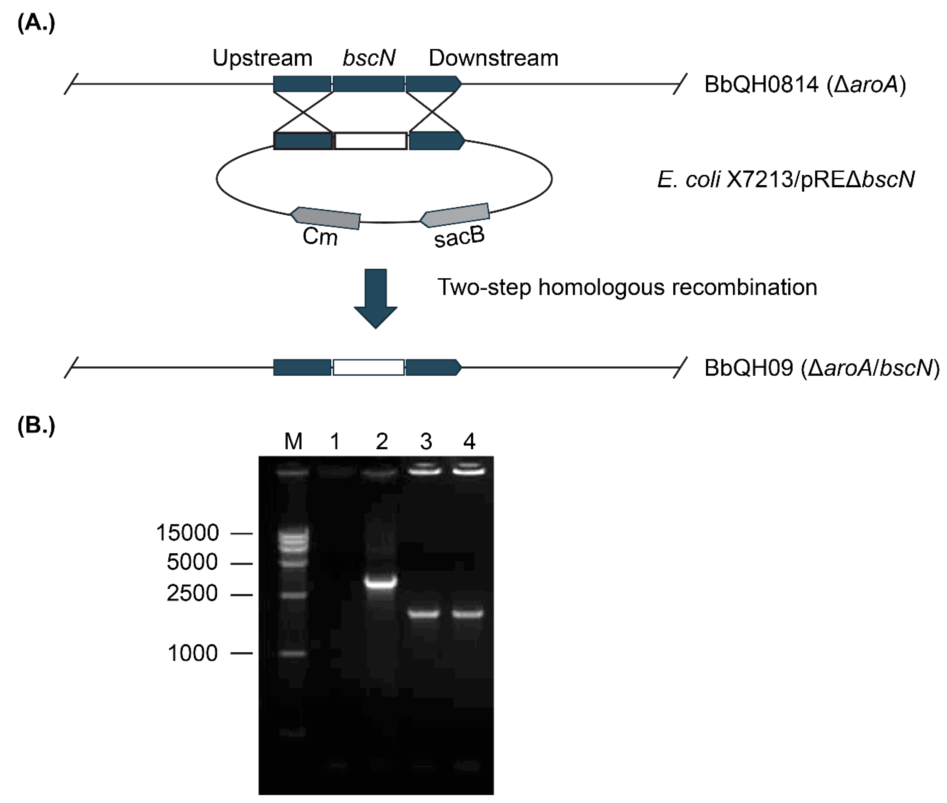
3.1. Construction of the Marker-Free aroA/bscN Double Deletion Strain QH09

To generate the marker-free aroA/bscN double deleted strain QH09, conjugation assay was performed between E. coli X7213 which contains the suicide plasmid pRE-ΔbscN and the aroA-deleted B. bronchiseptica QH0814. Ten single-crossover transconjugants capable of growing on CHL-supplemented agars were selected for determination of PCR assays with primers F1/F2, B3/B6, and Cm1/Cm2 after conjugation. Since the presence of the integrated plasmid, PCR assays using the genomic DNA of the putative B. bronchiseptica single-crossover transconjugants as a template yielded a 2661-bp product for the wild-type bscN, a 2120-bp product for the bscN mutant, and a 920-bp product for the Cm box. After the second crossover, PCR assays using the genomic DNA of the putative B. bronchiseptica mutants as a template only yielded 2120-bp product for the bscN mutant; the 2661-bp product (bscN gene) and the 920-bp product (Cm gene) were missing (Figure 1). DNA sequencing further confirmed that the deleted fragment was bscN, stability of the deletion evident from repeat PCR assays (Figure S1 in Supplementary Materials).

3.2. QH09 Showed Attenuated Virulence and Capacity of Colonization in Mice

To test the virulence attenuation of QH09, the LD50 value of the strain was calculated and compared with that of the parent strain QH0814 and the wild type strain HH0809. The results revealed that the LD50 of QH09 was 4.17 × 10⁸ CFU, which attenuates approximately 20-fold compared to that of QH0804 (1.90 × 10⁷ CFU), and approximately 225-fold to that of HH0809 (1.85 × 10⁶ CFU).

3.3. QH09 Was Safe to Piglets

Animal experiments showed that piglets challenged with either 3 × 10¹¹ CFU or 1 × 10¹¹ CFU of QH09 showed slight cough, deep breath, little depression, decreased appetite, and increased temperature (4041 °C), but all signs disappeared by 24–48 h (Table 2). No death was observed during the 30 days post infection. Dissection of the challenged piglets at 30 dpc did not reveal any observable lesions in lungs, tracheae, nasal cavities and other respiratory organs. However, the piglets challenged with the wild type strain HH0809 at 3 × 10¹¹ CFU exhibited severe clinical symptoms. The piglets became pyrexic (41–42 °C) within 24 dpc and the high body temperature lasted during the whole observation period. The other signs included labored breathing, cough, serious depression, appetite losing, and exacerbated signs including open mouth-breathing, red and swollen nasal mucosa, and disordered coats were also observed from 2 dpc. Mortality occurred from 3 dpc and all piglets died at 7 dpc. Before death, the piglets showed decreased temperature and visible symptoms of skin cyanosis at abdomen. Piglets challenged with HH0809 at 1 × 10¹¹ CFU displayed similar signs; two piglets died at 6 dpc and 7 dpc; the remaining one became thin and stiff. The gross lesions in the piglets died from an infection of HH0809 included hydropericardium and pleural effusion, edematous lungs with areas of hemorrhage and necrosis on their surfaces, edematous kidneys and spleens with hemorrhagic points on the surfaces, and swollen lymph nodes; there were bleeding spots in pericardium and epicardium, accompanied by fibrous pericarditis; the lobes of the lungs have sticky appearance towards intrathoracic wall; the tracheas were full of foamy mucus, the mucosa were edematous, congestive, and hemorrhagic. Histological check on lungs revealed that the alveolar walls of the pigs challenged with HH0809 broken; the alveolar epithelial cells were proliferous; there were many inflammatory cellular infiltrations in the lung tissue and there was serous exudation in alveolar cavities (Figure 2C). In comparison, damages on lung tissues of pigs challenged by QH09 are much slighter. The alveolar walls were intact; there was no exudation in alveolar cavities (Figure 2B). There were no visible lesions in the brains and other organs.

3.4. QH09 Induced High Levels of Antibodies and Conferred Protection Against Lethal Challenge

Results of ELISA assays showed that the titers of IgG, IgA, sIgA, IgG1, and IgG2a in piglets receiving live (QH09) or inactivated (HH0809) vaccines were significantly increased after immunization (Figure 3 and Table 3). The serum IgG titer induced by the attenuated vaccine QH09 was lower than that induced by the inactivated vaccine HH0809 (Figure 3A). However, the IgA titer in the BALF and serum induced by QH09 was significantly higher than that induced by HH0809 (Figure 3B). In particularly, a high level of sIgA was detected in the BALF from pigs immunized with QH09, but there was almost no sIgA titer detected in the BALF from the HH0809-immunized group (Figure 3C).
The protective ability of QH09 was tested and compared to that of the HH0809 inactivated vaccine by infection of HH0809 (2.0 × 10¹⁰ CFU) during the fifth week post-first immunization. After challenge, the piglets received PBS showed serious illness, including long last (≥7 days post infection), high temperature (40.5–41 °C), disordered coats, labor breathing, serious depression, and appetite losing. Two of the piglets died at 5 dpc and 7 dpc, the remaining two showed slow growth and finally became stiff. After dissection, the tracheae of the dead piglets were full of foamy mucus and the mucosal surface was edematous, congestive, and hemorrhagic; the lungs also showed same appearance with caseous exudate. Dissection on the survived piglets on the 30 dpc showed that nasal turbinates were withered and disappeared (Figure 4). The piglets immunized with the HH0809 inactivated vaccine also showed increased temperature (>40 °C), but the symptoms only lasted 5 dpc and then recovered. Mild clinical signs were observed as compared to the piglets immunized with PBS, one died piglet after infection exhibited edematous, congestive, and hemorrhagic lung, with some caseous exudate on it. Dissection of the survival piglets showed that the turbinates of two piglets also showed visible deformation (Figure 4). Piglets given a vaccination of QH09 showed higher temperature (approximately 40.5 °C) within the 5 days post challenge, but they recovered at day 6 post challenge. Only one piglet showed the difficulty in breathing, the other three showed slight coughs, but all of them recovered later. No death was observed and no appearance of visible lesions on the lungs and nasal cavities after dissection (Figure 4).

4. Discussion

Most pathogens enter the body through mucosal surfaces and, therefore, mucosal vaccines (mostly are live attenuated vaccines) that target the respiratory, gastrointestinal, or urogenital tracts are attractive as they stimulate local protection against infections [7]. This is really true for the vaccine against the infection caused by the respiratory pathogenic bacterium B. bronchiseptica which can efficiently colonize respiratory mucosa, as an attenuated live B. bronchiseptica vaccine often induces immunity and provides effective and long-lasting protection superior to those achieved by an inactivated vaccine and/or a sub-unit vaccine [3,6,11,12,13]. In addition, another advantage of attenuated live B. bronchiseptica vaccine is that could be administrated directly through nasal cavity, making vaccination more convenient. However, the greatest concern for an attenuated live vaccine is the safety of the animals after administration. In one of our previous studies, we developed an attenuated live B. bronchiseptica vaccine by knocking out its aroA gene, which encodes the 5-enolpyruvyl-shikimate-3-phosphate synthase that is crucial for the bacterial in vivo survival and growth [6]. While such strategy enables the attenuation without compromising the antigenicity, this B. bronchiseptica mutant might be not safe enough to keep all virulence factor-encoding genes inactive. Therefore, we knocked-out the bscN gene of the aroA-deletion vaccine we developed in our previous study [6], to increase the safety of the vaccine.
The bscN gene encodes an ATPase that provides energy for the secretion of effector proteins and BscN protein is required for the function of the T3SS apparatus [14]. It has been found that the in-frame deletion of bscN leads to decreased cytotoxicity of B. bronchiseptica to mammalian cells in vitro and the bscN deletion strain is defective for long-term tracheal colonization in rats [14]. These findings suggest that bscN involves in the virulence of B. bronchiseptica. In this study, the LD50 of the aroA/bscN double deletion B. bronchiseptica strain QH09 attenuates approximately 20-fold compared to that of QH0804 (4.17 × 10⁸ CFU vs. 1.90 × 10⁷ CFU). The clinical signs in piglets induced by the bscN deletion strain (Table 2) are less severe than those of parent strain, reported in our previous study [6]. All these findings suggest that the aroA/bscN double deletion strain is safer than the aroA deletion strain as a live vaccine candidate.
In addition to safety, the other parameters to determine an ideal vaccine candidate are the effectiveness and convenient administration [13]. While several types of anti-B. bronchiseptica vaccines such as the inactivated vaccines and/or the subunit vaccines including the recombinant OMPs or the novel adjuvant BfcA are commercially available or reported in laboratory phase [10,15,16], attenuated live vaccines are still proposed to be the preferable vaccines [17]. First, compared to the inactivated B. bronchiseptica vaccines and/or subunit vaccines which are often administered intramuscularly, the attenuated live B. bronchiseptica vaccines are administered through intranasal inoculation, which represents an easier way for administration [6,17,18,19]. Second but the most important reason, administration of the attenuated live B. bronchiseptica vaccine is capable of inducing mucosal immune response [6]. It has been documented that mucosal immune responses are most efficiently induced by administration of vaccines onto mucosal surfaces, whereas injected vaccines are generally poor inducers of mucosal immunity [4]. The production of sIgA represents a hallmark of the mucosal immune response [5,20]. In our study, immunization of the attenuated live B. bronchiseptica vaccine QH09 induced the production of IgG, and IgA antibodies in immunized pigs (Figure 4), suggesting that QH09 can induce immune responses effectively. In particular, administration of the attenuated live B. bronchiseptica vaccine QH09 induced a production of sIgA which was not detected in the BALF of the inactivated vaccine immunized-pigs (Figure 3), suggesting that vaccination with QH09 can also induce the mucosal immunity. It has been described that the sIgA antibodies play an important role in limiting the overgrowth of commensal microbes, and they also protect the host by binding to the surface of luminal microbes and toxins to prevent them from attaching to epithelial cells [5]. Therefore, the induction of the mucosal immunity and the production of the sIgA antibodies are crucial in order to prevent subsequent bacterial infections [11,17]. It is worthy of note that all of the results obtained from the present study are in agreement with those from the pigs immunized with the aroA deleted B. bronchiseptica QH0814 [6]. These findings suggest that vaccination of QH09 induces immune responses effectively and a deletion of the bscN gene does not alter its immune-stimulant efficacy. This could be also supported by the results from the protective test, as the vaccination of QH09 could protect the pigs against the lethal challenge of the wild type strain HH0809 and decrease the lesions on the turbinates (Figure 4).

5. Conclusions

To be concluded, an aroA/bscN double deleted B. bronchiseptica was generated in this study; this mutant showed lower capacity of colonization and virulence than the parent aroA deleted strain which encompasses all safety characteristics without compromising its efficacy. Vaccination of this aroA/bscN double deleted B. bronchiseptica activated the systemic and mucosal immunity in pigs and provided protection against lethal challenge, which represents an effective vaccine against B. bronchiseptica infections.

Supplementary Materials

The following are available online at https://www.mdpi.com/2076-393X/7/4/176/s1. Figure S1: PCR assays indicated the genetic stability of the aroA/bscN double deleted B. bronchiseptica QH09. The bacterium was passaged on blood-TSA for 50 passages and strains were selected every five passages to determine the deletion of the bscN gene PCR assays with primers B3/B6 listed in Table 1. M1: DL 15,000 DNA marker; M2: DL 2000 DNA marker; Lane 1: ddH2O as a negative control; Lane 2: the wild type strain HH0809 as a positive control; Lanes 3–12: colonies of OH09 selected from the 5th, 10th, 15th, 20th, 25th, 30th, 35th, 40th, 45th, and 50th passages, respectively.

Author Contributions

Conceptualization, Z.P. and B.W.; Methodology, W.A., Z.P., F.W., Y.Z., S.X., W.L., and L.H.; Writing—original draft preparation, W.A. and Z.P.; Writing—review and editing, Z.P., X.W., H.C., and B.W.; Supervision, Z.P., X.W., and B.W.; Project administration, Z.P. and B.W.; Funding acquisition, B.W. and H.C.

Funding

This research was funded by the Agricultural Science and Technology Innovation Program of Hubei Province, grant number 2018skjcx05, the Key Laboratory of Prevention and Control Agents for Animal Bacteriosis of the Ministry of Agriculture and Rural Affairs of China (grant number KLPCAAB-2019-03), and the earmarked fund for China Agriculture Research System, (grant number CARS-35). Zhong Peng was supported in part by China Postdoctoral Science Foundation (grant number: 2018M640719). The APC was funded by the Agricultural Science and Technology Innovation Program of Hubei Province (2018skjcx05).

Acknowledgments

We sincerely acknowledge Roy Curtiss III at Washington University School of Medicine, St. Louis, Missouri for the gift of the suicide plasmid pRE112 and E. coli X7213.

Conflicts of Interest

The authors declare no conflict of interest.

References

  1. Goodnow, R.A. Biology of Bordetella bronchiseptica. Microbiol. Rev. 1980, 44, 722–738. [Google Scholar] [PubMed]
  2. Cheong, Y.; Oh, C.; Lee, K.; Cho, K.H. Survey of porcine respiratory disease complex-associated pathogens among commercial pig farms in Korea via oral fluid method. J. Vet. Sci. 2017, 18, 283–289. [Google Scholar] [CrossRef] [PubMed]
  3. Matherne, C.M.; Steffen, E.K.; Wagner, J.E. Efficacy of commercial vaccines for protecting guinea pigs against Bordetella bronchiseptica pneumonia. Lab. Anim. Sci. 1987, 37, 191–194. [Google Scholar] [PubMed]
  4. Neutra, M.R.; Kozlowski, P.A. Mucosal vaccines: The promise and the challenge. Nat. Rev. Immunol. 2006, 6, 148–158. [Google Scholar] [CrossRef] [PubMed]
  5. Boyaka, P.N. Inducing Mucosal IgA: A Challenge for Vaccine Adjuvants and Delivery Systems. J. Immunol. 2017, 199, 9–16. [Google Scholar] [CrossRef] [PubMed]
  6. Zhang, Q.; Hu, R.; Hu, J.; He, H.; Tang, X.; Jin, M.; Chen, H.; Wu, B. aroA deleted Bordetella bronchiseptica inspiring robust mucosal immune response and provide full protection against intranasal challenge. Res. Vet. Sci. 2013, 94, 55–61. [Google Scholar] [CrossRef] [PubMed]
  7. Bernasconi, V.; Norling, K.; Bally, M.; Hook, F.; Lycke, N.Y. Mucosal Vaccine Development Based on Liposome Technology. J. Immunol. Res. 2016, 2016, 5482087. [Google Scholar] [CrossRef] [PubMed]
  8. Zhao, Z.; Xue, Y.; Wu, B.; Tang, X.; Hu, R.; Xu, Y.; Guo, A.; Chen, H. Subcutaneous vaccination with attenuated Salmonella enterica serovar Choleraesuis C500 expressing recombinant filamentous hemagglutinin and pertactin antigens protects mice against fatal infections with both S. enterica serovar Choleraesuis and Bordetella bronchiseptica. Infect. Immun. 2008, 76, 2157–2163. [Google Scholar] [CrossRef] [PubMed]
  9. Edwards, R.A.; Keller, L.H.; Schifferli, D.M. Improved allelic exchange vectors and their use to analyze 987P fimbria gene expression. Gene 1998, 207, 149–157. [Google Scholar] [CrossRef]
  10. Zhao, Z.; Xue, Y.; Tang, X.; Wu, B.; Cheng, X.; He, Q.; Zhang, C.; Guo, A.; Jin, M.; Chen, H. Immunogenicity of recombinant protective antigen and efficacy against intranasal challenge with Bordetella bronchiseptica. Vaccine 2009, 27, 2523–2528. [Google Scholar] [CrossRef] [PubMed]
  11. Wolfe, D.N.; Kirimanjeswara, G.S.; Goebel, E.M.; Harvill, E.T. Comparative role of immunoglobulin A in protective immunity against the Bordetellae. Infect. Immun. 2007, 75, 4416–4422. [Google Scholar] [CrossRef] [PubMed]
  12. Williams, J.; Laris, R.; Gray, A.W.; Jacobs, A.A. Studies of the efficacy of a novel intranasal vaccine against feline bordetellosis. Vet. Rec. 2002, 150, 439–442. [Google Scholar] [CrossRef] [PubMed]
  13. Mann, P.; Goebel, E.; Barbarich, J.; Pilione, M.; Kennett, M.; Harvill, E. Use of a genetically defined double mutant strain of Bordetella bronchiseptica lacking adenylate cyclase and type III secretion as a live vaccine. Infect. Immun. 2007, 75, 3665–3672. [Google Scholar] [CrossRef] [PubMed]
  14. Yuk, M.H.; Harvill, E.T.; Miller, J.F. The BvgAS virulence control system regulates type III secretion in Bordetella bronchiseptica. Mol. Microbiol. 1998, 28, 945–959. [Google Scholar] [CrossRef] [PubMed]
  15. Zhang, H.; Zhang, H.; Xiong, B.; Fan, G.; Cao, Z. Immunogenicity of recombinant outer membrane porin protein and protective efficacy against lethal challenge with Bordetella bronchiseptica in rabbits. J. Appl. Microbiol. 2019. [Google Scholar] [CrossRef] [PubMed]
  16. Zhao, Z.; Wang, C.; Xue, Y.; Ding, K.; Zhang, C.; Cheng, X.; Li, Y.; Liu, Y.; Wu, T. Immunogenicity and protective efficacy of pertactin recombinants against Bordetella bronchiseptica challenge. Wei Sheng Wu Xue Bao 2010, 50, 1239–1245. [Google Scholar] [PubMed]
  17. Yevsa, T.; Ebensen, T.; Fuchs, B.; Zygmunt, B.; Libanova, R.; Gross, R.; Schulze, K.; Guzman, C.A. Development and characterization of attenuated metabolic mutants of Bordetella bronchiseptica for applications in vaccinology. Environ. Microbiol. 2013, 15, 64–76. [Google Scholar] [CrossRef] [PubMed]
  18. McArthur, J.D.; West, N.P.; Cole, J.N.; Jungnitz, H.; Guzman, C.A.; Chin, J.; Lehrbach, P.R.; Djordjevic, S.P.; Walker, M.J. An aromatic amino acid auxotrophic mutant of Bordetella bronchiseptica is attenuated and immunogenic in a mouse model of infection. FEMS Microbiol. Lett. 2003, 221, 7–16. [Google Scholar] [CrossRef]
  19. Wang, C.; Liu, L.; Zhang, Z.; Yan, Z.; Yu, C.; Shao, M.; Jiang, X.; Chi, S.; Wei, K.; Zhu, R. Immunological and protective effects of Bordetella bronchiseptica subunit vaccines based on the recombinant N-terminal domain of dermonecrotic toxin. Int. Immunopharmacol. 2015, 28, 952–959. [Google Scholar] [CrossRef] [PubMed]
  20. Corthesy, B. Multi-faceted functions of secretory IgA at mucosal surfaces. Front. Immunol. 2013, 4, 185. [Google Scholar] [CrossRef] [PubMed]
Figure 1. Strategy for the construction of the marker-free aroA/bscN double deleted B. bronchiseptica strain QH09. (A) Schematic diagram showing the strategy for the construction of the aroA/bscN-deletion strain. (B) PCR determination of the bscN-deletion in the aroA-deletion strain QH0814. M: DL 15,000 DNA marker; Lane 1: ddH2O as the negative control; Lane 2: QH0814 as the positive control (2661-bp); Lanes 3–4: bscN-deletion (2120-bp).
Vaccines 07 00176 g001
Figure 2. Histological analysis on pig lungs challenged with QH09, HH0809, and PBS in the pig safety tests (bar 100 μm). Panel A is a lung histological section from a pig challenged with PBS; Panel B is a lung histological section from a pig challenged with QH09 at 3 × 10¹¹ CFU; Panel C is a lung histological section from a pig challenged with HH0809 at 1 × 10¹¹ CFU.
Vaccines 07 00176 g002
Figure 3. ELISA titers of IgG in serum (A), IgA in serum (B), sIgA in BALF (C), in piglets induced by the attenuated live vaccine (QH09), the inactivated vaccine (HH0809), and PBS. “*” p < 0.5.
Vaccines 07 00176 g003
Figure 4. Lesions on the nasal cavities of pigs immunized with different vaccines after infection. (A) lesions on the nasal cavities of pigs immunized with the attenuated live vaccine (QH09); (B) lesions on the nasal cavities of pigs immunized with the inactivated vaccine (HH0809); (C) lesions on the nasal cavities of pigs immunized with PBS; (D) the nasal cavities of the healthy pigs.
Vaccines 07 00176 g004
Table 1. Bacterial strains, plasmids, and primers used in this study.
Bacterial StrainsCharacteristicsSource
B. bronchiseptica HH0809Wild type isolate from a pig with atrophic rhinitis[7,8]
B. bronchiseptica QH0814Unmarked aroA deletion of HH0809[6]
B. bronchiseptica QH09Unmarked bscN deletion of QH0814This study
E. coli X7213 Thi-1 thr-1 leuB6 fhuA21 lacY1 glnV44 △asdA4 recA1 RP4 2-Tc::Mu[λpir] Kmr[9]
Plasmids
pBluescript II KS/SK (+)oriColE1 oriF1(+) bla lacZaOur collection
pRE112 (CmR)oriT oriV sacB1, CmR, counterselectable suicide plasmid[9]
pSKΔbscNbscN upstream and downstream fragment amplified from QH0814 ligated into pBluescript II KS/SK (+)This study
pREΔbscNbscN upstream and downstream fragment from pSKΔbscN ligated into pRE112This study
Primers
F15′- CCCCCGCACATTTCCGAACTTC -3′Part of flagellin (237 bp)
F25′- AGGCTCCCAAGAGAGAAAGGCTT -3′
B15′- CGGGAATTCATGCGTCAGTACCACTACATCAC -3′ (EcoR I)Part of bscN (1334 bp)
B25′- GCTAAGCTTTAGGATTCGGGTCCGATGATTTCAG -3′ (Hind III)
B35′- TAA GGTACC GCGCTTGCGCTGGTGCTGTC -3′ (Kpn I)bscN upstream flank (1148 bp)
B45′- TCA GAATTC ACGCGCACCCCCAGCTCC -3′ (EcoR I)
B55′- TGTGAATTC GACGGCCACATCGTGCTCT -3′ (EcoR I)bscN downstream flank (980 bp)
B65′- GGAGAGCTCCCTTCGCTTTCTCCTGTTCCAT -3′ (Sac I)
Cm15′- TAAATACCTGTGACGGAAGAT -3′Cm resistant gene (700 bp)
Cm25′- TATCACTTATTCAGGCGTAGC -3′
Table 2. Scores indicating clinic signs in pig safety test.
GroupPiglets ID24 hpc48 hpcFirst Week
Appetite aMood aCough aAppetite aSpirit aCough a
QH09 (3 × 10¹¹ CFU)1110000
2110000
3110000
QH09 (1 × 10¹¹ CFU)1110000
2110000
3110000
HH0809 (3 × 10¹¹ CFU)1333333
2333333
3333333
HH0809 (1 × 10¹¹ CFU)1333333
2333322
3333322
PBS1000000
2000000
3000000
MeanQH09 (3 × 10¹¹ CFU)1.0 ± 0.001.0 ± 0.000000
QH09 (1 × 10¹¹ CFU)1.0 ± 0.001.0 ± 0.000000
HH0809 (3 × 10¹¹ CFU)3.0 ± 0.003.0 ± 0.003.0 ± 0.003.0 ± 0.003.0 ± 0.003.0 ± 0.00
HH0809 (1 × 10¹¹ CFU)3.0 ± 0.003.0 ± 0.003.0 ± 0.003.0 ± 0.002.3 ± 0.472.3 ± 0.47
PBS000000
a Clinical signs were scored as: normal (grade 0); slight, yet obvious (grade 1); moderate with significant symptoms (grade 2); and severe symptoms (grade 3).
Table 3. IgG1 and IgG2a tiers induced by different vaccines at different days post vaccination.
Antibodies (Log10)Attenuated Live Vaccine (QH09)Inactivated Vaccine (HH0809)
14 Days (OD630)28 Days (OD630)14 Days (OD630)28 Days (OD630)
IgG11.83 ± 0.172.20 ± 0.213.83 ± 0. 0.304.47 ± 0.17
IgG2a2.60 ± 0.372.97 ± 0.173.13 ± 0.113.54 ± 0.15
IgG1/IgG2a0.700.741.221.26
Baseline titers are zero.

