Article

Prevalence and Clinical Implications of Ascites in Gastric Cancer Patients after Curative Surgery

1
Department of Surgery, Hanyang University College of Medicine, 222 Wangsimri-ro, Seongdong-gu, Seoul 04763, Korea
2
Department of Radiology, Hanyang University College of Medicine, 222 Wangsimri-ro, Seongdong-gu, Seoul 04763, Korea
*
Author to whom correspondence should be addressed.
J. Clin. Med. 2021, 10(16), 3557; https://doi.org/10.3390/jcm10163557
Submission received: 30 June 2021 / Revised: 5 August 2021 / Accepted: 11 August 2021 / Published: 13 August 2021
(This article belongs to the Special Issue Gastrointestinal Malignancies: Screening, Diagnosis, and Treatment)

Abstract

:
We aimed to determine the frequency and clinical significance of ascites that developed during the follow-up period in patients who underwent curative resection for gastric cancer. The study included 577 patients with gastric cancer who underwent curative gastrectomy. Among them, 184 showed ascites in postoperative follow-up images. Benign ascites was observed in 131 of 490 patients without recurrence, 48 patients (of 87) with recurrence had malignancy-related ascites, and the remaining 5 patients had ascites only prior to recurrence. In most patients without recurrence (97.7%) and in 50% of patients with malignancy-related ascites, the ascites was small in volume and located in the pelvic cavity at the time that it was first identified. However, with the exception of nine patients, malignancy-related pelvic ascites occurred simultaneously or after obvious recurrence. Of those nine patients who had minimal pelvic ascites before obvious recurrence, only one had a clear association with a malignancy-related ascites. In the multivariate analysis, an age of ≤45 was the only independent risk factor for the occurrence of benign ascites. A small volume of pelvic ascites fluid is common in young gastric cancer patients who do not have recurrence after gastrectomy, regardless of sex. It is rare for ascites to be the first manifestation of recurrence.

1. Introduction

Gastric cancer is one of the leading causes of cancer-related deaths worldwide [1] and most gastric cancer-related deaths are due to recurrence [2]. The peritoneal region is the most common site of gastric cancer recurrence and is associated with a poor prognosis [3,4,5]. Diagnosis of peritoneal metastasis is typically determined by a computed tomography [CT] scan. Ascites is one of the most common findings suggestive of peritoneal carcinomatosis; others include peritoneal thickening, nodularity, and contrast enhancement in CT [6,7,8,9]. Although there is little convincing evidence that intense surveillance improves survival, routine follow-up after curative resection for the early detection of recurrence in gastric cancer is considered general practice, as some research findings indicate that asymptomatic patients had longer post-recurrence and overall survival than symptomatic patients [10,11,12]. Physicians often encounter ascites in abdominal imaging during the post-gastrectomy follow-up period and there are concerns that this finding may indicate early peritoneal recurrence, especially in men, despite a lack of evidence otherwise. Preoperatively detected ascites in CT strongly suggests the presence of peritoneal metastasis and free cancer cells in patients with advanced gastric cancer [13,14]. However, the clinical significance of ascites detected by postoperative CT or other abdominal imaging during the follow-up period is not well-studied. In this study, we evaluated the frequency and clinical implications of ascites in patients who underwent curative surgery for gastric cancer.

2. Materials and Methods

A total of 634 patients with gastric cancer who underwent curative gastrectomy at the Hanyang University Seoul Hospital between January 2008 and December 2015 were selected from a prospective gastric cancer database. Fifty-seven patients were excluded for the following reasons: (1) mortality after surgery (n = 3); (2) synchronous or metachronous cancers (n = 14); (3) recurrent ascites due to liver cirrhosis or chronic kidney disease (n = 3); (4) ascites due to ileus (n = 1); and (5) follow-up loss or a short follow-up time of <12 months due to unknown causes (in patients without recurrence) after surgery (n = 35). A postoperative follow-up was conducted every 3–6 months for up to 5 years and annually thereafter. Standard clinical practice included evaluation by physical examination, laboratory tests including the measurement of tumor markers, radiologic imaging, and endoscopy. Imaging was conducted alternatively by abdominopelvic and chest CT and abdominal sonography. Medical records of the remaining 577 patients were retrospectively reviewed. The median period of follow-up was 61.0 months (range of 4.0–146.0 months).
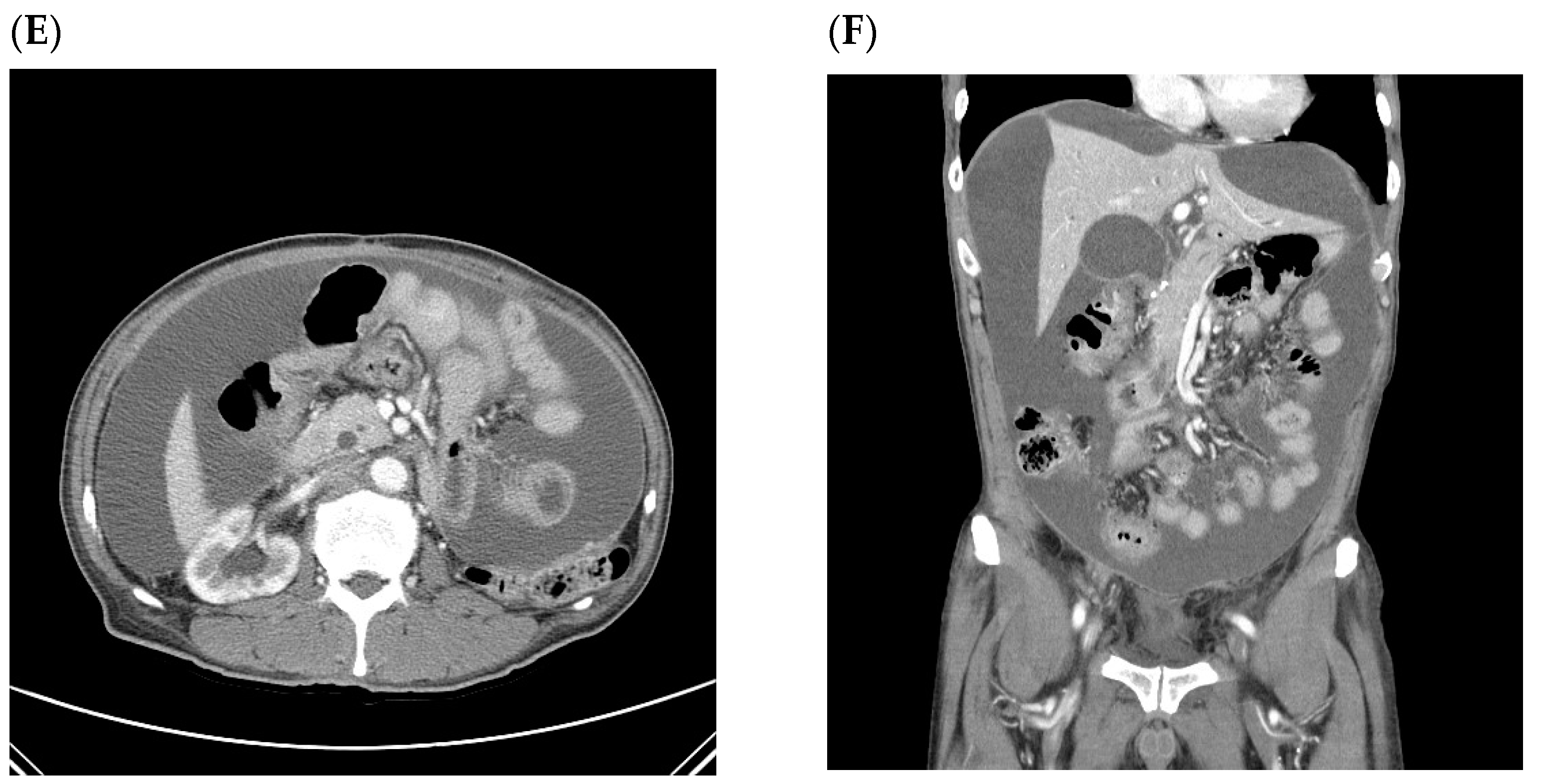
Ascites was primarily detected in CT imaging, having been initially identified by abdominal sonography in only one patient. Images were reviewed by at least two experienced radiologists and ascites was considered present when a low radiologic density of ≤10 Hounsfield units was found within the abdominal cavity outside the intra-abdominal or pelvic organs. Intraperitoneal fluid collection that occurred within 3 months after surgery was excluded to distinguish from postoperative changes in benign ascites. The volume of ascites fluid was estimated using ruler grids applied to CT images using the method described by Chang et al. [15]. A small degree of ascites was defined as a volume of <50 mL, moderate as 50–500 mL, and large as >500 mL (Figure 1).
Cases of benign ascites were those in which patients developed ascites without recurrence during the follow-up period. None of the patients had an interval of <12 months from identification of ascites to the follow-up conclusion, excluding eight patients whose follow-up period exceeded to 5 years without recurrence. If only ascites was present without symptoms or other findings suggesting intra-abdominal recurrence, the ascites was considered benign at that time and routine radiologic follow-up was performed according to the gastric cancer follow-up protocol of our hospital. Short-term radiologic tests including positron emission tomography and/or abdominopelvic CT were performed within 3 months in 40 patients with benign ascites for the following reasons: (1) an advanced-stage disease with newly detected ascites (n = 13); (2) a remaining or increased infiltration around the surgical site (n = 11); (3) combined intra-abdominal lymphadenopathy, ultimately confirmed as reactive lymph node enlargement by repeat tests (n = 7); (4) combined abnormal laboratory findings (n = 2) or levels of tumor markers (n = 1); (5) complaints of abdominal symptoms (n = 1); (6) the presence of portal vein thrombosis; (7) moderate volume of ascites fluid (n = 1); (8) an increased volume of ascites fluid compared to the findings immediately after surgery (n = 2); and (9) a liver cyst of increasing size (n = 1). The phrase “malignancy-related ascites” is used as a more appropriate descriptor than “malignant ascites” considering malignant cells were confirmed by ascites cytology in only some patients with recurrence. Intra-abdominal recurrence or peritoneal metastasis was diagnosed by serial changes in the CT and/or positron emission tomography performed when recurrence was suspected based on the CT. A histological examination of biopsy specimens or ascites cytology for patients with recurrence was performed whenever possible. Death due to disease progression was confirmed in all patients classified as having recurrence.
Statistical analyses were performed using SPSS version 22.0 (SPSS Inc., Chicago, IL, USA). Chi-squared tests and independent Student’s t-tests were used for comparisons between groups. A binary logistic regression model was used for multivariate analysis. The threshold for statistical significance was set at p ≤ 0.05.

3. Results

Of the 577 eligible patients, ascites was identified in 184 patients during the follow-up period. Among them, 131 patients had benign ascites, accounting for 26.7% of the 490 patients without recurrence. Of the 131 patients with benign ascites, 78 were male (78/328; 23.8% of all males without recurrence) and 53 were female (53/162; 32.7% of all females without recurrence). Ascites was observed in 53 (60.7%) of the 87 patients with recurrence. Patients fell into three groups: (1) 40 patients with malignancy-related ascites at the same time as the findings of peritoneal seeding or intra-abdominal recurrence, including one patient who on initial recurrence had ascites only; (2) eight patients with ascites presumed to be benign before obvious recurrence and considered to have malignancy-related ascites after the obvious recurrence; and (3) five patients with ascites before recurrence but no evidence of malignancy-related ascites upon recurrence. Groups (1) and (2) were classified as patients with malignancy-related ascites. Among 48 patients with malignancy-related ascites, ascites appeared prior to recurrence in eight patients (16.7%, Group (2)), simultaneous with the recurrence detection in 25 (48.1%) and following recurrence in 15 (31.3%) (Figure 2).
Comparisons between patients with and without recurrence are shown in Table 1. The T and N stages, type of resection, mean albumin and hemoglobin level, and history of adjuvant chemotherapy were significantly different between patients with and without recurrence. The most common location of ascites at first appearance was the pelvic cavity both in patients with (29/53, 54.7%) and without (128/131, 97.7%) recurrence, followed by the whole abdominal region (16/53, 30.2%), perihepatic area (6/53, 11.3%), and paracolic gutter (2/53, 3.8%) in patients with recurrence. In the remaining patients without recurrence, ascites was located in the perihepatic area (2/131, 1.5%) and the left abdominal cavity (1/131, 0.8%). Ascites fluid was small in volume and located in the pelvic cavity in most patients without recurrence (128/131, 97.7%). In the majority of patients with malignancy-related ascites, the volume of ascites fluid at first detection was small (33/48, 68.8%). Among 131 patients with benign ascites, repeatability was observed in 79 (60.3%). With the exception of one case, there was no difference in the amount and location of ascites after the initial detection in patients with recurrent benign ascites. The one exceptional case had a moderate amount of benign ascites in the perisplenic area (Figure 1C) and a small volume of pelvic ascites fluid on later examination. Median time for the first appearance of ascites was 10.5 months post-surgery (range of 3.0–108.0) in all patients with ascites, 9.0 months (range of 3.0–108.0) in those with benign ascites, and 11.5 months (range of 3.0–71.0) in those with recurrence. There was a significant difference in the mean age of males and females at the detection of benign ascites (58.8 ± 11.5 vs. 51.6 ± 12.4, p = 0.001). Repeatability (45 (57.0%) vs. 34 (65.4%), p = 0.366) and the history of adjuvant chemotherapy (32 (41.8%) vs. 18 (34.6%), p = 0.466) were not significantly different between males and females with benign ascites.
According to the presence or absence of ascites, sensitivity, specificity, accuracy, and positive and negative predictive values for intra-abdominal recurrence were 60.9%, 73.3%, 71.4%, 28.8%, and 91.3%, respectively. Positive and negative likelihood ratios were 2.28 and 0.53, respectively. Values for intra-abdominal recurrence were then calculated according to location of ascites (pelvic vs. other). Sensitivity, specificity, and positive and negative predictive values for the pelvic location were 54.7%, 2.3%, 18.5%, and 1.1%, respectively, and for other locations, 45.3%, 97.7%, 88.9%, and 81.5%, respectively. The positive and negative likelihood ratios for the pelvic location were 0.56 and 19.7, respectively, and 19.7 and 0.56 for other locations, respectively.
Risk factors for the occurrence of benign ascites were evaluated in recurrence-free patients (Table 2). Univariate analyses showed that younger age (≤45), a pN2–3 stage, and a history of adjuvant chemotherapy were associated with the occurrence of benign ascites. Patient sex (p = 0.065) and pT stage (p = 0.054), significant at the 0.1 level, were included with these factors in a multivariate analysis to evaluate the risk for benign ascites. Younger age (≤45) was the only independent risk factor associated with the occurrence of benign ascites post-surgery.
Table 3 shows the characteristics of the nine patients with undefined ascites at the first discovery. This group includes eight patients with ascites presumed to be benign before definitive recurrence and one patient with eventual recurrence who developed ascites without other evidence of peritoneal and/or intra-abdominal recurrence. Short-term follow-up abdominopelvic CT and ascites cytology examinations were conducted one month later due to increased ascites, and peritoneal recurrence was eventually confirmed in the latter patient mentioned previously (Figure 3). All of these patients were in an advanced stage of pathology and the ascites fluid was small in volume and located in the pelvic cavity at first appearance.

4. Discussion

In gastric cancer patients, the most common cause of ascites after surgery is thought to be intra-abdominal recurrence but this is not supported by our study. In this study, a small volume of pelvic ascites fluid that had no identified pathological cause was noted in a substantial number of patients, notably an incidental finding during regular follow-ups. Malignancy-related ascites was observed in only 28.8% of patients with a history of ascites.
Normal peritoneal fluid that keeps the peritoneum moist and smooth may accumulate in the deep region of the pelvis in both males and females [16]. It is more frequently observed in premenopausal females than in males or postmenopausal females. In premenopausal females, the fluid that accumulates in the pelvis is thought to originate from ovarian exudation and decreased absorption of peritoneal fluid due to adhesions caused by various factors including endometriosis [17,18]. In a board sense, this fluid is called “physiologic ascites”. The precise incidence of physiologic ascites has rarely been studied. According to Yoshikawa et al., a small amount of physiologic pelvic ascites was observed in 3.8% of healthy males and 16.8% of healthy postmenopausal females in pelvic magnetic resonance imaging conducted during health screenings [17]. In our study, benign ascites was identified in 23.8% of all male patients and 32.7% of all female patients without recurrence. There was no statistically significant difference in ascites detection between the sexes on either univariate or multivariate analyses. Peritoneal fluid accumulation seems to occur more often after gastric cancer surgery regardless of sex. A possible explanation for this is that the absorption capacity of the peritoneum decreases when it is infected or injured [19]. It is assumed that ascites accumulation increases after surgery due to peritoneal injury and adhesion. This reaction may be stronger in younger patients because an age of ≤45 was the only independent factor significantly linked to the occurrence of benign ascites in our study. In addition, young female patients are more likely to have ascites due to gynecological causes but those were not investigated in this study because of insufficient medical records. Therefore, the level of concern regarding recurrence is lower if the patient’s ascites has a gynecological cause.
Cheon et al. reported that a small amount of pelvic fluid was detected in follow-up CT after curative surgery for gastric cancer in 3.9% of male patients [20]. This incidence is quite low when compared with our study. The authors obtained data only from radiology reports without a review of all CT images, whereas in our study, all images were independently reviewed by two radiologists. In addition, their definition of a “small” volume of ascites fluid was less than 20 mL, while in our study, it was defined as less than 50 mL. These reasons may explain the discrepancy between the results of the two studies. Further prospective observation is required for clarification.
According to reports from South Korea as well as western countries, malignant tumors are the second most common pathologic cause of ascites, following portal hypertension due to cirrhosis [21,22]. Malignancy-related ascites typically develops in the setting of recurrent and/or advanced cancer. The primary pathophysiological mechanism of malignancy-related ascites is peritoneal carcinomatosis, which blocks the drainage of lymphatic channels and increases vascular permeability [23]. Along with peritoneal carcinomatosis, some tumors may metastasize in the liver, which can cause ascites because of the obstruction/compression of the portal veins, further leading to portal hypertension or liver failure. Other types of tumors such as lymphomas can cause lymph node obstruction with accumulation of chylous ascites [24]. In gastric cancer, multiple pathophysiological mechanisms of ascites formation can occur but the primary mechanism is peritoneal carcinomatosis. There are few reports considering ascites as the first manifestation of gastric cancer despite its being the first detected sign of intra-abdominal malignancy in 50% of patients with peritoneal carcinomatosis [23,25]. To date, there are no reports considering ascites as the first manifestation of recurrence in postgastrectomy gastric cancer patients. In our study, most cases of malignancy-related ascites were accompanied by other findings of recurrence or were discovered during disease progression. Malignancy-related ascites is indicated as a late manifestation of intra-abdominal metastasis after gastrectomy for gastric cancer, as in the first diagnosis of gastric cancer [26]. Therefore, a small amount of pelvic ascites cannot be excluded from consideration as an early indicator of peritoneal carcinomatosis. This judgment requires caution. In our study, ascites was the only initially detected sign of intra-abdominal recurrence in one patient (Table 3; Figure 3). Furthermore, among patients with ascites presumed to be benign before obvious recurrence, the possibility of malignancy-related ascites due to peritoneal recurrence cannot be excluded because of the relatively short time between the first occurrence of ascites and the recurrence in some cases (Pt. 1, 5, 6, and 8 in Table 3). All patients mentioned above were in far-advanced stages of the disease (Table 3). The possibility of recurrence should be suspected in all gastric cancer patients with advanced disease who develop ascites even when the characteristics of the ascites are similar to those of benign ascites and there are no definitive findings suggesting peritoneal metastasis.
CT is frequently used for the postoperative surveillance of patients with gastric carcinoma. CT allows for the detection of even small amounts of ascites and provides information that is difficult to obtain in ultrasonography [16]. It would be desirable if intra-abdominal recurrence could be predicted with CT-detected ascites; however, this does not seem to be possible. The likelihood ratios were not at appropriate levels in our study. According to previous reports, malignancy-related ascites is often loculated or septated, or may be absent in typical or dependent areas such as the pelvis [8,27]. Similarly, in our study, the specificity and positive and negative predictive values for intra-abdominal recurrence were improved when calculated for ascites at locations other than the pelvis. In terms of the volume of the ascites fluid, that of >50 mL in a preoperative CT was found to be related to peritoneal metastasis in gastric cancer patients in a recently published study [13]. Additionally, in our study, malignancy-related ascites fluid volumes were larger (>50 mL are considered moderate and large amounts) than benign ascites at the time of initial detection. However, except for some patients with undefined ascites, all patients with malignancy-related ascites exhibited definitive peritoneal metastasis or intra-abdominal recurrence in the CT when ascites was first detected. Ascites alone therefore seems to be inappropriate as a diagnostic marker for intra-abdominal or peritoneal recurrence in postgastrectomy gastric cancer patients.

5. Conclusions

The presence of small-volume pelvic ascites fluid in follow-up images has minimal clinical significance in the majority of patients who undergo gastrectomy for gastric cancer. This phenomenon is more common in younger patients, regardless of sex. Although the presence of malignant ascites alone in the pelvic cavity can precede obvious intra-abdominal recurrence involving peritoneal seeding, ascites is more likely to be an indicator of disease progression in patients with recurrent gastric cancer.

Author Contributions

Conceptualization, J.-H.L. and S.-J.K.; methodology, M.K., J.-H.L. and S.-J.K.; software, J.-H.L.; validation, J.-H.L., S.-J.K., M.K. and B.-K.K.; formal analysis, J.-H.L.; investigation, J.-H.L., M.K. and B.-K.K.; resources, S.-J.K. and B.-K.K.; data curation, J.-H.L.; writing—original draft preparation, J.-H.L.; writing—review and editing, J.-H.L., S.-J.K., M.K. and B.-K.K.; visualization, J.-H.L.; supervision, S.-J.K.; project administration, J.-H.L.; funding acquisition, J.-H.L. All authors have read and agreed to the published version of the manuscript.

Funding

This work was supported by the research fund of Hanyang University (HY-202100000000767).

Institutional Review Board Statement

The study was conducted according to the guidelines of the Declaration of Helsinki and was approved by the Institutional Review Board (or Ethics Committee) of the Hanyang University hospital (protocol code 2021-03-050-004 and date of approval 23 April 2021).

Informed Consent Statement

Patient consent was waived due to the minimal risk of a retrospective study.

Data Availability Statement

The data presented in this study are available on request from the corresponding author.

Conflicts of Interest

The authors declare no conflict of interest.

References

  1. Jemal, A.; Bray, F.; Center, M.M.; Ferlay, J.; Ward, E.; Forman, D. Global cancer statistics. CA Cancer J. Clin. 2011, 61, 69–90. [Google Scholar] [CrossRef] [Green Version]
  2. Kim, C.D.; Chang, M.C.; Roh, H.R.; Chae, G.B.; Yang, D.H.; Choi, W.J. Factors influencing recurrence after curative resection for advanced gastric cancer. J. Korean Surg. Soc. 2003, 65, 301–308. [Google Scholar]
  3. Yoo, C.H.; Noh, S.H.; Shin, D.W.; Choi, S.H.; Min, J.S. Recurrence following curative resection for gastric carcinoma. Br. J. Surg. 2000, 87, 236–242. [Google Scholar] [CrossRef] [PubMed]
  4. Chu, D.Z.; Lang, N.P.; Thompson, C.; Osteen, P.K.; Westbrook, K.C. Peritoneal carcinomatosis in nongynecologic malignancy. A prospective study of prognostic factors. Cancer 1989, 63, 364–367. [Google Scholar]
  5. Sadeghi, B.; Arvieux, C.; Glehen, O.; Beaujard, A.C.; Rivoire, M.; Baulieux, J.; Fontaumard, E.; Brachet, A.; Caillot, J.L.; Faure, J.L.; et al. Peritoneal carcinomatosis from non-gynecologic malignancies: Results of the EVOCAPE 1 multicentric prospective study. Cancer 2000, 88, 358–363. [Google Scholar] [CrossRef]
  6. Raptopoulos, V.; Gourtsoyiannis, N. Peritoneal carcinomatosis. Eur. Radiol. 2001, 11, 2195–2206. [Google Scholar] [CrossRef]
  7. Kim, S.J.; Kim, H.H.; Kim, Y.H.; Hwang, S.H.; Lee, H.S.; Park, D.J.; Kim, S.Y.; Lee, K.H. Peritoneal metastasis: Detection with 16-or 64-detector row CT in patients undergoing surgery for gastric cancer. Radiology 2009, 253, 407–415. [Google Scholar] [CrossRef] [PubMed]
  8. Friedman, A.C.; Sohotra, P.; Radecki, P.D. CT manifestations of peritoneal carcinomatosis. Am. J. Roentgenol. 1988, 150, 1035–1041. [Google Scholar]
  9. Lee, H.J.; Kim, M.J.; Lim, J.S.; Kim, K.W. Follow up CT findings of various types of recurrence after curative gastric surgery. J. Korean Radiol. Soc. 2007, 57, 553–562. [Google Scholar] [CrossRef]
  10. Bennett, J.J.; Gonen, M.; D’Angelica, M.; Jaques, D.P.; Brennan, M.F.; Coit, D.G. Is detection of asymptomatic recurrence after curative resection associated with improved survival in patients with gastric cancer? J. Am. Coll Surg. 2005, 201, 503–510. [Google Scholar] [CrossRef]
  11. Kim, J.H.; Jang, Y.J.; Park, S.S.; Park, S.H.; Mok, Y.J. Benefit of post-operative surveillance for recurrence after curative resection for gastric cancer. J. Gastrointest. Surg. 2010, 14, 969–976. [Google Scholar] [CrossRef] [PubMed]
  12. Lee, J.H.; Lim, J.K.; Kim, M.G.; Kwon, S.J. The influence of post-operative surveillance on the prognosis after curative surgery for gastric cancer. Hepatogastroenterology 2014, 61, 2123–2132. [Google Scholar] [PubMed]
  13. Kim, S.H.; Choi, Y.H.; Kim, J.W.; Oh, S.; Lee, S.; Kim, B.G.; Lee, K.L. Clinical significance of computed tomography-detected ascites in gastric cancer patients with peritoneal metastases. Medicine 2018, 97, e9343. [Google Scholar] [CrossRef] [PubMed]
  14. Yajima, K.; Kanda, T.; Ohashi, M.; Wakai, T.; Nakagawa, S.; Sasamoto, R.; Hatakeyama, K. Clinical and diagnostic significance of preoperative computed tomography findings of ascites in patients with advanced gastric cancer. Am. J. Surg. 2006, 192, 185–190. [Google Scholar] [CrossRef]
  15. Chang, D.K.; Kim, J.W.; Kim, B.K.; Lee, K.L.; Song, C.S.; Han, J.K.; Song, I.S. Clinical significance of CT-defined minimal ascites in patients with gastric cancer. World J. Gastroenterol. 2005, 11, 6587–6592. [Google Scholar] [CrossRef]
  16. Healy, J.C.; Reznek, R.H. The peritoneum, mesenteries and omenta: Normal anatomy and pathological processes. Eur. Radiol. 1998, 8, 886–900. [Google Scholar] [CrossRef] [PubMed]
  17. Yoshikawa, T.; Hayashi, N.; Maeda, E.; Matsuda, I.; Sasaki, H.; Ohtsu, H.; Ohtomo, K. Peritoneal fluid accumulation in healthy men and postmenopausal women: Evaluation on pelvic MRI. Am. J. Roentgenol. 2013, 200, 1181–1185. [Google Scholar] [CrossRef]
  18. Koninckx, P.R.; Renaer, M.; Brosens, I.A. Origin of peritoneal fluid in women: An ovarian exudation product. Br. J. Obstet. Gynaecol. 1980, 87, 177–183. [Google Scholar] [CrossRef]
  19. Verger, C.; Luger, A.; Moore, H.L.; Nolph, K.D. Acute changes in peritoneal morphology and transport properties with infectious peritonitis and mechanical injury. Kidney Int. 1983, 23, 823–831. [Google Scholar] [CrossRef] [Green Version]
  20. Cheon, H.J.; Ryeom, H.K.; Bae, J.H.; Jang, Y.J.; Kim, G.C.; Kim, J.H.; Shin, K.M.; Chung, H.Y. Clinical significance of a small amount of isolated pelvic free fluid at multidetector CT in male patients after curative surgery for gastric carcinoma. J. Korean Soc. Radiol. 2014, 71, 69–74. [Google Scholar] [CrossRef] [Green Version]
  21. Hwangbo, Y.; Jung, J.H.; Shim, J.; Kim, B.H.; Jung, S.H.; Lee, C.K.; Jang, J.Y.; Dong, S.H.; Kim, H.J.; Chang, Y.W.; et al. Etiologic and laboratory analyses of ascites in patients who underwent diagnostic paracentesis. Korean J. Hepatol. 2007, 13, 185–195. [Google Scholar] [PubMed]
  22. Runyon, B.A. Management of adult patients with ascites caused by cirrhosis. Hepatology 1998, 27, 264. [Google Scholar] [CrossRef]
  23. Sangisetty, S.L.; Miner, T.J. Malignant ascites: A review of prognostic factors, pathophysiology and therapeutic measures. World J. Gastrointest. Surg. 2012, 4, 87–95. [Google Scholar] [CrossRef]
  24. Press, O.W.; Press, N.O.; Kaufman, S.D. Evaluation and management of chylous ascites. Ann. Intern. Med. 1982, 96, 358. [Google Scholar] [CrossRef] [PubMed]
  25. Duarte, I.; Outerelo, C. Gastric cancer presenting as isolated ascites: A diagnostic challenge. Eur. J. Case Rep. Intern. Med. 2019, 6, 001141. [Google Scholar] [PubMed]
  26. Maeda, H.; Kobayashi, M.; Sakamoto, J. Evaluation and treatment of malignant ascites secondary to gastric cancer. World J. Gastroenterol. 2015, 21, 10936–10947. [Google Scholar] [CrossRef]
  27. Kim, K.W.; Choi, B.I.; Han, J.K.; Kim, T.K.; Kim, A.Y.; Lee, H.J.; Kim, Y.H.; Choi, J.; Do, K.; Kim, H.C.; et al. Postoperative anatomic and pathologic findings at CT following gastrectomy. Radiographics 2002, 22, 323–336. [Google Scholar] [CrossRef] [PubMed]
Figure 1. Typical ascites imaging in CT: (A) benign small pelvic ascites; (B) malignant small pelvic ascites with peritoneal thickening; (C) benign moderate ascites in perisplenic area (left abdomen); (D) malignant moderate ascites; and (E,F) alignant large ascites. Yellow stars indicate ascites.
Jcm 10 03557 g001aJcm 10 03557 g001b
Figure 2. Study population.: +, presence; −, absence.
Jcm 10 03557 g002
Figure 3. CT images of a patient on initial recurrence of malignant ascites without other CT findings related to peritoneal seeding, later confirmed by cytology. A small volume of pelvic ascites fluid was observed at first appearance (A). Follow-up CT (B) showed an increased volume of ascites fluid. Yellow stars indicate ascites.
Jcm 10 03557 g003
Table 1. Clinicopathologic features.
Ascites without RecurrenceAscites with Recurrencep
n (%)n (%)
Sex 0.869
 Male78 (60.3)31 (58.5)
 Female53 (39.7)22 (41.5)
Age (years ± SD)55.9 ± 12.360.0 ± 12.10.046
Type of resection <0.001
 Partial gastrectomy105 (80.2)26 (49.1)
 Total gastrectomy26 (19.8)27 (50.9)
Surgical approach <0.001
 Open89 (67.9)52 (98.1)
 Laparoscopy42 (32.1)1 (1.9)
pT stage <0.001
 T180 (61.1)4 (7.5)
 T212 (9.2)4 (7.5)
 T318 (13.7)24 (45.3)
 T421 (16.0)21 (39.6)
pN stage <0.001
 N080 (61.1)8 (15.1)
 N123 (17.6)5 (9.4)
 N219 (14.5)7 (13.2)
 N39 (6.9)33 (62.3)
Nutritional parameters
 Albumin (g/dL ± SD)4.2 ± 0.44.0 ± 0.6<0.001
 Hemoglobin (g/dL ± SD)12.3 ± 1.411.4 ± 1.4<0.001
Adjuvant chemotherapy <0.001
 No80 (61.1)8 (15.1)
 Yes51 (38.9)45 (84.9)
Location of ascites at first appearance <0.001
 Pelvic cavity only128 (97.7)29 (54.7)
 Whole abdomen0 (0)16 (30.2)
 Other3 (2.3)8 (15.1)
Volume of ascites at first appearance <0.001
 Small130 (99.2)38 (71.7)
 Moderate1 (0.8)10 (18.9)
 Large0 (0)5 (9.4)
Repeatability
 No52 (39.7)
 Yes79 (60.3)
Timing of first appearance (months after surgery)9.0 (range of 3.0–108.0)11.5 (range of 3.0–71.0)
“Ascites with recurrence” includes patients with malignancy-related ascites (n = 40), patients in whom ascites before recurrence were reclassified as malignancy-related ascites at a later stage (n = 8), and patients with benign ascites before recurrence but with no evidence of malignancy-related ascites after recurrence (n = 5). Abbreviations: SD, standard deviation. In the case of ascites without recurrence.
Table 2. Risk factors related to the occurrence of benign ascites in disease-free patients.
Univariate Analysis Multivariate Analysis
Ascites (−), n (%)Ascites (+), n (%)p-ValueHazard Ratio95% CIp-Value
Sex 0.065
 Male249 (75.9)79 (24.1) 1
 Female110 (67.9)52 (32.1) 1.4360.926–2.2270.106
Age (years) <0.001
 ≤459 (32.1)19 (67.9) 6.4652.803–14.915<0.001
 >45350 (75.8)112 (24.2) 1
Type of surgery 0.211
 Total gastrectomy306 (74.5)105 (25.5)
 Partial gastrectomy53 (67.1)26 (32.9)
 Surgical approach 0.581
 Open252 (74.0)89 (26.0)
 Laparoscopy106 (71.6)42 (28.4)
Depth of invasion 0.054
 pT1–2283 (75.5)92 (24.5) 1
 pT3–476 (66.1)39 (33.9) 0.8890.435–1.8190.748
Lymph node metastasis
 pN0–1310 (75.1)103 (24.9)0.0491
 pN2–349 (63.6)28 (36.4) 1.0880.553–2.1400.806
Adjuvant chemotherapy 0.003
 No268 (77)80 (23.0) 1
 Yes91 (64.1)51 (35.9) 2.0980.985–4.4700.055
+, presence; −, absence. Abbreviation: CI, confidence interval.
Table 3. Characteristics of patients with temporarily undefined ascites.
Age/SexType of OperationTN StageCharacteristic of AscitesTiming of Malignant AscitesSite of First RecurrenceInterval between Surgery and Recurrence (Month)Interval between First Appearance of Benign Ascites and Recurrence (Month)
Patients with ascites presumed to be benign before confirmation of recurrence (n = 8)
Pt 165/MPGT3N3asmall pelvic cavitySimultaneous with recurrenceT colon (increased ascites in the pelvic cavity)44.48.1
Pt 253/FTGT4bN3bsmall pelvic cavitySimultaneous with recurrencePeritoneum (increased ascites, peritoneal thickening, bowel obstruction, and Krukenberg tumors)18.213.2
Pt 340/FTGT4aN3asmall pelvic cavitySimultaneous with recurrencePeritoneum (nodularity and increased ascites)73.270.3
Pt 459/FTGT4aN3asmall pelvic cavitySimultaneous with recurrencePeritoneum (increased ascites and peritoneal thickening)25.221.7
Pt 539/FTGT3N3bsmall pelvic cavitySimultaneous with recurrencePeritoneum (Krukenberg tumors, nodularity, and increased ascites)12.19
Pt 657/FTGT3N3asmall pelvic cavitySimultaneous with recurrencePeritoneum (bowel obstruction and increased ascites)9.84.4
Pt 761/FTGT3N1small pelvic cavitySimultaneous with recurrencePeritoneum (increased ascites, T colon, and mesentery LNs)29.626
Pt 868/FPGT4aN2small pelvic cavitySimultaneous with recurrencePeritoneum (increased ascites and peritoneal thickening)11.75.3
A patient who first recurred with malignant ascites without other CT findings related to peritoneal seeding (later confirmed by cytology) (n = 1)
Pt 949/FTGT3N3asmall pelvic cavity
Abbreviations: PG, partial gastrectomy; TG, total gastrectomy; and CT, computed tomography.
Publisher’s Note: MDPI stays neutral with regard to jurisdictional claims in published maps and institutional affiliations.