Different Background: Natural Killer Cell Profiles in Secondary versus Primary Recurrent Pregnancy Loss

 , and
, and
Abstract
:1. Introduction
2. Materials and Methods
2.1. Study Population
2.2. Analysis of Peripheral Lymphocytes and NK-Cell Subsets
2.3. Statistics
3. Results
3.1. Study Population
3.2. Peripheral Lymphocyte Subpopulation in Controls
3.3. Peripheral Lymphocytes and Subsets in Patients and Controls
4. Discussion
Author Contributions
Funding
Institutional Review Board Statement
Informed Consent Statement
Data Availability Statement
Acknowledgments
Conflicts of Interest
Appendix A


| Antibodies and Dyes | Conjugate | Clone | Company | Catalogue Number |
|---|---|---|---|---|
| CD3 | BV510 | HIT3a | BD | 564713 |
| CD3 | FITC | HIT3a | BD | 555339 |
| CD4 | BV786 | SK3 (Leu3a) | BD | 563877 |
| CD4 | PE-CF594 | RPA-T4 | BD | 562281 |
| CD8 | FITC | HIT8a | BD | 555634 |
| CD14 | BV711 | MᴪP9 | BD | 563372 |
| CD14 | FITC | M5E2 | BD | 555397 |
| CD16 | BV711 | 3G8 | BD | 563127 |
| CD16 | PE | 3G8 | BD | 555407 |
| CD19 | FITC | HIB19 | BD | 555412 |
| CD19 | PE-Cy7 | HIB19 | ThermoFisher | 25-0199-42 |
| CD25 | BV605 | BC96 | BioLegend | 302632 |
| CD45 | APC-H7 | 2D1 | BD | 560178 |
| CD56 | APC | AF12-7H3 | Miltenyi | 130-113-305 |
| CD57 | PE-CF594 | NK-1 | BD | 562488 |
| CD62L | BV650 | DREG-56 | BD | 563808 |
| CD64 | BV421 | 10.1 | BD | 562872 |
| CD127 | PE | HIL-7R-M21 | BD | 557938 |
| CD314 (NKG2D) | PE | BAT221 | Miltenyi | 130-092-672 |
| CD335 (NKp46) | BV421 | 9E2/NKp46 (9-E2) | BD | 564065 |
| FoxP3 | APC | PCH101 | eBioscience | 17-4776-42 |
| FVD | ef506 | eBioscience | 65-0866-14 | |
| 7AAD | BD | 559925 |
References
- Carrington, B.; Sacks, G.; Regan, L. Recurrent miscarriage: Pathophysiology and outcome. Curr. Opin. Obstet. Gynecol. 2005, 17, 591–597. [Google Scholar] [CrossRef] [PubMed]
- The ESHRE Guideline Group on RPL; Atik, R.B.; Christiansen, O.B.; Elson, J.; Kolte, A.M.; Lewis, S.; Middeldorp, S.; Nelen, W.; Peramo, B.; Quenby, S.; et al. Eshre guideline: Recurrent pregnancy loss. Hum. Reprod. Open 2018, 2018, hoy004. [Google Scholar]
- Christiansen, O.B.; Steffensen, R.; Nielsen, H.S.; Varming, K. Multifactorial etiology of recurrent miscarriage and its scientific and clinical implications. Gynecol. Obstet. Investig. 2008, 66, 257–267. [Google Scholar] [CrossRef] [PubMed]
- Laird, S.M.; Tuckerman, E.M.; Cork, B.A.; Linjawi, S.; Blakemore, A.I.; Li, T.C. A review of immune cells and molecules in women with recurrent miscarriage. Hum. Reprod. Update 2003, 9, 163–174. [Google Scholar] [CrossRef] [PubMed] [Green Version]
- Seshadri, S.; Sunkara, S.K. Natural killer cells in female infertility and recurrent miscarriage: A systematic review and meta-analysis. Hum. Reprod. Update 2014, 20, 429–438. [Google Scholar] [CrossRef] [PubMed]
- Hanna, J.; Goldman-Wohl, D.; Hamani, Y.; Avraham, I.; Greenfield, C.; Natanson-Yaron, S.; Prus, D.; Cohen-Daniel, L.; Arnon, T.I.; Manaster, I.; et al. Decidual nk cells regulate key developmental processes at the human fetal-maternal interface. Nat. Med. 2006, 12, 1065–1074. [Google Scholar] [CrossRef] [PubMed]
- Tang, A.W.; Alfirevic, Z.; Quenby, S. Natural killer cells and pregnancy outcomes in women with recurrent miscarriage and infertility: A systematic review. Hum. Reprod. 2011, 26, 1971–1980. [Google Scholar] [CrossRef] [PubMed] [Green Version]
- Tuckerman, E.; Mariee, N.; Prakash, A.; Li, T.C.; Laird, S. Uterine natural killer cells in peri-implantation endometrium from women with repeated implantation failure after ivf. J. Reprod. Immunol. 2010, 87, 60–66. [Google Scholar] [CrossRef] [PubMed]
- Teles, A.; Zenclussen, A.C.; Schumacher, A. Regulatory t cells are baby’s best friends. Am. J. Reprod. Immunol. 2013, 69, 331–339. [Google Scholar] [CrossRef]
- Craenmehr, M.H.; Heidt, S.; Eikmans, M.; Claas, F.H. What is wrong with the regulatory t cells and foetomaternal tolerance in women with recurrent miscarriages? HLA 2016, 87, 69–78. [Google Scholar] [CrossRef]
- Leber, A.; Teles, A.; Zenclussen, A.C. Regulatory t cells and their role in pregnancy. Am. J. Reprod. Immunol. 2010, 63, 445–459. [Google Scholar] [CrossRef] [PubMed]
- Jin, L.P.; Chen, Q.Y.; Zhang, T.; Guo, P.F.; Li, D.J. The cd4+cd25 bright regulatory t cells and ctla-4 expression in peripheral and decidual lymphocytes are down-regulated in human miscarriage. Clin. Immunol. 2009, 133, 402–410. [Google Scholar] [CrossRef] [PubMed]
- Robertson, M.J.; Ritz, J. Biology and clinical relevance of human natural killer cells. Blood 1990, 76, 2421–2438. [Google Scholar] [CrossRef] [PubMed] [Green Version]
- Kwak-Kim, J.; Gilman-Sachs, A. Clinical implication of natural killer cells and reproduction. Am. J. Reprod. Immunol. 2008, 59, 388–400. [Google Scholar] [CrossRef] [PubMed]
- Laird, S.M.; Mariee, N.; Wei, L.; Li, T.C. Measurements of cd56+ cells in peripheral blood and endometrium by flow cytometry and immunohistochemical staining in situ. Hum. Reprod. 2011, 26, 1331–1337. [Google Scholar] [CrossRef] [PubMed] [Green Version]
- Bulmer, J.N.; Lash, G.E. Uterine natural killer cells: Time for a re-appraisal? F1000Res 2019, 8, F1000 Faculty Rev-1999. [Google Scholar] [CrossRef]
- Juelke, K.; Killig, M.; Luetke-Eversloh, M.; Parente, E.; Gruen, J.; Morandi, B.; Ferlazzo, G.; Thiel, A.; Schmitt-Knosalla, I.; Romagnani, C. Cd62l expression identifies a unique subset of polyfunctional cd56dim nk cells. Blood 2010, 116, 1299–1307. [Google Scholar] [CrossRef] [PubMed] [Green Version]
- Draghi, M.; Pashine, A.; Sanjanwala, B.; Gendzekhadze, K.; Cantoni, C.; Cosman, D.; Moretta, A.; Valiante, N.M.; Parham, P. Nkp46 and nkg2d recognition of infected dendritic cells is necessary for nk cell activation in the human response to influenza infection. J. Immunol. 2007, 178, 2688–2698. [Google Scholar] [CrossRef] [Green Version]
- Bjorkstrom, N.K.; Riese, P.; Heuts, F.; Andersson, S.; Fauriat, C.; Ivarsson, M.A.; Bjorklund, A.T.; Flodstrom-Tullberg, M.; Michaelsson, J.; Rottenberg, M.E.; et al. Expression patterns of nkg2a, kir, and cd57 define a process of cd56dim nk-cell differentiation uncoupled from nk-cell education. Blood 2010, 116, 3853–3864. [Google Scholar] [CrossRef] [Green Version]
- Kuon, R.J.; Vomstein, K.; Weber, M.; Muller, F.; Seitz, C.; Wallwiener, S.; Strowitzki, T.; Schleussner, E.; Markert, U.R.; Daniel, V.; et al. The “killer cell story” in recurrent miscarriage: Association between activated peripheral lymphocytes and uterine natural killer cells. J. Reprod. Immunol. 2017, 119, 9–14. [Google Scholar] [CrossRef]
- Toth, B.; Vomstein, K.; Togawa, R.; Bottcher, B.; Hudalla, H.; Strowitzki, T.; Daniel, V.; Kuon, R.J. The impact of previous live births on peripheral and uterine natural killer cells in patients with recurrent miscarriage. Reprod. Biol. Endocrinol. 2019, 17, 72. [Google Scholar] [CrossRef] [PubMed] [Green Version]
- Toth, B.; Wurfel, W.; Bohlmann, M.; Zschocke, J.; Rudnik-Schoneborn, S.; Nawroth, F.; Schleussner, E.; Rogenhofer, N.; Wischmann, T.; von Wolff, M.; et al. Recurrent miscarriage: Diagnostic and therapeutic procedures. Guideline of the dggg, oeggg and sggg (s2k-level, awmf registry number 015/050). Geburtshilfe Frauenheilkd 2018, 78, 364–381. [Google Scholar] [CrossRef] [PubMed] [Green Version]
- Practice Committee of the American Society for Reproductive Medicine. Evaluation and treatment of recurrent pregnancy loss: A committee opinion. Fertil. Steril. 2012, 98, 1103–1111. [Google Scholar] [CrossRef] [PubMed]
- RCOG. Green-Top Guideline No. 17 the Investigation and Treatment of Couples with Recurrent First-Trimester and Second-Trimester Miscarriage; RCOG: London, UK, 2011. [Google Scholar]
- Nielsen, H.S.; Steffensen, R.; Varming, K.; Van Halteren, A.G.; Spierings, E.; Ryder, L.P.; Goulmy, E.; Christiansen, O.B. Association of hy-restricting hla class ii alleles with pregnancy outcome in patients with recurrent miscarriage subsequent to a firstborn boy. Hum. Mol. Genet. 2009, 18, 1684–1691. [Google Scholar] [CrossRef] [PubMed]
- Shakhar, K.; Ben-Eliyahu, S.; Loewenthal, R.; Rosenne, E.; Carp, H. Differences in number and activity of peripheral natural killer cells in primary versus secondary recurrent miscarriage. Fertil. Steril. 2003, 80, 368–375. [Google Scholar] [CrossRef]
- Michel, T.; Poli, A.; Cuapio, A.; Briquemont, B.; Iserentant, G.; Ollert, M.; Zimmer, J. Human cd56bright nk cells: An update. J. Immunol. 2016, 196, 2923–2931. [Google Scholar] [CrossRef] [Green Version]
- Cichocki, F.; Schlums, H.; Theorell, J.; Tesi, B.; Miller, J.S.; Ljunggren, H.-G.; Bryceson, Y.T. Diversification and functional specialization of human nk cell subsets. In Natural Killer Cells; Vivier, E., Di Santo, J., Moretta, A., Eds.; Springer International Publishing: Cham, Switzerland, 2016; pp. 63–93. [Google Scholar]
- Biassoni, R.; Cantoni, C.; Marras, D.; Giron-Michel, J.; Falco, M.; Moretta, L.; Dimasi, N. Human natural killer cell receptors: Insights into their molecular function and structure. J. Cell Mol. Med. 2003, 7, 376–387. [Google Scholar] [CrossRef] [Green Version]
- Moretta, A.; Bottino, C.; Vitale, M.; Pende, D.; Cantoni, C.; Mingari, M.C.; Biassoni, R.; Moretta, L. Activating receptors and coreceptors involved in human natural killer cell-mediated cytolysis. Annu. Rev. Immunol. 2001, 19, 197–223. [Google Scholar] [CrossRef]
- Zhang, Y.; Zhao, A.; Wang, X.; Shi, G.; Jin, H.; Lin, Q. Expressions of natural cytotoxicity receptors and nkg2d on decidual natural killer cells in patients having spontaneous abortions. Fertil. Steril. 2008, 90, 1931–1937. [Google Scholar] [CrossRef]
- Fukui, A.; Ntrivalas, E.; Gilman-Sachs, A.; Kwak-Kim, J.; Lee, S.K.; Levine, R.; Beaman, K. Expression of natural cytotoxicity receptors and a2v-atpase on peripheral blood nk cell subsets in women with recurrent spontaneous abortions and implantation failures. Am. J. Reprod. Immunol. 2006, 56, 312–320. [Google Scholar] [CrossRef]
- Ariga, H.; Ohto, H.; Busch, M.P.; Imamura, S.; Watson, R.; Reed, W.; Lee, T.H. Kinetics of fetal cellular and cell-free DNA in the maternal circulation during and after pregnancy: Implications for noninvasive prenatal diagnosis. Transfusion 2001, 41, 1524–1530. [Google Scholar] [CrossRef] [PubMed]
- Bianchi, D.W.; Zickwolf, G.K.; Weil, G.J.; Sylvester, S.; DeMaria, M.A. Male fetal progenitor cells persist in maternal blood for as long as 27 years postpartum. Proc. Natl. Acad. Sci. USA 1996, 93, 705–708. [Google Scholar] [CrossRef] [PubMed] [Green Version]
- Kuon, R.J.; Schaumann, J.; Goeggl, T.; Strowitzki, T.; Sadeghi, M.; Opelz, G.; Daniel, V.; Toth, B. Patients with idiopathic recurrent miscarriage show higher levels of dr+ activated t-cells that are less responsive to mitogens. J. Reprod. Immunol. 2015, 112, 82–87. [Google Scholar] [CrossRef] [PubMed]
- Weintraub, A.Y.; Sheiner, E.; Bashiri, A.; Shoham-Vardi, I.; Mazor, M. Is there a higher prevalence of pregnancy complications in a live-birth preceding the appearance of recurrent abortions? Arch. Gynecol. Obstet. 2005, 271, 350–354. [Google Scholar] [CrossRef] [PubMed]
- Nielsen, H.S.; Mortensen, L.; Nygaard, U.; Schnor, O.; Christiansen, O.B.; Andersen, A.M. Brothers and reduction of the birth weight of later-born siblings. Am. J. Epidemiol. 2008, 167, 480–484. [Google Scholar] [CrossRef] [PubMed] [Green Version]
- Nielsen, H.S.; Mogensen, M.; Steffensen, R.; Kruse, C.; Christiansen, O.B. Indications of anti-hy immunity in recurrent placental abruption. J. Reprod. Immunol. 2007, 75, 63–69. [Google Scholar] [CrossRef] [PubMed]
- Mikkelsen, A.P.; Egerup, P.; Ebert, J.F.M.; Kolte, A.M.; Nielsen, H.S.; Lidegaard, Ø. Pregnancy loss and cancer risk: A nationwide observational study. EClinicalMedicine 2019, 15, 80–88. [Google Scholar] [CrossRef] [PubMed] [Green Version]
- Braem, M.G.; Onland-Moret, N.C.; Schouten, L.J.; Kruitwagen, R.F.; Lukanova, A.; Allen, N.E.; Wark, P.A.; Tjønneland, A.; Hansen, L.; Braüner, C.M.; et al. Multiple miscarriages are associated with the risk of ovarian cancer: Results from the european prospective investigation into cancer and nutrition. PLoS ONE 2012, 7, e37141. [Google Scholar] [CrossRef] [PubMed] [Green Version]
- Orange, J.S. How i manage natural killer cell deficiency. J. Clin. Immunol. 2020, 40, 13–23. [Google Scholar] [CrossRef]
- Egerup, P.; Lindschou, J.; Gluud, C.; Christiansen, O.B.; ImmuReM IPD Study Group. The effects of intravenous immunoglobulins in women with recurrent miscarriages: A systematic review of randomised trials with meta-analyses and trial sequential analyses including individual patient data. PLoS ONE 2015, 10, e0141588. [Google Scholar] [CrossRef] [Green Version]
- Carp, H.J.; Toder, V.; Torchinsky, A.; Portuguese, S.; Lipitz, S.; Gazit, E.; Mashiach, S. Allogenic leukocyte immunization after five or more miscarriages. Recurrent miscarriage immunotherapy trialists group. Hum. Reprod. 1997, 12, 250–255. [Google Scholar] [CrossRef] [PubMed] [Green Version]
- Choi, J.; Lee, S.J.; Lee, Y.A.; Maeng, H.G.; Lee, J.K.; Kang, Y.W. Reference values for peripheral blood lymphocyte subsets in a healthy korean population. Immune Netw. 2014, 14, 289–295. [Google Scholar] [CrossRef] [PubMed] [Green Version]
- Ilavska, S.; Horvathova, M.; Szabova, M.; Nemessanyi, T.; Jahnova, E.; Tulinska, J.; Liskova, A.; Wsolova, L.; Staruchova, M.; Volkovova, K. Association between the human immune response and body mass index. Hum. Immunol. 2012, 73, 480–485. [Google Scholar] [CrossRef] [PubMed]
- Valiathan, R.; Deeb, K.; Diamante, M.; Ashman, M.; Sachdeva, N.; Asthana, D. Reference ranges of lymphocyte subsets in healthy adults and adolescents with special mention of t cell maturation subsets in adults of south florida. Immunobiology 2014, 219, 487–496. [Google Scholar] [CrossRef] [PubMed]
- Nielsen, H.S. Secondary recurrent miscarriage and h-y immunity. Hum. Reprod. Update 2011, 17, 558–574. [Google Scholar] [CrossRef] [PubMed] [Green Version]
| Characteristics | pRPL (n = 46) | sRPL (n = 24) | pCtrl (n = 60) | sCtrl (n = 25) | p |
|---|---|---|---|---|---|
| Age a | 35.5 ± 5.4 | 35.2 ± 4.4 | 24.8 ± 3.1 | 33.4 ± 6.4 | <0.001 |
| BMI a | 25.7 ± 4.9 | 25.3 ± 5.1 | 22.0 ± 3.5 | 23.0 ± 3.7 | <0.001 |
| Gravidity b | 3(0/8) | 4(3/8) | 0 | 1(1/3) | <0.001 |
| Parity b | 0(0/1) | 1(1/3) | 0 | 1(1/3) | <0.001 |
| No. of miscarriages b | 3(2/8) | 3(2/6) | 0 | 0 | <0.001 |
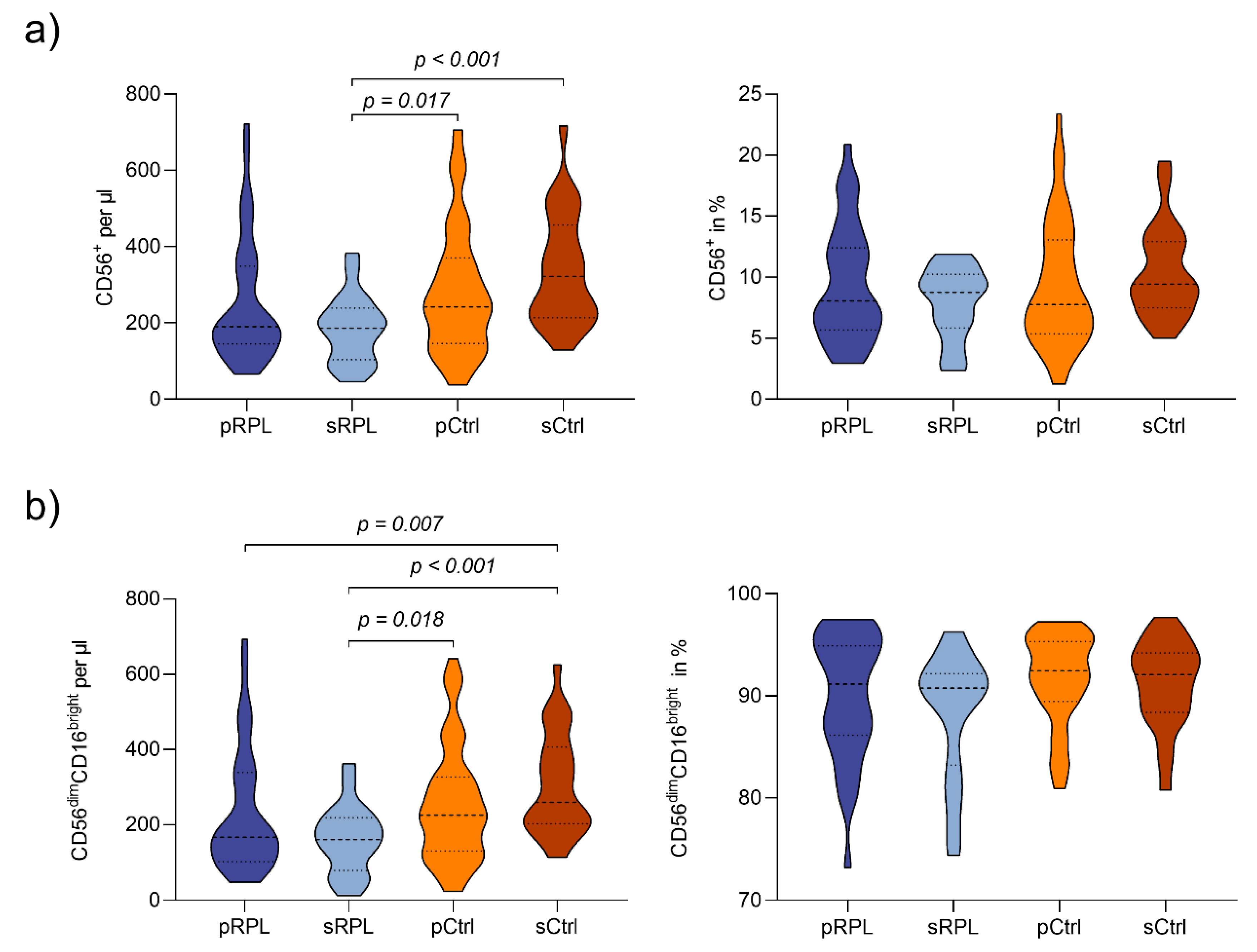
| Peripheral Lymphocytes Subpopulations | Unit | pRPL (n = 46) | sRPL (n = 25) | pCtrl (n = 60) | sCtrl (n = 25) | p | |
|---|---|---|---|---|---|---|---|
| CD45+ | /µL | 2734 ± 721 | 2358 ± 699 | 3067 ± 738 | 2897 ± 751 | 0.002 3 | |
| CD45 | CD56+ | /µL % | 253.3 ± 161.0 9.5 ± 4.7 | 184.7 ± 88.8 7.9 ± 2.9 | 282.5 ± 165.8 9.4 ± 5.0 | 339.8 ± 147.0 10.4 ± 3.8 | 0.003 4,5 0.327 |
| CD56brightCD16dim | /µL % | 16.7 ± 6.9 7.8 ± 4.7 | 15.8 ± 6.5 9.2 ± 4.5 | 15.9 ± 6.3 6.4 ± 3.3 | 21.4 ± 9.6 6.2 ± 2.4 | 0.114 0.063 | |
| CD56dimCD16bright | /µL % | 232.2 ± 161.4 91.9 ± 4.2 | 162.6 ± 90.3 88.2 ± 6.1 | 254.6 ± 152.6 91.9 ± 4.2 | 308.8 ± 134.8 90.7 ± 4.6 | 0.003 3,4,5 0.122 | |
| CD56+CD3+NKT | /µL % | 61.9 ± 48.3 2.3 ± 1.8 | 65.2 ± 55.4 2.1 ± 1.4 | 73.0 ± 51.6 2.6 ± 2.0 | 84.6 ± 67.4 2.9 ± 2.2 | 0.496 0.772 | |
| CD4+CD25+FoxP3+ | % | 2.0 ± 1.0 | 2.0 ± 1.0 | 1.5 ± 0.7 | 1.7 ± 0.5 | 0.035 2 | |
| CD56brightCD16dim | CD57+ | % | 2.5 ± 4.5 | 1.6 ± 2.1 | 2.3 ± 5.2 | 1.3 ± 3.2 | 0.434 |
| CD62L+ | % | 91.9 ± 7.8 | 94.2 ± 5.1 | 94.8 ± 5.4 | 94.4 ± 5.4 | 0.111 | |
| NKG2D+ | % | 92.3 ± 7.6 | 92.5 ± 6.9 | 91.2 ± 8.7 | 96.0 ± 4.8 | 0.234 | |
| NKp46+ | % | 96.3 ± 3.9 | 95.6 ± 4.1 | 97.2 ± 1.6 | 98.2 ± 0.9 | 0.030 1,4,5 | |
| CD56dimCD16bright | CD57+ | % | 34.3 ± 16.3 | 41.5 ± 15.5 | 32 ± 14.9 | 32.3 ± 15.2 | 0.077 |
| CD62L+ | % | 27.3 ± 11.2 | 28.8 ± 12.1 | 29.6 ± 11.3 | 31.8 ± 12.3 | 0.483 | |
| NKG2D+ | % | 95.6 ± 3.7 | 95.3 ± 4.4 | 96.9 ± 2.2 | 97.2 ± 3.0 | 0.029 4,5 | |
| NKp46+ | % | 63.7 ± 20.0 | 56.3 ± 23.4 | 77.1 ± 11.7 | 81.5 ± 7.5 | <0.001 2,3,4,5 | |
Publisher’s Note: MDPI stays neutral with regard to jurisdictional claims in published maps and institutional affiliations. |
© 2021 by the authors. Licensee MDPI, Basel, Switzerland. This article is an open access article distributed under the terms and conditions of the Creative Commons Attribution (CC BY) license (http://creativecommons.org/licenses/by/4.0/).
Share and Cite
Strobel, L.; Vomstein, K.; Kyvelidou, C.; Hofer-Tollinger, S.; Feil, K.; Kuon, R.-J.; Ebner, S.; Troppmair, J.; Toth, B. Different Background: Natural Killer Cell Profiles in Secondary versus Primary Recurrent Pregnancy Loss. J. Clin. Med. 2021, 10, 194. https://doi.org/10.3390/jcm10020194
Strobel L, Vomstein K, Kyvelidou C, Hofer-Tollinger S, Feil K, Kuon R-J, Ebner S, Troppmair J, Toth B. Different Background: Natural Killer Cell Profiles in Secondary versus Primary Recurrent Pregnancy Loss. Journal of Clinical Medicine. 2021; 10(2):194. https://doi.org/10.3390/jcm10020194
Chicago/Turabian StyleStrobel, Laura, Kilian Vomstein, Christiana Kyvelidou, Susanne Hofer-Tollinger, Katharina Feil, Ruben-Jeremias Kuon, Susanne Ebner, Jakob Troppmair, and Bettina Toth. 2021. "Different Background: Natural Killer Cell Profiles in Secondary versus Primary Recurrent Pregnancy Loss" Journal of Clinical Medicine 10, no. 2: 194. https://doi.org/10.3390/jcm10020194