Article

Negligible Correlation between Radiographic Measurements and Clinical Outcomes in Patients Following Primary Reverse Total Shoulder Arthroplasty

1
Department of Orthopaedic Surgery, University of Connecticut, Farmington, CT 06030, USA
2
Department of Orthopaedic Sports Medicine, Technical University of Munich, 81675 Munich, Germany
3
Department of Orthopaedic Surgery, Juntendo University, Tokio 113-8421, Japan
4
UNC Orthopaedics, University of North Carolina at Chapel Hill, Chapel Hill, NC 27599, USA
5
Southern Oregon Orthopedics, Medford, OR 97504, USA
6
The Cleveland Shoulder Institute, Beachwood, OH 44194, USA
7
Department of Orthopaedic Sports Medicine, University of Arizona College of Medicine, Tucson, AZ 85006, USA
8
Dupage Medical Group, Elmhurst, IL 60126, USA
9
Arthroscopy and Orthopedic Sportsmedicine, ATOS Orthoparc Clinic, 50858 Cologne, Germany
*
Author to whom correspondence should be addressed.
J. Clin. Med. 2021, 10(4), 809; https://doi.org/10.3390/jcm10040809
Submission received: 25 January 2021 / Revised: 8 February 2021 / Accepted: 11 February 2021 / Published: 17 February 2021
(This article belongs to the Special Issue Shoulder Arthroplasty: State of the Art and Future Perspectives)

Abstract

:
Previous attempts to measure lateralization, distalization or inclination after reverse total shoulder arthroplasty (rTSA) and to correlate them with clinical outcomes have been made in the past years. However, this is considered to be too demanding and challenging for daily clinical practice. Additionally, the reported findings were obtained from heterogeneous rTSA cohorts using 145° and 155° designs and are limited in external validity. The purpose of this study was to investigate the prognostic preoperative and postoperative radiographic factors affecting clinical outcomes in patients following rTSA using a 135° prosthesis design. In a multi-center design, patients undergoing primary rTSA using a 135° design were included. Radiographic analysis included center of rotation (COR), acromiohumeral distance (AHD), lateral humeral offset (LHO), distalization shoulder angle (DSA), lateralization shoulder angle (LSA), critical shoulder angle (CSA), and glenoid and baseplate inclination. Radiographic measurements were correlated to clinical and functional outcomes, including the American Shoulder and Elbow Surgeons (ASES), Simple Shoulder Test (STT), Single Assessment Numeric Evaluation (SANE), and Visual Analogue Scale (VAS) score, active forward elevation (AFE), external rotation (AER), and abduction (AABD), at a minimum 2-year follow-up. There was a significant correlation between both DSA (r = 0.299; p = 0.020) and LSA (r = −0.276; p = 0.033) and the degree of AFE at final follow-up. However, no correlation between DSA (r = 0.133; p = 0.317) and LSA (r = −0.096; p = 0.471) and AER was observed. Postoperative AHD demonstrated a significant correlation with final AFE (r = 0.398; p = 0.002) and SST (r = 0.293; p = 0.025). Further, postoperative LHO showed a significant correlation with ASES (r = −0.281; p = 0.030) and LSA showed a significant correlation with ASES (r = −0.327; p = 0.011), SANE (r = −0.308, p = 0.012), SST (r = −0.410; p = 0.001), and VAS (r = 0.272; p = 0.034) at terminal follow-up. All other correlations were found to be non-significant (p > 0.05, respectively). Negligible correlations between pre- and postoperative radiographic measurements and clinical outcomes following primary rTSA using a 135° prosthesis design were demonstrated; however, these observations are of limited predictive value for outcomes following rTSA. Subsequently, there remains a debate regarding the ideal placement of the components during rTSA to most sufficiently restore active ROM while minimizing complications such as component loosening and scapular notching. Additionally, as the data from this study show, there is still a considerable lack of data in assessing radiographic prosthesis positioning in correlation to clinical outcomes. As such, the importance of radiographic measurements and their correlation with clinical and functional outcomes following rTSA may be limited.

1. Introduction

In the past years, the prevalence and clinical use of reverse total shoulder arthroplasty (rTSA) in the USA has dramatically increased by 40.8%, with 30,850 procedures being performed in 2013 compared to 21,916 in 2011 [1]. First designed by Paul Grammont in 1985 for the treatment of arthritic shoulders with severe cuff insufficiency [2,3], the rationale of rTSA was to medialize the center of rotation, and to distalize the humerus relative to the acromion, resulting in increased deltoid muscle tension in an attempt to facilitate active forward elevation (AFE) [4]. The initial design with a humeral inclination of 155° showed promising long-term functional outcomes, however it failed to restore active external rotation (AER) and led to significant scapular notching, which has been reported to occur in 74% to 88% of cases [2,3,5,6,7,8].
Thus, recent studies have focused on significant design modifications to improve active range of motion (ROM) by increasing lateralization on the glenoid side, implementing a more anatomic humeral inclination of 135°, and decreasing distalization of the humeral shaft [5,6,9,10,11,12]. As a result, Boileau et al. demonstrated that lateralization of the glenoid improved postoperative AER, and subsequently decreased the risk of scapular notching [5]. However, the increased use of rTSAs still elicits high rates of postoperative complications, occurring in 39% to 59% of cases [13,14]. However, of interest, Mahendraraj et al. recently showed that the distalization shoulder angle (DSA) and lateralization shoulder angle (LSA) may be reproducible measures, but seem to have only marginal correlation with postoperative clinical outcomes. As such, further investigations into the prognostic utility of minimally cumbersome rTSA measurement methodologies are warranted [15].
As intraoperative implant positioning has been shown to influence complication rates, attempts have been made to correlate pre- and postoperative radiographic measurements to clinical and functional outcomes [16,17]. However, these measurements and their correlation to outcomes in patients following rTSA are controversial among shoulder surgeons, while current evidence on the importance of these measurements is still lacking. Previous attempts to measure distalization of the humerus as well as medialization of the center of rotation have been considered to be too demanding for daily clinical practice [18,19], which has led Boutsiadis et al. to introduce more reproducible measurements and to evaluate their impact on postoperative clinical outcomes [20]. The authors showed that a lateralization shoulder angle (LSA) of 75° to 95° was correlated wirh increased AER, whereas a distalization shoulder angle (DSA) of 40° to 65° was correlated with increased AFE. However, the reported findings were obtained from a heterogeneous rTSA cohort using 145° and 155° designs and were limited in external validity and due to small sample sizes. Further, Jeon et al. found insufficient AFE in patients with increased postoperative lateral humeral offset (LHO) [21]. However, this was only observed when rTSAs were performed using medialized implants, to increase the force on the anterior deltoid (in patients with severe cuff tear arthropathy). As such, data on patients undergoing rTSA using a 135° prosthesis design remain limited.
The purpose of this study was to determine prognostic radiographic factors affecting clinical and functional outcomes in patients undergoing primary rTSA using a design with a humeral inclination of 135°. The authors hypothesized that there would be no significant correlation between radiographic measurements and clinical and functional outcomes following primary rTSA.

2. Methods

2.1. Study Design

A retrospective multi-center review was conducted on rTSA cases performed by 5 independent surgeons from 5 separate institutions between June 2013 and January 2018. Institutional review board permission was obtained prior to initiation of the study (IRB 17-202-2). Patients who underwent primary rTSA using an implant with a 135° humeral inclination for the treatment of cuff tear arthropathy or primary glenohumeral arthritis with a minimum follow-up of 2 years were included in the study. Patients were excluded if they underwent rTSA using a 155° prothesis design, were revision cases, had concomitant fractures of the humeral head or glenoid requiring surgery, or had neurovascular injuries.

2.2. Outcome Measures

American Shoulder and Elbow Surgeons (ASES), Simple Shoulder Test (SST), Single Assessment Numeric Evaluation (SANE) and Visual Analogue Scale (VAS) scores were collected preoperatively and at final follow-up [22,23]. Furthermore, range of motion, consisting of active forward elevation (AFE), active abduction (AABD) and active external rotation (AER), were recorded preoperatively and at final follow-up.

2.3. Surgical Procedure

All surgical procedures were performed utilizing a uniform implant design (Univers Reverse; Arthrex Inc., Naples, FL, USA). A deltopectoral approach was used in all cases. Subscapularis repair was based on surgeon preference. On the glenoid side, this system provides a 36, 39, or 42 mm glenosphere with neutral, 4 mm lateralized, or 2.5 mm inferior eccentric options. Inclination angle, glenosphere size and offset were based on intraoperative deltoid tension, implant stability, and notching according to surgeon’s preference. On the humeral side, the prosthesis has a modular cup which allows the surgeon to implant the component with either 135° or 155° of inclination. All humeral components were implanted by press-fitting. No patients required bone grafting.

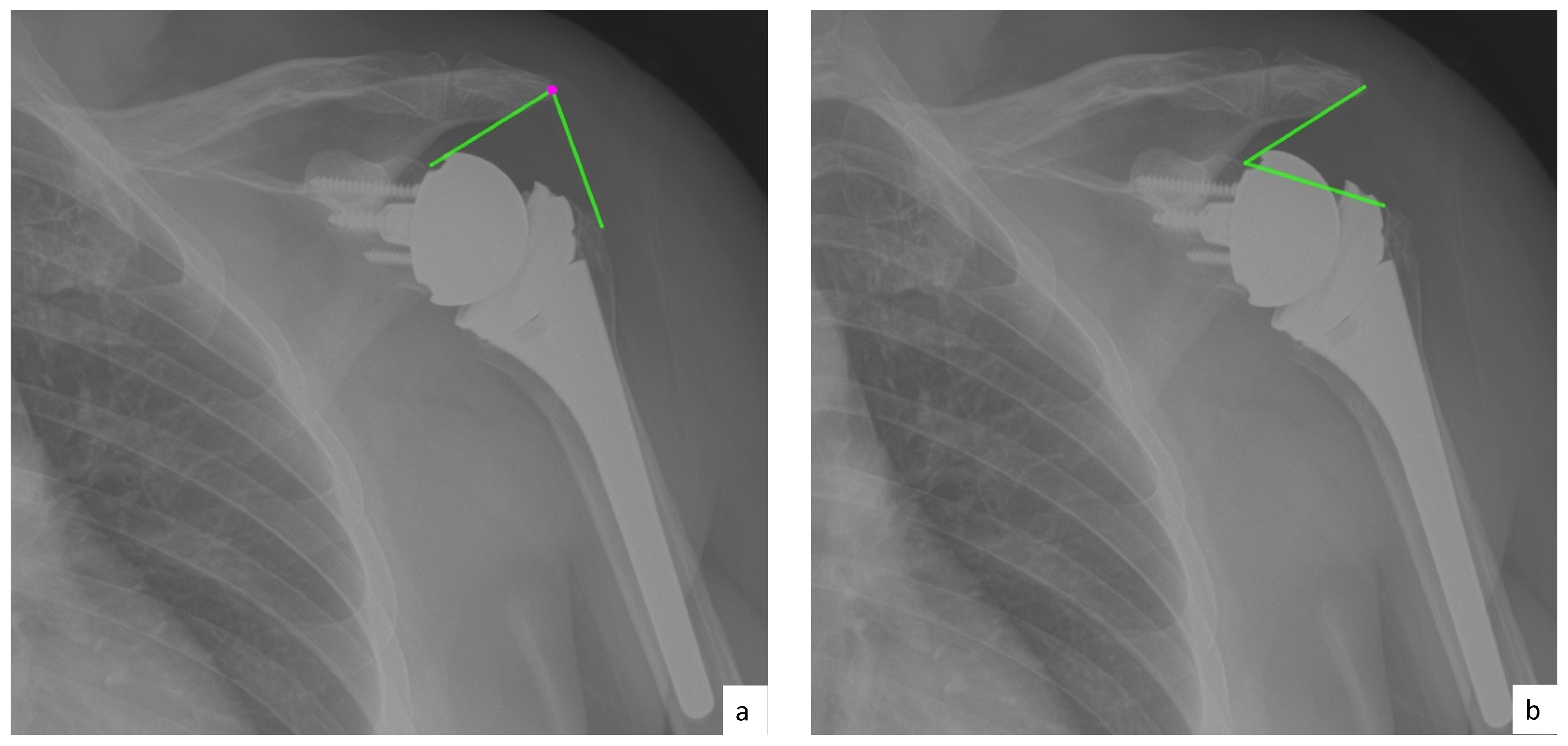
2.4. Radiographic Evaluation

All patients had standard preoperative and postoperative radiographs (true anteriorposterior, y view and axillary view). Radiographic assessment was performed by two independent viewers blinded to patient outcomes. Radiographic measurements were performed on standard anteroposterior (AP) view performed at the most recent preoperative and last postoperative visit. Preoperative measurements included center of rotation (COR), critical shoulder angle (CSA) acromiohumeral distance (AHD), lateral humeral offset (LHO), and glenoid inclination (GI). Postoperative measurements included AHD, LHO, baseplate inclination (BI), distalization shoulder angle (DSA), and lateralization shoulder angle (LSA) [20,21,24,25,26].
AHD was measured by calculating the perpendicular distance between the most lateral portion of the undersurface of the acromion and a line parallel to the superior border of the greater tuberosity [21] (Figure 1). LHO was measured by determining the distance from the AHD line to the most lateral projection of the greater tuberosity [21] (Figure 2). LSA was measured by drawing a line from the superior glenoid tubercle to the most lateral border of the acromion and a second line from the most lateral border of the acromion to the most lateral border of the greater tuberosity. The angle between these two lines formed the LSA [20] (Figure 3a). DSA was measured by drawing a line between the most lateral border of the acromion and the superior glenoid tubercle and drawing a second line to connect the superior glenoid tubercle with the most superior border of the greater tuberosity. The angle between these two lines formed the DSA [20] (Figure 3b). Glenoid and baseplate were determined as the angle between the floor of the supraspinatus fossa and the glenoid fossa [25] (Figure 4). COR was measured by determining the best fit circle flush to the articular surface, identifying the center of the circle in the humeral head, and then measuring the distance of the perpendicular line between the center of the humeral head and the midpoint of the line connecting the superior and inferior glenoid tubercles [24] (Figure 5b). CSA was measured by a line from the superior pole to the inferior pole of the glenoid and a line from the inferior pole to the lateral edge of the acromion [26] (Figure 5a). In addition, scapular notching was graded according to the Nerot–Sirveaux classification and severity of preoperative cuff tear arthropathy was evaluated according to the Hamada classification [27,28].

2.5. Statistical Analysis

Descriptive statistics including mean and standard deviation for continuous variables and frequency and proportion for categorical variables were calculated to characterize the study groups. The relationships between clinical outcome measures and radiographic measurements were examined graphically with scatterplots and with Pearson correlation coefficients (rho). The effect of DSA and LSA on postoperative forward elevation was examined using a mixed effects linear model to account for patients nesting within surgeon’s practices. An interclass correlation coefficient (ICC) was calculated to determine reproducibility of the radiographic measurements. A p-value < 0.05 was considered statistically significant. All analyses were performed with Stata statistical software (StataCorp. 2017. Stata Statistical Software: Release 15. College Station, TX: StataCorp LLC).

3. Results

3.1. Subjects

Ninety-four rTSAs meeting the study criteria were performed during the study period. Of those, 61 were available at final follow-up (Figure 6). The mean age of patients was 69.2 ± 8.2 years (range: 53–88) with a mean follow-up of 3.1 ± 0.7 years (range: 2.0–4.2) years. Most patients were female (55.7%). Patient demographics are demonstrated in Table 1.

3.2. Clinical Outcome

Overall, there was significant improvement in all clinical outcome measures from pre- to postoperative. SST improved from 2.5 ± 1.8pre to 8.0 ± 2.6post, SANE improved from 28.9 ± 22.7pre to 80.7 ± 20.1post, VAS improved from 6.0 ± 2.2 pre to 1.4 ± 2.3post, ASES improved from 37 ± 14.5pre to 78.1 ± 21.6post. At final follow-up, there was no significant difference in SST, SANE, VAS, and ASES when comparing patient populations of the different institutions.
In addition, there was significant improvement in ROM from pre-to postoperative. AFE improved from 92 ± 36°pre to 131 ± 27°post, AABD improved from 69 ± 35°pre to 109 ± 38°post, AER improved from 29 ± 18°pre to 42 ± 19°post at final follow-up (p < 0.01, respectively). When comparing ROM of patients at the different institutions, no significant difference was found for AFE, AABD, or AER.

3.3. Inter-Rater Reliability of Radiographic Analysis

Inter-rater reliability was calculated for COR, Pre-CSA, Pre-AHD, Post-AHD, DSA, Pre-LHO, Post-LHO, LSA, glenoid inclination, baseplate inclination, Hamada and Notching grades. Reliability was found to be good for most of the radiographic measurements. However, Pre-AHD (ICC = 0.37; CI: 0.18–0.55) showed only poor reliability. Moderate to good ICC was found for COR (ICC = 0.68; CI: 0.51–0.8), DSA (ICC = 0.66; CI: 0.32–0.82) and glenoid inclination (ICC = 0.66; CI: 0.47–0.79). Mean values of radiographic measurements with corresponding inter-rater reliability are demonstrated in Table 2.

3.4. Correlation between Preoperative Radiographic Measurements and Clinical Outcomes

Pre-AHD was found to have a significant correlation with final AER (p = 0.016; r = 0.314). Additionally, Pre-LHO showed a significant correlation with final ASES (p = 0.032; r = −0.277). COR, CSA, and glenoid inclination had no significant influence on clinical outcomes at terminal follow-up (p > 0.05, respectively) (Appendix A Table A1).

3.5. Correlation between Lateralization and Clinical Outcomes

Post-LHO was found to significantly correlate with final ASES (p = 0.03; r = −0.281). Further, there was a significant correlation of LSA with final SST (p = 0.001; r = −0.41), final pain score (p = 0.034; r = 0.272), final SANE (p = 0.018, r = −0.308), and final ASES (p = 0.011; r = −0.327). Further, there was a significant correlation between LSA and final AFE (p = 0.033; r = −0.276). Correlations of LSA with final AER (p = 0.471; r = −0.096) and AABD (p = 0.824; r = 0.030) were found to be non-significant (Appendix A Table A1).

3.6. Correlation between Distalization and Clinical Outcomes

Post-AHD had a significant correlation with final SST (p = 0.025; r = 0.293). On the contrary, DSA showed no significant correlation to any clinical outcome measures. Post-AHD demonstrated a significant correlation to final AER (p = 0.002; r = 0.398). In addition, DSA significantly influenced final AFE (p = 0.02; r = 0.299). No significant correlations were found between DSA and final AER (p = 0.317; r = 0.133) and AABD (p = 0.283; r = 0.145).

3.7. Prediction of Active ROM

The highest degree in AFE was observed in patients presenting with a postoperative DSA between 40° and 60°. Patients with an AFE < 100° (n = 5) were further shown to have a DSA smaller than 40° (Figure 7). When looking at the LSA, patients with an AFE < 100° (n = 4) had an LSA greater than 95°. The highest degree in AFE was observed in patients having an LSA between 75° to 95° (Figure 8). However, there was no statistically significant correlation between distalization (p = 0.317) and lateralization (p = 0.471) to AER at final follow-up.

4. Discussion

The most important finding of this study was that there was only a negligible correlation between radiographic measurements and clinical and functional outcomes following primary rTSA using a design with a humeral inclination of 135°. Even though statistically significant correlations between postoperative outcomes scores and radiographic measurements were found, these observations are of limited predictive value for outcomes following rTSA. The data gathered from this multi-center study indicate that the importance of radiographic measurements and their correlation with outcomes following rTSA may be limited.
In the postoperative setting, lateralization in rTSA can be expressed by different radiographic variables, including LHO, LSA, and COR. In their retrospective study, Jeon et al. demonstrated that an increased postoperative LHO was found to be a significant risk factor for poor restoration of postoperative AFE, when using an implant designed to be medialized [21]. In contrast, the data from this study showed that in a cohort using a lateralized implant, no significant relationship between preoperative and postoperative LHO and AFE could be demonstrated. However, in this study, post-LHO was found to significantly influence final ASES score, which may be of limited predictive value, as this finding did not allow for drawing a definite conclusion.
Increasing the lateralization of the COR in rTSA using a medialized implant design has been shown to result in greater active ROM [9]. As only few studies have focused on measuring COR in lateralized implants [18,29], the authors from this study could not find a significant relationship between COR, clinical outcomes, and final active ROM when using a lateralized rTSA design. Similar to a previous study by Boutsiadis et al. [20], who reported that patients achieved the highest degree in postoperative AFE and AABD with a DSA between 40° and 65° and the highest degree in AER when having a LSA of 75° to 95°, the findings of this study demonstrated a significant correlation between LSA and AFE as well as DSA and AFE. Additionally, the highest degree in AER was noted in patients having LSA values between 75° and 95°; however, a direct correlation of LSA and DSA with final AER and AABD could not be confirmed [20]. This may be explained by existing differences in implant designs being used, as all included patients uniformly underwent rTSA using a lateralized design with a humeral inclination of 135°. In contrast, Boutsiadis et al. included patients with two different implant designs (145° and 155° humeral inclination) [20].
Additionally, a positive correlation between LSA and DSA could be shown, which is consistent with the current literature [20]. In a lateralized rTSA design, a lower LSA, which corresponds to a more medialized implant, is associated with a larger DSA, indicating a greater distance between the acromion and humerus. To this, the findings from this study suggest that a LSA greater than 100° correlates with a DSA of less than 40°, thus reducing final AFE. Considering the current literature, lateralization of rTSA has been shown to increase postoperative AFE and AER by restoring the anatomic COR, while optimizing recruitment of the muscle fibers [6,30,31]. However, LSA was noted to be between 75° and 95° for optimal implant lateralization, with excessive lateralization resulting in less active ROM.
Increasing distalization, in order to improve tension on the deltoid muscle, has been shown to play an important role in rTSA [4]. In a computer-based model using different humeral offset and stem designs, Lädermann et al. demonstrated a strong positive linear relationship between AHD and AFE and AABD [6]. Furthermore, the authors showed that AHD decreased by 6 mm when switching from a 155° inlay design to a 135° onlay design. Even though it was shown that a higher AHD, expressed as arm lengthening, was related to a higher degree in AFE [6], the exact amount of arm lengthening remains inconclusive [6,18,32,33].
First introduced by Moor et al. [26], the CSA has been reported to be a reproducible radiographic index. As a larger CSA has been found to be associated with degenerative rotator cuff tears, there is still limited knowledge regarding its influence on rTSA [34]. Even though Roberson et al. [34] reported improved AFE in patients with a lower CSA, no significant relationship between CSA and clinical outcomes scores or final active ROM was found in this study.
Taking these findings into account, there remains a debate regarding the ideal placement of the components during rTSA to most sufficiently restore active ROM while minimizing complications such as component loosening and scapular notching. Additionally, as this study further verified, there is still a considerable lack of data in assessing radiographic prosthesis positioning in correlation with clinical outcomes. This may lead shoulder surgeons to overestimate current data and the importance of radiographic measurements and their correlation with outcomes following rTSA.
There are several limitations to the study. First, the study cohort was not randomized. Second, although outcomes were collected prospectively, data were reviewed retrospectively, which could create selection bias. Third, all radiographic measurements are highly dependent on patient orientation during radiographic imaging, as angles and distances are influenced by the position of the scapula as well as rotation of the humerus. However, this reflects daily clinical practice, as radiographic imaging, even if standardized, can show significant variances. Fourth, the multi-center design of this study including five surgeons from different sites leads to differences in implant positioning and intraoperative and postoperative outcomes. However, for the purpose of this study, the authors intended to demonstrate that even with high experience and expertise in this field, the observations and findings from this study and its subsequent comparison to the current literature should be interpreted with careful attention.

5. Conclusions

Negligible correlations between pre- and postoperative radiographic measurements and clinical outcomes following primary rTSA using a 135° prosthesis design were demonstrated. However, these observations are of limited predictive value for outcomes following rTSA. Subsequently, there remains a debate regarding the ideal placement of the components during rTSA to most sufficiently restore active ROM while minimizing complications such as component loosening and scapular notching. Additionally, as the data from this study showed, there is still a considerable lack of data in assessing radiographic prosthesis positioning in correlation to clinical outcomes. As such, the importance of radiographic measurements and their correlation with clinical and functional outcomes following rTSA may be limited.

Author Contributions

D.P.B. and L.N.M. wrote the manuscript. D.M. contributed to study design and data conception. M.P.C. contributed to the statistical analysis. J.B.B. helped with data conception. R.A.C. helped with interpretation of data and radiographic measurements. A.D.M., A.A.R., P.J.D., R.G., E.L. helped with data conception and analysis. K.B. served as important reviewer and helped with data interpretation. All authors have read and agreed to the published version of the manuscript.

Funding

The University of Connecticut Health Center/UConn Musculoskeletal Institute has received direct funding and material support from Arthrex Inc. (Naples. Fl). The company had no influence on study design, data collection, or interpretation of the results or the final manuscript.

Institutional Review Board Statement

Ethical approval was obtained via Human Research Determination Form to the institutional review board (IRB) of the University of Connecticut (IRB 17-202-2).

Informed Consent Statement

Informed consent was obtained from all subjects involved in the study.

Data Availability Statement

The data presented in this study are available on request from the corresponding author. The data are not publicly available due to ethical reasons.

Conflicts of Interest

Authors B.D.P., M.D., M.L.M. and B.J.B., C.A.R. declare that they have no conflict of interest. A.D.M. reports research grants from Arthrex Inc., is a consultant for Arthrex Inc. and receives royalties from Arthrex Inc. D.P.J. is a consultant for Arthrex Inc. and receives royalties from Arthrex Inc. L.E. is a consultant for Arthrex Inc. and receives royalties from Arthrex Inc. C.M.P. receives personal fees from Arthroscopy Association of North America (AANA). Gobezie R is a consultant for Arthrex Inc. and receives royalties from Arthrex Inc. R.A.R. receives material or orther financial support from AANA; is a board or committee member of Amercian Shoulder and Elbow Surgeons; Receives financial and material support from Arthrex Inc. ansd receives royalties from Arthrex Inc; receives other financial or material support from Mayor League Baseball; is an Editorial or governing board for Orthopedics and a board or committee member for Orthopedics Today; receives research support from Paragen Technologies and holds stock or stock options for Paragen Technologies; Is an editorial or governing board for SAGE; receives royalties and material support by Saunders/Mosby-Elsevier; receives royalties or material support for SLACK incorporated and is a editorial or governing board for SLACK incorporated; is an editorial or governing board for Wolters Kluwer Health. K Beitzel is a consultant for Arthrex Inc. and receives royalties from Arthrex Inc.

Appendix A

Table A1. Table showing correlation between radiographic analysis and final clinical outcome scores.
ASESSANESSTVASFEABDER
CORr−0.0790.0800.0060.1540.170−0.176−0.011
p−value0.5470.5450.9660.2350.1940.1900.937
Pre CSAr−0.0350.056−0.0560.051−0.211−0.212−0.187
p−value0.7940.6720.6770.6970.1050.1130.156
Pre AHDr0.0850.1420.124−0.059−0.051−0.0150.314
p−value0.5180.2840.3490.6520.6970.9100.016
Pre LHOr−0.277−0.243−0.2510.177−0.048−0.0370.035
p−value0.0320.0640.0550.1730.7160.7830.790
Post−AHDr0.1500.1790.293−0.1350.3980.1110.233
p−value0.2530.1740.0250.2990.0020.4110.075
Post LHOr−0.281−0.215−0.1970.1930.0860.045−0.003
p−value0.0300.1020.1350.1360.5130.7390.985
DSAr0.1690.0990.234−0.1450.2990.1450.133
p−value0.1980.4560.0750.2660.0200.2830.317
LSAr−0.327−0.308−0.4100.272−0.2760.030−0.096
p−value0.0110.0120.0010.0340.0330.8240.471
Inclination Glenoind.r−0.066−0.156−0.0950.1320.0720.1760.104
p−value0.6140.2380.4730.3100.5830.1910.435
Inclination Baseplater−0.1210.038−0.1020.1230.1220.1060.050
p−value0.3560.7760.4420.3470.3530.4330.710
Hamadar−0.0090.067−0.0310.0130.156−0.141−0.289
p−value0.9470.6130.8170.9190.2330.2970.026
Notchingr−0.030−0.1650.0420.151−0.246−0.133−0.214
p−value0.8180.2130.7540.2440.0580.3250.104
Significant values (p < 0.05) are highlighted. Abbreviation: COR = center of rotation; Pre CSA = preoperative critical shoulder angle; Pre AHD = preoperative acromiohumeral distance; Post AHD = postoperative acromiohumeral distance; DSA = distalization shoulder angle; Pre LHO = preoperative lateral humeral offset; Post LHO = postoperative lateral humeral; LSA = lateralization shoulder angle; FE = postoperative forward elevation; ABD = postoperative abduction; ER = postoperative external rotation; ASES = Postoperative American Shoulder and Elbow Surgeons; SST = Postoperative Simple Shoulder Test; VAS = Postoperative Visual Analogue Scale; SANE = Postoperative Single Assessment Numeric Evaluation.

References

  1. Routman, H.D.; Flurin, P.H.; Wright, T.W.; Zuckerman, J.D.; Hamilton, M.A.; Roche, C.P. Reverse Shoulder Arthroplasty Prosthesis Design Classification System. Bull. NYU Hosp. Jt. Dis. 2015, 73, S5–S14. [Google Scholar]
  2. Grammont, P. Etude et réalisation d’une nouvelle prothèse d’épaule. Rheumatologie 1987, 39, 27–38. [Google Scholar]
  3. Grammont, P.M.; Baulot, E. Delta shoulder prosthesis for rotator cuff rupture. Orthopedics 1993, 16, 65–68. [Google Scholar] [CrossRef]
  4. Boileau, P.; Watkinson, D.J.; Hatzidakis, A.M.; Balg, F. Grammont reverse prosthesis: Design, rationale, and biomechanics. J. Shoulder Elbow Surg. 2005, 14, 147s–161s. [Google Scholar] [CrossRef] [PubMed]
  5. Boileau, P.; Moineau, G.; Roussanne, Y.; O’Shea, K. Bony increased-offset reversed shoulder arthroplasty: Minimizing scapular impingement while maximizing glenoid fixation. Clin. Orthop. Relat. Res. 2011, 469, 2558–2567. [Google Scholar] [CrossRef] [Green Version]
  6. Ladermann, A.; Denard, P.J.; Boileau, P. Effect of humeral stem design on humeral position and range of motion in reverse shoulder arthroplasty. Int. Orthop. 2015, 39, 2205–2213. [Google Scholar] [CrossRef] [Green Version]
  7. Bacle, G.; Nove-Josserand, L.; Garaud, P.; Walch, G. Long-Term Outcomes of Reverse Total Shoulder Arthroplasty: A Follow-up of a Previous Study. J. Bone Jt. Surg. Am. 2017, 99, 454–461. [Google Scholar] [CrossRef]
  8. Mizuno, N.; Denard, P.J.; Raiss, P.; Walch, G. The clinical and radiographical results of reverse total shoulder arthroplasty with eccentric glenosphere. Int. Orthop. 2012, 36, 1647–1653. [Google Scholar] [CrossRef] [PubMed] [Green Version]
  9. Gutierrez, S.; Levy, J.C.; Frankle, M.A. Evaluation of abduction range of motion and avoidance of inferior scapular impingement in a reverse shoulder model. J. Shoulder Elbow Surg. 2008, 17, 608–615. [Google Scholar] [CrossRef]
  10. Berliner, J.L.; Regalado-Magdos, A.; Ma, C.B.; Feeley, B.T. Biomechanics of reverse total shoulder arthroplasty. J. Shoulder Elbow Surg. 2015, 24, 150–160. [Google Scholar] [CrossRef] [PubMed]
  11. Frankle, M.; Levy, J.C.; Pupello, D. The reverse shoulder prosthesis for glenohumeral arthritis associated with severe rotator cuff deficiency. a minimum two-year follow-up study of sixty patients surgical technique. J. Bone Jt. Surg. Am. 2006, 88, 178–190. [Google Scholar] [CrossRef]
  12. Katz, D.; Valenti, P.; Kany, J.; Elkholti, K.; Werthel, J.D. Does lateralisation of the centre of rotation in reverse shoulder arthroplasty avoid scapular notching? Clinical and radiological review of one hundred and forty cases with forty five months of follow-up. Int. Orthop. 2016, 40, 99–108. [Google Scholar] [CrossRef] [Green Version]
  13. Ernstbrunner, L.; Suter, A.; Catanzaro, S.; Rahm, S.; Gerber, C. Reverse Total Shoulder Arthroplasty for Massive, Irreparable Rotator Cuff Tears Before the Age of 60 Years: Long-Term Results. J. Bone Jt. Surg. Am. 2017, 99, 1721–1729. [Google Scholar] [CrossRef] [PubMed] [Green Version]
  14. Gerber, C.; Canonica, S.; Catanzaro, S.; Ernstbrunner, L. Longitudinal observational study of reverse total shoulder arthroplasty for irreparable rotator cuff dysfunction: Results after 15 years. J. Shoulder Elbow Surg. 2018, 27, 838. [Google Scholar] [CrossRef]
  15. Mahendraraj, K.A.; Colliton, E.; Muniz, A.; Menendez, M.E.; Jawa, A. Assessing the validity of the distalization and lateralization shoulder angles following reverse total shoulder arthroplasty. Semin. Arthroplast. JSES 2020. [Google Scholar] [CrossRef]
  16. Li, X.; Knutson, Z.; Choi, D. Effects of glenosphere positioning on impingement-free internal and external rotation after reverse total shoulder arthroplasty. J. Shoulder Elbow Surg. 2013, 22, 807–813. [Google Scholar] [CrossRef]
  17. Berhouet, J.; Garaud, P.; Favard, L. Evaluation of the role of glenosphere design and humeral component retroversion in avoiding scapular notching during reverse shoulder arthroplasty. J. Shoulder Elbow Surg. 2014, 23, 151–158. [Google Scholar] [CrossRef] [PubMed]
  18. Jobin, C.M.; Brown, G.D.; Bahu, M.J. Reverse total shoulder arthroplasty for cuff tear arthropathy: The clinical effect of deltoid lengthening and center of rotation medialization. J. Shoulder Elbow Surg. 2012, 21, 1269–1277. [Google Scholar] [CrossRef] [PubMed]
  19. Marcoin, A.; Ferrier, A.; Blasco, L.; De Boissieu, P.; Nerot, C.; Ohl, X. Reproducibility of a new method for measuring lowering and medialisation of the humerus after reverse shoulder arthroplasty. Int. Orthop. 2018, 42, 141–147. [Google Scholar] [CrossRef]
  20. Boutsiadis, A.; Lenoir, H.; Denard, P.J. The lateralization and distalization shoulder angles are important determinants of clinical outcomes in reverse shoulder arthroplasty. J. Shoulder Elbow Surg. 2018, 27, 1226–1234. [Google Scholar] [CrossRef]
  21. Jeon, Y.S.; Rhee, Y.G. Factors associated with poor active anterior elevation after reverse total shoulder arthroplasty. J. Shoulder Elbow Surg. 2018, 27, 786–793. [Google Scholar] [CrossRef]
  22. Richards, R.R.; An, K.N.; Bigliani, L.U. A standardized method for the assessment of shoulder function. J. Shoulder Elbow Surg. 1994, 3, 347–352. [Google Scholar] [CrossRef]
  23. Lippitt, S. A practical tool for evaluating shoulder function. The Simple Shoulder Test. Shoulder A Balance Mobil. Stab. 1993, 501–518. [Google Scholar]
  24. Rhee, S.M.; Lee, J.D.; Park, Y.B.; Yoo, J.C.; Oh, J.H. Prognostic Radiological Factors Affecting Clinical Outcomes of Reverse Shoulder Arthroplasty in the Korean Population. Clin. Orthop. Surg. 2019, 11, 112–119. [Google Scholar] [CrossRef] [PubMed]
  25. Maurer, A.; Fucentese, S.F.; Pfirrmann, C.W. Assessment of glenoid inclination on routine clinical radiographs and computed tomography examinations of the shoulder. J. Shoulder Elbow Surg. 2012, 21, 1096–1103. [Google Scholar] [CrossRef] [Green Version]
  26. Moor, B.K.; Bouaicha, S.; Rothenfluh, D.A.; Sukthankar, A.; Gerber, C. Is there an association between the individual anatomy of the scapula and the development of rotator cuff tears or osteoarthritis of the glenohumeral joint?: A radiological study of the critical shoulder angle. Bone Jt. J. 2013, 95, 935–941. [Google Scholar] [CrossRef]
  27. Sirveaux, F.; Favard, L.; Oudet, D.; Huquet, D.; Walch, G.; Mole, D. Grammont inverted total shoulder arthroplasty in the treatment of glenohumeral osteoarthritis with massive rupture of the cuff: Results of a multicentre study of 80 shoulders. J. Bone Jt. Surg. Br. Vol. 2004, 86, 388–395. [Google Scholar] [CrossRef]
  28. Hamada, K.; Yamanaka, K.; Uchiyama, Y.; Mikasa, T.; Mikasa, M. A radiographic classification of massive rotator cuff tear arthritis. Clin. Orthop. Relat. Res. 2011, 469, 2452–2460. [Google Scholar] [CrossRef] [PubMed] [Green Version]
  29. Sabesan, V.J.; Lombardo, D.; Josserand, D. The effect of deltoid lengthening on functional outcome for reverse shoulder arthroplasty. Musculoskelet. Surg. 2016, 100, 127–132. [Google Scholar] [CrossRef]
  30. Werner, B.S.; Chaoui, J.; Walch, G. The influence of humeral neck shaft angle and glenoid lateralization on range of motion in reverse shoulder arthroplasty. J. Shoulder Elbow Surg. 2017, 26, 1726–1731. [Google Scholar] [CrossRef]
  31. Greiner, S.; Schmidt, C.; Konig, C.; Perka, C.; Herrmann, S. Lateralized reverse shoulder arthroplasty maintains rotational function of the remaining rotator cuff. Clin. Orthop. Relat. Res. 2013, 471, 940–946. [Google Scholar] [CrossRef] [Green Version]
  32. Gutierrez, S.; Comiskey, C.A.; Luo, Z.P.; Pupello, D.R.; Frankle, M.A. Range of impingement-free abduction and adduction deficit after reverse shoulder arthroplasty. Hierarchy of surgical and implant-design-related factors. J. Bone Jt. Surg. Am. 2008, 90, 2606–2615. [Google Scholar] [CrossRef] [PubMed]
  33. Schwartz, D.G.; Cottrell, B.J.; Teusink, M.J. Factors that predict postoperative motion in patients treated with reverse shoulder arthroplasty. J. Shoulder Elbow Surg. 2014, 23, 1289–1295. [Google Scholar] [CrossRef]
  34. Roberson, T.A.; Shanley, E.; Abildgaard, J.T. The influence of radiographic markers of biomechanical variables on outcomes in reverse shoulder arthroplasty. JSES Open Access 2019, 3, 59–64. [Google Scholar] [CrossRef] [Green Version]
Figure 1. (a) Preoperative acromiohumeral distance (AHD; green line); (b) postoperative acromiohumeral distance (AHD; green line).
Jcm 10 00809 g001
Figure 2. (a) Preoperative lateral humeral offset (LHO; green line); (b) postoperative lateral humeral offset (LHO; green line).
Jcm 10 00809 g002
Figure 3. (a) Lateralization shoulder angle (LSA); (b) distalization shoulder angle (DSA).
Jcm 10 00809 g003
Figure 4. (a) Glenoid inclination (green angle); (b) baseplate inclination (green line).
Jcm 10 00809 g004
Figure 5. (a) Critical shoulder angle (CSA; green angle); (b) center of rotation (COI; green line).
Jcm 10 00809 g005
Figure 6. Flowchart displaying inclusion criteria.
Jcm 10 00809 g006
Figure 7. Scatterplot showing the linear correlation between DSA and LSA for final AFE. Good final AFE could be seen for DSA between 40 and 60° and LSA between 80 and 100°; Abbreviations: DSA = distalization shoulder angle; LSA = lateralization shoulder angle; AFE = active forward elevation.
Jcm 10 00809 g007
Figure 8. Scatterplot showing the linear correlation between LSA and DSA for final AFE. Good final AFE could be seen for DSA between 40 and 60° and LSA between 80 and 100°. Abbreviations: DSA = distalization shoulder angle; LSA = lateralization shoulder angle; AFE = active forward elevation.
Jcm 10 00809 g008
Table 1. Patient demographics (N = 61).
n%
Sex
Male2744.3
Female3455.7
Mean age ± SD (years)69.2 ± 8.2
Mean follow-up ± SD (years)3.1 ± 0.7
Dominant Arm Involved3252.0
Right Shoulders3354.0
BMI29.9 ± 7.0
Table 2. Inter-rater reliability for all radiographic measurements and mean values for radiographic analysis.
Mean ± SDICCICC 95% CIReliability
COR20.9 ± 3.9 mm0.68[0.51, 0.8]Moderate-Good
Pre CSA35.2 ± 4.5 deg0.9[0.9, 0.94]Good
Pre AHD5.1 ± 3.2 mm0.37[0.18, 0.55]Poor
Post AHD26.3 ± 9.5 mm0.88[0.82, 0.93]Good
DSA38.6 ± 9.6 deg0.66[0.32, 0.82]Moderate-Good
Pre LHO9.9 ± 5.7 mm0.86[0.79, 0.91]Good
Post LHO9.5 ± 6 mm0.84[0.75, 0.89]Good
LSA89.2 ± 11.9 deg0.84[0.73, 0.9]Good
Glenoid inclination81.2 ± 6.8 deg0.66[0.47, 0.79]Moderate-Good
Baseplate inclination83.2 ± 6.4 deg0.79[0.69, 0.86]Good
Abbreviation: COR = center of rotation; Pre CSA = preoperative critical shoulder angle; Pre AHD = preoperative acromiohumeral distance; Post AHD = postoperative acromiohumeral distance; DSA = distalization shoulder angle; Pre LHO = preoperative lateral humeral offset; Post LHO = postoperative lateral humeral; LSA = lateralization shoulder angle.
Publisher’s Note: MDPI stays neutral with regard to jurisdictional claims in published maps and institutional affiliations.

