
Optic Nerve Drusen Evaluation: A Comparison between Ultrasound and OCT

 , ,
, ,  and
and 
Abstract
:1. Introduction
2. Materials and Methods
2.1. Patients Selection
2.2. Instruments and Methods
2.3. Statistical Analysis
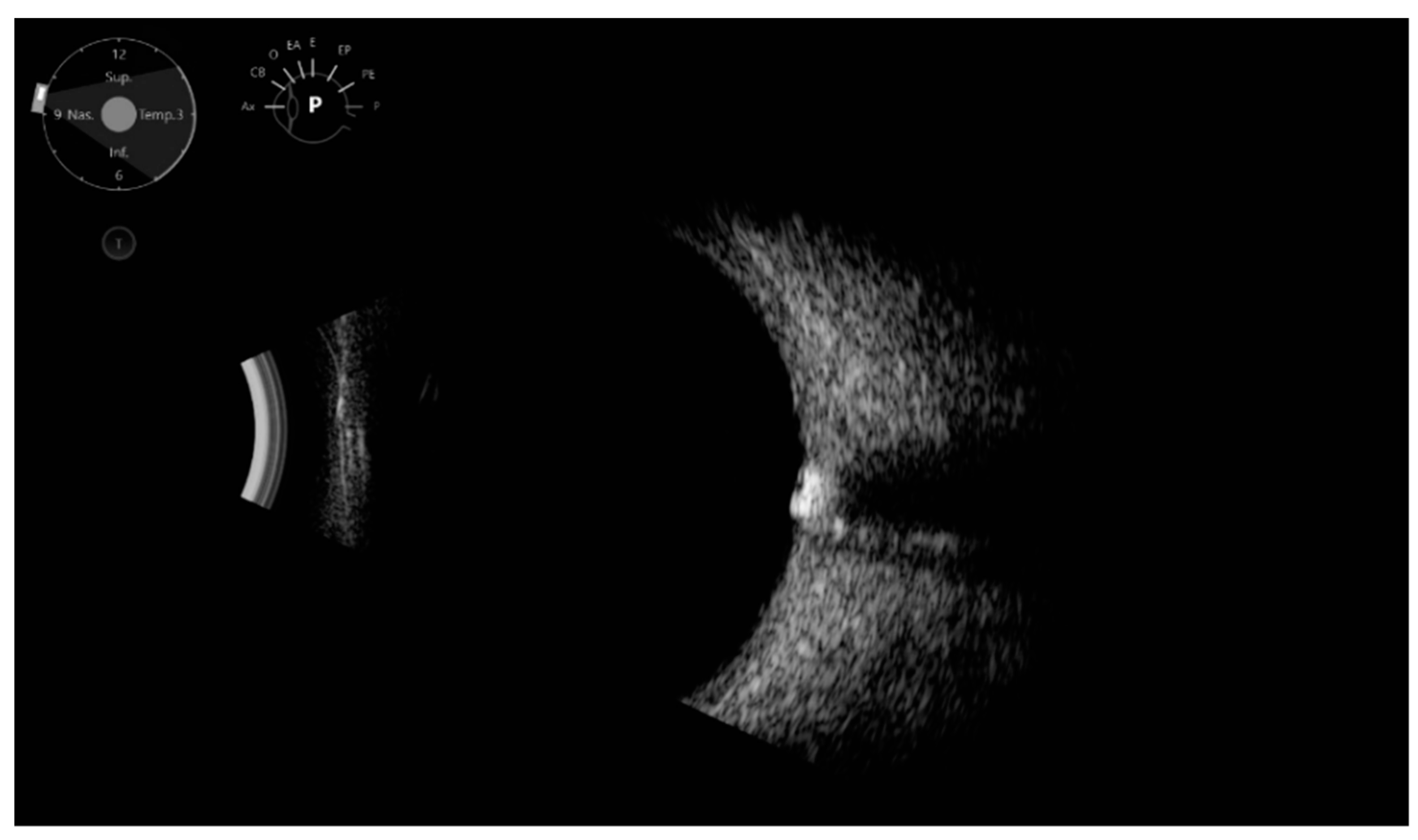
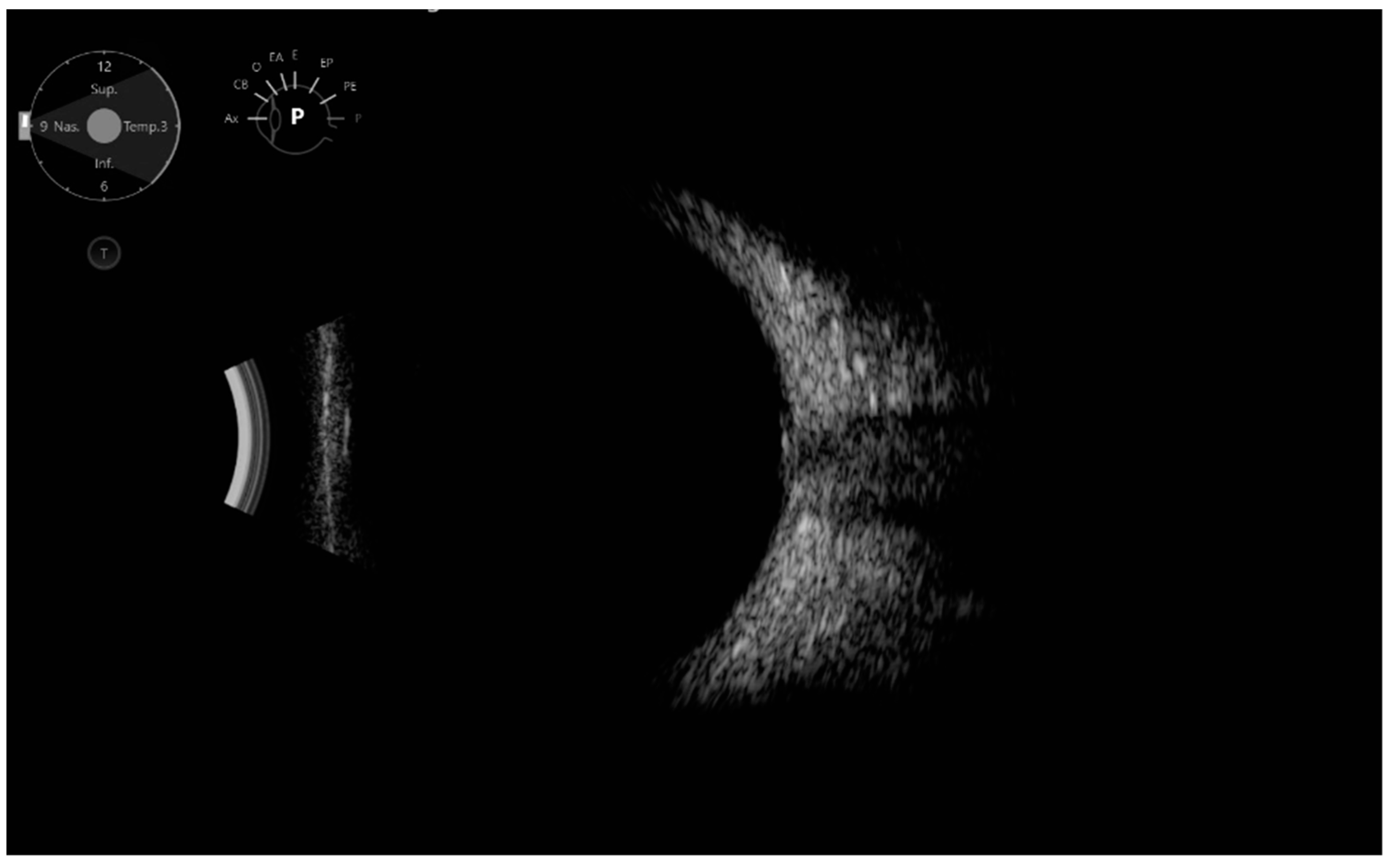
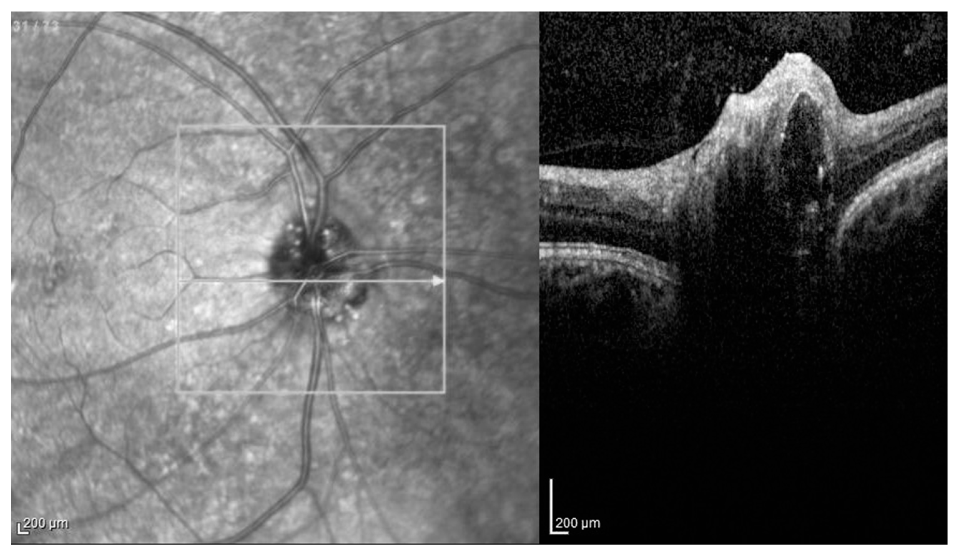
3. Results
- Large buried: In two cases, we found both ODD and PHOMS; in two cases, OCT could not detect either ODD or PHOMS;
- Large superficial: In 3 cases, we found only ODD; in 3 cases, only PHOMS; in 14 cases, both ODD and PHOMS; and in 4 cases, OCT could not detect either ODD or PHOMS;
- Point-like buried: In one case, we found only ODD; in two cases, only PHOMS; in one case, both ODD and PHOMS; and in two cases, OCT could not detect either ODD or PHOMS;
- Point-like superficial: In 3 cases, we found only ODD; in 20 cases, only PHOMS; in 18 cases, both ODD and PHOMS; and in 8 cases, OCT could not detect either ODD or PHOMS;
- Point-like mixed superficial and buried: In two cases, we found both ODD and PHOMS, and in one case, OCT could not detect either ODD or PHOMS;
4. Discussion
Author Contributions
Funding
Institutional Review Board Statement
Informed Consent Statement
Data Availability Statement
Conflicts of Interest
References
- Vitiello, L.; De Bernardo, M.; Nuzio, S.G.; Mandato, C.; Rosa, N.; Vajro, P. Pediatric liver diseases and ocular changes: What hepatologists and ophthalmologists should know and share with each other. Dig. Liver Dis. 2020, 52, 1–8. [Google Scholar] [CrossRef] [PubMed] [Green Version]
- De Bernardo, M.; Vitiello, L.; Rosa, N. Ocular ultrasonography to detect intracranial pressure in aneurysmal subarachnoid hemorrhage. Ann. Clin. Transl. Neurol. 2020, 7, 1459–1460. [Google Scholar] [CrossRef] [PubMed]
- De Bernardo, M.; Vitiello, L.; Rosa, N. A-scan ultrasonography and optic nerve sheath diameter assessment during acute elevations in intra-abdominal pressure. Surgery 2020, 167, 1023–1024. [Google Scholar] [CrossRef] [PubMed]
- Hayreh, S.S. Pathogenesis of optic disc edema in raised intracranial pressure. Prog. Retin. Eye Res. 2016, 50, 108–144. [Google Scholar] [CrossRef] [Green Version]
- Saenz, R.; Cheng, H.; Prager, T.C.; Frishman, L.J.; Tang, R.A. Use of A-scan Ultrasound and Optical Coherence Tomography to Differentiate Papilledema From Pseudopapilledema. Optom. Vis. Sci. 2017, 94, 1081–1089. [Google Scholar] [CrossRef]
- Seddon, J.M.; Dossett, J.P.; Widjajahakim, R.; Rosner, B. Association Between Perifoveal Drusen Burden Determined by OCT and Genetic Risk in Early and Intermediate Age-Related Macular Degeneration. Investig. Ophthalmol. Vis. Sci. 2019, 60, 4469–4478. [Google Scholar] [CrossRef] [Green Version]
- Lam, B.L.; Morais, C.G., Jr.; Pasol, J. Drusen of the optic disc. Curr. Neurol. Neurosci. Rep. 2008, 8, 404–408. [Google Scholar] [CrossRef]
- Sato, T.; Mrejen, S.; Spaide, R.F. Multimodal imaging of optic disc drusen. Am. J. Ophthalmol. 2013, 156, 275–282. [Google Scholar] [CrossRef]
- Rajagopal, R.; Mitchell, E.; Sylvester, C.; Lope, L.A.; Nischal, K.K. Detection of Optic Disc Drusen in Children Using Ultrasound through the Lens and Avoiding the Lens-Point of Care Ultrasound Technique of Evaluation Revisited. J. Clin. Med. 2019, 8, 1449. [Google Scholar] [CrossRef] [Green Version]
- De Bernardo, M.; Altieri, V.; Coppola, A.; Gioia, M.; Rosa, N. Choroidal evaluation in patients under alpha-lytic therapy. Graefe’s Arch. Clin. Exp. Ophthalmol. 2020, 258, 2729–2736. [Google Scholar] [CrossRef]
- Merchant, K.Y.; Su, D.; Park, S.C.; Qayum, S.; Banik, R.; Liebmann, J.M.; Ritch, R. Enhanced depth imaging optical coherence tomography of optic nerve head drusen. Ophthalmology 2013, 120, 1409–1414. [Google Scholar] [CrossRef] [PubMed]
- Leon, M.; Hutchinson, A.K.; Lenhart, P.D.; Lambert, S.R. The cost-effectiveness of different strategies to evaluate optic disc drusen in children. J AAPOS 2014, 18, 449–452. [Google Scholar] [CrossRef] [PubMed] [Green Version]
- Silverman, A.L.; Tatham, A.J.; Medeiros, F.A.; Weinreb, R.N. Assessment of optic nerve head drusen using enhanced depth imaging and swept source optical coherence tomography. J. Neuroophthalmol. 2014, 34, 198–205. [Google Scholar] [CrossRef] [Green Version]
- Jia, X.; Bao, T.; Wang, S.; Jiang, T.; Zhong, Z.; Zhang, Y.; Li, Q.; Zhu, X. Diagnostic Value of Systematic Imaging Examination in Embedded Optic Disc Drusen in Adolescents with Mild Visual Impairment. J. Ophthalmol. 2020, 2020, 6973587. [Google Scholar] [CrossRef] [Green Version]
- Allegrini, D.; Pagano, L.; Ferrara, M.; Borgia, A.; Sorrentino, T.; Montesano, G.; Angi, M.; Romano, M.R. Optic disc drusen: A systematic review: Up-to-date and future perspective. Int. Ophthalmol. 2020, 40, 2119–2127. [Google Scholar] [CrossRef] [PubMed]
- Flores-Rodríguez, P.; Gili, P.; Martín-Ríos, M.D. Sensitivity and specificity of time-domain and spectral-domain optical coherence tomography in differentiating optic nerve head drusen and optic disc oedema. Ophthalmic Physiol. Opt. 2012, 32, 213–221. [Google Scholar] [CrossRef] [PubMed]
- Gili, P.; Kim-Yeon, N.; de Manuel-Triantafilo, S.; Modamio-Gardeta, L.; Leal-González, M.; Yangüela, J. Diagnosis of optic nerve head drusen using enhanced depth imaging optical coherence tomography. Eur. J. Ophthalmol. 2021, 31, 3476–3482. [Google Scholar] [CrossRef] [PubMed]
- Malmqvist, L.; Bursztyn, L.; Costello, F.; Digre, K.; Fraser, J.A.; Fraser, C.; Katz, B.; Lawlor, M.; Petzold, A.; Sibony, P.; et al. The optic disc drusen studies consortium recommendations for diagnosis of optic disc drusen using optical coherence tomography. J. Neuroophthalmol. 2018, 38, 299–307. [Google Scholar] [CrossRef]
- Lee, K.M.; Woo, S.J.; Hwang, J.M. Differentiation of optic nerve head drusen and optic disc edema with spectral-domain optical coherence tomography. Ophthalmology 2011, 118, 971–977. [Google Scholar] [CrossRef]
- Costello, F.; Malmqvist, L.; Hamann, S. The Role of Optical Coherence Tomography in Differentiating Optic Disc Drusen from Optic Disc Edema. Asia-Pac. J. Ophthalmol. 2018, 7, 271–279. [Google Scholar]
- Caramoy, A.; Engel, L.; Koch, K.R.; Kirchhof, B.; Cursiefen, C.; Heindl, L.M. Multiple imaging modalities for the detection of optic nerve head drusen: Is echography still mandatory? Acta Ophthalmol. 2017, 95, 320–323. [Google Scholar] [CrossRef] [PubMed]
- Kulkarni, K.M.; Pasol, J.; Rosa, P.R.; Lam, B.L. Differentiating mild papilledema and buried optic nerve head drusen using spectral domain optical coherence tomography. Ophthalmology 2014, 121, 959–963. [Google Scholar] [CrossRef] [PubMed] [Green Version]
- Tuğcu, B.; Özdemir, H. Imaging Methods in the Diagnosis of Optic Disc Drusen. Turk. J. Ophthalmol. 2016, 46, 232–236. [Google Scholar] [CrossRef] [PubMed]
- Traber, G.L.; Weber, K.P.; Sabah, M.; Keane, P.A.; Plant, G.T. Enhanced depth imaging optical coherence tomography of optic nerve head drusen: A comparison of cases with and without visual field loss. Ophthalmology 2017, 124, 66–73. [Google Scholar] [CrossRef] [Green Version]
- Fraser, J.A.; Sibony, P.A.; Petzold, A.; Thaung, C.; Hamann, S.; ODDS Consortium. Peripapillary Hyper-reflective Ovoid Mass-like Structure (PHOMS): An Optical Coherence Tomography Marker of Axoplasmic Stasis in the Optic Nerve Head. J Neuroophthalmol. 2021, 41, 431–441. [Google Scholar] [CrossRef]
- Cekic, S.; Stanković-Babić, G.; Visnjić, Z.; Jovanovic, I.; Risimic, D. Optic disc abnormalities—Diagnosis, evolution and influence on visual acuity. Bosn. J. Basic Med. Sci. 2010, 10, 125–132. [Google Scholar] [CrossRef] [Green Version]
- Capasso, L.; De Bernardo, M.; Vitiello, L.; Rosa, N. Ultrasound Options for Measuring Optic Nerve Sheath Diameter in Children. Pediatr. Crit. Care Med. 2021, 22, e329–e330. [Google Scholar] [CrossRef]
- De Bernardo, M.; Vitiello, L.; Rosa, N. Ultrasound optic nerve sheath diameter evaluation in patients undergoing robot-assisted laparoscopic pelvic surgery. J. Robot. Surg. 2019, 13, 709–710. [Google Scholar] [CrossRef]
- De Bernardo, M.; Vitiello, L.; Rosa, N. Optic Nerve Evaluation in Idiopathic Intracranial Hypertension. AJNR Am. J. Neuroradiol. 2019, 40, E36. [Google Scholar] [CrossRef]
- De Bernardo, M.; Vitiello, L.; Rosa, N. Ocular Ultrasound Assessment to Estimate the Risk of Increased Intracranial Pressure after Traumatic Brain Injury in Prehospital Setting. Prehosp. Emerg. Care 2019, 23, 746–747. [Google Scholar] [CrossRef]

Publisher’s Note: MDPI stays neutral with regard to jurisdictional claims in published maps and institutional affiliations. |
© 2022 by the authors. Licensee MDPI, Basel, Switzerland. This article is an open access article distributed under the terms and conditions of the Creative Commons Attribution (CC BY) license (https://creativecommons.org/licenses/by/4.0/).
Share and Cite
Rosa, N.; De Bernardo, M.; Abbinante, G.; Vecchio, G.; Cione, F.; Capasso, L. Optic Nerve Drusen Evaluation: A Comparison between Ultrasound and OCT. J. Clin. Med. 2022, 11, 3715. https://doi.org/10.3390/jcm11133715
Rosa N, De Bernardo M, Abbinante G, Vecchio G, Cione F, Capasso L. Optic Nerve Drusen Evaluation: A Comparison between Ultrasound and OCT. Journal of Clinical Medicine. 2022; 11(13):3715. https://doi.org/10.3390/jcm11133715
Chicago/Turabian StyleRosa, Nicola, Maddalena De Bernardo, Giulia Abbinante, Gianluca Vecchio, Ferdinando Cione, and Luigi Capasso. 2022. "Optic Nerve Drusen Evaluation: A Comparison between Ultrasound and OCT" Journal of Clinical Medicine 11, no. 13: 3715. https://doi.org/10.3390/jcm11133715