Article

REM-Predominant Obstructive Sleep Apnea in Patients with Coronary Artery Disease

1
Department of Pulmonary Medicine, Koç University Hospital Istanbul, 34010 Zeytinburnu, Turkey
2
Research Center for Translational Medicine, Koç University School of Medicine, 34450 Istanbul, Turkey
3
Division of Pulmonary, Allergy, and Critical Care Medicine, University of Pittsburgh School of Medicine, Pittsburgh, PA 15213, USA
4
Division of Sleep and Circadian Disorders, Brigham and Women’s Hospital, Harvard Medical School, Boston, MA 02115, USA
5
Department of Molecular and Clinical Medicine, Institute of Medicine, Sahlgrenska Academy, University of Gothenburg, 405 30 Gothenburg, Sweden
6
Department of Clinical Sciences, Respiratory Medicine and Allergology, Faculty of Medicine, Lund University, 221 84 Lund, Sweden
*
Authors to whom correspondence should be addressed.
J. Clin. Med. 2022, 11(15), 4402; https://doi.org/10.3390/jcm11154402
Submission received: 22 May 2022 / Revised: 20 July 2022 / Accepted: 26 July 2022 / Published: 28 July 2022

Abstract

:
Obstructive sleep apnea (OSA) is common in adults with coronary artery disease (CAD). OSA that occurs predominantly during rapid-eye movement (REM) sleep has been identified as a specific phenotype (REM-predominant OSA) in sleep clinic cohorts. We aimed to examine the association of REM-predominant OSA with excessive sleepiness, functional outcomes, mood, and quality of life in a CAD cohort, of whom 286 OSA patients with total sleep time ≥ 240 min, and REM sleep ≥ 30 min, were included. REM-predominant OSA was defined as a REM-apnea-hypopnea-index (AHI) /non-REM (NREM) AHI ≥ 2. In all, 73 (25.5%) had REM-predominant OSA. They were more likely to be female (26.0% vs. 9.9%; p = 0.001), and more obese (42.5% vs. 24.4%; p = 0.003) but had less severe OSA in terms of AHI (median 22.6/h vs. 36.6/h; p < 0.001) compared to the patients with non-stage specific OSA. In adjusted logistic regression models, female sex (odds ratio [OR] 4.64, 95% confidence interval [CI] 1.85–11.64), body-mass-index (BMI; OR 1.17; 95% CI 1.07–1.28) and AHI (OR 0.93, 95% CI 0.91–0.95) were associated with REM-predominant OSA. In univariate linear regression models, there was a dose-response relationship between REM-AHI and Zung Self-rated Depression Scale but not excessive sleepiness, functional outcomes, and anxiety scores. Among the Short Form-36 subdomains, Vitality, Mental Health, and Mental Component Summary (MCS) scores were inversely correlated with REM-AHI. In multivariate linear models, only MCS remained significantly associated with REM-AHI after adjustment for age, BMI, and sex (β-coefficient −2.20, %95 CI [−0.56, −0.03]; p = 0.028). To conclude, female sex and BMI were related to REM-predominant OSA in this revascularized cohort. MCS was inversely associated with REM-AHI in the multivariate model.

1. Introduction

Obstructive sleep apnea (OSA) is a chronic condition characterized by repetitive episodes of cessation of airflow and arousals from sleep and intermittent hypoxemia [1]. Exhibition of OSA differs significantly between individuals in clinical cohorts; i.e., not all OSA patients demonstrate excessive daytime sleepiness (EDS) and increased risk of cardiovascular morbidity [2]. Further exploration of these differences is therefore crucial for a better understanding of the features, prognosis, and cardiovascular outcomes in patients with OSA.
Rapid eye movement (REM) sleep, which typically accounts for 20–25% of total sleep time, is associated with distinct physiological variations that influence upper airway function [3]. During REM sleep, there is a tendency for upper airway collapse due to cholinergic-mediated suppression of genioglossus activity [4,5]. REM sleep is also associated with increased hemodynamic variability, sympathetic activity, and myocardial demand [6]. OSA that occurs predominantly during REM sleep has been identified as a specific phenotype (REM-predominant OSA) and has been reported to occur among 14% to 36% of all OSA cases [7]. OSA events during REM sleep are typically longer, more frequent, and are related to greater oxyhemoglobin desaturation than events during non-REM sleep [6]. Patients with REM-predominant OSA tend to be younger women and have a less severe OSA in terms of the apnea-hypopnea index (AHI) [3]. Changes in sleep architecture regarding REM sleep have been associated with depressive mood [8,9] and anxiety [10], but not with impairment in quality of life [11]. REM-predominant OSA has been independently related to prevalent and incident hypertension as well as impaired glucose metabolism [12,13]. A dose-response relationship between REM-AHI and non-dipping blood pressure has also been reported [14].
Coronary artery disease (CAD) is one of the most common conditions associated with increased morbidity and mortality [10]. The occurrence of OSA among patients with CAD is very high (50% as compared to 10–20% in the general adult population) [2], and OSA patients have an increased risk for incident CAD compared to individuals without OSA [2,15]. Moreover, major adverse cardiovascular and cerebrovascular events have been reported to be more common in patients with CAD with concomitant OSA [16]. Less is known regarding the determinants of REM predominant OSA and its associations with EDS, functional outcomes, anxiety, depression, and quality of life in adults with CAD.
The “Randomized Intervention with CPAP in Coronary Artery Disease and Obstructive Sleep Apnea” (RICCADSA) trial primarily addressed the impact of CPAP on cardiovascular outcomes in revascularized patients with CAD and concomitant OSA. In the current secondary analysis, we aimed to determine the occurrence of REM-predominant OSA and its association with daytime symptoms, depressive mood, anxiety, and quality of life at baseline in the RICCADSA cohort.

2. Materials and Methods

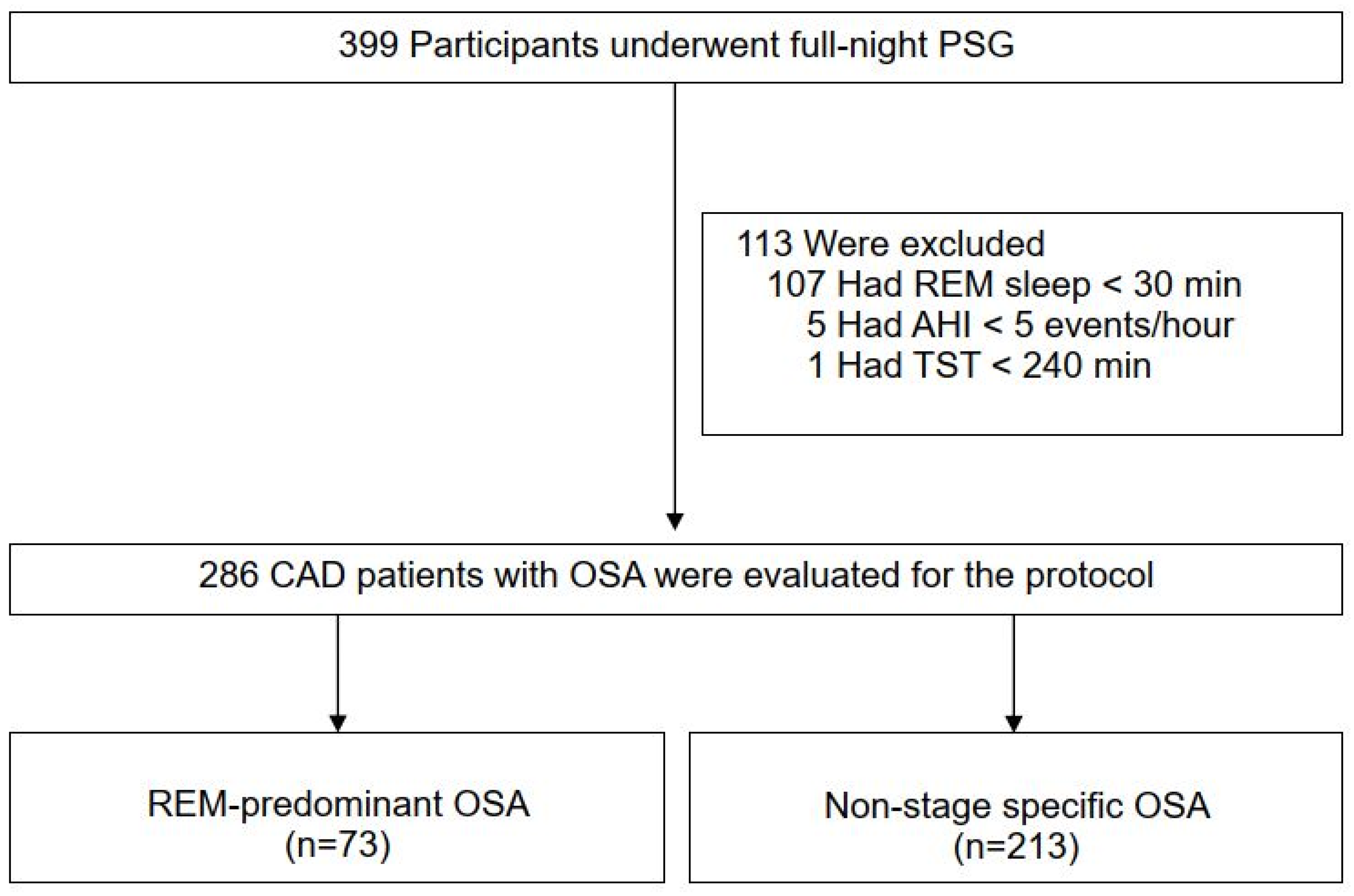
2.1. Study Design and Participants

The RICCADSA cohort has been described in detail previously [15,17]. In brief, patients with CAD who underwent percutaneous coronary intervention (PCI) or coronary artery bypass grafting (CABG) in the Skaraborg County of West Götaland, Sweden, were recruited for the main trial between 2005 and 2010, and the follow-up for the primary outcomes was completed in May 2013 [15]. As illustrated in Figure 1, 399 of 511 patients had OSA based on an Apnea-Hypopnea-Index (AHI) ≥15/h on a home sleep apnea testing (HSAT) in the entire RICCADSA cohort. A polysomnography (PSG) was conducted in a hospital in the OSA group before the study started [17]. For the current protocol, only the patients with a REM sleep duration of at least 30 min were included. REM-predominant OSA was defined as a REM-AHI/non-REM-AHI ≥ 2 [3]. None of the patients were on treatment for OSA.

2.2. Questionnaires

Epworth sleepiness scale (ESS): The ESS questionnaire [18] was used to determine subjective daytime sleepiness. As previously described, the ESS questionnaire contains eight questions to address the possibility of dozing off under eight different conditions in the past month. A cut-off value of 10 out of 24 was used for categorizing the patients with excessive daytime sleepiness in the RICCADSA trial [17].
Zung Self-rated Depression Scale (SDS): The Zung SDS is a commonly accepted survey that provides both a total score and a categorical rating of depression [19]. In brief, 20 questions are included with a score ranging from 1 to 4 points, which gives a total raw score from 20 to 80. As previously described in detail, the raw score is multiplied by 1.25, and the total result ranges from 25 to 100. The subjects with a score below 50 were categorized as normal, and the patients with a score of 50 or more were classified as having depression [19].
Zung Self-rated Anxiety Scale (SAS): The Zung SAS is a Likert scale that compromises 20 items to measure physiological and psychological symptoms [20]. As in the Zung SDS, each item is rated on a 4-point scale from 1 (none, or a little of the time) to 4 (most, or all of the time), and the raw scores (range 20–80) are converted into index scores (range 25–100) by multiplying 1.25 [20]. Individuals with a SAS score of at least 45 points were defined as having clinically significant anxiety [21].
Functional Outcomes of the Sleep Questionnaire (FOSQ): FOSQ is a survey regarding sleep-related symptoms, divided into five dimensions, including activity level, vigilance, intimacy, and sexual relationships, general productivity, and social outcome [22]. Item responses range from no difficulty (4 points) to extreme difficulty (1 point). The total score is calculated as the sum of the subscale scores, with a total score ranging from 5 to 20. Lower scores suggest function impairment, and the clinically significant impairment in the FOSQ is defined as a total score of fewer than 17.9 points [23].
Short Form-36 Health Survey (SF-36): Data regarding the Health-Related Quality of Life (HRQoL) were collected with the SF-36, which measures eight different domains (Physical Functioning, Role Physical, Bodily Pain, General Health, Vitality, Social Functioning, Role Emotional, Mental Health) and two summary scores (Physical Component Summary [PCS], Mental Component Summary [MCS]) [24]. An SF-36 questionnaire is a subjective tool that reflects the individual’s well-being preceding four weeks prior to the time of the survey. Domain and summary component scores range from 0 to 100; higher scores correspond to better health status or well-being.

2.3. Sleep Recordings

For the HSATs, the Embletta® Portable Digital System device (Embla, Broomfield, CO, USA) was used. As explained previously [15], the HSAT system included a nasal pressure detector, two respiratory inductance plethysmography belts (RIP) for thoracoabdominal movements and body position, and a finger pulse-oximeter for heart rate and oxyhemoglobin saturation (SpO2). Apnea was defined as at least ≥90% cessation of airflow and hypopnea was defined as a 50% reduction, at least, in thoracoabdominal movement and nasal pressure amplitude for ≥10 s [25]. Additionally, the total number of significant falls in SpO2 (≥4% from the immediately preceding baseline) was scored and the oxygen desaturation index (ODI) was determined as the number of significant desaturations per hour of estimated sleep. Obstructive events with a clear reduction in RIP belts and in the nasal pressure amplitude for at least 10 s were also recorded as hypopneas if there was a significant desaturation [25]. For the overnight PSG in-hospital for the OSA group, before the study started, a computerized recording system (Embla A10®, Embla, Broomfield, CO, USA) was used. As previously described [17], the PSG system included sleep monitoring through three-channel electroencephalography (EEG [C4/A1, C3/A2, CZ/A1]), two-channel electrooculography (EOG), one-channel submental electromyography (EMG), bilateral tibial EMG and two-lead electrocardiogram (ECG) in addition to the cardiorespiratory channels as described for the Embletta system above [17]. PSG recordings were scored by an observer blinded to clinical data and baseline screening results from the previous HSAT recordings. Obstructive events on the PSGs were scored according to the same criteria applied to the HSATs.

2.4. Comorbidities

As described previously, baseline anthropometrics, smoking habits, and medical history of the entire study population were obtained from the medical records [15]. Obesity was defined as a body mass index (BMI) ≥ 30 kg/m2, and abdominal obesity was defined as the waist-to-hip ratio (WHR) ≥ 0.9 for men and WHR ≥ 0.8 for women, respectively [26].

2.5. Statistical Analysis

The Shapiro–Wilk test was used to test the normality assumption of the current data for all variables. Descriptive data are shown as means and standard deviation (SD), or median with interquartile ranges (IQR) for continuous variables, and as a percentage for categorical variables. For comparison between groups, an independent sampled student t-test or, when appropriate, the Mann–Whitney U test was used. The Chi-square test was used for the comparison of categorical variables. Pearson’s correlation analysis was used to determine the relationship between REM-AHI levels and the other continuous variables. A logistic regression model was applied to determine variables associated with REM predominant OSA. Age, BMI, sex, percentage of stage 3 of non-REM-sleep (slow-wave sleep [SWS]) and AHI (Model 1) or ODI (Model 2), as well as significant associates (if any) in the univariate analysis, were entered into the multivariate models. Linear univariate regression analysis was used to evaluate the association between REM-AHI and the continuous variables of the ESS, FOSQ, SF-36, Zung SDS, and SAS scores, respectively, and the significant results were entered into the multivariate models with age, BMI, and female sex covariates. The significant variables associated with REM-AHI were tested separately against non-REM-AHI to address if the REM-AHI-related associations were exclusive for the REM sleep stage. All statistical tests were two-sided, odds ratios (ORs) with 95% confidence interval (CI) were reported, and a p-value < 0.05 was considered significant. Statistical analysis was performed using the Statistical Package for Social Sciences, version 22.0 for the Windows® system (SPSS® Inc., Chicago, IL, USA).

3. Results

A total of 399 participants with OSA were eligible for the current protocol. After excluding patients with REM sleep duration less than 30 min (n = 107), total sleep time less than 4 h (n = 1), and AHI less than 5 events/h on the PSG (n = 5), 286 (mean age 63.7 ± 7.9 years, 86.1% men) remained as the final study population (Figure 1).
In all, 73 (25.5%) of the study cohort had REM-predominant OSA. As shown in Table 1, there were more women among the participants with REM-predominant OSA compared to those with non-stage-specific OSA (26.0% vs. 9.9%; p = 0.001). Average BMI was significantly higher, and obesity was more common (42.5% vs. 25.4; p = 0.003) in the REM-predominant group, whereas there were no significant differences between the groups regarding age and comorbidities.
Table 2 demonstrates that sleep efficiency, as well as the duration and proportion of slow-wave sleep (SWS), was significantly higher in the REM-predominant group, whereas the non-stage specific OSA group had more severe OSA in terms of total AHI and oxygenation indices. Per selection criteria requiring at least 30 min of REM sleep, the REM-dependent OSA group tended to have longer REM sleep time and significantly more severe REM-AHI than the non-stage specific group.
As shown in Table 3, there were no significant differences between the groups regarding EDS, functional outcomes, mood, and HRQoL measures at baseline.
In unadjusted logistic regression analyzes, there were significant positive relationships between REM-predominant OSA and female sex, obesity, BMI, and SWS, whereas AHI and ODI were inversely correlated (Table 4). In the adjusted models, female sex, obesity, and BMI, as well as AHI and ODI, remained significant determinants of REM-predominant OSA (Table 4).
As illustrated in Figure 2A, there was significant inverse relationship between REM-AHI and age (r = −0.124, p = 0.035), whereas the correlation with BMI was positive (Figure 2B; r = 0.351, p < 0.001).
Other significant correlates of REM-AHI were Zung SDS scores (Figure 3A; r = 0.154, p = 0.010) as well as the SF-36 subdomains, Vitality (Figure 3B; r = −0.126, p = 0.022) (Figure 3B), Mental Health (Figure 3C, r = −0.148, p = 0.013), and MCS (Figure 3D, r = −0.142, p = 0.013). No significant associations were found between REM-AHI and other SF-36 domains as well as ESS, FOSQ scores, and Zung SAS scores (data not shown).
In the adjusted models, the association of REM-AHI with Zung SDS, Vitality, and Mental Health scores disappeared. However, the relationship with MCS remained significant after including age, BMI, and female sex into the model (β-coefficient −2.20, %95 CI −0.56, −0.03; p = 0.028) (Table 5). Non-REM-AHI was not associated with the aforementioned variables (r = 0.101, p = 0.094 for Zung SDS; r = −0.080, p = 0.181 for Vitality; r = −0.106, p = 0.077 for Mental Health; and r = −0.101, p = 0.098 for MCS).

4. Discussion

The current analysis demonstrated that REM-predominant OSA was significantly associated with female sex and obesity in this revascularized CAD cohort. Despite a lower degree of OSA in terms of AHI and oxygenation indices, the patients with REM-predominant OSA had similar characteristics regarding comorbidities, EDS, functional outcomes, mood, and QoL measures as did the patients with non-stage specific OSA. Further analyzes revealed significant associations of the REM-AHI with depressive mood scores as well as SF-36 subdomains Vitality and Mental Health, which all, however, disappeared in the multivariate models. Only the SF-domain MCS remained significantly associated with REM-AHI independent of age, BMI, and female sex.
To our knowledge, this is the first study to evaluate the association of REM-predominant OSA with excessive sleepiness, functional outcomes, anxiety, and depression, as well as the quality of life measures in a CAD cohort. In prior literature, Zinchuk and colleagues have reported that adults with REM-predominant OSA in sleep clinic cohorts are usually younger women with more efficient sleep and longer total sleep time compared to patients with non-stage specific OSA [3]. Our results suggesting an inverse relationship between REM-AHI and age as well as the dominance of female sex among the REM-predominant OSA supports those findings to be valid also in patients with CAD. Our results are in line with the findings from prior sleep-clinic-based studies, which showed that REM-predominant OSA was not related to excessive sleepiness as measured by multiple sleep latency tests [27,28], as well as with the findings from the large population-based Sleep Heart Health Study, which showed no relationship between REM-AHI and ESS scores after adjustment for confounding factors such as age, sex, and race [11].
Few studies addressed the functional outcomes in patients with REM-predominant OSA. Though the definition of hypopneas, as well as REM-predominant OSA, differed, the total FOSQ score tended to be lower among patients with REM-predominant OSA compared to non-stage specific OSA in 2 studies [29,30]. Our results do not support those findings, which might reflect the differences in clinical characteristics of sleep clinics vs. cardiac cohorts, as the patients from sleep clinics are usually symptomatic and have lower FOSQ scores at baseline. The average total FOSQ score was around 12 units in both studies [29,30], whereas our cohort demonstrated values of around 18 units, which suggests that many patients with CAD are with concomitant OSA are asymptomatic and do not have impairment in sleep-related functional outcomes.
Changes in sleep architecture regarding REM sleep have been associated with depressive mood [8,9] and anxiety [10]. In general, sleep pattern changes among patients with depression consist of impaired sleep onset and maintenance, early morning awakening, as well as reduced deep sleep and disinhibition of REM sleep [8]. Sleep disturbances are prevalent in some mental diseases, such as anxiety disorders, and anxiety disorders have been reported to be more common in REM-related OSA in a sleep clinic cohort [10], but we did not observe any difference in the anxiety scores in our CAD cohort. Of note, the Hospital Anxiety-Depression Scale was used in that study [10], whereas Zung SDS and Zung SAS scores were conducted in the current protocol. We found a significant association between REM-AHI and the Zung SDS scores, but this association disappeared after adjustment for the female sex.
Prior studies regarding the association of REM-predominant OSA with the QoL measures have not been consistent. The findings from the Sleep Heart Health Study [11] suggested that NREM-AHI but not REM-AHI was significantly associated with impairment in the PCS and MCS after adjustment for the confounding factors. The inclusion of individuals with a REM sleep time of less than 30 min, as well as the statistical analysis including both REM-AHI and NREM-AHI in the same model (collinearity), might explain some of the differences between our findings and the results of that population-based study [11]. We found significant linear correlations between REM-AHI and Mental Health and Vitality scores, but these associations were not significant anymore in the multivariate analysis. Notwithstanding, the association of the REM-AHI with the MCS remained significant after adjustment for age, BMI, and female sex. Given the absence of a significant association between non-REM-AHI and MCS, our findings suggest that the decline in MCS is exclusive to the REM sleep stage.
It should be noted that there have been several definitions of REM-dependent OSA in literature: (I) overall AHI ≥ 5 and REM-AHI/NREM-AHI ratio ≥ 2; (II) overall AHI ≥ 5, REM-AHI/NREM-AHI ratio ≥ 2, and NREM-AHI < 15; or (III) overall AHI ≥ 5, REM-AHI/NREM-AHI ratio ≥ 2, NREM-AHI < 15, and at least 10.5 min of REM sleep duration [31]. There have also been studies including only subjects with at least 30 min of recorded REM sleep [32] to reduce the possibility of exaggerating the effect of REM-predominant OSA in individuals for short REM duration, which is also applied in the current study. However, the strict criterion brings a potential bias, as the excluded patients are probably the ones with severe OSA (overall AHI ≥ 30), who otherwise would belong to the non-stage-specific OSA.
Notwithstanding, the significant association of REM-AHI with the MCS despite the lower degree of OSA severity highlights the need for a more complete characterization of each patient’s pathophysiology and a sex-stratified approach in the management of patients with CAD with concomitant OSA. This may be particularly important for increasing adherence to CPAP treatment to cover the early morning hours before awakening, thus treating the whole REM sleep period for better MCS outcomes. Given that there is a decrease in upper respiratory muscle tone in the REM period [33], which results in lower respiratory drive, and that longer periods of respiratory events cause more severe hypoxemia [34], a dose-response relationship between REM-AHI and hypertension has also been suggested [35]. However, more research with larger studies considering individual risk factor profiling in refining treatment strategies in OSA phenotypes is still urged.
We should acknowledge certain limitations. Firstly, the power estimate for the entire RICCADSA cohort was conducted for the primary outcome and not for the secondary outcomes assessed in this subprotocol. Secondly, the EDS definition was based on the ESS threshold, which may not be precise in a CAD population. Thirdly, our results are not generalizable to adults with OSA in the general population or in sleep clinic cohorts.

5. Conclusions

To conclude, female sex and BMI were related to REM-predominant OSA in this revascularized cohort. MCS was inversely associated with REM-AHI in the multivariate model. Further research is needed to establish better whether these patients will obtain any benefit from long-term CPAP therapy and whether sex-stratified treatment strategies should be applied for patients with CAD and REM-predominant OSA.

Author Contributions

Y.P. designed the study and performed the patient recruitment and clinical follow-ups. B.B., Y.C. and Y.P. performed the statistical analysis. B.B., Y.C. and Y.P. prepared the manuscript and drafted the article. All authors (B.B., Y.C., J.N., P.J.S.J. and Y.P.) interpreted the data. Y.P. obtained study funding and takes full responsibility for the work, including the study design, access to data, and the decision to submit and publish the manuscript. All authors have read and agreed to the published version of the manuscript.

Funding

The main RICCADSA trial is supported by grants from the Swedish Research Council (521-2011-537 and 521-2013-3439); the Swedish Heart-Lung Foundation (20080592, 20090708 and 20100664); the “Agreement concerning research and education of doctors” of Västra Götalandsregionen (ALFGBG-11538 and ALFGBG-150801), Research fund at Skaraborg Hospital (VGSKAS-4731, VGSKAS-5908, VGSKAS-9134, VGSKAS-14781, VGSKAS-40271, and VGSKAS-116431); Skaraborg Research and Development Council (VGFOUSKB-46371); the Heart Foundation of Kärnsjukhuset; ResMed Foundation; and ResMed Ltd. ResMed Sweden provided some of the sleep recording devices and technical support. None of the funders had any direct influence on the design of the study, the analysis of the data, data collection, drafting of the manuscript, or the decision to publish.

Institutional Review Board Statement

The study was conducted according to the guidelines of the Declaration of Helsinki and approved by the Regional Ethical Review Board in Gothenburg (approval nr 207-05; 13 September 2005; amendment T744-10; 26 November 2010; amendment T512-11; 16 June 2011). 2.2. The trial was registered with ClinicalTrials.gov (NCT 00519597) as well as with the national researchweb.org (FoU i Sverige—Research and development in Sweden; nr VGSKAS-4731; 04.29.2005).

Informed Consent Statement

All patients provided written informed consent.

Data Availability Statement

Individual participant data that underlie the results reported in this article can be obtained by contacting the principal investigator of the RICCADSA trial; [email protected].

Conflicts of Interest

Y.P. received institutional grants from ResMed for the main RICCADSA trial. P.S. reports personnel fees from Emmi Solutions, Jazz Pharmaceuticals, Itamar Medical, Separation Design Group, Belluscura, grants and personal fees from ResMed, Inspire Medical Systems, grants from Philips-Respironics, National Football League, and PinMed, other from Biologix, outside the submitted work; B.B., Y.C. and J.N. report no conflict of interest.

References

  1. Patil, S.P.; Ayappa, I.A.; Caples, S.M.; Kimoff, R.J.; Patel, S.R.; Harrod, C.G. Treatment of Adult Obstructive Sleep Apnea with Positive Airway Pressure: An American Academy of Sleep Medicine Clinical Practice Guideline. J. Clin. Sleep Med. 2019, 15, 335–343. [Google Scholar] [CrossRef] [Green Version]
  2. Peker, Y.; Balcan, B. Cardiovascular outcomes of continuous positive airway pressure therapy for obstructive sleep apnea. J. Thorac. Dis. 2018, 10, S4262–S4279. [Google Scholar] [CrossRef] [PubMed] [Green Version]
  3. Zinchuk, A.V.; Gentry, M.J.; Concato, J.; Yaggi, H.K. Phenotypes in obstructive sleep apnea: A definition, examples and evolution of approaches. Sleep Med. Rev. 2017, 35, 113–123. [Google Scholar] [CrossRef] [PubMed]
  4. Owens, R.L.; Edwards, B.A.; Eckert, D.J.; Jordan, A.S.; Sands, S.A.; Malhotra, A.; White, D.P.; Loring, S.H.; Butler, J.P.; Wellman, A. An Integrative Model of Physiological Traits Can be Used to Predict Obstructive Sleep Apnea and Response to Non Positive Airway Pressure Therapy. Sleep 2015, 38, 961–970. [Google Scholar] [CrossRef] [PubMed] [Green Version]
  5. Strollo, P.J., Jr.; Soose, R.J.; Maurer, J.T.; de Vries, N.; Cornelius, J.; Froymovich, O.; Hanson, R.D.; Padhya, T.A.; Steward, D.L.; Gillespie, M.B.; et al. Upper-airway stimulation for obstructive sleep apnea. N. Engl. J. Med. 2014, 370, 139–149. [Google Scholar] [CrossRef] [PubMed] [Green Version]
  6. Somers, V.K.; Dyken, M.E.; Clary, M.P.; Abboud, F.M. Sympathetic neural mechanisms in obstructive sleep apnea. J. Clin. Investig. 1995, 96, 1897–1904. [Google Scholar] [CrossRef] [PubMed] [Green Version]
  7. Mokhlesi, B.; Punjabi, N.M. “REM-related” obstructive sleep apnea: An epiphenomenon or a clinically important entity? Sleep 2012, 35, 5–7. [Google Scholar] [CrossRef] [PubMed] [Green Version]
  8. Palagini, L.; Baglioni, C.; Ciapparelli, A.; Gemignani, A.; Riemann, D. REM sleep dysregulation in depression: State of the art. Sleep Med. Rev. 2013, 17, 377–390. [Google Scholar] [CrossRef]
  9. Harlev, D.; Ravona-Springer, R.; Nuriel, Y.; Fruchter, E. Sleep Monitoring Using WatchPAT Device to Predict Recurrence of Major Depression in Patients at High Risk for Major Depression Disorder Recurrence: A Case Report. Front. Psychiatry 2021, 12, 572660. [Google Scholar] [CrossRef]
  10. Geckil, A.A.; Ermis, H. The relationship between anxiety, depression, daytime sleepiness in the REM-related mild OSAS and the NREM-related mild OSAS. Sleep Breath. = Schlaf. Atm. 2020, 24, 71–75. [Google Scholar] [CrossRef]
  11. Chami, H.A.; Baldwin, C.M.; Silverman, A.; Zhang, Y.; Rapoport, D.; Punjabi, N.M.; Gottlieb, D.J. Sleepiness, quality of life, and sleep maintenance in REM versus non-REM sleep-disordered breathing. Am. J. Respir. Crit. Care Med. 2010, 181, 997–1002. [Google Scholar] [CrossRef] [Green Version]
  12. Chami, H.A.; Gottlieb, D.J.; Redline, S.; Punjabi, N.M. Association between Glucose Metabolism and Sleep-disordered Breathing during REM Sleep. Am. J. Respir. Crit. Care Med. 2015, 192, 1118–1126. [Google Scholar] [CrossRef] [Green Version]
  13. Appleton, S.L.; Vakulin, A.; Martin, S.A.; Lang, C.J.; Wittert, G.A.; Taylor, A.W.; McEvoy, R.D.; Antic, N.A.; Catcheside, P.G.; Adams, R.J. Hypertension Is Associated with Undiagnosed OSA during Rapid Eye Movement Sleep. Chest 2016, 150, 495–505. [Google Scholar] [CrossRef]
  14. Mokhlesi, B.; Hagen, E.W.; Finn, L.A.; Hla, K.M.; Carter, J.R.; Peppard, P.E. Obstructive sleep apnoea during REM sleep and incident non-dipping of nocturnal blood pressure: A longitudinal analysis of the Wisconsin Sleep Cohort. Thorax 2015, 70, 1062–1069. [Google Scholar] [CrossRef] [Green Version]
  15. Peker, Y.; Glantz, H.; Eulenburg, C.; Wegscheider, K.; Herlitz, J.; Thunstrom, E. Effect of Positive Airway Pressure on Cardiovascular Outcomes in Coronary Artery Disease Patients with Nonsleepy Obstructive Sleep Apnea. The RICCADSA Randomized Controlled Trial. Am. J. Respir. Crit. Care Med. 2016, 194, 613–620. [Google Scholar] [CrossRef]
  16. Peker, Y.; Thunström, E.; Glantz, H.; Eulenburg, C. Effect of Obstructive Sleep Apnea and CPAP Treatment on Cardiovascular Outcomes in Acute Coronary Syndrome in the RICCADSA Trial. J. Clin. Med. 2020, 9, 4051. [Google Scholar] [CrossRef]
  17. Peker, Y.; Glantz, H.; Thunstrom, E.; Kallryd, A.; Herlitz, J.; Ejdeback, J. Rationale and design of the Randomized Intervention with CPAP in Coronary Artery Disease and Sleep Apnoea—RICCADSA trial. Scand. Cardiovasc. J. 2009, 43, 24–31. [Google Scholar] [CrossRef]
  18. Johns, M.W. A new method for measuring daytime sleepiness: The Epworth sleepiness scale. Sleep 1991, 14, 540–545. [Google Scholar] [CrossRef] [Green Version]
  19. Zung, W.W.; Richards, C.B.; Short, M.J. Self-rating depression scale in an outpatient clinic. Further validation of the SDS. Arch. Gen. Psychiatry 1965, 13, 508–515. [Google Scholar] [CrossRef] [PubMed]
  20. Zung, W.W. A rating instrument for anxiety disorders. Psychosomatics 1971, 12, 371–379. [Google Scholar] [CrossRef]
  21. Dunstan, D.A.; Scott, N.; Todd, A.K. Screening for anxiety and depression: Reassessing the utility of the Zung scales. BMC Psychiatry 2017, 17, 329. [Google Scholar] [CrossRef]
  22. Weaver, T.E.; Laizner, A.M.; Evans, L.K.; Maislin, G.; Chugh, D.K.; Lyon, K.; Smith, P.L.; Schwartz, A.R.; Redline, S.; Pack, A.I.; et al. An instrument to measure functional status outcomes for disorders of excessive sleepiness. Sleep 1997, 20, 835–843. [Google Scholar] [PubMed]
  23. Weaver, T.E.; Maislin, G.; Dinges, D.F.; Bloxham, T.; George, C.F.; Greenberg, H.; Kader, G.; Mahowald, M.; Younger, J.; Pack, A.I. Relationship between hours of CPAP use and achieving normal levels of sleepiness and daily functioning. Sleep 2007, 30, 711–719. [Google Scholar] [CrossRef] [PubMed] [Green Version]
  24. Ware, J.E., Jr. SF-36 health survey update. Spine 2000, 25, 3130–3139. [Google Scholar] [CrossRef] [PubMed]
  25. Sleep-related breathing disorders in adults: Recommendations for syndrome definition and measurement techniques in clinical research. The Report of an American Academy of Sleep Medicine Task Force. Sleep 1999, 22, 667–689. [CrossRef]
  26. Obesity: Preventing and Managing the Global Epidemic; WHO Technical Report Series 894; World Health Organization: Geneva, Switzerland, 2000; 252p.
  27. Chervin, R.D.; Aldrich, M.S. The relation between multiple sleep latency test findings and the frequency of apneic events in REM and non-REM sleep. Chest 1998, 113, 980–984. [Google Scholar] [CrossRef] [Green Version]
  28. Punjabi, N.M.; Bandeen-Roche, K.; Marx, J.J.; Neubauer, D.N.; Smith, P.L.; Schwartz, A.R. The association between daytime sleepiness and sleep-disordered breathing in NREM and REM sleep. Sleep 2002, 25, 307–314. [Google Scholar]
  29. Duce, B.; Kulkas, A.; Langton, C.; Töyräs, J.; Hukins, C. The prevalence of REM-related obstructive sleep apnoea is reduced by the AASM 2012 hypopnoea criteria. Sleep Breath. Schlaf. Atm. 2018, 22, 57–64. [Google Scholar] [CrossRef]
  30. Hu, X.Y.; Cho, J.G.; Perri, R.; Ting, T.; Al Oweidat, K.; Lambert, S.; Wheatley, J. CPAP treatment in REM-related obstructive sleep apnea: A distinct clinical phenotype of sleep disordered breathing. Sleep Breath. Schlaf. Atm. 2021, 25, 1875–1884. [Google Scholar] [CrossRef]
  31. Conwell, W.; Patel, B.; Doeing, D.; Pamidi, S.; Knutson, K.L.; Ghods, F.; Mokhlesi, B. Prevalence, clinical features, and CPAP adherence in REM-related sleep-disordered breathing: A cross-sectional analysis of a large clinical population. Sleep Breath. Schlaf. Atm. 2012, 16, 519–526. [Google Scholar] [CrossRef]
  32. Mokhlesi, B.; Finn, L.A.; Hagen, E.W.; Young, T.; Hla, K.M.; Van Cauter, E.; Peppard, P.E. Obstructive sleep apnea during REM sleep and hypertension. results of the Wisconsin Sleep Cohort. Am. J. Respir. Crit. Care Med. 2014, 190, 1158–1167. [Google Scholar] [CrossRef] [Green Version]
  33. McSharry, D.G.; Saboisky, J.P.; Deyoung, P.; Jordan, A.S.; Trinder, J.; Smales, E.; Hess, L.; Chamberlin, N.L.; Malhotra, A. Physiological mechanisms of upper airway hypotonia during REM sleep. Sleep 2014, 37, 561–569. [Google Scholar] [CrossRef] [Green Version]
  34. Trinder, J.; Kleiman, J.; Carrington, M.; Smith, S.; Breen, S.; Tan, N.; Kim, Y. Autonomic activity during human sleep as a function of time and sleep stage. J. Sleep Res. 2001, 10, 253–264. [Google Scholar] [CrossRef] [Green Version]
  35. Pamidi, S.; Knutson, K.L.; Ghods, F.; Mokhlesi, B. Depressive symptoms and obesity as predictors of sleepiness and quality of life in patients with REM-related obstructive sleep apnea: Cross-sectional analysis of a large clinical population. Sleep Med. 2011, 12, 827–831. [Google Scholar] [CrossRef]
Figure 1. Consort flow chart of the study sample. Definition of abbreviations: AHI = apnea-hypopnea index; CAD = coronary artery disease; OSA = obstructive sleep apnea; PSG = polysomnography; REM =rapid eye movements; TST = total sleep time.
Jcm 11 04402 g001
Figure 2. (A). Pearson correlation between REM-AHI levels and age, (B). Pearson correlation analysis between REM-AHI and BMI. Definition of abbreviations: AHI = apnea-hypopnea-index; BMI = body mass index; REM = rapid eye movements.
Jcm 11 04402 g002
Figure 3. (A). Pearson analysis between REM-AHI and Zung Self-rated Depression Scale score. (B). Pearson analysis between REM-AHI and Vitality Score. (C). Pearson analysis between REM-AHI and Mental Health score. (D). Pearson analysis between REM-AHI and Mental Component Summary score. Definition of abbreviations: AHI = apnea-hypopnea-index; REM = rapid eye movements.
Jcm 11 04402 g003
Table 1. Baseline demographic and clinical characteristics of the study population (n = 286).
REM-Predominant OSA
N = 73
Non-Stage Specific
OSA
N = 213
p-Value
Age, years63.2 ± 7.563.8 ± 8.10.539
Age ≥ 65 years, %43.843.20.924
Female sex, %26.09.90.001
BMI, kg/m229.0 (26.3–32.2)28.1 (25.8–29.9)0.020
Obesity, %42.524.40.003
Current smoker, %16.416.41.000
CABG at baseline, %20.523.90.552
AMI at baseline, %43.851.20.279
Former revascularization, %24.321.00.554
Hypertension,%64.460.60.563
History of atrial fibrillation, %11.016.90.225
Diabetes mellitus, %24.722.10.648
History of stroke,%8.27.60.861
Pulmonary disease,%5.56.10.846
Anti-depressive medication, %5.63.40.419
Definitions of abbreviations: AMI = acute myocardial infarction; BMI = body mass index; CABG = coronary artery by-pass grafting; OSA = obstructive sleep apnea; REM = rapid eye movements.
Table 2. Polysomnographic findings of the study population (n = 286).
REM-Predominant OSA
N = 73
Non-Stage Specific OSA
N = 213
p-Value
TST, min430.5 (374.8–469.3)413.3 (371.7–451.5)0.320
Sleep Onset, min10.0 (5.3–14.1)9.3 (5.7–16.6)0.702
Sleep Efficiency, % of TST84.6 (78.3–91.0)82.4 (75.5–88.3)0.041
SWS, min32.5 (3.5–59.5)22.0 (0.5–47.3)0.039
SWS, % of TST7.8 (0.9–15.3)5.6 (0.1–11.5)0.044
REM sleep, min63.5 (47.5–79.8)59.5 (42.0–71.8)0.077
REM sleep, % of TST15.7 (12.3–19.4)14.4 (10.9–18.0)0.151
AHI, events/hour22.6 (13.9–31.9)36.6 (24.4–55.7)<0.001
REM—AHI, events/hour53.5 (33.4–69.0)34.5 (18.2–56.8)<0.001
Non-REM—AHI, events/hour16.1 (9.7–27.4)38.1 (25.9–57.1)<0.001
ODI, events/hour10.2 (6.7–19.9)19.2 (9.2–31.5)<0.001
Average SpO2 %94.0 (93.2–95.1)93.9 (92.8–94.8)0.098
Nadir SpO2 %82.0 (78.0–86.5)84.0 (79.0–87.0)0.289
SpO2 < 90%, min5.1 (1.2–16.9)5.1 (1.0–24.0)0.473
SpO2 < 90%, % of TST1.5 (0.3–4.8)1.2 (0.2–5.3)0.820
Heart rate, beats/min58.8 (53.4–64.8)57.0 (51.3–63.2)0.167
Definitions of abbreviations: AHI = apnea-hypopnea-index; ODI = oxygen desaturation index; OSA = obstructive sleep apnea; REM = rapid eye movements; SpO2 = oxyhemoglobin saturation; SWS = slow wave sleep (stage 3 of non-REM sleep); TST = total sleep time.
Table 3. Comparison of excessive sleepiness, functional outcomes, depression, anxiety, and quality of life measures of the study participants (n = 286) polysomnographic findings of the study population (n = 286).
REM-Predominant OSA
N = 73
Non-Stage Specific
OSA
N = 213
p-Value
ESS score8 (4.0–11.0)8.0 (5.0–11.0)0.504
EDS (ESS score ≥ 10), %42.539.90.598
FOSQ scores
 General Productivity3.8 (3.6–4.0)4.0 (3.7–4.0)0.216
 Social Outcome4.0 (4.0–4.0)4.0 (4.0–4.0)0.580
 Activity level3.8 (3.2–3.9)3.7(3.3–3.9)0.872
 Vigilance3.7 (3.4–4.0)3.7 (3.3–4.0)0.891
 Intimate relationship3.5 (3.0–4.0)3.8 (3.0–4.0)0.988
 Total score18.5 (17.6–19.4)18.7 (17.3–19.6)0.594
Zung SDS score38.8 (33.4–50.0)41.3 (35.0–50.0)0.244
Depression (Zung SDS score ≥ 50), %31.428.30.618
Zung SAS score36.3 (31.3–43.8)38.8 (33.8–41.3)0.211
Anxiety (Zung SAS score ≥ 45), %21.415.90.294
SF-36 domains
 Physical Functioning85.0 (65.0–95.0)83.3 (68.3–90.0)0.577
 Role Physical75.0 (25.0–100.0)75.0 (25.0–100.0)0.575
 Bodily Pain84.0 (61.0–100.0)74.0 (51.0–100.0)0.090
 General Health70.0 (57.0–82.0)67.0 (52.0–82.0)0.610
 Vitality70.0 (55.0–85.0)70.0 (50.0–85.0)0.782
 Social Functioning100.0 (87.5–100.0)100.0 (75.0–100.0)0.230
 Role Emotional100.0 (66.7–100.0)100.0 (66.7–100.0)0.307
 Mental Health88.0 (75.0–96.0)88.0 (72.0–96.0)0.379
 PCS46.2 (39.4–52.9)46.0 (37.6–52.6)0.690
 MCS55.1 (46.9–58.7)54.2 (45.3–57.9)0.335
Definitions of abbreviations: ESS = Epworth sleepiness scale; FOSQ = functional outcomes of sleep questionnaire; MCS = mental component summary; OSA = obstructive sleep apnea; PCS = physical component summary; REM = rapid eye movements; SAS = self-rating anxiety scale; SDS = self-rating depression scale; SF = short form.
Table 4. Unadjusted and adjusted ORs (95% CIs) for variables associated with REM predominant OSA.
VariablesOR95 CI%p-Value
Unadjusted
Age, years0.980.96–1.020.538
Female sex3.221.61–6.410.001
BMI1.091.02–1.160.011
Obesity2.291.30–3.990.004
ESS0.960.90–1.030.316
EDS (ESS ≥ 10)1.160.67–1.980.598
SWS, % of TST1.041.01–1.080.015
AHI, events/h0.950.93–0.97<0.001
ODI, events/h0.960.94–0.98<0.001
Nadir SpO2 %0.980.95–1.020.346
SpO2 < 90%, min0.990.98–1.000.098
SpO2 < 90%, % of TST0.970.93–1.010.115
Current smoker1.000.48–2.050.999
Baseline AMI0.750.44–1.270.280
Hypertension1.180.68–2.050.563
History of atrial fibrillation0.610.27–1.370.228
Diabetes mellitus1.160.62–2.160.649
Stroke1.090.41–2.900.861
Pulmonary disease0.890.28–2.830.846
Zung SDS score0.990.96–1.010.297
Zung SAS score0.980.94–1.010.217
PCS1.010.98–1.040.507
MCS1.010.99–1.040.355
Adjusted
Model 1
Age, years0.980.94–1.020.374
Female sex vs. Male sex4.641.85–11.640.001
BMI, kg/m21.171.07–1.28<0.001
SWS, % of TST0.990.96–1.030.716
AHI, events/h0.930.91–0.95<0.001
Model 2
Age, years0.980.94–1.020.359
Female sex vs. Male sex2.961.30–6.720.009
BMI, kg/m21.141.05–1.230.003
SWS, % of TST1.010.98–1.050.606
ODI, events/h0.950.92–0.97<0.001
Definitions of abbreviations: AMI = acute myocardial infarction; AHI = apnea-hypopnea-index; BMI = body mass index; CI = confident interval; ESS = Epworth sleepiness scale; MCS = mental component summary; ODI = oxygen desaturation index; OSA = obstructive sleep apnea; PCS = physical component summary; REM = rapid eye movements; SAS = self-rating anxiety scale; SDS = self-rating depression scale; SF = short form; SWS = slow wave sleep (stage 3 of non-REM sleep).
Table 5. Variables associated with REM-AHI in adjusted linear regression analyzes.
β Coefficient95% CIp-Value
Model 1
 Age−0.04−0.46, −0.250.544
 BMI0.301.15, 2.61<0.001
 Zung SDS score0.120.01, 0.570.045
Model 2
 Age−0.08−0.59, 0.110.177
 BMI0.270.95, 2.38<0.001
 Zung SDS score0.09−0.05, 0.500.115
 Female sex0.237.95, 23.70<0.001
Model 3
 Age−0.08−0.59, 0.120.185
 BMI0.270.99–2.42<0.001
 Vitality−0.07−0.19, 0.05<0.001
 Female sex0.249.30, 25.010.231
Model 4
 Age−0.08−0.59, 0.120.192
 BMI0.271.00, 2.42<0.001
 Mental Health−0.09−0.28, 0.020.094
 Female sex0.248.99, 24.64<0.001
Model 5
 Age−0.08−0.61, 0.110.168
 BMI0.281.06, 2.49<0.001
 MCS−0.12−0.55, −0.020.033
 Female sex0.259.81, 25.82<0.001
Definition of abbreviations: AHI = apnea-hypopnea-index; BMI = body mass index; CI = confident interval; MCS = mental component summary; OSA = obstructive sleep apnea; REM = rapid eye movements; SDS = self-rating depression scale.
Publisher’s Note: MDPI stays neutral with regard to jurisdictional claims in published maps and institutional affiliations.

