Article

Determinants of Arterial Pressure of Oxygen and Carbon Dioxide in Patients Supported by Veno-Arterial ECMO

1
Department of Anaesthesiology and Critical Care Medicine, Dijon University Medical Centre, F-21000 Dijon, France
2
Department of Anaesthesiology and Critical Care Medicine, University of Medicine and Pharmacy “Carol Davila”, 020021 Bucharest, Romania
3
LNC UMR1231, University of Burgundy and Franche-Comté, F-21000 Dijon, France
4
Cardiac Surgery Department, Dijon University Hospital, F-21000 Dijon, France
*
Author to whom correspondence should be addressed.
J. Clin. Med. 2022, 11(17), 5228; https://doi.org/10.3390/jcm11175228
Submission received: 3 June 2022 / Revised: 28 August 2022 / Accepted: 1 September 2022 / Published: 4 September 2022
(This article belongs to the Section Intensive Care)

Abstract

:
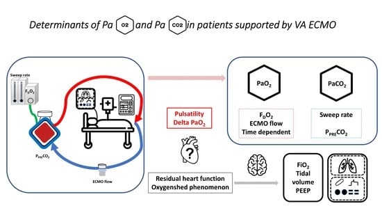
Background: The present study aimed to assess the determinants of arterial partial pressure of oxygen (PaO2) and carbon dioxide (PaCO2) in the early phase of veno-arterial extracorporeal membrane oxygenation (VA ECMO) support. Even though the guidelines considered both the risks of hypoxemia and hyperoxemia during ECMO support, there are a lack of data concerning the patients supported by VA ECMO. Methods: This is a retrospective, monocentric, observational cohort study in a university-affiliated cardiac intensive care unit. Hemodynamic parameters, ECMO parameters, ventilator settings, and blood gas analyses were collected at several time points during the first 48 h of VA ECMO support. For each timepoint, the blood samples were drawn simultaneously from the right radial artery catheter, VA ECMO venous line (before the oxygenator), and from VA ECMO arterial line (after the oxygenator). Univariate followed by multivariate mixed-model analyses were performed for longitudinal data analyses. Results: Forty-five patients with femoro-femoral peripheral VA ECMO were included. In multivariate analysis, the patients’ PaO2 was independently associated with QEC, FDO2, and time of measurement. The patients’ PaCO2 was associated with the sweep rate flow and the PpreCO2. Conclusions: During acute VA ECMO support, the main determinants of patient oxygenation are determined by VA ECMO parameters.

Graphical Abstract

1. Introduction

The use of veno-arterial extracorporeal membrane oxygenation (VA ECMO) has improved refractory cardiogenic shock mortality [1,2]. VA ECMO is used for bridge-to-recovery, bridge-to-transplantation, or bridge-to-decision purposes [3,4]. Several recommendations have been published relative to the indications, management and weaning of VA ECMO [5]. However, at bedside, physicians still have to manage the challenges related to interactions between the retrograde flow of the extracorporeal circuit and the native homeostatic systems [6,7,8,9,10]. Some important questions are still not addressed.
Firstly, the determinants of patient’s blood gases (PaO2 and PaCO2) have never been evaluated in patients on VA ECMO support. Several studies have evaluated the determinants of oxygenation and carbon dioxide clearance in the setting of veno-venous ECMO (VV ECMO), as it is used for respiratory indications [11,12,13,14]. Nevertheless, no study has ever been performed in patients supported by VA ECMO. An extrapolation of VV ECMO to VA ECMO is possible, but it might not reflect the hemodynamic effect of the retrograde aortic flow, particularly with commonly used femoral canulation [6].
Secondly, this hemodynamic phenomenon brings into question whether the oxygenated VA ECMO flow can reach the proximal aortic arch during the first hours of support, altering the difference between the PpostO2and theright radial artery PaO2 (delta-PaO2). A clear answer is difficult, considering that the maintaining of an optimal equilibrium between VA ECMO and the patient’s native heart and lung function is challenging, according to the medical conditions evolution and illness phase [9].
Recently, the Extracorporeal Life Support Organization (ELSO) published guidelines that consider the risks of both hypoxemia and hyperoxemia during ECMO support [15]. The ELSO experts recommend having a PpostO2 value of 150 mmHg to counterbalance the risk of hyperoxemia and to minimize the risk of hypoxemia [15]. The ELSO guidelines suggest targeting post membrane-normocapnia, but without providing a cut-off value [15]. A review of the published studies found out that these recommendations were proposed without clear published data [16]. In the absence of published data, a further question would be if the PpostO2 cutoff value of 150 mmHg proposed by ELSO guidelines can prevent hyperoxemia or hypoxemia.
The present study was designed to assess the determinants of blood gases (PaO2 and PaCO2) and determinants of and delta-PaO2 in patients supported by VA ECMO. The secondary aim was to evaluate the concordance with the actual ELSO guidelines.

2. Materials and Methods

2.1. Study Design

We performed a retrospective, observational, single-center study in a university-affiliated cardiac intensive care unit (ICU). Because all data were collected retrospectively from the standard medical chart, this study was not considered as involving human participants (according to French law). Nevertheless, this study was approved by the institutional review board (Ethics Committee of the French Society of Anaesthesia and Critical Care, IRB10252018179). Inclusion criteria were: (i) patients supported by peripheral femoro-femoral VA ECMO, (ii) right radial arterial cannula, and (iii) available pre/post membrane blood gas measurements. We excluded (i) pediatric patients and (ii) patients with femoral arterial catheter for invasive blood pressure monitoring. The patients with femoral arterial catheter for invasive blood pressure monitoring were excluded in order to have a homogenous cohort with the same site for arterial blood measurements.

2.2. Patients’ Management

All patients underwent femoro-femoral peripheral VA ECMO. The indications, protocols for implementation, VA ECMO management, and patient management have been described elsewhere [17,18,19]. Briefly, the VA ECMO circuit consists of a polymethylpentene hollow-fiber oxygenator (Maquet oxygenator or EOS Livanova), a magnetically levitated centrifugal pump, and heparin-coated tubing. The VA ECMO circuit is primed with isotonic saline solution. A dose of unfractionated heparin is administered in the absence of contraindications before VA ECMO implantation. The initial ECMO flow rate (QEC) is gradually increased until it corresponds to a cardiac index above 2.5 L min−1 m−2. Then, QEC is adapted to blood pressure, arterial lactate clearance, echocardiographic parameters, native heart function, and arterio-venous CO2 difference [20]. The fraction of inspired oxygen is progressively increased as needed to prevent ischemia-reperfusion injury. Sweep gas flow rate is initially placed at a ratio of 1:1 to the QEC, and then adjusted according to blood gases. All patients were mechanically ventilated during the study period. Patients were ventilated in the controlled volume mode with tidal volume of 5 to 8 mL kg−1 (predicted ideal body weight), respiratory rate between 5 and 12 respiratory rate per minute, and positive end-expiratory pressure (PEEP) between 5 and 12 cm H2O, in order to obtain plateau pressure below 25 cm H2O. All patients were continuously administered unfractionated heparin 4 h after VA ECMO initiation. The heparin dose was adjusted at least twice daily according to heparinemia, targeting an anti-factor Xa activity between 0.15 and 0.3 UI mL−1. Packed red blood cells were transfused to maintain hemoglobin level between 8 and 10 g dL−1.

2.3. Data Collection

All of the data were collected from the patient medical charts. These included demographic data, comorbidities, medical aetiology, circulatory parameters (systolic, mean and diastolic arterial pressure, heart rate, arterial line pulsatility), VA ECMO parameters (oxygen fraction (FDO2), sweep flow rate), ventilator settings (FiO2, minute volume, PEEP), vasopressor and inotropic therapy, blood gas analysis, ICU length of stay, hospital length of stay, and death. Blood gas analysis was noted at baseline (immediately after ECMO initiation), and at 12 h, 24 h, and 48 h after the initiation of VA ECMO support. For each time point, the blood samples were drawn simultaneously from the radial artery catheter, from the VA ECMO venous line (before the oxygenator), and from the VA ECMO arterial line (after the oxygenator). The arterial line pulsatility was defined as a pulse pressure ≥ 20 mmHg [21]. We calculated the gradient between PpostO2 and radial artery partial pressure (delta-PaO2) as the absolute difference between PpostO2 and patient’s radial artery PaO2.

2.4. Statistical Analyses

Continuous variables were expressed as medians with [25–75%] interquartile range (IQR), or as means ± standard deviation (SD), as appropriate. Categorical variables were expressed as numbers (percentage).
The determinants of PaO2, PaCO2, and delta-PaO2 were evaluated using a mixed effects generalized linear model approach. A random “subject” effect was used in these longitudinal analyses. Firstly, each of the collected variables of interest was introduced as fixed effect variable in a univariate mixed effect model with patient artery measured PaO2, PaCO2, or delta-PaO2, as dependent variables. Secondly, the statistically significant and clinically pertinent variables in univariate analysis were included in multivariate mixed effects generalized linear model models with patient artery measured PaO2, PaCO2, or delta-PaO2, as dependent variables.
The concordance with ELSO guidelines was evaluated based on the blood determinations of post-membrane PO2, because the ELSO experts recommend having a PpostO2 value of 150 mmHg and did not provide any cut-off value for post-membrane PCO2. Based on the PpostO2 the determinations were classified as ELSO concordant (<150 mmHg) or not. Furthermore, the patients’ PaO2 right radial artery determinations were classified as hyperoxemic (>120 mmHg) or as hypoxemic (<70 mmHg). The association the ELSO concordance (yes/no), hyperoxemia (yes/no), or hypoxemia (yes/no) was evaluated using an exploratory logistical regression model.
The statistical analyses were performed using RStudio (Version 1.1.447—© 2022–2018 RStudio, Inc. RStudio: Integrated Development for R. RStudio, PBC, Boston, MA, USA). The threshold for statistical significance was set at p < 0.05.

3. Results

The patients’ characteristics are shown in Table 1.
Forty-five patients were included in the study analyses. The mean age was 59 ± 12 years, and they were mostly male (77%), with a baseline SOFA score of 13 ± 2. The main indications for VA ECMO support were cardiopulmonary resuscitation (CPR), post-acute myocardial infarction cardiogenic shock, and post-cardiotomy cardiogenic shock. The description of patients’ hemodynamic and respiratory characteristics at each timepoint of the study is presented in Supplementary Table S1.

3.1. Determinants of Arterial Partial Pressure of Oxygen (PaO2)

The univariate analyses found that the patients’ PaO2 was associated with the ventilator FiO2, QEC, sweep rate, FDO2, pre-ECMO-membrane pH and post-ECMO-membrane pH, PaO2, and HCO3. The multivariate analysis demonstrated that QEC, the FDO2, and the time point of the measurement were the only variables independently associated with patient PaO2 (Table 2).

3.2. Determinants of Arterial Partial Pressure of Dioxide Carbon (PaCO2)

In univariate analyses we found that the patients’ PaCO2 was associated with pre- and post-membrane pressure of gases, but not with the ventilatory settings. In multivariate analysis, the sweep rate flow and the PpreCO2 were independently associated with patients’ PaCO2 (Table 3). Ventilatory setting were not associated with patients’ PaCO2.

3.3. Determinants of Delta-PaO2 (Difference between Post-Membrane PO2 and PaO2)

The median delta-PaO2 was 11 [−14–67] mmHg. In univariate analyses, delta-PaO2 was associated with arterial line pulsatility, pre-membrane pH, SpreO2, PpreO2, PpreCO2, post-membrane pH, PpostO2, PpostCO2, SpostO2, and FDO2. The multivariate analysis demonstrated that FDO2 and arterial pulsatility were significantly associated with delta-PaO2 (Supplementary Table S2).
The timing of measurement was not independently associated with the delta-PaO2.

3.4. Accordance with ELSO Guidelines

Fifty-nine (33%) blood determinations of PpostO2 were adequate (<150 mmHg), according to the recent ELSO recommendations. Of these 59 determinations, 22 (37%) had a radial artery PaO2 over 120 mmHg (hyperoxemia) and 14 (24%) a radial artery PaO2 lower than 70 mmHg (hypoxemia).
In all cohort, the concordance with ELSO guidelines was protective of patient hyperoxemia (OR 0.15, 95%CI 0.01-0.31, p < 0.001), but it was also associated with a risk of patient hypoxemia (OR 6.78, 95%CI 2.1–21.8, p = 0.001).

4. Discussion

Our findings can be summarized as following: (i) VA ECMO settings were the main contributor to patient oxygenation and CO2 removal during the first 48 h of support, (ii) the ventilator parameters had no significant clinical effect on oxygenation or CO2 removal during the acute phase of VA ECMO support, (iii) a patient’s pulsatile arterial flow pattern was associated with higher delta-PaO2, and (iv) maintaining a PpostO2 lower than 150 mmHg can be associated with an increased risk of hypoxemia.
Our study demonstrated that VA ECMO oxygenation parameters, and particularly ECMO membrane oxygenation parameters (FDO2, sweep flow rate), are the main determinants of patients’ PaO2. These results are similar to those demonstrated in VV ECMO, where patients’ oxygenation was dependent on the ratio between VV ECMO QEC and patient cardiac output, and VV ECMO oxygenation parameters [11]. The importance of VA ECMO PO2 might also suggest that the so-called oxygenshed phenomenon is not a very significant concern in the acute phase [22]. Even though we did not specifically measure it, our findings suggest that the retrograde aortic flow of VA ECMO was able to reach the proximal aortic arch (i.e., right radial artery). We did not find an association between hemoglobin and oxygenation, as described by some authors for VV ECMO [12,23,24]. This can be explained by the hemoglobin value, which was not very low in the patients from our cohort, and the QEC which was high, thus maintaining oxygen delivery [23]. We also found that oxygenation performance of the ECMO membrane decreases over time, even during the relatively short time period of 48 h, and despite anticoagulation. This observation, which confirms a prior study in VV ECMO patients [25], is explained by clot formation and cellular deposit on the fibers of the membrane [25,26].
Similarly to oxygenation, patient’s CO2 removal mainly depends on VA ECMO. However, due to CO2 different physical properties, there are some notable differences. Interestingly, the PpreCO2 was independently associated with PaCO2. The role of pre-membrane CO2 seemed similar in a previous experimental VV ECMO study [13]. This finding underlies the importance of the patient’s metabolic status, even during acute shock. The importance of the sweep gas flow rate has already been described for VV ECMO [11]. The same study and various others have reported that QEC and FiO2 have little effect on CO2 removal [11,27], but this is inconsistent with other experimental studies [13]. Contrary to oxygenation, time had no impact on membrane CO2 removal, at least during the first 48 h. This finding is explained by the high diffusion coefficient of CO2.
The gradient of oxygenation (delta-PaO2) between the ECMO PpostO2 and the patient’s PaO2 was influenced by ECMO FDO2 and arterial pulsatility. These results emphasize the main role of ECMO parameters, and particularly the residual cardiac function of the patient. A low pulse pressure during VA reflects a low native cardiac output [28]. Retrograde aortic flow into the aorta during VA ECMO can cause upper body hypoxemia in relation to the competitive flows between the native blood flow and the VA ECMO blood flow (QEC), particularly in the case of the commonly used peripheral femoro-femoral cannulation [29,30,31]. Several mixed blood zones have been observed because the mixed blood zone location depends on native heart and lung functionality and on the hemodynamic support provided by VA ECMO [32]. It is worth noting that 50% of the patients in our study had arterial pulsatility, and that PaO2 measured from the radial artery is a sensitive tool that is readily available at bedside to detect oxygenation imbalance [33]. The fact that arterial pulsatility was positively associated with the gradient of oxygenation between the ECMO membrane in our cohort reflects the effect of cardiac function on the oxygenation of the higher body part. Thus, this gradient could be analyzed as a variable reflecting the ratio between native cardiac/lung function and VA ECMO retrograde flow in the oxygenation of the body. In case of a pulsatility index over 20 mmHg, physicians should be aware of the risk of the oxygenshed phenomenon, but further studies are needed to confirm this point.
In our study, only one third of the PpostO2 determinations were in concordance with the ELSO cut-off of <150 mmHg. This rather low concordance level might be explained by the fact that the patients were managed before the issue of this recommendations. The ELSO concordance was associated with patients’ hypoxemia. Our cohort limited size did not allow for a complex analysis including all the possible confounding factors. The association with patient’s hypoxemia might be simply due a more severe hemodynamic status. However, this result confirms the need for large trials on this topic.

4.1. Clinical Implications

This study provided the first data of the determinants of patients’ blood gases during acute VA ECMO support, answering a both hemodynamic and ventilatory question. During the acute phase of circulatory VA ECMO support, patient oxygenation and CO2 removal is often overlooked. However, these factors are of importance for several reasons. Both hyperoxemia and hypoxemia are associated with worse outcomes [34]. Moreover, changes in PaCO2 levels affect neurological outcomes [35]. In VA ECMO-supported patients, blood oxygenation and CO2 removal are difficult to predict at bedside. Our results reflect real life for many patients who were hyperoxemic or hypoxemic.
When implementing VA ECMO, the ECMO team should be aware of the parameters of the VA ECMO sweep gas blender setting in regard to QEC. Hyperoxia affects outcomes and mortality. FDO2 should, therefore, be probably started at 0.6 because the probability of hypoxemia is low at this setting, since the PaO2 is over 100 mmHg in most cases [35], and because QEC is higher than native cardiac output. Because sweep gas flow is the main determinant of patient PaCO2, it should be carefully adapted to PaCO2 and to patient metabolic status. In our cohort, the median PaCO2 was low, with a ratio of sweep gas flow to QEC that was probably too high. We observed slow changes in sweep gas flow, and PaCO2 changes were within acceptable values. At the implantation of VA ECMO, sweep gas flow should probably not be too high and could be set at a ratio of 0.7 to QEC. Considering both our results and a priori data, physicians should probably measure blood gases as early as possible to adapt VA ECMO sweep gas blender setting parameters.
None of the ventilator parameters were associated with oxygenation or CO2 removal because QEC was high, and up to 50% of patients had very low cardiac output with no blood pulsatility. In this context, physicians should focus on the parameters of the VA ECMO to avoid anomalies in oxygenation and CO2 removal. Ventilator parameters, such as respiratory rate, ventilatory FiO2, or PEEP, were not associated with patient homeostasis. Nevertheless, studies have demonstrated that ventilatory parameters should be set to avoid lung trauma [36]. In other words, physicians should consider using protective ventilation with low FiO2 and low respiratory rate in patients supported by VA ECMO without any concerns about patient oxygenation or CO2 removal. This decision should be taken also based on the delta-PaO2 and the pulsatility that indicate the cardiac contribution to the systemic circulation.
This approach becomes even more important during the weaning process, where the decrease in QEC and the restoration of native cardiac blood flow through pulmonary circulation may favor competitive flow, and thus, local hypoxemia and tissue hypoperfusion [37]. In this context, the gradient between PpostO2 and patient’s PaO2 (delta-PaO2) could provide information on competing flow. In addition, most patients supported by VA ECMO undergo early extubation [17]. In this case, clinical examination and pulse oximetry may help to detect hypoxemia and/or “harlequin syndrome” during the decrease in VA ECMO support and the weaning process. Furthermore, in light of the recent ELSO recommendations preventing deleterious hypoxemia, the clinician should also be aware of the risk of patients’ hypoxemia.

4.2. Limits

The study is limited by its retrospective, observational, exploratory design, and causality cannot be inferred. Considering the high complexity of the ICU clinical situations requiring VA ECMO support, our study design is limited. We included only patients on femoro-femoral VA ECMO. Some important cardio-respiratory parameters were not available for the analysis, such as the native cardiac output for all patients or the native respiratory function and pulmonary shunt [27]. Furthermore, we did not evaluate middle and long-term lung function or patient outcomes. Best ventilation parameters was not an objective of the study [38]. These results are limited to the acute phase (i.e., the first 48 h) and cannot be extrapolated to later phases. However, the acute phase is highly critical because association between hyperoxemia, hypoxia, CO2 removal, and clinical outcomes has been demonstrated [39,40].

5. Conclusions

During the early VA ECMO support, the main determinants of patients’ oxygenation and carbon dioxide removal are the parameters the VA ECMO. QEC and FDO2 are associated with patient oxygenation, whereas sweep gas flow and PpreCO2 are associated with PaCO2. Further studies in patients with varying QEC and during the weaning process may lead to a better understanding of the interactions between QEC and native flow.

Supplementary Materials

The following supporting information can be downloaded at: https://www.mdpi.com/article/10.3390/jcm11175228/s1, Table S1: Patients’ characteristics at each timepoint (baseline, H12, H24, and H48); Table S2: Mixed generalized linear model with a random “subject” for longitudinal analysis of the variables associated with delta-PaO2 (the difference between PpostO2–and patient’s right radial artery PaO2). A univariate analysis was followed by a multivariate analysis.

Author Contributions

Conception and study design: S.A. and P.-G.G.; Patient recruitment: S.A., V.B., B.D., V.D., M.-C.M., O.B. and P.-G.G.; Data analysis: S.A. and M.N.; Interpretation of data: P.-G.G., M.N., S.A. and B.B.; Writing up the first draft of the paper: S.A. and P.-G.G.; Critical reviewing: B.B., M.-C.M. and O.B. All authors have read and agreed to the published version of the manuscript.

Funding

This research received no external funding.

Institutional Review Board Statement

The study was conducted in accordance with the Declaration of Helsinki, and approved by the Ethics Committee of the French Society of Anaesthesia and Critical Care, IRB10252018179.

Informed Consent Statement

Specific patient consent was waived due to the retrospective observational study design.

Data Availability Statement

Data are available upon reasonable request upon the corresponding author.

Acknowledgments

We are grateful to Suzanne Rankin for proofreading and reviewing the English manuscript.

Conflicts of Interest

The authors declare no conflict of interest.

References

  1. Hamm, C.W.; Bassand, J.-P.; Agewall, S.; Bax, J.; Boersma, E.; Bueno, H.; Caso, P.; Dudek, D.; Gielen, S.; Huber, K.; et al. ESC Guidelines for the management of acute coronary syndromes in patients presenting without persistent ST-segment elevation: The Task Force for the management of acute coronary syndromes (ACS) in patients presenting without persistent ST-segment elevation of the European Society of Cardiology (ESC). Eur. Heart J. 2011, 32, 2999–3054. [Google Scholar] [CrossRef]
  2. Wong, A.S.K.; Sin, S.W.C. Short-term mechanical circulatory support (intra-aortic balloon pump, Impella, extracorporeal membrane oxygenation, TandemHeart): A review. Ann. Transl. Med. 2020, 8, 829. [Google Scholar] [CrossRef]
  3. Welker, C.; Huang, J.; Ramakrishna, H. Analysis of the 2020 EACTS/ELSO/STS/AATS Expert Guidelines on the Management of Adult Postcardiotomy Extracorporeal Life Support. J. Cardiothorac. Vasc. Anesth. 2022, 36, 2207–2219. [Google Scholar] [CrossRef]
  4. Tsangaris, A.; Alexy, T.; Kalra, R.; Kosmopoulos, M.; Elliott, A.; Bartos, J.A.; Yannopoulos, D. Overview of Veno-Arterial Extracorporeal Membrane Oxygenation (VA-ECMO) Support for the Management of Cardiogenic Shock. Front. Cardiovasc. Med. 2021, 8, 686558. [Google Scholar] [CrossRef]
  5. Koeltz, A.; Gendron, N.; Ajzenberg, N.; Longrois, D. How to Manage Thrombocytopenia with ECLS: A Proposal of Clinical Reasoning Tools. J. Extra-Corpor. Technol. 2018, 50, 256–259. [Google Scholar]
  6. Geyer, M.; Gohrbandt, B.; Sagoschen, I.; Hartmann, T.; Post, F.; Vahl, C.-F.; Münzel, T. Pitfalls of cannulation for extracorporeal life support: Review of the literature and illustrative case presentation. J. Artif. Organs 2018, 21, 8–16. [Google Scholar] [CrossRef]
  7. Guimaron, S.; Laverdure, F.; Andrei, S.; Kortchinsky, T.; Thès, J.; Stéphan, F. Reimplantation of Venoarterial Extracorporeal Membrane Oxygenation (ECMO) After Withdrawal Failure. J. Cardiothorac. Vasc. Anesth. 2019, 33, 2360–2361. [Google Scholar] [CrossRef]
  8. Andrei, S.; Nguyen, M.; Berthoud, V.; Morgant, M.-C.; Bouhemad, B.; Guinot, P.-G.; ECMORIX Study Group. Evaluation of the Oxiris Membrane in Cardiogenic Shock Requiring Extracorporeal Membrane Oxygenation Support: Study Protocol for a Single Center, Single-Blind, Randomized Controlled Trial. Front. Cardiovasc. Med. 2021, 8, 738496. [Google Scholar] [CrossRef]
  9. Chung, M.; Shiloh, A.L.; Carlese, A. Monitoring of the adult patient on venoarterial extracorporeal membrane oxygenation. Sci. World J. 2014, 2014, 393258. [Google Scholar] [CrossRef]
  10. Gu, K.; Zhang, Z.; Gao, B.; Chang, Y.; Wan, F. Hemodynamic effects of perfusion level of peripheral ECMO on cardiovascular system. Biomed. Eng. OnLine 2018, 17, 59. [Google Scholar] [CrossRef]
  11. Schmidt, M.; Tachon, G.; Devilliers, C.; Muller, G.; Hekimian, G.; Bréchot, N.; Merceron, S.; Luyt, C.E.; Trouillet, J.-L.; Chastre, J.; et al. Blood oxygenation and decarboxylation determinants during venovenous ECMO for respiratory failure in adults. Intensiv. Care Med. 2013, 39, 838–846. [Google Scholar] [CrossRef]
  12. Messai, E.; Bouguerra, A.; Harmelin, G.; Di Lascio, G.; Bonizzoli, M.; Bonacchi, M. A numerical model of blood oxygenation during veno-venous ECMO: Analysis of the interplay between blood oxygenation and its delivery parameters. J. Clin. Monit. Comput. 2016, 30, 327–332. [Google Scholar] [CrossRef]
  13. Park, M.; Costa, E.L.V.; Maciel, A.T.; Silva, D.P.E.; Friedrich, N.; Barbosa, E.V.S.; Hirota, A.S.; Schettino, G.; Azevedo, L.C.P. Determinants of oxygen and carbon dioxide transfer during extracorporeal membrane oxygenation in an experimental model of multiple organ dysfunction syndrome. PLoS ONE 2013, 8, e54954. [Google Scholar] [CrossRef]
  14. Spinelli, E.; Colussi, G.; Dal Santo, G.; Scotti, E.; Marongiu, I.; Garbelli, E.; Mazzucco, A.; Dondossola, D.; Maia, R.; Battistin, M.; et al. Atelectasis, Shunt, and Worsening Oxygenation Following Reduction of Respiratory Rate in Healthy Pigs Undergoing ECMO: An Experimental Lung Imaging Study. Front. Physiol. 2021, 12, 663313. [Google Scholar] [CrossRef]
  15. Lorusso, R.; Shekar, K.; MacLaren, G.; Schmidt, M.; Pellegrino, V.; Meyns, B.; Haft, J.; Vercaemst, L.; Pappalardo, F.; Bermudez, C.; et al. ELSO Interim Guidelines for Venoarterial Extracorporeal Membrane Oxygenation in Adult Cardiac Patients. ASAIO J. 2021, 67, 827–844. [Google Scholar] [CrossRef]
  16. Winiszewski, H.; Guinot, P.-G.; Schmidt, M.; Besch, G.; Piton, G.; Perrotti, A.; Lorusso, R.; Kimmoun, A.; Capellier, G. Optimizing PO2 during peripheral veno-arterial ECMO: A narrative review. Crit. Care 2022, 26, 226. [Google Scholar] [CrossRef]
  17. Ellouze, O.; Lamirel, J.; Perrot, J.; Missaoui, A.; Daily, T.; Aho, S.; Petrosyan, A.; Guinot, P.G.; Bouchot, O.; Bouhemad, B. Extubation of patients undergoing extracorporeal life support. A retrospective study. Perfusion 2019, 34, 50–57. [Google Scholar] [CrossRef]
  18. Ellouze, O.; Abbad, X.; Constandache, T.; Missaoui, A.; Berthoud, V.; Daily, T.; Aho, S.; Bouchot, O.; Bouhemad, B.; Guinot, P.-G. Risk Factors of Bleeding in Patients Undergoing Venoarterial Extracorporeal Membrane Oxygenation. Ann. Thorac. Surg. 2021, 111, 623–628. [Google Scholar] [CrossRef]
  19. Ellouze, O.; Soudry Faure, A.; Radhouani, M.; Abou-Arab, O.; Besnier, E.; Moussa, M.; Cransac, A.; Ksiazek, E.; Fischer, M.-O.; Mertes, P.M.; et al. Levosimendan in venoarterial ECMO weaning. Rational and design of a randomized double blind multicentre trial. ESC Heart Fail. 2021, 8, 3339–3347. [Google Scholar] [CrossRef]
  20. Ellouze, O.; Nguyen, M.; Missaoui, A.; Berthoud, V.; Aho, S.; Bouchot, O.; Guinot, P.G.; Bouhemad, B. Prognosis Value of Early Veno Arterial PCO2 Difference in Patients Under Peripheral Veno Arterial Extracorporeal Membrane Oxygenation. Shock 2020, 54, 744–750. [Google Scholar] [CrossRef]
  21. Schmidt, M.; Burrell, A.; Roberts, L.; Bailey, M.; Sheldrake, J.; Rycus, P.T.; Hodgson, C.; Scheinkestel, C.; Cooper, D.J.; Thiagarajan, R.R.; et al. Predicting survival after ECMO for refractory cardiogenic shock: The survival after veno-arterial-ECMO (SAVE)-score. Eur. Heart J. 2015, 36, 2246–2256. [Google Scholar] [CrossRef] [Green Version]
  22. Zhang, Q.; Gao, B.; Chang, Y. The numerical study on the effects of cardiac function on the aortic oxygen distribution. Med. Biol. Eng. Comput. 2018, 56, 1305–1313. [Google Scholar] [CrossRef]
  23. Spinelli, E.; Bartlett, R.H. Relationship between hemoglobin concentration and extracorporeal blood flow as determinants of oxygen delivery during venovenous extracorporeal membrane oxygenation: A mathematical model. ASAIO J. 2014, 60, 688–693. [Google Scholar] [CrossRef]
  24. Moussa, M.D.; Beyls, C.; Lamer, A.; Roksic, S.; Juthier, F.; Leroy, G.; Petitgand, V.; Rousse, N.; Decoene, C.; Dupré, C.; et al. Early hyperoxia and 28-day mortality in patients on venoarterial ECMO support for refractory cardiogenic shock: A bicenter retrospective propensity score-weighted analysis. Crit. Care 2022, 26, 257. [Google Scholar] [CrossRef]
  25. Lubnow, M.; Philipp, A.; Foltan, M.; Enger, T.B.; Lunz, D.; Bein, T.; Haneya, A.; Schmid, C.; Riegger, G.; Müller, T.; et al. Technical Complications during Veno-Venous Extracorporeal Membrane Oxygenation and Their Relevance Predicting a System-Exchange—Retrospective Analysis of 265 Cases. PLoS ONE 2014, 9, e112316. [Google Scholar] [CrossRef]
  26. Qi, J.; Gao, S.; Liu, G.; Yan, S.; Zhang, M.; Yan, W.; Zhang, Q.; Teng, Y.; Wang, J.; Zhou, C.; et al. An Ovine Model of Awake Veno-Arterial Extracorporeal Membrane Oxygenation. Front. Vet. Sci. 2021, 8, 809487. [Google Scholar] [CrossRef]
  27. Park, M.; Mendes, P.V.; Costa, E.L.V.; Barbosa, E.V.S.; Hirota, A.S.; Azevedo, L.C.P. Factors associated with blood oxygen partial pressure and carbon dioxide partial pressure regulation during respiratory extracorporeal membrane oxygenation support: Data from a swine model. Rev. Bras. Ter. Intensiv. 2016, 28, 11–18. [Google Scholar] [CrossRef]
  28. Mourad, M.; Eliet, J.; Zeroual, N.; Saour, M.; Sentenac, P.; Manna, F.; Molinari, N.; Gandet, T.; Colson, P.H.; Gaudard, P. Pulse pressure and end-tidal carbon dioxide for monitoring low native cardiac output during veno-arterial ECLS: A prospective observational study. Crit. Care 2020, 24, 569. [Google Scholar] [CrossRef]
  29. Rupprecht, L.; Lunz, D.; Philipp, A.; Lubnow, M.; Schmid, C. Pitfalls in percutaneous ECMO cannulation. Heart Lung Vessel. 2015, 7, 320–326. [Google Scholar]
  30. Ghalayini, M.; Brun, P.-Y.; Augustin, P.; Guivarch, E.; Dilly, M.P.; Provenchere, S.; Mordant, P.; Castier, Y.; Montravers, P.; Longrois, D. Esmolol Corrects Severe Hypoxemia in Patients with Femoro-Femoral Venoarterial Extracorporeal Life Support for Lung Transplantation. J. Extra-Corpor. Technol. 2016, 48, 113–121. [Google Scholar]
  31. Andrei, S.; Tran-Dinh, A.; Provenchere, S.; Lortat-Jacob, B.; Ghodbane, W.; Montravers, P.; Longrois, D. A quantified description of the interactions between the native cardiovascular system and femoro-femoral versus femoro-axillary extracorporeal life support using descending thoracic aorta velocity time integral. Artif. Organs 2019, 43, 647–655. [Google Scholar] [CrossRef]
  32. Stevens, M.C.; Callaghan, F.M.; Forrest, P.; Bannon, P.G.; Grieve, S.M. Flow mixing during peripheral veno-arterial extra corporeal membrane oxygenation—A simulation study. J. Biomech. 2017, 55, 64–70. [Google Scholar] [CrossRef]
  33. Avgerinos, D.V.; DeBois, W.; Voevidko, L.; Salemi, A. Regional variation in arterial saturation and oxygen delivery during venoarterial extracorporeal membrane oxygenation. J. Extra-Corpor. Technol. 2013, 45, 183–186. [Google Scholar]
  34. Justus, A.; Burrell, A.; Anstey, C.; Cornmell, G.; Brodie, D.; Shekar, K. The Association of Oxygenation, Carbon Dioxide Removal, and Mechanical Ventilation Practices on Survival During Venoarterial Extracorporeal Membrane Oxygenation. Front. Med. 2021, 8, 756280. [Google Scholar] [CrossRef]
  35. Cavayas, Y.A.; Munshi, L.; Del Sorbo, L.; Fan, E. The Early Change in PaCO2 after Extracorporeal Membrane Oxygenation Initiation Is Associated with Neurological Complications. Am. J. Respir. Crit. Care Med. 2020, 201, 1525–1535. [Google Scholar] [CrossRef]
  36. Amado-Rodríguez, L.; Del Busto, C.; López-Alonso, I.; Parra, D.; Mayordomo-Colunga, J.; Arias-Guillén, M.; Albillos-Almaraz, R.; Martín-Vicente, P.; López-Martínez, C.; Huidobro, C.; et al. Biotrauma during ultra-low tidal volume ventilation and venoarterial extracorporeal membrane oxygenation in cardiogenic shock: A randomized crossover clinical trial. Ann. Intensiv. Care 2021, 11, 132. [Google Scholar] [CrossRef]
  37. Lüsebrink, E.; Stremmel, C.; Stark, K.; Joskowiak, D.; Czermak, T.; Born, F.; Kupka, D.; Scherer, C.; Orban, M.; Petzold, T.; et al. Update on Weaning from Veno-Arterial Extracorporeal Membrane Oxygenation. J. Clin. Med. 2020, 9, 992. [Google Scholar] [CrossRef]
  38. Chiu, L.-C.; Kao, K.-C. Mechanical Ventilation during Extracorporeal Membrane Oxygenation in Acute Respiratory Distress Syndrome: A Narrative Review. J. Clin. Med. 2021, 10, 4953. [Google Scholar] [CrossRef]
  39. Chang, W.-T.; Wang, C.-H.; Lai, C.-H.; Yu, H.-Y.; Chou, N.-K.; Wang, C.-H.; Huang, S.-C.; Tsai, P.-R.; Chou, F.-J.; Tsai, M.-S.; et al. Optimal Arterial Blood Oxygen Tension in the Early Postresuscitation Phase of Extracorporeal Cardiopulmonary Resuscitation: A 15-Year Retrospective Observational Study. Crit. Care Med. 2019, 47, 1549–1556. [Google Scholar] [CrossRef]
  40. Al-Kawaz, M.N.; Canner, J.; Caturegli, G.; Kannapadi, N.; Balucani, C.; Shelley, L.; Kim, B.S.; Choi, C.W.; Geocadin, R.G.; Whitman, G.; et al. Duration of Hyperoxia and Neurologic Outcomes in Patients Undergoing Extracorporeal Membrane Oxygenation. Crit. Care Med. 2021, 49, e968–e977. [Google Scholar] [CrossRef]
Table 1. Patients’ demographic and hemodynamic characteristics at baseline.
VariablesAll Cohort (n = 45)
Age (years), mean ± SD58 ± 11
Female gender, n (%)10 (22)
BMI (kg m−2), mean ± SD28.2 ± 5
SOFA score, mean ± SD13 ± 2
SAPS 2 score, mean ± SD69 ± 22
Pulsatility (yes), n (%)21 (47)
Vasoactive and inotropic agents
Norepinephrine, n (%)29 (64)
Dobutamine, n (%)12 (27)
Epinephrine, n (%)26 (58)
Indication for VA-ECMO, n (%)
Cardiac Arrest12 (27)
Post cardiotomy shock15 (33)
Medical cardiogenic shock15 (34)
Drug intoxication3 (7)
ECMO baseline parameters
-
QEC (L min−1), median [IQR]
4.1 [3.7–4.8]
-
Sweep rate (L min−1), median [IQR]
4.5 [4–5.4]
-
FDO2 (%), median [IQR]
80 [70–100]
Ventilatory parameters
-
FiO2 (%), median [IQR]
60 [50–100]
-
Respiratory rate (min−1)
14 [12–16]
-
Tidal volume (mL Kg−1), mean ± SD
5.8 [5.1–6.4]
-
PEEP, median [IQR]
5 [5,6]
28-day mortality, n (%)28 (62)
Abbreviations: BMI—body mass index; SOFA—sequential organ failure assessment; SAPS—simplified acute physiologic score; ECMO—extracorporeal membrane oxygenation; VA—veno-arterial; FiO2—fraction of inspired oxygen; FDO2—fraction of membrane oxygen; PEEP—positive end-expiratory pressure; SD—standard deviation; IQR—interquartile range.
Table 2. Mixed generalized linear model with random “subject” for longitudinal analysis of the variables associated with right radial artery PaO2. A univariate analysis was followed by a multivariate analysis.
VariablesUnivariate ModelMultivariate Model
EstimateStd Errorp-ValueEstimateStd Errorp-Value
Norepinephrine (yes)−20.218.10.267
Dobutamine (yes)−2318.50.216
Epinephrine (yes)21190.273
Pulsatility (yes)−6.316.60.702
Hemoglobin (g dL−1)−6.540.112
Ventilatory parameters
FiO2 (%)0.990.470.0370.330.440.442
Minute volume (mL kg−1)0.210.420.614
PEEP (cmH20)−64.20.151
ECMO parameters
QEC−23.810.20.020−23.69.870.018
% of theoretical flow−0.970.450.03
Sweep rate14.55.40.0075.115.440.348
FDO23.10.47<0.0012.330.52<0.001
Pre-membrane blood gases
pH−22459<0.001−2381600.139
PO20.260.230.267
PCO2−0.0510.957
HCO3−−0.370.60.539
Blood Saturation1.10.540.040.890.490.069
Post-membrane blood gases
pH−188620.00245.41650.784
PO20.560.06<0.001
PCO2−1.31.090.245
HCO3−−5.41.80.0031.192.190.588
Blood saturation (%)1.81.070.093
Time point of measurement −20.57.60.007
Abbreviations: ECMO—extracorporeal membrane oxygenation; VA—veno-arterial; FiO2—fraction of inspired oxygen; FDO2—fraction of membrane oxygen; PEEP—positive end-expiratory pressure.
Table 3. Mixed generalized linear model with random “subject” for longitudinal analysis of the variables associated with right radial artery PaCO2. A univariate analysis was followed by a multivariate analysis.
VariablesUnivariate ModelMultivariate Model
EstimateStd Errorp-ValueEstimateStd Errorp-Value
Norepinephrine (yes)−0.191.370.892
Dobutamine (yes)−1.891.420.185
Epinephrine (yes)1.711.430.237
Pulsatility (yes)−2.131.240.087
Hemoglobin (g dL−1)−0.220.30.45
Ventilatory parameters
FiO2 (%)−0.060.030.11
Minute volume (mL kg−1)0.0020.020.93
PEEP (cmH20)0.010.310.966
VA ECMO parameters
QEC0.610.780.439
Sweep rate flow0.50.410.2270.660.280.022
FDO21.450.040.833
Pre-membrane blood gases
pH−154.50.0019.279.30.322
PO2−0.0080.010.63
PCO20.660.05<0.0010.720.06<0.001
HCO3−0.0050.040.895
Blood saturation−0.090.040.030.060.030.043
Post-membrane blood gases
pH−16.74.6<0.001−59.440.597
PO2−0.010.0050.008−0.0060.0030.078
PCO20.720.057<0.001
HCO3−0.170.140.224
Blood saturation−0.040.080.552
Time point of measurement −0.30.40.448
Abbreviations: ECMO—extracorporeal membrane oxygenation; VA—veno-arterial; FiO2—fraction of inspired oxygen; FDO2—fraction of membrane oxygen; PEEP—positive end-expiratory pressure.
Publisher’s Note: MDPI stays neutral with regard to jurisdictional claims in published maps and institutional affiliations.