


High Accuracy and Safety of Intraoperative CT-Guided Navigation for Transpedicular Screw Placement in Revision Spinal Surgery
 , and
, and 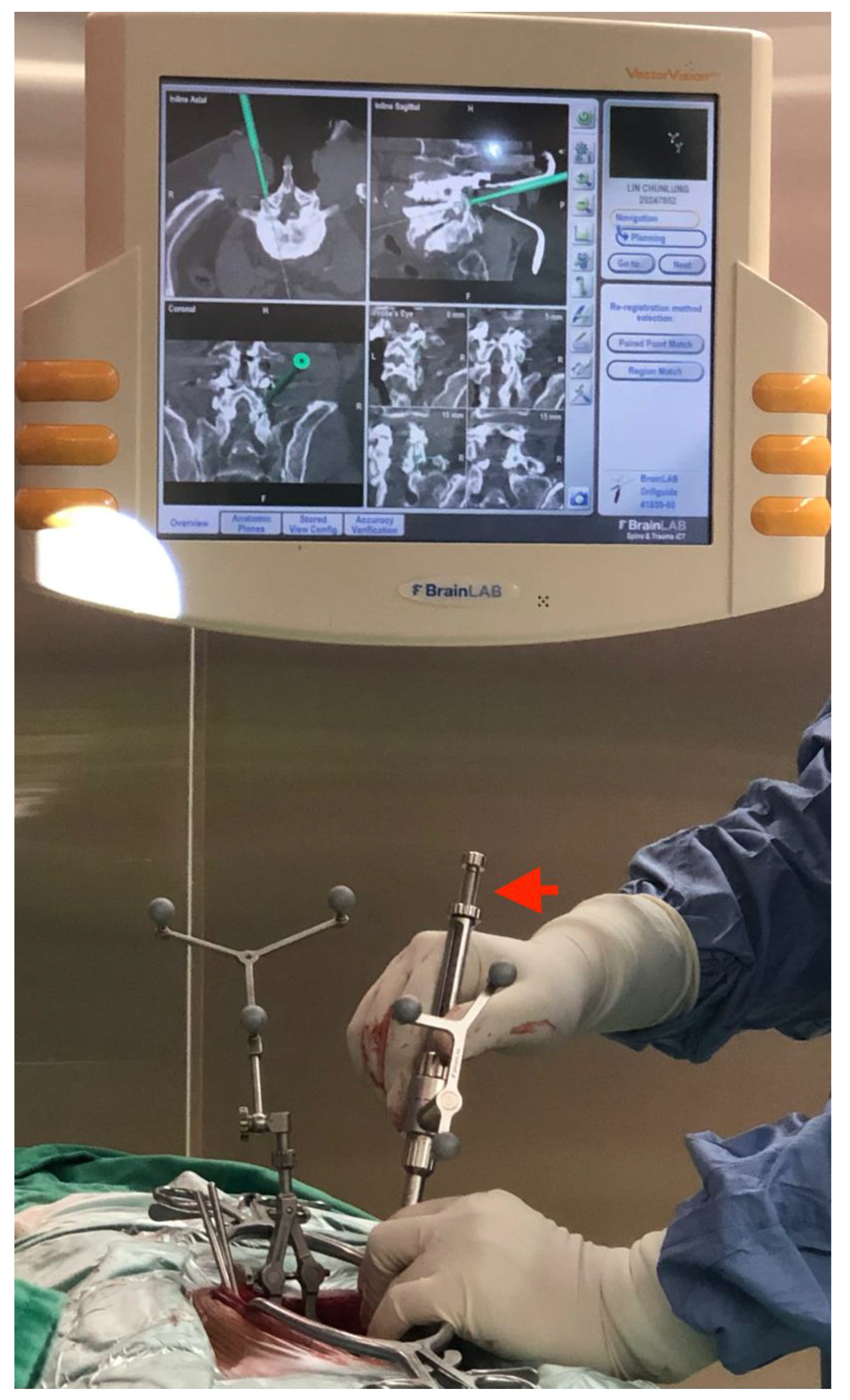
Abstract
1. Introduction
2. Materials and Methods
2.1. Surgical Technique
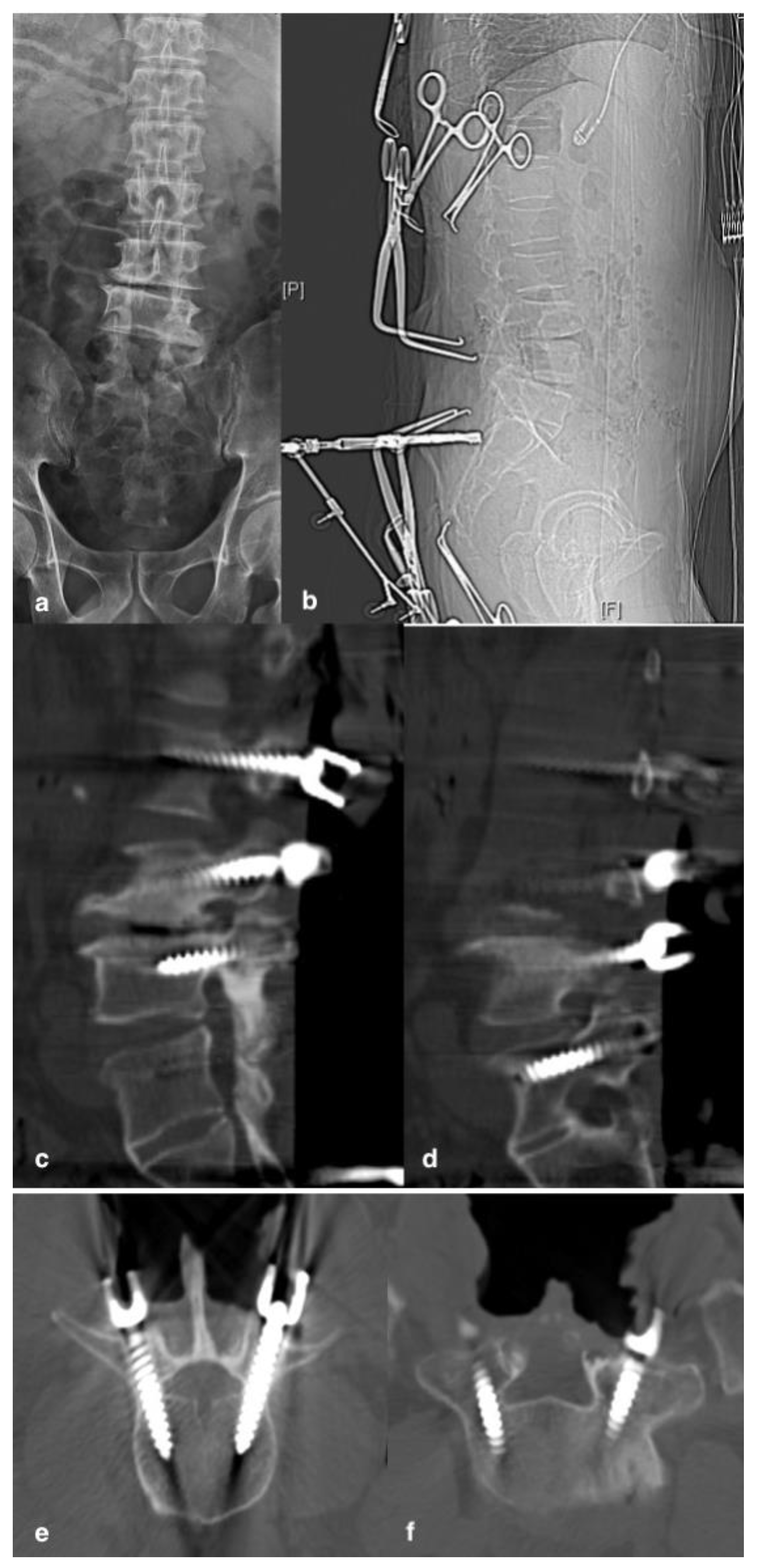
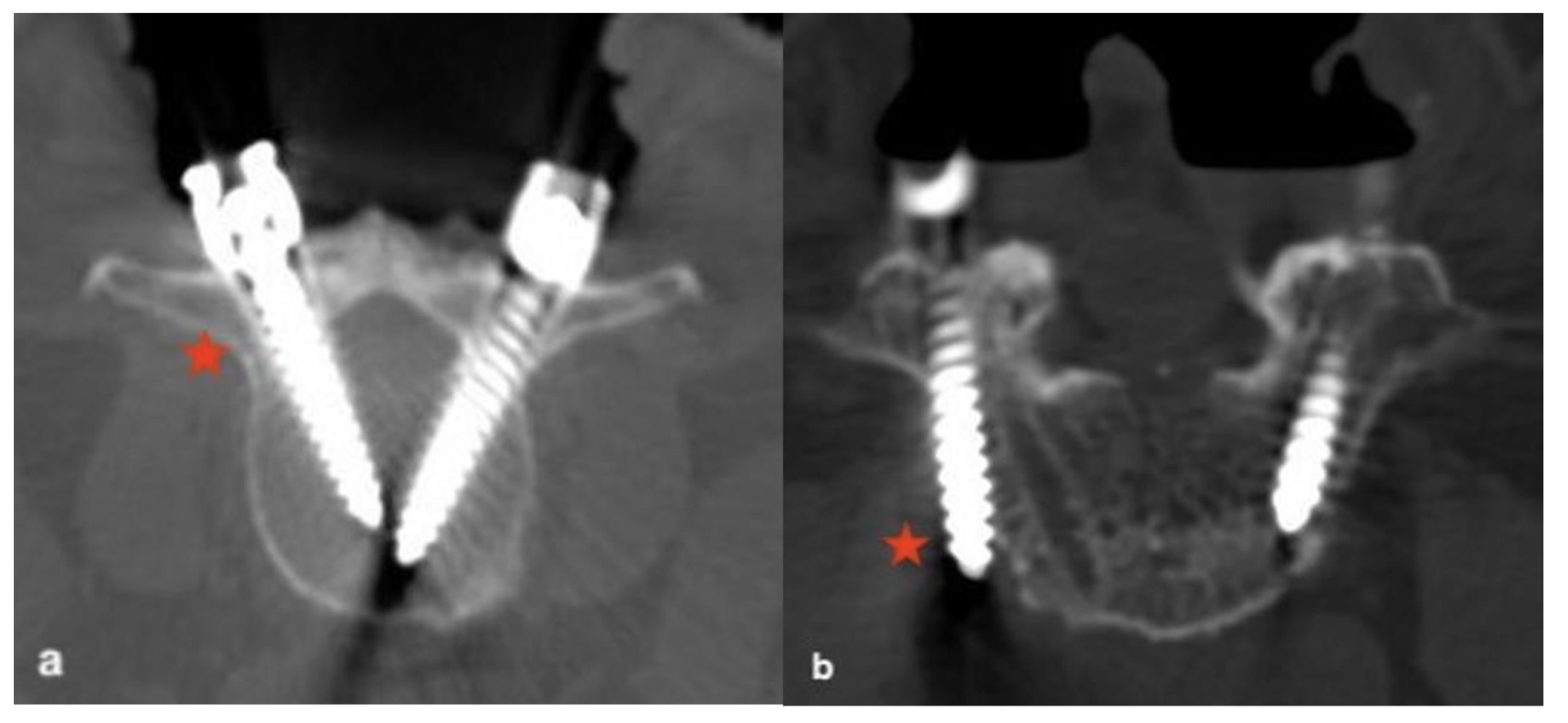
2.2. Assessment of TPS Placement
2.3. Radiographic and Clinical Analyses
2.4. Statistical Analysis
3. Results
4. Discussion
5. Conclusions
Supplementary Materials
Author Contributions
Funding
Institutional Review Board Statement
Informed Consent Statement
Data Availability Statement
Conflicts of Interest
References
- Suk, S.; Lee, S.M.; Chung, E.R.; Kim, J.H.; Kim, S.S. Selective thoracic fusion with segmental pedicle screw fixation in the treatment of thoracic idiopathic scoliosis: More than 5-year follow-up. Spine 2005, 30, 1602–1609. [Google Scholar] [CrossRef] [PubMed]
- Kim, Y.W.; Lenke, L.G.; Kim, Y.J.; Bridwell, K.H.; Kim, Y.B.; Watanabe, K.; Watanabe, K. Free-hand TPS placement during revision spinal surgery analysis of 552 screws. Spine 2008, 33, 1141–1148. [Google Scholar] [CrossRef] [PubMed]
- Kim, Y.J.; Lenke, L.G.; Cheh, G.; Riew, K.D. Evaluation of pedicle screw placement in the deformed spine using intraoperative plain radiographs: A comparison with computerized tomography. Spine 2005, 30, 2084–2088. [Google Scholar] [CrossRef] [PubMed]
- Hicks, J.M.; Singla, A.; Shen, F.H.; Arlet, V. Complications of pedicle screw fixation in scoliosis surgery: A systematic review. Spine 2010, 35, E465–E470. [Google Scholar] [CrossRef] [PubMed]
- Acosta, F.L., Jr.; Thompson, T.L.; Campbell, S.; Weinstein, P.R.; Ames, C.P. Use of intraoperative isocentric C-arm 3D fluoroscopy for sextant percutaneous pedicle screw placement: Case report and review of the literature. Spine J. 2005, 5, 339–343. [Google Scholar] [CrossRef] [PubMed]
- Holly, L.T.; Foley, K.T. Three-dimensional fluoroscopy-guided percutaneous thoracolumbar pedicle screw placement technical note. J. Neurosurg. Spine 2003, 99, 324–329. [Google Scholar] [CrossRef] [PubMed]
- Oertel, M.F.; Hobart, J.; Stein, M.; Schreiber, V.; Scharbrodt, W. Clinical and methodological precision of spinal navigation assisted by 3D intraoperative O-arm radiographic imaging. J. Neurosurg. Spine 2011, 14, 532–536. [Google Scholar] [CrossRef] [PubMed]
- Silbermann, J.; Riese, F.; Allam, Y.; Reichert, T.; Koeppert, H.; Gutberlet, M. Computed tomography assessment of pedicle screw placement in lumbar and sacral spine: Comparison between free-hand and O-arm based navigation techniques. Eur. Spine J. 2011, 20, 875–881. [Google Scholar] [CrossRef] [PubMed]
- Lee, M.H.; Lin, M.H.; Weng, H.H.; Cheng, W.C.; Tsai, Y.H.; Wang, T.C.; Yang, J.T. Feasibility of Intraoperative Computed Tomography Navigation System for Pedicle Screw Insertion of the Thoracolumbar Spine. J. Spinal. Disord Tech. 2013, 26, E183–E187. [Google Scholar] [CrossRef] [PubMed]
- Lee, C.Y.; Wu, M.H.; Li, Y.Y.; Cheng, C.C.; Hsu, C.H.; Huang, T.J.; Hsu, R.W. Intraoperative computed tomography navigation for transpedicular screw fixation to treat unstable thoracic and lumbar spine fractures. Medicine 2015, 94, e757. [Google Scholar] [CrossRef] [PubMed]
- Hsieh, J.C.; Drazin, D.; Firempong, A.O.; Pashman, R.; Johnson, P.; Kim, T.T. Accuracy of intraoperative computed tomography image-guided surgery in placing pedicle and pelvic screws for primary versus revision spine surgery. Neurosurg. Focus 2014, 36, e2. [Google Scholar] [CrossRef] [PubMed]
- Li, Y.Y.; Lee, C.Y.; Wu, M.H.; Huang, T.J.; Cheng, C.C.; Lee, C.Y. Intraoperative computed tomography navigation for transpedicular screw in posterior instrumentation and correction of adolescent idiopathic scoliosis. Formosan. J. Musculoskelet Disord 2016, 7, 57–62. [Google Scholar]
- Tsai, T.H.; Tzou, R.D.; Su, Y.F.; Wu, C.H.; Tsai, C.Y.; Lin, C.L. Pedicle screw placement accuracy of bone-mounted miniature robot system. Medicine 2017, 96, e5835. [Google Scholar] [CrossRef]
- Laine, T.; Makitalo, K.; Schlenzka, D.; Tallroth, K.; Poussa, M.; Alho, A. Accuracy of pedicle screw placement: A prospective CT study in 30 consecutive low back patients. Eur. Spine J. 1997, 6, 402–405. [Google Scholar] [CrossRef] [PubMed]
- Castro, W.H.; Halm, H.; Jerosch, J.; Malms, J.; Steinbeck, J.; Blasius, S. Accuracy of pedicle screw placement in lumbar vertebrae. Spine 1996, 21, 1320–1324. [Google Scholar] [CrossRef] [PubMed]
- Carbone, J.J.; Tortolani, P.J.; Quartararo, L.G. Fluoroscopically assisted pedicle screw fixation for thoracic and thoracolumbar injuries: Technique and short-term complications. Spine 2003, 28, 91–97. [Google Scholar] [CrossRef] [PubMed]
- Lehman, R.A.; Lenke, L.G.; Keeler, K.A.; Kim, Y.J.; Cheh, G. Computed tomography evaluation of pedicle screws placed in the pediatric deformed spine over an 8-year period. Spine 2007, 32, 2679–2684. [Google Scholar] [CrossRef] [PubMed]
- Samdani, A.F.; Ranade, A.; Saldanha, V.; Yondorf, M.Z. Learning curve for placement of thoracic pedicle screws in the deformed spine. Neurosurgery 2010, 66, 290–295. [Google Scholar] [CrossRef] [PubMed]
- Hecht, N.; Yassin, H.; Czabanka, M.; Fohre, B.; Arden, K.; Liebig, T.; Vajkoczy, P. Intraoperative computed tomography versus 3D C-arm imaging for navigated spinal instrumentation. Spine 2018, 43, 370–377. [Google Scholar] [CrossRef] [PubMed]
- Scarone, P.; Vincenzo, G.; Distefano, D.; Grande, F.D.; Cianfoni, A.; Presilla, S.; Reinert, M. Use of the Airo mobile intraoperative. CT system versus the O-arm for transpedicular screw fixation in the thoracic and lumbar spine: A retrospective cohort study of 263 patients. J. Neurosurg. Spine 2018, 29, 397–406. [Google Scholar] [CrossRef] [PubMed]


Publisher’s Note: MDPI stays neutral with regard to jurisdictional claims in published maps and institutional affiliations. |
© 2022 by the authors. Licensee MDPI, Basel, Switzerland. This article is an open access article distributed under the terms and conditions of the Creative Commons Attribution (CC BY) license (https://creativecommons.org/licenses/by/4.0/).
Share and Cite
Li, Y.-Y.; Chen, S.-H.; Huang, K.-C.; Lee, C.-Y.; Cheng, C.-C.; Lee, C.-Y.; Wu, M.-H.; Huang, T.-J. High Accuracy and Safety of Intraoperative CT-Guided Navigation for Transpedicular Screw Placement in Revision Spinal Surgery. J. Clin. Med. 2022, 11, 5853. https://doi.org/10.3390/jcm11195853
Li Y-Y, Chen S-H, Huang K-C, Lee C-Y, Cheng C-C, Lee C-Y, Wu M-H, Huang T-J. High Accuracy and Safety of Intraoperative CT-Guided Navigation for Transpedicular Screw Placement in Revision Spinal Surgery. Journal of Clinical Medicine. 2022; 11(19):5853. https://doi.org/10.3390/jcm11195853
Chicago/Turabian StyleLi, Yen-Yao, Shih-Hao Chen, Kuo-Chin Huang, Chien-Yin Lee, Chin-Chang Cheng, Ching-Yu Lee, Meng-Huang Wu, and Tsung-Jen Huang. 2022. "High Accuracy and Safety of Intraoperative CT-Guided Navigation for Transpedicular Screw Placement in Revision Spinal Surgery" Journal of Clinical Medicine 11, no. 19: 5853. https://doi.org/10.3390/jcm11195853
APA StyleLi, Y.-Y., Chen, S.-H., Huang, K.-C., Lee, C.-Y., Cheng, C.-C., Lee, C.-Y., Wu, M.-H., & Huang, T.-J. (2022). High Accuracy and Safety of Intraoperative CT-Guided Navigation for Transpedicular Screw Placement in Revision Spinal Surgery. Journal of Clinical Medicine, 11(19), 5853. https://doi.org/10.3390/jcm11195853