Article

Serum Levels of Selected IL-1 Family Cytokines in Patients with Morphea

by
Paulina Szczepanik-Kułak
*,
Małgorzata Michalska-Jakubus
,
Małgorzata Kowal
and
Dorota Krasowska
Chair and Department of Dermatology, Venerology and Paediatric Dermatology, Medical University of Lublin, 20-081 Lublin, Poland
*
Author to whom correspondence should be addressed.
J. Clin. Med. 2022, 11(21), 6375; https://doi.org/10.3390/jcm11216375
Submission received: 29 August 2022 / Revised: 22 September 2022 / Accepted: 27 October 2022 / Published: 28 October 2022
(This article belongs to the Special Issue Skin Inflammation: Symptoms, Causes, Diagnosis and Treatment)

Abstract

:
Morphea/localized scleroderma (LoS) represents an inflammatory-sclerotic skin disease, the pathogenesis of which is not fully understood. Given the important role of IL-1 family cytokines in the development and therapy of inflammatory diseases, including systemic sclerosis, we analyzed the clinical significance of serum levels of selected IL-1 family cytokines (IL-1α, IL-1β, IL-18, IL-33, IL-37 and IL-38) in LoS patients (n = 30) using the standardized disease assessment tools and comparison to healthy controls (n = 28). We also compared the pre- and post-treatment concentrations, i.e., before and after systemic (glucocorticosteroids and/or methotrexate) and/or topical (topical glucocorticosteroids and/or calcineurin inhibitors). Our findings did not reveal significant differences in baseline IL-1α, IL-1β, IL-18, IL-33, IL-37 and IL-38 levels between LoS group and HCs; however, after treatment, there were marked changes in concentrations of IL-1α and IL-33 within LoS group as well as in comparison to HCs. We also found significant negative correlations between PGA-A and IL-1α concentration as well as between mLoSSI and IL-1α after treatment. Furthermore, we showed an inverse correlation of baseline IL-1β levels with mLoSSI scores of borderline significance. We believe that IL-1α and IL-33, as well as Il-1β, may be potential mediators and targets of interest in LoS.

1. Introduction

Morphea, also termed localized scleroderma (LoS), is a chronic disease associated with fibrosis of the skin and possibly underlying tissues, which has the potential to progress to significant morbidity [1]. The incidence of morphea is estimated at 27 cases/1,000,000, with a marked female predominance [2,3]. There are two peaks of incidence of LoS, one between the ages of 7 and 11 and the other between the ages of 40 and 50 [4]. The pathogenesis of LoS is still not fully known [1]. The interaction of triggering factors with the individual susceptibility appears to have the major role in the disease development, resulting in the activation of both innate and acquired responses with immunoinflammatory and profibrotic actions involving the epidermis and dermis. Furthermore, in the light of evidence, including the high prevalence of an individual or family history of autoimmune disease and the presence of autoantibodies as well as certain human leukocyte antigen subtypes, morphea is believed to have an autoimmune nature [5,6].
The clinical features of LoS are heterogeneous and classified into five subtypes: limited, generalized, linear, deep and mixed [2]. In general, morphea is characterized by a consecutive stage of an active phase characterized by inflammatory or inflammatory-sclerotic, erythematous or indurative lesions with active “lilac” ring, which appeared or enlarged in the last month, followed by an inactive postinflammatory lesions with hyperpigmentation and dermal atrophy [4,7]. A variety of methods is available for the treatment of LoS [8]. For mild, superficial lesions limited to the skin, topical treatments and UV phototherapy are recommended, while for generalized, linear or deep types, systemic immunomodulatory therapies, such as glucocorticosteroids (GCS) or methotrexate (MTX), are usually introduced. In severe cases, early implementation of therapy, at the stage of inflammatory lesions, allows to avoid serious complications and disfigurement [8].
For the assessment of disease severity, the “localized scleroderma cutaneous assessment tool” (LoSCAT) in combination with Physician’s Global Assessment (PGA) are recommended [9]. LoSCAT includes the “modified localized scleroderma severity index” (mLoSSI) and the “localized scleroderma skin damage index” (LoSDI). mLoSSI incorporates features of disease activity or severity, such as the appearance of new skin lesions or enlargement of existing lesions in the past month, together with erythema and induration of the skin. LoSDI assesses tissue damage, reflected by atrophy of the skin and subcutaneous tissue, as well as pigmentary alterations [10].
To date, the search for laboratory biomarkers for LoS has not been much of a success.
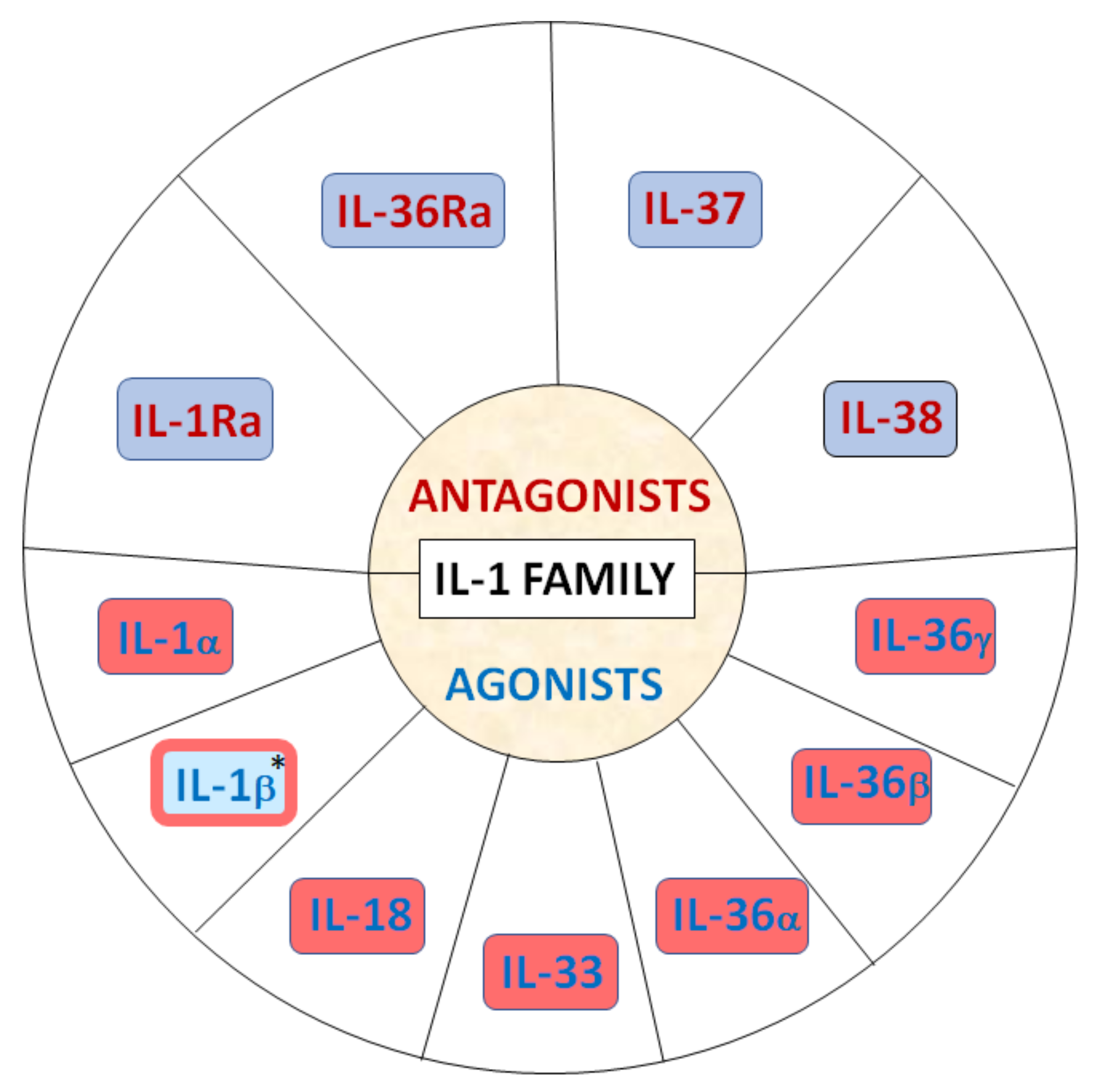
The IL-1 cytokine family comprises 11 members, including seven ligands with broad pro-inflammatory and profibrotic activities (IL-1α, IL-1β, IL-18, IL-33, IL-36α, IL-36β and IL-36γ), three receptor antagonists and one anti-inflammatory cytokine (Figure 1) [11,12,13,14].
IL-1 is critical to the pathogenesis of a variety of human diseases and IL-1 targeted therapies have been successfully employed to treat a range of inflammatory conditions such as rheumatoid arthritis. The role of the IL-1 family in LoS has not been extensively investigated to date [15,16,17,18,19,20,21,22,23,24,25,26,27]. Of note, involvement of the IL-1 family seems to be well established in the pathogenesis of systemic sclerosis (SSc), including development of inflammation and fibrosis in both the skin and underlying tissues, as well as internal organs [28,29,30].
Thus, considering the presence of some resemblances between SSc and LoS, including the similarity of in the histological pattern of skin [31], we aimed to evaluate the clinical relevance of selected serum levels of IL-1 family cytokines, IL-1α, IL-1β, IL-18, IL-33, IL-37 and IL-38 in a modest-sized single-center cohort of well-characterized and prospectively followed LoS patients and healthy controls, including comparison before and after the initiation of therapy with the use of the standardized disease assessment tools.

2. Materials and Methods

2.1. The Study Group

The study included 30 LoS patients and 28 healthy controls (HCs), without significant difference in gender or age. All patients were hospitalized in the Department of Dermatology, Venereology, and Pediatric Dermatology Medical University of Lublin. The diagnosis of LoS was confirmed histopathologically in all cases. Informed consent was obtained from each individual before starting any procedures. The study protocol complies with the ethical guidelines of the 1975 Declaration of Helsinki. The study was approved by the local ethics committee (KE-0254/162/06/2022).

2.2. Clinical Assessment of Studied Individuals with LoS

First, we determined each patient’s demographics (age and sex) and age at onset of LoS diagnosis. Next, we performed a physical examination, establishing the subtype of LoS, as well as characteristics of lesions, such as activity (PGA-A, mLoSSI), damage (PGA-D, LoSDI) and body surface area (BSA). In addition, the type of LoS treatment (systemic/local) along with autoimmune co-morbidities were specified.
The following parameters were then assessed once again after completion of the therapeutic process: PGA-A, mLoSSI, PGA-D, LoSDI, BSA.
We also evaluated the presence of ANA autoantibodies and their pattern (detected by indirect immunofluorescence using human laryngeal epidermoid carcinoma cell line type 2, Hep-2).

2.3. Assessment of Serum Concentrations of IL-1 Family Cytokines in LoS Patients and HCs

Peripheral blood samples (10 mL) were collected after overnight fasting—in HCs once and in LoS patients twice—before and after treatment. The blood samples were centrifuged for 15–20 min at 1000× g and stored at −80 °C until testing. The concentrations of following IL-1 family cytokines in serum were determined using enzyme-linked immunosorbent assay (ELISA) kits according to the manufacturer’s protocol:
  • IL-1α: Quantikine™ELISA Human IL-1α/IL-1F1, R&D Systems, Inc., Minneapolis, MN, USA;
  • IL-1β: Human Interleukin-1 Beta ELISA, BioVendor–Laboratorni medicina a.s., Brno, Czech Republic;
  • IL-18: Quantikine™ELISA Human Total IL-18/IL-1F4, R&D Systems, Inc., Minneapolis, MN, USA;
  • IL-33: Quantikine™ELISA Human IL-33, R&D Systems, Inc., Minneapolis, MN, USA;
  • IL-37: ELISA Kit For Interleukin 1 Zeta (IL1z), Cloud-Clone Corp., Katy, Texas, USA;
  • IL-38: ELISA Kit For Interleukin 1 Theta (IL1q), Cloud-Clone Corp., Katy, Texas, USA.

2.4. Statistical Methods

The statistical analysis was performed using the Statistica version 10.0 software (StatSoft Inc., Tulsa, OK, USA) for Windows.
The following statistical tests were used:
  • Pearson’s chi-square test to compare categorical variables, including gender, between LoS patients and HCs;
  • Mann–Whitney’s U test to compare age, cytokine concentrations between LoS patients and HCs as well as the difference in cytokine concentrations in response to systemic (immunomodulatory) treatment in LoS patients;
  • The Wilcoxon rank-sum test to compare cytokine concentrations as well as markers of disease-targeted measures before and after treatment in patients with LoS;
  • Spearman’s correlation coefficient to correlate markers of disease and particular interleukin levels before and after treatment in patients with LoS;
  • McNemar test to assess alteration in disease-targeted measures following treatment.
The p value < 0.05 was considered statistically significant.

3. Results

Basic characteristics of LoS patients and HCs are shown in Table 1 and Table 2.

3.1. Clinical Characteristics of the Study LoS Group

The mean age of patients with LoS at the time of diagnosis was 45.4 years. The middle-aged patients predominated (median 51.5 y.o.); however, the study group also included pediatric cases (min. 10 y.o.). At baseline, we found active LoS in 28 patients.
A considerable proportion of patients had co-occurring autoimmune diseases, most commonly vulvar lichen sclerosus (n = 8), vitiligo (n = 3) and Hashimoto’s disease (n = 2). All patients had positive ANA antibody titers, in the range of 1:80 to 1:1280, with a dominant nuclear speckled pattern (n = 25, 83.3%), less frequently nucleolar pattern (n = 5, 16.7%). In all cases, commercially available ENA/blot panels were found to be negative. Systemic therapy, in the form of GCS and MTX, was used in 13 patients (43.3%), while 17 patients (46.7%) did not require this type of therapy. All patients included in the study were treated with topical medications, topical GCS and/or calcineurin inhibitors (CI). None of the patients were on phototherapy. The data are summarized in Table 3.

3.2. Baseline and Post-Treatment Cytokines Levels in Patients with LoS and HCs

Baseline (pre-treatment) levels of analyzed cytokines in all of the LoS patients did not differ significantly when compared to HCs (Table 4).
After completion of the treatment, we noted an increase in IL-1α levels in our patients, and the measured concentrations were significantly higher in comparison to the HCs (p = 0.0440), whereas IL-33 concentrations were found to be markedly lower in LoS group after treatment in comparison to HCs (p = 0.0005) (Table 5).
When comparing cytokines levels before and after treatment within LoS group, the mean and median IL-33 concentration was found to be significantly higher before therapy (p = 0.0110) (Table 6).
Of note, there was no significant difference in changes in cytokines levels between LoS patients on or without systemic therapy (p > 0.05) (Table 7).

3.3. Baseline and Post-Treatment Cytokines Levels in LoS Patients in Relation to Disease-Targeted Measures

In 26 of 28 patients with primarily active lesions, there was a switch to inactive phase after treatment. The decrease in the number of patients with active lesions after therapy was statistically significant (p < 0.0001). Mean and median disease activity (PGA-A), damage (PGA-D), mLoSSI value, LoSDI and BSA also significantly decreased after treatment (p < 0.05) (Table 8).
There was no significant correlation between any of disease-targeted measures and baseline levels of analyzed cytokines (p > 0.05) (Table 9).
After treatment, significant negative correlations were shown between PGA-A and IL-1α concentration (p = 0.0338), as well as between mLoSSI and IL-1α (p = 0.0338) (Table 10).

4. Discussion

To the best of our knowledge, this study is the first to investigate the extensive profile of IL-1 family in LoS as biomarkers of the disease and possible therapy-targeted measure.
Our findings did not reveal significant differences in baseline IL-1α, IL-1β, IL-18, IL-33, IL-37 and IL-38 levels between LoS group and HCs; however, after treatment, there were marked changes in concentrations of IL-1α and IL-33 either within LoS group as well as in comparison to HCs. Specifically, in contrast to baseline measures, after completion of therapy we obtained statistically higher values of serum IL-1α in patients with LoS than in controls, although an absolute increase in these levels within LoS group were only close to significant, probably due to a relatively small sample size. On the contrary, IL-33 concentrations significantly decreased in LoS patients under treatment and were significantly lower in post-treated LoS individuals when compared to HCs.
In addition, our results showed some important associations of IL-1 members with certain disease-targeted measures. Particularly noteworthy, we found that IL-1α levels may have a potential strong association with disease activity and severity. In post-treated LoS patients IL-1α concentrations negatively correlated with both mLoSSi and PGA-A scores. With respect to serum levels of other analyzed cytokines, no significant association was found in terms of clinical variables in the study group; however, it is worth noting the inverse correlation of baseline IL-1β levels with mLoSSI scores with borderline significance.
In light of these results, it seems that IL-1α and IL-33, as well as Il-1β, may be potential mediators and targets of interest in LoS. These IL-1 family members are acknowledged to play a role in inflammation and have been suggested to be involved in autoimmune diseases, including systemic lupus erythematosus and rheumatoid arthritis as well as systemic sclerosis (SSc) [28,32,33,34,35]. Moreover, both IL-1α and IL-1β have been shown to directly stimulate fibroblasts proliferation and collagen synthesis and fibroblasts are induced to secrete a range of inflammatory cytokines in response to IL-1α and IL-1β [36,37,38]. IL-1β has also been found to participate in the differentiation of Th17 cells that may play a crucial role in the development of tissue fibrosis [28,32,39]. Of note, it was demonstrated that SSc patients with coexisting LoS-like lesions exhibited overexpression of IL-1α in the epidermis of both LoS-like and typical SSc lesions [31]. On this background, our findings of lower IL-1α levels in LoS patients after treatment and their negative correlation with both mLoSSi and PGA-A scores as well as inverse association of IL-1β with mLoSSi seem controversial. However, there is a large body of literature also demonstrating an inhibitory effect of IL-1α and IL-1β on collagen synthesis [40,41]. Surprisingly, it has been shown that IL-1α increases the mRNA expression of matrix metalloproteinases (MMPs), namely, MMP-1 and MMP-3, and induces MMP-7 resulting in collagen degradation [42]. Of note, some previous studies reported increase in MMPs in LoS after treatment and their correlation with clinical improvement [43,44], but direct experimental studies on MMPs and IL-1 in LoS are lacking.
Another possible explanation for increased post-treatment levels of IL-1α in line with improvement in disease activity and severity scores is the direct effect of applied therapies. However, data on the effects of systemic treatment on the levels of serum IL-1 family cytokines are scarce and unavailable for LoS; the paradoxical proinflammatory properties of MTX in the form of increased IL-1 as measured by secreted protein and level of gene expression have been demonstrating in vitro on human monocytic cell line U937, which is an effect that seems to be at odds with the generally anti-inflammatory activity of this drug [45]. In contrast, regarding systemic GCS, there are reports of down-regulation of IL-1α [46,47]. However, immunosuppressive and anti-inflammatory agents such as glucocorticoids may simultaneously induce increased expression of IL-1R2 (type 2 receptor for IL-1) that acts as negative regulator of the IL-1 system, modulating IL-1α availability for the signaling receptor. Interestingly, this anti-IL-1 effect seems predominantly local [32,48]. Thus, in relation to our results, it may be speculated that observed post-treatment increase in IL-1α is a kind of feedback compensatory reaction to the blocking effect of IL-1R2, making IL-1α an interesting ambiguous mediator in LoS. However, future experimental studies are warranted to elucidate this phenomenon.
IL-33 is structurally related to IL-1β and is known to have a crucial role in immune and inflammatory reactions. Recent articles have also described IL-33 as a cytokine with emerging pro-fibrotic potential depending on targets such as IL-13, TGF-β, IFN-γ and TLR/NF-κB signaling pathways [35]. Subcutaneous injection of IL-33 in mice resulted in the development of cutaneous fibrosis, similar to that observed in patients SSc, including dermal mast cells and skin-infiltrating neutrophils through the suppression of Th1-mediated contact hypersensitivity responses [35]. Additionally, IL-33 may function as an alarmin that alerts the immune system after endothelial or epithelial cell damage during infection, physical stress, or trauma and skin trauma as well as endothelial cell damage have been proposed as crucial events in the development of LoS lesions [49,50,51]. Moreover, MTX treatment has been shown to decline skin and blood IL-33 levels in psoriatic patients [52].
Thus, with respect to these observations, our finding of significant decrease in serum IL-33 concentrations in LoS patients under treatment may highlight IL-33 as the possible pathogenic mediator in LoS as well as a candidate for future targeted therapies. However, since we did not found correlations with measures of disease activity or severity, further research on larger cohort is required to clarify this contradiction.
Currently, no comparable clinical studies are available for LoS. Thus, the results are difficult to discuss. However, some essential data may be obtained from research of other diseases that may be applicable to LoS, particularly SSc that shares common inflammatory and immunologic pathways.
However, the expression levels of IL-1β and IL-1α were found significantly up-regulated in the lesional skin tissue in SSc [31,53], the serum levels of IL-1α and IL-1β are somewhat controversial. Similarl to our findings in LoS, Lin et al. [54], in line with some other studies, did not observe a significant difference in serum concentrations of IL-1α and IL-1β between SSc and HCs, although some authors have reported their elevated serum levels in SSc patients [55,56,57,58,59]. With respect to clinical variables, observations for SSc are also puzzling. In contrast to our results in LoS cohort, serum IL-1β, but not IL-1α, was positively correlated with the severity of skin involvement in SSc measured by modified Rodnan skin score (mRSS), suggesting a potential role of this cytokine in SSc fibrotic complications, but serum levels of both IL-1α and IL-1β positively correlated with carbon monoxide transfer coefficient and patients with high serum IL-1β had higher DLCO, suggesting a reduced risk of lung fibrosis and PAH [54].
Recently, an increasing number of studies have shown the potential role of IL-33 in SSc [28,35]. In contrast to our findings in LoS patients, serum IL-33 levels were reported to be elevated in patients with SSc compared with healthy controls and correlated with the extent of skin sclerosis as well as with the severity of pulmonary fibrosis [60]. In one recent study of IL-1α, IL-1β, IL-18 and IL-33 in a relatively modest-sized Chinese SSc cohort, only serum IL-1β and IL-33 were found to be higher in SSc in multivariable analysis; however, no clinical associations with any of these cytokines were found [59].
In conclusion, our observations may highlight the potential relevance of certain IL-1 family members, namely, IL-1α, IL-1β and, in particular, IL-33, as interesting pathogenic mediators in LoS as well as the feasibility of their use in clinical applications. However, the puzzling results obtained for IL-1α and IL-1β emphasize the need for future experimental and clinical research to clarify their role in LoS.
We are aware of several limitations in our study, including a relatively small sample size and the restriction to a single-center population, which are likely to limit the statistical power. Moreover, due to small number of patients with inactive phase of LoS lesions at baseline, their selection as separate group for statistical analysis and comparisons was unavailable. Therefore, replication in multi-center studies with a greater number of enrolled individuals will be beneficial.
Author Contributions

Conceptualization, P.S.-K. and D.K.; methodology P.S.-K., M.K., M.M.-J. and D.K.; software, P.S.-K. and M.K.; validation, M.M.-J. and D.K.; formal analysis, P.S.-K. and M.K.; investigation, P.S.-K. and M.K.; resources, P.S.-K. and M.K.; data curation, P.S.-K. and M.K.; writing—original draft preparation, P.S.-K.; writing—review and editing, M.M.-J. and D.K.; visualization, P.S.-K., M.M.-J. and D.K.; supervision, M.M.-J. and D.K.; project administration, D.K.; funding acquisition, D.K. All authors have read and agreed to the published version of the manuscript.

Funding

This research received no external funding.

Institutional Review Board Statement

The study was conducted according to the guidelines of the Declaration of Helsinki and approved by the Institutional Ethics Committee of Medical University of Lublin, Poland (KE-0254/162/06/2022).

Informed Consent Statement

Written informed consent has been obtained from the patient(s) to publish this paper.

Data Availability Statement

Data sharing not applicable.

Conflicts of Interest

The authors declare no conflict of interest.

References

  1. LaChance, A.H.; Goldman, N.; Kassamali, B.; Vleugels, R.A. Immunologic underpinnings and treatment of morphea. Expert Rev. Clin. Immunol. 2022, 4, 1–23. [Google Scholar] [CrossRef] [PubMed]
  2. Kreuter, A.; Krieg, T.; Worm, M.; Wenzel, J.; Moinzadeh, P.; Kuhn, A.; Aberer, E.; Scharffetter-Kochanek, K.; Horneff, G.; Reil, E.; et al. German guidelines for the diagnosis and therapy of localized scleroderma. JDDG J. der Dtsch. Dermatol. Ges. 2016, 14, 199–216. [Google Scholar] [CrossRef] [PubMed] [Green Version]
  3. Silman, A.; Jannini, S.; Symmons, D.; Bacon, P. An epidemiological study of scleroderma in the West Midlands. Rheumatology 1988, 27, 286–290. [Google Scholar] [CrossRef] [PubMed]
  4. Krasowska, D.; Rudnicka, L.; Dańczak-Pazdrowska, A.; Chodorowska, G.; Woźniacka, A.; Lis-Święty, A.; Czuwara, J.; Maj, J.; Majewski, S.; Sysa-Jędrzejowska, A.; et al. Localized scleroderma (morphea). Diagnostic and therapeutic recommendations of the Polish Dermatological Society. Dermatol. Rev. 2019, 106, 333–353. [Google Scholar] [CrossRef]
  5. Saracino, A.; Denton, C.; Orteu, C. The molecular pathogenesis of morphoea: From genetics to future treatment targets. Br. J. Dermatol. 2017, 177, 34–46. [Google Scholar] [CrossRef] [PubMed] [Green Version]
  6. Wolska-Gawron, K.; Bartosińska, J.; Krasowska, D. MicroRNA in localized scleroderma: A review of literature. Arch. Dermatol. Res. 2020, 312, 317–324. [Google Scholar] [CrossRef] [PubMed] [Green Version]
  7. Abbas, L.; Joseph, A.; Kunzler, E.; Jacobe, H.T. Morphea: Progress to date and the road ahead. Ann. Transl. Med. 2021, 9, 437. [Google Scholar] [CrossRef]
  8. Knobler, R.; Moinzadeh, P.; Hunzelmann, N.; Kreuter, A.; Cozzio, A.; Mouthon, L.; Cutolo, M.; Rongioletti, F.; Denton, C.; Rudnicka, L.; et al. European Dermatology Forum S1-guideline on the diagnosis and treatment of sclerosing diseases of the skin, Part 1: Localized scleroderma, systemic sclerosis and overlap syndromes. J. Eur. Acad. Dermatol. Venereol. 2017, 31, 1401–1424. [Google Scholar] [CrossRef]
  9. Arkachaisri, T.; Vilaiyuk, S.; Torok, K.S.; Medsger, T.A., Jr. Development and initial validation of the Localized Scleroderma Skin Damage Index and Physician Global Assessment of disease Damage: A proof-of-concept study. Rheumatology 2010, 49, 373–381. [Google Scholar] [CrossRef] [Green Version]
  10. Skrzypek-Salamon, A.; Lis-Święty, A.; Ranosz-Janicka, I.; Brzezińska-Wcisło, L. Localized Scleroderma Cutaneous Assessment Tool (LoSCAT) adapted for use in adult patients: Report from an initial validation study. Health Qual. Life Outcomes 2018, 16, 185. [Google Scholar] [CrossRef]
  11. Sims, J.E.; Smith, D.E. The IL-1 family: Regulators of immunity. Nat. Rev. Immunol. 2010, 10, 89–102. [Google Scholar] [CrossRef]
  12. Dinarello, C.; Arend, W.; Sims, J.; Smith, D.; Blumberg, H.; O’Neill, L.; Goldbach-Mansky, R.; Pizarro, T.; Hoffman, H.; Bufler, P.; et al. IL-1 family nomenclature. Nat. Immunol. 2010, 11, 973. [Google Scholar] [CrossRef] [Green Version]
  13. Yazdi, A.S.; Ghoreschi, K. The Interleukin-1 Family. Adv. Exp. Med. Biol. 2016, 941, 21–29. [Google Scholar] [CrossRef]
  14. Dinarello, C.A. Overview of the IL-1 family in innate inflammation and acquired immunity. Immunol. Rev. 2018, 281, 8–27. [Google Scholar] [CrossRef] [Green Version]
  15. Kurzinski, K.; Torok, K.S. Cytokine profiles in localized scleroderma and relationship to clinical features. Cytokine 2011, 55, 157–164. [Google Scholar] [CrossRef] [Green Version]
  16. Hasegawa, M.; Sato, S.; Nagaoka, T.; Fujimoto, M.; Takehara, K. Serum Levels of Tumor Necrosis Factor and Interleukin-13 are Elevated in Patients with Localized Scleroderma. Dermatology 2003, 207, 141–147. [Google Scholar] [CrossRef]
  17. Ihn, H.; Sato, S.; Fujimoto, M.; Kikuchi, K.; Takehara, K. Demonstration of interleukin 8 in serum samples of patients with localized scleroderma. Arch. Dermatol. 1994, 130, 1327–1328. [Google Scholar] [CrossRef]
  18. Uziel, Y.; Krafchik, B.R.; Feldman, B.; Silverman, E.D.; Rubin, L.A.; Laxer, R.M. Serum levels of soluble interleukin-2 receptor. Arthritis Care Res. 1994, 37, 898–901. [Google Scholar] [CrossRef]
  19. Ihn, H.; Fujimoto, M.; Sato, S.; Kikuchi, K.; Igarashi, A.; Soma, Y.; Takehara, K. Increased levels of circulating intercellular adhesion molecule-1 in patients with localized scleroderma. J. Am. Acad. Dermatol. 1994, 31, 591–595. [Google Scholar] [CrossRef]
  20. Ihn, H.; Sato, S.; Fujimoto, M.; Kikuchi, K.; Takehara, K. Demonstration of interleukin-2, interleukin-4 and interleukin-6 in sera from patients with localized scleroderma. Arch. Dermatol. Res. 1995, 287, 439–442. [Google Scholar] [CrossRef]
  21. Higley, H.; Bs, K.P.; Chu, S.; Waegell, W.; Vancheeswaran, R.; Black, C. Immunocytochemical localization and serological detection of transforming growth factor β1. Association with type I procollagen and inflammatory cell markers in diffuse and limited systemic sclerosis, morphea, and Raynaud’s phenomenon. Arthritis Care Res. 1994, 37, 278–288. [Google Scholar] [CrossRef] [PubMed]
  22. Ihn, H.; Sato, S.; Fujimoto, M.; Kikuchi, K.; Takehara, K. Clinical significance of serum levels of soluble interleukin-2 receptor in patients with localized scleroderma. Br. J. Dermatol. 1996, 134, 843–847. [Google Scholar] [CrossRef] [PubMed]
  23. Nagaoka, T.; Sato, S.; Hasegawa, M.; Ihn, H.; Takehara, K. Serum levels of soluble interleukin 6 receptor and soluble gp130 are elevated in patients with localized scleroderma. J. Rheumatol. 2000, 27, 1917–1921. [Google Scholar] [PubMed]
  24. Budzyńska-Włodarczyk, J.; Michalska-Jakubus, M.M.; Kowal, M.; Krasowska, D. Evaluation of serum concentrations of the selected cytokines in patients with localized scleroderma. Adv. Dermatol. Allergol. 2016, 33, 47–51. [Google Scholar] [CrossRef]
  25. Magee, K.; Kelsey, C.E.; Kurzinski, K.L.; Ho, J.; Mlakar, L.R.; Feghali-Bostwick, C.; Torok, K.S. Interferon-gamma inducible protein-10 as a potential biomarker in localized scleroderma. Arthritis Res. Ther. 2013, 15, R188. [Google Scholar] [CrossRef] [PubMed] [Green Version]
  26. Torok, K.S.; Kurzinski, K.; Kelsey, C.; Yabes, J.; Magee, K.; Vallejo, A.N.; Medsger, T.; Feghali-Bostwick, C.A. Peripheral blood cytokine and chemokine profiles in juvenile localized scleroderma: T-helper cell-associated cytokine profiles. Semin. Arthritis Rheum. 2015, 45, 284–293. [Google Scholar] [CrossRef] [Green Version]
  27. Dańczak-Pazdrowska, A.; Kowalczyk, M.J.; Szramka-Pawlak, B.; Gornowicz-Porowska, J.; Szewczyk, A.; Silny, W.; Olewicz-Gawlik, A.; Molińska-Glura, M.; Żaba, R.; Hrycaj, P. Clinical immunology Interleukin 1β in morphea. Central Eur. J. Immunol. 2012, 3, 247–252. [Google Scholar] [CrossRef] [Green Version]
  28. Xu, D.; Mu, R.; Wei, X. The Roles of IL-1 Family Cytokines in the Pathogenesis of Systemic Sclerosis. Front. Immunol. 2019, 10, 2025. [Google Scholar] [CrossRef] [Green Version]
  29. Zhang, L.; Yan, J.-W.; Wang, Y.-J.; Wan, Y.-N.; Wang, B.-X.; Tao, J.-H.; Chen, B.; Li, B.-Z.; Yang, G.-J.; Wang, J. Association of Interleukin 1 Family with Systemic Sclerosis. Inflammation 2014, 37, 1213–1220. [Google Scholar] [CrossRef]
  30. De Luca, G.; Cavalli, G.; Campochiaro, C.; Bruni, C.; Tomelleri, A.; Dagna, L.; Matucci-Cerinic, M. Interleukin-1 and Systemic Sclerosis: Getting to the Heart of Cardiac Involvement. Front. Immunol. 2021, 12, 653950. [Google Scholar] [CrossRef]
  31. Saigusa, R.; Asano, Y.; Yamashita, T.; Takahashi, T.; Nakamura, K.; Miura, S.; Ichimura, Y.; Toyama, T.; Taniguchi, T.; Sumida, H.; et al. Systemic sclerosis complicated with localized scleroderma-like lesions induced by Köbner phenomenon. J. Dermatol. Sci. 2017, 89, 282–289. [Google Scholar] [CrossRef] [PubMed] [Green Version]
  32. Cavalli, G.; Colafrancesco, S.; Emmi, G.; Imazio, M.; Lopalco, G.; Maggio, M.C.; Sota, J.; Dinarello, C.A. Interleukin 1α: A comprehensive review on the role of IL-1α in the pathogenesis and treatment of autoimmune and inflammatory diseases. Autoimmun. Rev. 2021, 20, 102763. [Google Scholar] [CrossRef]
  33. Kageyama, Y.; Torikai, E.; Tsujimura, K.; Kobayashi, M. Involvement of IL-33 in the pathogenesis of rheumatoid arthritis: The effect of etanercept on the serum levels of IL-33. Mod. Rheumatol. 2011, 22, 89–93. [Google Scholar] [CrossRef] [PubMed]
  34. Mok, M.Y.; Huang, F.P.; Ip, W.K.; Lo, Y.; Wong, F.Y.; Chan, E.Y.T.; Lam, K.; Xu, D. Serum levels of IL-33 and soluble ST2 and their association with disease activity in systemic lupus erythematosus. Rheumatology 2009, 49, 520–527. [Google Scholar] [CrossRef] [PubMed] [Green Version]
  35. Li, L.; Zhu, H.; Zuo, X. Interleukin-33 in Systemic Sclerosis: Expression and Pathogenesis. Front. Immunol. 2018, 9, 2663. [Google Scholar] [CrossRef] [Green Version]
  36. Kawaguchi, Y.; Hara, M.; Wright, T.M. Endogenous IL-1α from systemic sclerosis fibroblasts induces IL-6 and PDGF-A. J. Clin. Investig. 1999, 103, 1253–1260. [Google Scholar] [CrossRef] [Green Version]
  37. Wynn, T.A.; Ramalingam, T.R. Mechanisms of fibrosis: Therapeutic translation for fibrotic disease. Nat. Med. 2012, 18, 1028–1040. [Google Scholar] [CrossRef] [Green Version]
  38. Kirk, T.Z.; Mayes, M.D. IL-1 Rescues Scleroderma Myofibroblasts from Serum-Starvation-Induced Cell Death. Biochem. Biophys. Res. Commun. 1999, 255, 129–132. [Google Scholar] [CrossRef]
  39. Santarlasci, V.; Cosmi, L.; Maggi, L.; Liotta, F.; Annunziato, F. IL-1 and T Helper Immune Responses. Front. Immunol. 2013, 15, 182. [Google Scholar] [CrossRef] [Green Version]
  40. Xiao, H.; Ji, A.M.; Li, Z.L.; Song, X.D.; Su, D.; Chen, A.H. Interleukin-1β inhibits collagen synthesis and promotes its decomposition in cultured cardiac fibroblasts. Sheng Li Xue Bao 2008, 60, 355–361. [Google Scholar]
  41. Bhatnagar, R.; Penfornis, H.; Mauviel, A.; Loyau, G.; Saklatvala, J.; Pujol, J.P. Interleukin-1 inhibits the synthesis of collagen by fibroblasts. Biochem. Int. 1986, 13, 709–720. [Google Scholar]
  42. Wisithphrom, K.; Murray, P.E.; Windsor, L.J. Interleukin-1 α Alters the Expression of Matrix Metalloproteinases and Collagen Degradation by Pulp Fibroblasts. J. Endod. 2006, 32, 186–192. [Google Scholar] [CrossRef]
  43. Gruss, C.; Reed, J.; Altmeyer, P.; McNutt, N.; Kerscher, M. Induction of interstitial collagenase (MMP-1) by UVA-1 phototherapy in morphea fibroblasts. Lancet 1997, 350, 1295–1296. [Google Scholar] [CrossRef]
  44. Malewska-Woźniak, A.; Lodyga, M.; Adamski, Z. Concentrations of metalloproteinase-1 in patients with morphea treated with phototherapy: A preliminary study. Adv. Dermatol. Allergol. 2022, 38. [Google Scholar] [CrossRef]
  45. Olsen, N.J.; Spurlock, C.F., 3rd; Aune, T.M. Methotrexate induces production of IL-1 and IL-6 in the monocytic cell line U937. Arthritis Res. Ther. 2014, 16, R17. [Google Scholar] [CrossRef] [Green Version]
  46. Amano, Y.; Lee, S.W.; Allison, A.C. Inhibition by glucocorticoids of the formation of interleukin-1 alpha, interleukin-1 beta, and interleukin-6: Mediation by decreased mRNA stability. Mol. Pharmacol. 1993, 43, 176–182. [Google Scholar]
  47. Andersson, A.K.; Chaduvula, M.; Atkinson, S.E.; Khanolkar-Young, S.; Jain, S.; Suneetha, L.; Suneetha, S.; Lockwood, D.N.J. Effects of Prednisolone Treatment on Cytokine Expression in Patients with Leprosy Type 1 Reactions. Infect. Immun. 2005, 73, 3725–3733. [Google Scholar] [CrossRef] [Green Version]
  48. Peters, V.A.; Joesting, J.J.; Freund, G.G. IL-1 receptor 2 (IL-1R2) and its role in immune regulation. Brain, Behav. Immun. 2013, 32, 1–8. [Google Scholar] [CrossRef] [Green Version]
  49. Chan, B.C.L.; Lam, C.W.K.; Tam, L.-S.; Wong, C.K. IL33: Roles in Allergic Inflammation and Therapeutic Perspectives. Front. Immunol. 2019, 10, 364. [Google Scholar] [CrossRef] [Green Version]
  50. Grabell, D.; Hsieh, C.; Andrew, R.; Martires, K.; Kim, A.; Vasquez, R.; Jacobe, H. The role of skin trauma in the distribution of morphea lesions: A cross-sectional survey of the Morphea in Adults and Children cohort IV. J. Am. Acad. Dermatol. 2014, 71, 493–498. [Google Scholar] [CrossRef] [Green Version]
  51. Sartori-Valinotti, J.C.; Tollefson, M.M.; Reed, A.M. Updates on Morphea: Role of Vascular Injury and Advances in Treatment. Autoimmune Dis. 2013, 2013, 467808. [Google Scholar] [CrossRef] [PubMed] [Green Version]
  52. Meephansan, J.; Subpayasarn, U.; Ponnikorn, S.; Chakkavittumrong, P.; Juntongjin, P.; Komine, M.; Ohtsuki, M.; Poovorawan, Y. Methotrexate, but not narrowband ultraviolet B radiation, suppresses interleukin-33 mRNA levels in psoriatic plaques and protein levels in serum of patients with psoriasis. J. Dermatol. 2017, 45, 322–325. [Google Scholar] [CrossRef] [PubMed] [Green Version]
  53. Martínez-Godínez, M.A.; Cruz-Domínguez, M.P.; Jara, L.J.; Domínguez-López, A.; Jarillo-Luna, R.A.; Vera-Lastra, O.; Montes-Cortés, D.H.; Campos-Rodríguez, R.; López-Sánchez, D.M.; Mejía-Barradas, C.M.; et al. Expression of NLRP3 inflammasome, cytokines and vascular mediators in the skin of systemic sclerosis patients. Isr. Med. Assoc. J. IMAJ 2015, 17, 5–10. [Google Scholar] [PubMed]
  54. Lin, E.; Vincent, F.B.; Sahhar, J.; Ngian, G.; Kandane-Rathnayake, R.; Mende, R.; Morand, E.F.; Lang, T.; Harris, J. Analysis of serum interleukin(IL)-1α, IL -1β and IL -18 in patients with systemic sclerosis. Clin. Transl. Immunol. 2019, 8, e1045. [Google Scholar] [CrossRef] [PubMed] [Green Version]
  55. Duan, H.; Fleming, J.; Pritchard, D.K.; Amon, L.M.; Xue, J.; Arnett, H.A.; Chen, G.; Breen, P.; Buckner, J.H.; Molitor, J.A.; et al. Combined analysis of monocyte and lymphocyte messenger RNA expression with serum protein profiles in patients with scleroderma. Arthritis Care Res. 2008, 58, 1465–1474. [Google Scholar] [CrossRef]
  56. Maekawa, T.; Jinnin, M.; Ohtsuki, M.; Ihn, H. Serum levels of interleukin-1α in patients with systemic sclerosis. J. Dermatol. 2013, 40, 98–101. [Google Scholar] [CrossRef]
  57. Needleman, B.W.; Wigley, F.M.; Stair, R.W. Interleukin-1, Interleukin-2, Interleukin-4, Interleukin-6, Tumor Necrosis Factor α, and Interferon-γ Levels in Sera from Patients with Scleroderma. Arthritis Care Res. 1992, 35, 67–72. [Google Scholar] [CrossRef]
  58. Hussein, M.R.; Hassan, H.I.; Hofny, E.R.M.; Elkholy, M.; Fatehy, N.A.; Elmoniem, A.; El-Din, A.M.E.; Afifi, O.A.; Rashed, H.G. Alterations of mononuclear inflammatory cells, CD4/CD8+ T cells, interleukin 1, and tumour necrosis factor in the bronchoalveolar lavage fluid, peripheral blood, and skin of patients with systemic sclerosis. J. Clin. Pathol. 2005, 58, 178–184. [Google Scholar] [CrossRef] [Green Version]
  59. Zhang, Y.-J.; Zhang, Q.; Yang, G.-J.; Tao, J.-H.; Wu, G.-C.; Huang, X.-L.; Duan, Y.; Li, X.-P.; Ye, D.-Q.; Wang, J. Elevated serum levels of interleukin-1β and interleukin-33 in patients with systemic sclerosis in Chinese population. Z Rheumatol. 2018, 77, 151–159. [Google Scholar] [CrossRef]
  60. Yanaba, K.; Yoshizaki, A.; Asano, Y.; Kadono, T.; Sato, S. Serum IL-33 levels are raised in patients with systemic sclerosis: Association with extent of skin sclerosis and severity of pulmonary fibrosis. Clin. Rheumatol. 2011, 30, 825–830. [Google Scholar] [CrossRef]
Figure 1. IL-1 family antagonists (highlighted in red font) and agonists (highlighted in blue font). Pro-inflammatory cytokines shown on red background and anti-inflammatory on blue background. * IL-1β may function both inflammatory and anti-inflammatory, depending on the specific receptor.
Jcm 11 06375 g001
Table 1. Gender structure of LoS patients and HCs.
GenderGroupχ2p
LoS PatientsControl
n%n%
Woman2686.72175.01.280.2574
Man413.3725.0
Summary30100.028100.0
χ2—Pearson’s chi-square test value.
Table 2. Age structure of LoS patients and HCs.
GroupnAge [Years]Zp
MeanSDMedianMin.Max.
LoS patients3053.518.861.511751.190.2334
Control2848.518.754.51175
Z—Mann–Whitney U test value, SD—standard deviation.
Table 3. Clinical characteristics of patients with LoS.
Feature
LoS onset (years)Mean ± SD45.4 ± 17.8
Median (min.–max.)51.5 (10–73)
n%
Active diseaseYes2893.3
No26.67
Autoimmune co-morbiditiesYes1343.3
No1756.7
LoS typeGeneralized *2583.3
Linear **516.7
ANA (IIF) Positive30100.0
ANA pattern (IIF)Speckled2583.3
Nucleolar516.7
Systemic treatmentYes1343.3
No1756.7
Topical treatmentYes30100.0
No00.0
SD—standard deviation; IIF—indirect immunofluorescence; * four or more foci of skin sclerosis with a diameter of over 3 cm that localize in at least two anatomical areas; ** linear lesion confined to one body area.
Table 4. Comparison of baseline (before systemic and topical or only topical treatment) cytokines concentrations in LoS patients and HCs.
CytokineGroupnMeanSDMedianMin.Max.Zp
IL-1αLoS300.570.230.530.211.101.470.1417
Control280.510.290.480.211.83
IL-1βLoS300.742.520.000.0012.530.220.8282
Control282.319.760.000.0051.73
IL-18LoS30159.363.0145.171.6298.4−0.970.3308
Control28192.8111.6163.774.6621.9
IL-33LoS300.881.270.390.005.50−1.370.1697
Control281.101.100.550.003.52
IL-37LoS300.412.240.000.0012.29−0.670.5035
Control283.1713.960.000.0072.70
IL-38LoS300.412.240.000.0012.29−0.670.5035
Control283.1713.960.000.0072.70
Z—Mann–Whitney U test value, SD—standard deviation.
Table 5. Comparison of post-treatment (after systemic and topical or only topical treatment) cytokines concentrations in LoS patients and HCs.
CytokineGroupnMeanSDMedianMin.Max.Zp
IL-1αLoS300.760.490.580.212.072.010.0440
Control280.510.290.480.211.83
IL-1βLoS300.140.500.000.002.72−0.710.4766
Control282.319.760.000.0051.73
IL-18LoS30160.950.1166.070.8281.2−0.630.5285
Control28192.8111.6163.774.6621.9
IL-33LoS300.390.690.020.002.32−3.460.0005
Control281.101.100.550.003.52
IL-37LoS301.628.890.000.0048.67−0.630.5297
Control283.1713.960.000.0072.70
IL-38LoS301.628.890.000.0048.67−0.630.5297
Control283.1713.960.000.0072.70
SD—standard deviation; Z—Mann–Whitney U test value; p—probability level; Bold indicates statistically significant, p < 0.05.
Table 6. Changes in cytokines concentrations in patients with LoS before (baseline) and after treatment.
Cytokine nMeanSDMedianMin.Max.Zp
IL-1αBaseline300.570.230.530.211.101.730.0840
Post-treatment300.760.490.580.212.07
IL-1βBaseline300.742.520.000.0012.530.600.5509
Post-treatment300.140.500.000.002.72
IL-18Baseline30159.363.0145.171.6298.40.130.8936
Post-treatment30160.950.1166.070.8281.2
IL-33Baseline300.881.270.390.005.502.540.0110
Post-treatment300.390.690.020.002.32
IL-37Baseline300.412.240.000.0012.290.450.6547
Post-treatment301.628.890.000.0048.67
IL-38Baseline300.412.240.000.0012.290.450.6547
Post-treatment301.628.890.000.0048.67
SD—standard deviation; Z—Wilcoxon rank-sum test value; p—probability level; Bold indicates statistically significant, p < 0.05.
Table 7. Comparison of changes in cytokines concentrations in LoS patients depending on receiving systemic therapy.
CytokineSystemic TreatmentnMeanSDMedianMin.Max.Zp
IL-1αYes130.230.530.09−0.651.160.310.7536
No170.150.440.09−0.511.05
IL-1βYes13−0.030.080.00−0.220.10−0.570.5700
No17−1.033.390.00−12.531.74
IL-18Yes13−3.2240.04−11.29−68.1889.91−0.670.5031
No175.2937.898.30−71.4060.97
IL-33Yes13−0.541.28−0.18−2.321.86−0.250.8009
No17−0.451.23−0.12−3.902.32
IL-37Yes13−0.953.410.00−12.290.00−1.350.1755
No172.8611.810.000.0048.67
IL-38Yes13−0.953.410.00−12.290.00−1.350.1755
No172.8611.810.000.0048.67
SD—standard deviation; p—probability level; Z—Mann–Whitney U test value.
Table 8. Results of disease-targeted measures at baseline and after treatment (only topical or combined systemic GKS/MTX and topical).
Feature nMeanSDMedianMin.Max.Zp
PGA-ABaseline1561.0021.4860.0010.00100.003.410.0007
Post-treatment154.0011.210.000.0040.00
PGA-DBaseline1531.6720.5025.0010.0070.003.410.0007
Post-treatment1512.337.5310.005.0030.00
mLoSSIBaseline1522.4719.3013.001.0059.003.410.0007
Post-treatment150.802.240.000.008.00
LoSDIBaseline157.735.208.000.0018.002.980.0029
Post-treatment154.272.894.001.0011.00
BSA (%)Baseline158.537.866.002.0033.003.180.0015
Post-treatment156.807.264.002.0030.00
SD—standard deviation; Z—Wilcoxon rank-sum test value; p—probability level; Bold indicates statistically significant, p < 0.05.
Table 9. Correlations between disease-targeted measures and levels of selected cytokines in LoS group before treatment.
Before Treatment
VariablesnRsp
PGA-AIL-1α300.2220.2392
IL-1β30−0.1970.2960
IL-18300.1120.5555
IL-33300.3080.0979
IL-37300.2490.1849
IL-38300.2490.1849
PGA-DIL-1α30−0.0820.6672
IL-1β300.1170.5375
IL-18300.0440.8170
IL-33300.2340.2123
IL-37300.0220.9096
IL-38300.0220.9096
mLoSSIIL-1α300.2570.1703
IL-1β30−0.3370.0689
IL-1830−0.0700.7138
IL-3330−0.0340.8577
IL-37300.1500.4277
IL-38300.1500.4277
LoSDIIL-1α300.0510.7898
IL-1β30−0.0690.7187
IL-18300.1000.5989
IL-33300.1810.3389
IL-3730−0.2050.2766
IL-3830−0.2050.2766
BSA (%)IL-1α300.1910.3111
IL-1β300.1070.5753
IL-18300.2020.2839
IL-33300.2280.2257
IL-37300.2480.1863
IL-38300.2480.1863
Rs—value of Spearman rank correlation coefficient.
Table 10. Correlations between disease-targeted measures and levels of selected cytokines in LoS group after treatment.
After Treatment
VariablesnRsp
PGA-AIL-1α30−0.3890.0338
IL-1β30−0.1710.3653
IL-18300.2960.1119
IL-33300.0380.8437
IL-3730−0.0500.7946
IL-3830−0.0500.7946
PGA-DIL-1α30−0.0200.9155
IL-1β30−0.2660.1556
IL-1830−0.0600.7533
IL-3330−0.1200.5279
IL-3730−0.2340.2141
IL-3830−0.2340.2141
mLoSSIIL-1α30−0.3890.0338
IL-1β30−0.1710.3653
IL-18300.2960.1119
IL-33300.0380.8437
IL-3730−0.0500.7946
IL-3830−0.0500.7946
LoSDIIL-1α300.1300.4934
IL-1β30−0.1540.4175
IL-1830−0.3090.0961
IL-3330−0.1240.5142
IL-37300.2280.2251
IL-38300.2280.2251
BSA (%)IL-1α30−0.1090.5678
IL-1β30−0.1170.5375
IL-1830−0.1960.2986
IL-3330−0.0970.6083
IL-37300.1410.4572
IL-38300.1410.4572
Rs—value of Spearman rank correlation coefficient; p—probability level; Bold indicates statistically significant, p < 0.05.
Publisher’s Note: MDPI stays neutral with regard to jurisdictional claims in published maps and institutional affiliations.