Article

Changes in Stress-Strain Index and Corneal Biomechanics in Granular Corneal Dystrophy

by
Hamidreza Heidari
1,
Hamed Momeni-Moghaddam
2,*,
Khosrow Jadidi
3,
Shiva Pirhadi
3,4 and
Majid Moshirfar
5,6,7,*
1
Rehabilitation Research Center, Iran University of Medical Sciences, Tehran 14496-14535, Iran
2
Rehabilitation Sciences Research Center, Zahedan University of Medical Sciences, Zahedan 43463-98167, Iran
3
Vision Health Research Center, Semnan University of Medical Sciences, Semnan 35147-99442, Iran
4
Department of Biomedical Engineering, Science and Research Branch, Islamic Azad University, Tehran 14496-14535, Iran
5
Hoopes Vision Research Center, Hoopes Vision, 11820 S. State St. #200, Draper, UT 84020, USA
6
John A. Moran Eye Center, University of Utah School of Medicine, Salt Lake City, UT 84132, USA
7
Utah Lions Eye Bank, Murray, UT 84107, USA
*
Authors to whom correspondence should be addressed.
J. Clin. Med. 2022, 11(21), 6571; https://doi.org/10.3390/jcm11216571
Submission received: 10 October 2022 / Revised: 31 October 2022 / Accepted: 3 November 2022 / Published: 5 November 2022
(This article belongs to the Section Ophthalmology)

Abstract

:
Background: The aim of this study was to assess stress-strain index (SSI) and corneal biomechanical parameters in eyes with granular corneal dystrophy (GCD). Methods: This case-control study included 12 eyes of 12 patients with GCD (mean age 45.2 ± 18.7 years) and 20 eyes of 20 healthy individuals (mean age 54.4 ± 3.8 years). In addition to SSI, dynamic corneal response (DCR) parameters were assessed at the first and second applanation, including length (AL1, AL2), velocity (AV1, AV2), time (AT1, AT2), and deformation amplitude (DA A1, DA A2), and at the highest concavity (HC) phase, including DA, peak distance (PD), radius (HCR), and DA ratio (DAR 1 and 2 mm), by Corvis ST. Central corneal thickness (CCT) and biomechanically corrected intraocular pressure (bIOP) were considered covariates in comparing DCR parameters between the two groups. Results: SSI was statistically significantly lower in eyes with GCD than in normal eyes (p = 0.04). The corneal velocity towards the first applanation was 0.02 m/s faster in the GCD eyes AV1 (0.15 ± 0.02 vs. 0.13 ± 0.02 m/s, p < 0.001) and IR (7.48 ± 1.01 vs. 6.80 ± 1.22 mm, p = 0.003) parameters were significantly higher in the GDC group, while AT1 (7.33 ± 0.66 vs. 7.47 ± 0.36 ms, p = 0.002) and HCR (7.42 ± 0.76 vs. 8.20 ± 1.08 mm, p = 0.014) were significantly lower in the normal group. Conclusions: GCD led to a change in biomechanical properties of the cornea. SSI refers to fewer stiff corneas in GDC than normal.

1. Introduction

Granular corneal dystrophy (GCD) is a stromal dystrophy with an autosomal dominant inheritance pattern [1]. Its onset is in the first decade of life and regardless of its type, it was reported as a common dystrophy in clinical practice [2,3,4,5]. GCD is usually characterized by central anterior stromal snowflake- or breadcrumb-like deposits, which progress slowly by increasing in the number of deposits and their peripheral spreading while the limbus remains uninvolved [2,3,4,5]. Consequently, due to the reduced corneal transparency secondary to opacification, the corrected distance visual acuity (CDVA) is diminished [6]. Recent studies showed that these opacities migrate from the epithelium to the inner layers of the stroma; however, GCD is classified as a stromal dystrophy [7,8].
Studies reported changes in the corneal biomechanical parameters in different types of corneal dystrophies [9,10,11]. Kamiya et al. mentioned no difference in the biomechanical parameters evaluated using an ocular response analyzer (ORA) between GCD and normal eyes [9]. As we know, there are two instruments available for in vivo clinical assessment of corneal biomechanics: ORA (Reichert Ophthalmic Instruments, Buffalo, NY, USA) and Corvis ST (OCULUS Optikgeräte GmbH; Wetzlar, Germany) [12]. Due to some limitations of ORA, including the limited corneal area assessed and variable air pressure dependent based on the first applanation pressure (P1) applied, Corvis ST produces acceptable measurements with adequate reliability in an 8 mm area along the horizontal meridian [13,14].
Phototherapeutic keratectomy (PTK) and penetrating keratoplasty (PKP) are the most recommended visual rehabilitation methods for GCD cases with significant corneal opacities [15,16,17,18]. In addition to the histopathological changes in GCD, these surgical interventions may affect corneal biomechanical strength [19,20] and increase the rate of ectasia reoccurrence after surgery [21,22], so the assessment of corneal biomechanics in this group of disorders seems reasonable. Therefore, the current study was designed to compare corneal biomechanical parameters assessed using the Scheimpflug technology in GCD patients with healthy subjects.

2. Materials and Methods

This retrospective cross-sectional study included 12 eyes of 12 GCD patients and 20 eyes of 20 normal subjects. The sample size was calculated based on the mean and standard deviation of corneal biomechanical parameters obtained from a pilot study including 10 eyes in each group, considering a 95% confidence interval and 80% statistical power.
GCD was diagnosed based on the clinical finding of the presence of round whitish deposits in the anterior stroma in slit-lamp biomicroscopy using low-level light intensity in the retro-illumination technique, which was confirmed by an experienced corneal specialist (K.J.) (Figure 1). Although the accurate diagnosis of various types of GCD is almost impossible without appropriate genetic and histopathological tests [2,3], in the present study, clinical diagnosis was also aided by anterior segment optical coherence tomography (AS-OCT, Tomey CASIA SS-1000, Tomey Corporation, Nagoya, Japan). All steps of this study followed the tenets of the Declaration of Helsinki and a local ethics committee approved the study protocol (Code No.: IR.SEMUMS.REC.1401.043).
The inclusion criterion in the study group was clinically confirmed GCD. The control group was selected from the normal eye subjects who had visited the Vision Health Research Center, Tehran, Iran. In both groups, subjects with a corneal scar, moderate or severe dry eye, pterygium, chalazion, smoking history, pregnancy at the time of examination, history of corneal or ocular surgery, systemic or topical medications, contact lenses worn during the 2 weeks (for soft) or 4 weeks (for gas permeable lenses) before assessment, or history of any ocular or systemic disorders affecting the cornea, such as diabetes mellitus and collagen vascular disorders, were excluded from the study.
Along with a comprehensive ophthalmic examination, including visual acuity measurement, cycloplegic refraction, and anterior and posterior segment evaluations, corneal biomechanical parameters were assessed by Corvis ST for all subjects by a qualified and experienced technician during a single visit between 16:00 and 20:00 to avoid any bias.
The variables extracted from the Corvis ST printout are shown in Table 1 [23].
In addition to the above variables, a composite Corvis ST screening parameter known as the Corvis biomechanical index (CBI) was recorded. This index combines dynamic corneal response parameters with the pachymetric profile of the cornea along the horizontal meridian.
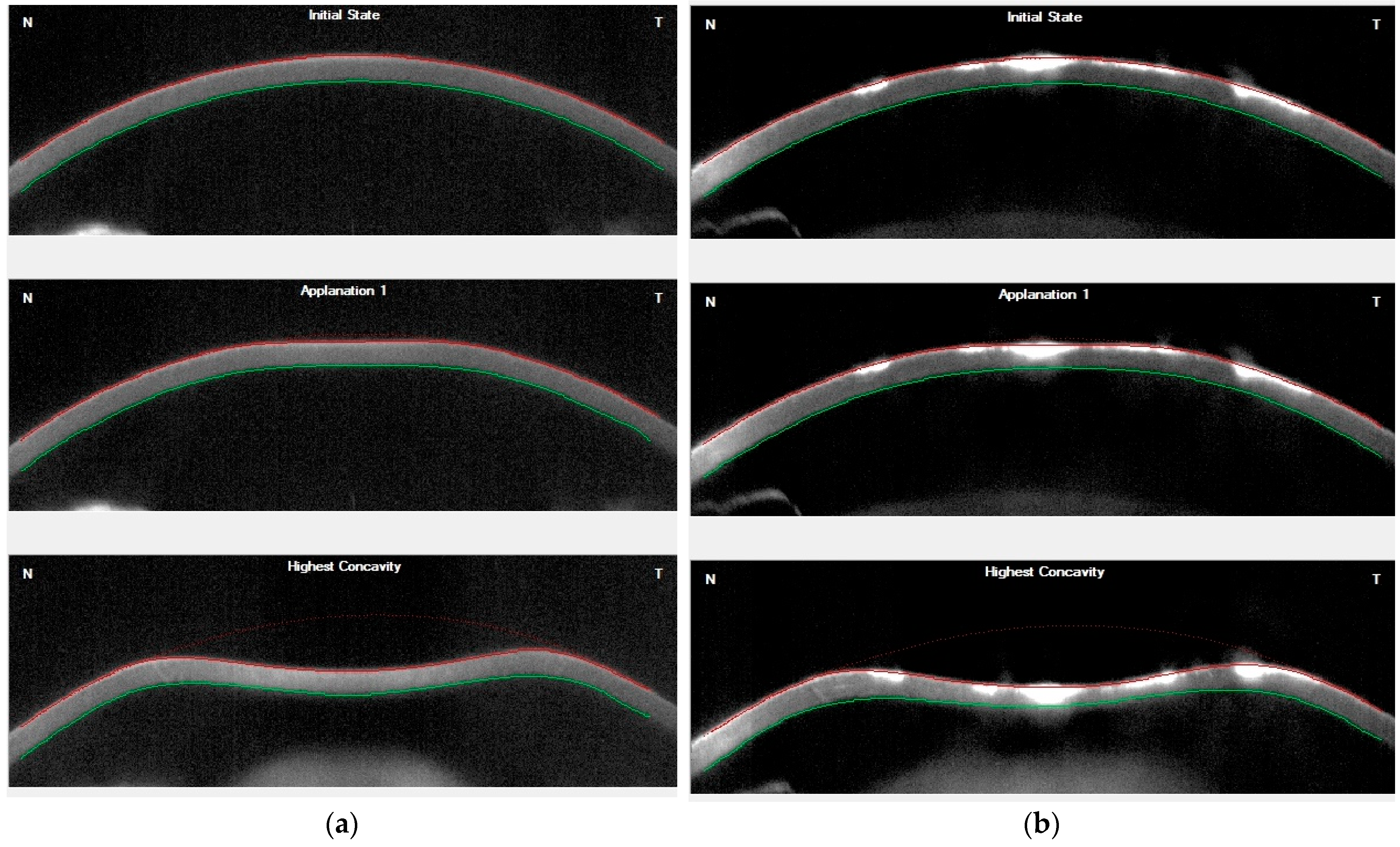
Example of Corvis tomograms recorded in a normal case in the control group and a subject with GCD are shown in Figure 2.
Data were analyzed using SPSS software version 17. The normality of quantitative variables was checked by the Kolmogorov–Smirnov test. An independent sample t-test was used to compare age, CCT, and bIOP between the two groups. A general linear model was used to compare corneal biomechanical parameters in the normal and GCD groups, while CCT, bIOP, and age were considered covariates. Pairwise comparisons were performed using the Dunn–Bonferroni post hoc test. A p-value less than 0.05 was considered significant statistically.

3. Results

Forty-five percent of participants in the GCD group and fifty percent in the control group were males, with no significant difference in sex distribution between the two groups (p = 0.751).
The mean age was 43.6 ± 19.4 years (range: 14–75 years) in the GCD group and 45.7 ± 15.9 years (range: 20–63 years) in the control group, with no statistically significant difference between the normal and GCD subjects (p = 0.710).
The mean and standard deviation of CCT, IOPnct, and bIOP in GCD and normal eyes were 561.45 ± 68.12 and 542.65 ± 36.45 µm (p = 0.291), 18.62 ± 5.41 and 16.17 ± 2.90 mm Hg (p = 0.085), and 17.33 ± 5.69 and 15.53 ± 2.57 mm Hg (p = 0.210), respectively.
The mean and standard deviation of standard and newer corneal biomechanical parameters of Corvis ST in the two groups are shown in Table 2 while CCT, bIOP, and age were considered covariates (Table 2).
Among all assessed biomechanical parameters, only AV1 (p < 0.001), AT1 (p = 0.002), HCR (p = 0.014), IR (p = 0.003), and SSI (p = 0.04) showed a statistically significant difference between the two groups using the independent samples t-test. The mean difference (GCD–Normal) was 0.02 m/s for AV1, −0.14 ms for AT1, −0.78 mm for HCR, 0.68 mm−1 for IR, and 0.11 for SSI.
Comparing the Ambrósio relational thickness to the horizontal profile (ARTh) in the two groups showed a statistically significant difference (p < 0.001), with mean values of 426.13 ± 206.23 microns (95% CI: 344.95 to 507.31) and 651.63 ± 168.90 microns (95% CI: 572.63 to 730.62) in GCD and normal eyes, respectively.

4. Discussion

To the best of our knowledge, few studies evaluated the corneal biomechanical properties using the Scheimpflug technology in granular corneal dystrophy (GCD). The main distinguishing feature of this study is the comparison of the newly introduced parameter of the stress-strain index in corneas with granular dystrophy with normal ones. Given that the biomechanical characteristics of the cornea as well as the dynamic corneal response of Corvis ST are affected by central corneal thickness, intraocular pressure, and age [12,24,25,26,27], the effect of these parameters in the comparison of corneal biomechanical parameters between these groups was controlled by considering them as covariates. The two groups were also homogeneous in terms of gender and age. The present study showed that, among all the assessed biomechanical parameters, only AV1 and AT1 (related to the first applanation phase), HCR and IR (related to the highest concavity phase), and SSI showed a statistically significant difference between the GCD and normal eyes. The corneal velocity towards the first applanation was 0.02 m/s faster in the GCD eyes and the first applanation phase was created 0.14 ms earlier than the normal eyes. These two findings point to the more elastic nature of the cornea, which has been reported in cases of keratoconus or corneal ectasia [28]. The lower corneal radius of curvature at the highest concavity phase also confirmed lower corneal strength and less rigidity of the cornea in the GCD group. The literature also shows that, in cases of corneal ectasia and with increasing severity, the integrated inverse radius (IR) increases [28,29]; this finding in GCD eyes agrees with previous reports. IR in the study group was 0.68 mm−1 higher than in normal eyes.
The stress-strain index (SSI) was introduced as a newer in vivo parameter to predict the biomechanical behavior of the cornea in terms of the material properties used in its structure [30]. Based on previous studies, the stress-stain behavior of biological tissue, such as the cornea, is nonlinear [31,32]. While biomechanical characteristics and corneal deformation response are affected by IOP and CCT [33,34,35], SSI was reported to be independent of these two parameters and only shows a significant positive correlation with age [30]. Moreover, it was mentioned as a better predictor of corneal stiffness, and values greater than or less than 1.0 indicate a biomechanically stiffer or softer cornea, respectively [36].
CBI was reported as a screening index to detect early ectasia even before the appearance of geometric changes in the cornea and higher values indicate a more likely presence of ectasia and less corneal stiffness [37,38]. The present study was designed on corneal biomechanical changes in a type of corneal dystrophy; however, the results show that GCD can cause an abnormal response in corneal biomechanical behavior compared to healthy corneas. This finding supports the higher CBI reported in cases with GCD compared to controls [39,40].
Histological evaluation showed that corneal collagen fibrils are maximally compressed in Bowman’s membrane and this layer plays a major role in corneal stability [41]. Therefore, it is expected that the biomechanical strength of the cornea in GCD is reduced secondary to the breaks in this layer due to the histopathological nature of the disease [19,20]. A lower SSI in GCD compared to normal corneas confirmed these changes in involved corneas. In addition, the present findings are in line with other studies that simultaneously reported the association of GCD with keratoconus [42,43,44,45,46].
Based on these findings and the presence of biomechanical instability of the corneas in GCD compared to normal corneas, including biomechanical evaluation in preoperative investigations is clinically valuable for deciding on the type and method of surgery in these patients to reduce the risk of iatrogenic ectasia following surgical interventions [21,22].
One study focusing on corneal biomechanical parameters in GCD was that of Kamiya et al., who used ORA to evaluate the biomechanical characteristics of GCD eyes before and after phototherapeutic keratectomy [9]. It was reported that the obtained ORA parameters (10.2 ± 2.2 mmHg for CH, 10.3 ± 2.0 mmHg for CRF) were similar to those of normal eyes (10.2 ± 1.3 mmHg for CH, 10.2 ± 1.7 mmHg for CRF) with equal CCT [9]. Considering the previously stated major drawbacks of ORA in evaluating corneal biomechanics, more studies are needed to confirm their findings.
A recent study that compared DCR parameters measured using Corvis ST in GCD and normal eyes reported higher A1DA and lower SPA1 in the GCD group [40], while these parameters were not statistically significant in the present study. In addition to the age difference, this difference can be attributed to racial differences (Chinese versus Iranian). Although their work points to abnormal corneal biomechanical behavior in GCD, unlike the present study, they did not evaluate the newly developed SSI parameter, which has been reported to be a better predictor of structural corneal strength.
In the present study, there was no significant difference in mean CCT between the two groups, while ARTh was significantly lower in the study group than the normal group, indicating an abnormal thickness profile or pachymetric progression in GCD patients, leading to a biomechanically softer cornea [47].
One limitation of the present study was the small sample size. A further study with greater numbers of patients is required to confirm our preliminary findings. Another challenge in our study is that some may argue for other covariates, such as the extent of the pathology, the number of deposits, or the area of the affected cornea, i.e., central, peripheral, or diffuse; however, the current sample size is too small to perform such classification and analysis. A study design considering the effect of these variables is suggested for future research.

5. Conclusions

GCD appears to alter corneal biomechanical characteristics assessed using Scheimpflug technology by Corvis ST. The corneal velocity towards the first applanation and the time taken to reach this phase, the corneal radius of curvature at the highest concavity phase, and the stress-strain index indicate less rigidity of the cornea in the GCD group. Until the availability of new biomechanical evaluation devices, such as the Brillouin microscopy in clinical practice, which can examine the biomechanics separately in each corneal layer, further studies are needed to confirm the present findings and the changes in the corneal biomechanical characteristics following different keratoplasty techniques in eyes with corneal granular dystrophy.

Author Contributions

Conceptualization, H.H., H.M.-M. and K.J.; methodology, H.M.-M., H.H., S.P. and M.M.; software, H.M.-M., K.J. and H.H.; validation, H.H., H.M.-M., K.J., S.P. and M.M.; formal analysis, H.M.-M. and M.M.; investigation, H.H. and S.P.; resources, K.J.; data curation, H.H., H.M.-M. and M.M.; writing—original draft preparation, H.H., H.M.-M., K.J., S.P. and M.M.; writing—review and editing, H.H., H.M.-M., K.J., S.P. and M.M.; visualization, H.H., H.M.-M. and K.J.; supervision, H.M.-M. and K.J.; project administration, H.H. and S.P. All authors have read and agreed to the published version of the manuscript.

Funding

This research received no external funding.

Institutional Review Board Statement

The study was conducted in accordance with the Declaration of Helsinki, and approved by the Ethics Committee of Semnan University of Medical Sciences, Semnan, Iran (protocol code IR.SEMUMS.REC.1401.043).

Informed Consent Statement

Informed consent was obtained from all subjects involved in the study. Written informed consent has been obtained from the patient(s) to publish this paper.

Data Availability Statement

All data generated or analyzed during this study are included in this published article.

Acknowledgments

The authors thank the personnel of Vision Health Research Center and the participants who made this study possible.

Conflicts of Interest

The authors declare no conflict of interest.

References

  1. Bourges, J.-L. Corneal dystrophies. J. Fr. D’ophtalmol. 2017, 40, e177–e192. [Google Scholar] [CrossRef] [PubMed]
  2. Poulaki, V.; Colby, K. (Eds.) Genetics of Anterior and Stromal Corneal Dystrophies, Seminars in Ophthalmology; Taylor & Francis: Abingdon, UK, 2008. [Google Scholar]
  3. Santos, L.N.; Fernandes, B.F.; de Moura, L.R.; Cheema, D.P.; Maloney, S.; Logan, P.; Burnier, M.N., Jr. Histopathologic study of corneal stromal dystrophies: A 10-year experience. Cornea 2007, 26, 1027–1031. [Google Scholar] [CrossRef] [PubMed]
  4. Lang, G.K.; Naumann, G. The frequency of corneal dystrophies requiring keratoplasty in Europe and the USA. Cornea 1987, 6, 209–211. [Google Scholar] [CrossRef]
  5. Musch, D.C.; Niziol, L.M.; Stein, J.D.; Kamyar, R.M.; Sugar, A. Prevalence of corneal dystrophies in the United States: Estimates from claims data. Investig. Ophthalmol. Vis. Sci. 2011, 52, 6959–6963. [Google Scholar] [CrossRef] [PubMed] [Green Version]
  6. Feizi, S.; Pakravan, M.; Baradaran-Rafiee, A.-R.; Yazdani, S. Granular corneal dystrophy manifesting after radial keratotomy. Cornea 2007, 26, 1267–1269. [Google Scholar] [CrossRef] [Green Version]
  7. Rodrigues, M.M.; Gaster, R.N.; Pratt, M.V. Unusual superficial confluent form of granular corneal dystrophy. Ophthalmology 1983, 90, 1507–1511. [Google Scholar] [CrossRef]
  8. Haddad, R.; Font, R.L.; Fine, B. Unusual superficial variant of granular dystrophy of the cornea. Am. J. Ophthalmol. 1977, 83, 213–218. [Google Scholar] [CrossRef]
  9. Kamiya, K.; Shimizu, K.; Ohmoto, F. The changes in corneal biomechanical parameters after phototherapeutic keratectomy in eyes with granular corneal dystrophy. Eye 2009, 23, 1790–1795. [Google Scholar] [CrossRef] [Green Version]
  10. del Buey, M.A.; Cristóbal, J.A.; Ascaso, F.J.; Lavilla, L.; Lanchares, E. Biomechanical properties of the cornea in Fuchs’ corneal dystrophy. Investig. Ophthalmol. Vis. Sci. 2009, 50, 3199–3202. [Google Scholar] [CrossRef]
  11. Feizi, S.; Karjou, Z.; Abbasi, H.; Javadi, M.A.; Azari, A.A. Characterization of in vivo biomechanical properties in macular corneal dystrophy. Am. J. Ophthalmol. 2020, 215, 8–13. [Google Scholar] [CrossRef]
  12. Piñero, D.P.; Alcón, N. In vivo characterization of corneal biomechanics. J. Cataract Refract. Surg. 2014, 40, 870–887. [Google Scholar] [CrossRef] [PubMed]
  13. Roberts, C.J. Concepts and misconceptions in corneal biomechanics. J. Cataract Refract. Surg. 2014, 40, 862–869. [Google Scholar] [CrossRef] [PubMed]
  14. Ali, N.Q.; Patel, D.V.; McGhee, C.N. Biomechanical responses of healthy and keratoconic corneas measured using a noncontact scheimpflug-based tonometer. Investig. Ophthalmol. Vis. Sci. 2014, 55, 3651–3659. [Google Scholar] [CrossRef] [PubMed]
  15. Das, S.; Langenbucher, A.; Seitz, B. Excimer Laser Phototherapeutic Keratectomy for Granular and Lattice Corneal Dystrophy: A Comparative Study; Slack Incorporated Thorofare: San Francisco, CA, USA, 2005. [Google Scholar]
  16. Fagerholm, P. Phototherapeutic keratectomy: 12 years of experience. Acta Ophthalmol. Scand. 2003, 81, 19–32. [Google Scholar] [CrossRef]
  17. Stasi, K.; Chuck, R.S. Update on phototherapeutic keratectomy. Curr. Opin. Ophthalmol. 2009, 20, 272–275. [Google Scholar] [CrossRef]
  18. Rathi, V.M.; Vyas, S.P.; Sangwan, V.S. Phototherapeutic keratectomy. Indian J. Ophthalmol. 2012, 60, 5. [Google Scholar] [CrossRef]
  19. Szentmáry, N.; Seitz, B.; Langenbucher, A.; Schlötzer-Schrehardt, U.; Hofmann-Rummelt, C.; Naumann, G.O. Histologic and ultrastructural changes in corneas with granular and macular dystrophy after excimer laser phototherapeutic keratectomy. Cornea 2006, 25, 257–263. [Google Scholar] [CrossRef]
  20. Seitz, B.; Behrens, A.; Fischer, M.; Langenbucher, A.; Naumann, G.O. Morphometric analysis of deposits in granular and lattice corneal dystrophy: Histopathologic implications for phototherapeutic keratectomy. Cornea 2004, 23, 380–385. [Google Scholar] [CrossRef]
  21. Dogru, M.; Katakami, C.; Nishida, T.; Yamanaka, A. Alteration of the ocular surface with recurrence of granular/avellino corneal dystrophy after phototherapeutic keratectomy: Report of five cases and literature review. Ophthalmology 2001, 108, 810–817. [Google Scholar] [CrossRef]
  22. Miyata, K.; Takahashi, T.; Tomidokoro, A.; Ono, K.; Oshika, T. Iatrogenic keratectasia after phototherapeutic keratectomy. Br. J. Ophthalmol. 2001, 85, 238. [Google Scholar] [CrossRef]
  23. Moshirfar, M.; Motlagh, M.N.; Murri, M.S.; Momeni-Moghaddam, H.; Ronquillo, Y.C.; Hoopes, P.C. Advances in biomechanical parameters for screening of refractive surgery candidates: A review of the literature, part III. Med. Hypothesis Discov. Innov. Ophthalmol. 2019, 8, 219. [Google Scholar] [PubMed]
  24. McMonnies, C.W. Assessing corneal hysteresis using the ocular response analyzer. Optom. Vis. Sci. 2012, 89, E343–E349. [Google Scholar] [CrossRef] [PubMed]
  25. Wong, Y.-Z.; Lam, A.K. The roles of cornea and axial length in corneal hysteresis among emmetropes and high myopes: A pilot study. Curr. Eye Res. 2015, 40, 282–289. [Google Scholar] [CrossRef]
  26. Huseynova, T.; Waring IV, G.O.; Roberts, C.; Krueger, R.R.; Tomita, M. Corneal biomechanics as a function of intraocular pressure and pachymetry by dynamic infrared signal and Scheimpflug imaging analysis in normal eyes. Am. J. Ophthalmol. 2014, 157, 885–893. [Google Scholar] [CrossRef] [PubMed]
  27. Miki, A.; Maeda, N.; Ikuno, Y.; Asai, T.; Hara, C.; Nishida, K. Factors associated with corneal deformation responses measured with a dynamic Scheimpflug analyzer. Investig. Ophthalmol. Vis. Sci. 2017, 58, 538–544. [Google Scholar] [CrossRef] [Green Version]
  28. Sedaghat, M.R.; Momeni-Moghaddam, H.; Ambrósio, R., Jr.; Heidari, H.R.; Maddah, N.; Danesh, Z.; Sabzi, F. Diagnostic Ability of Corneal Shape and Biomechanical Parameters for Detecting Frank Keratoconus. Cornea 2018, 37, 1025–1034. [Google Scholar] [CrossRef]
  29. Sedaghat, M.R.; Momeni-Moghaddam, H.; Roberts, C.J.; Maddah, N.; Ambrósio, R., Jr.; Hosseini, S.R. Corneal biomechanical parameters in keratoconus eyes with abnormal elevation on the back corneal surface only versus both back and front surfaces. Sci. Rep. 2021, 11, 11971. [Google Scholar] [CrossRef]
  30. Eliasy, A.; Chen, K.J.; Vinciguerra, R.; Lopes, B.T.; Abass, A.; Vinciguerra, P.; Ambrósio, R., Jr.; Roberts, C.J.; Elsheikh, A. Determination of Corneal Biomechanical Behavior in-vivo for Healthy Eyes Using CorVis ST Tonometry: Stress-Strain Index. Front. Bioeng. Biotechnol. 2019, 7, 105. [Google Scholar] [CrossRef] [Green Version]
  31. Elsheikh, A.; Wang, D.; Brown, M.; Rama, P.; Campanelli, M.; Pye, D. Assessment of corneal biomechanical properties and their variation with age. Curr. Eye Res. 2007, 32, 11–19. [Google Scholar] [CrossRef]
  32. Ethier, C.R.; Johnson, M.; Ruberti, J. Ocular biomechanics and biotransport. Annu. Rev. Biomed. Eng. 2004, 6, 249–273. [Google Scholar] [CrossRef]
  33. Valbon, B.F.; Ambrósio, R., Jr.; Fontes, B.M.; Luz, A.; Roberts, C.J.; Alves, M.R. Ocular biomechanical metrics by CorVis ST in healthy Brazilian patients. J. Refract. Surg. 2014, 30, 468–473. [Google Scholar] [CrossRef] [PubMed]
  34. Elsheikh, A.; Wang, D.; Pye, D. Determination of the modulus of elasticity of the human cornea. J. Refract. Surg. 2007, 23, 808–818. [Google Scholar] [CrossRef] [PubMed]
  35. Asaoka, R.; Nakakura, S.; Tabuchi, H.; Murata, H.; Nakao, Y.; Ihara, N.; Rimayanti, U.; Aihara, M.; Kiuchi, Y. The Relationship between Corvis ST Tonometry Measured Corneal Parameters and Intraocular Pressure, Corneal Thickness and Corneal Curvature. PLoS ONE 2015, 10, e0140385. [Google Scholar] [CrossRef] [PubMed]
  36. Kenia, V.P.; Kenia, R.V.; Pirdankar, O.H. Short term changes in corneal stress-strain index and other corneal biomechanical parameters post-laser in situ keratomileusis. Indian J. Ophthalmol. 2021, 69, 2650–2656. [Google Scholar]
  37. Vinciguerra, R.; Ambrósio, R., Jr.; Roberts, C.J.; Azzolini, C.; Vinciguerra, P. Biomechanical Characterization of Subclinical Keratoconus Without Topographic or Tomographic Abnormalities. J. Refract. Surg. 2017, 33, 399–407. [Google Scholar] [CrossRef] [Green Version]
  38. Sedaghat, M.R.; Momeni-Moghaddam, H.; Heravian, J.; Ansari, A.; Shayanfar, H.; Moshirfar, M. Detection ability of corneal biomechanical parameters for early diagnosis of ectasia. Eye 2022. [Google Scholar] [CrossRef]
  39. Tanikawa, A.; Soma, T.; Miki, A.; Koh, S.; Kitaguchi, Y.; Maeda, N.; Oie, Y.; Kawasaki, S.; Nishida, K. Assessment of the corneal biomechanical features of granular corneal dystrophy type 2 using dynamic ultra-high-speed Scheimpflug imaging. Graefes Arch Clin. Exp. Ophthalmol. 2022. [Google Scholar] [CrossRef]
  40. Wu, M.; Han, J.; Wang, X.; Shao, T.; Wang, Y. The alterations of corneal biomechanics in adult patients with corneal dystrophy. Eye 2022. [Google Scholar] [CrossRef]
  41. Dawson, D.G.; Grossniklaus, H.E. Biomechanical and wound healing characteristics of corneas after excimer laser keratorefractive surgery: Is there a difference between advanced surface ablation and sub-Bowman’s keratomileusis? J. Refract. Surg. 2008, 24, S90. [Google Scholar]
  42. Igarashi, S.; Makita, Y.; Hikichi, T.; Mori, F.; Hanada, K.; Yoshida, A. Association of keratoconus and Avellino corneal dystrophy. Br. J. Ophthalmol. 2003, 87, 367–368. [Google Scholar] [CrossRef] [Green Version]
  43. Mitsui, M.; Sakimoto, T.; Sawa, M.; Katami, M. Familial case of keratoconus with corneal granular dystrophy. Jpn. J. Ophthalmol. 1998, 42, 385–388. [Google Scholar] [CrossRef]
  44. Wollensak, G.; Green, W.R.; Temprano, J. Keratoconus associated with corneal granular dystrophy in a patient of Italian origin. Cornea 2002, 21, 121–122. [Google Scholar] [CrossRef] [PubMed]
  45. Wilson, C.M.; D’Ath, P.J.; Parmar, D.N.; Sykakis, E. Keratoconus and granular dystrophy. Case Rep. 2014, 2014, bcr2014205584. [Google Scholar] [CrossRef] [PubMed] [Green Version]
  46. Jadidi, K.; Mosavi, S.A.; Morovvati, S. Familial case of keratoconus with corneal granular dystrophy in a family of Iranian origin. J. Clin. Exp. Ophthalmol. 2014, 5, 2. [Google Scholar]
  47. Vinciguerra, R.; Ambrósio Jr, R.; Elsheikh, A.; Roberts, C.J.; Lopes, B.; Morenghi, E.; Azzolini, C.; Vinciguerra, P. Detection of keratoconus with a new biomechanical index. J. Refract. Surg. 2016, 32, 803–810. [Google Scholar] [CrossRef] [PubMed]
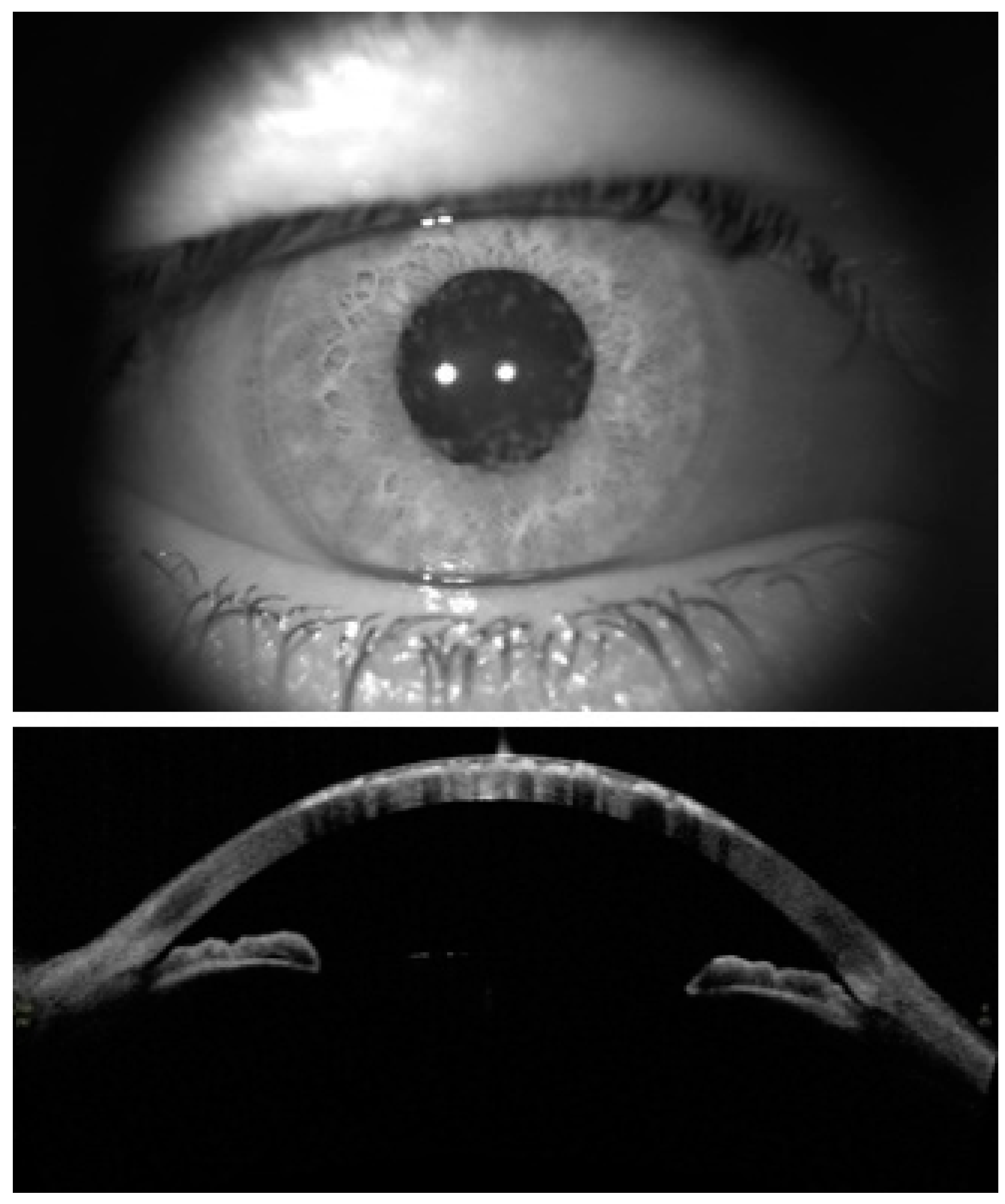
Figure 1. Slit-lamp biomicroscopy (diffuse illumination) (upper image) and anterior segment OCT (AS-OCT) images recorded from a case with GCD in the current study (lower image).
Jcm 11 06571 g001
Figure 2. Corvis tomograms in (a) a normal subject; (b) a subject with granular corneal dystrophy (GCD).
Jcm 11 06571 g002
Table 1. Parameters measured by Corvis ST included in this study.
Abbreviation (Unit)Complete NameDescription of Specific Corvis ST Parameters
AL1 (mm)Applanation length at the first flatteningCord diameter of the flattened cornea at the first applanation
AL2 (mm)Applanation length at the second flatteningCord diameter of the flattened cornea at the second applanation
A1V (m/s)First applanation velocityInward velocity of the cornea at the first applanation
A2V (m/s)Second applanation velocityOutward velocity of the cornea at the second applanation
A1T (ms)Time at the first applanationTime from initiation of air puff until the first applanation
A2T (ms)Time at the second applanationTime from initiation of air puff until the second applanation
A1DA (mm)Deformation amplitude at the first applanationCorneal displacement from the natural state until the first applanation
A2DA (mm)Deformation amplitude at the second applanationCorneal displacement at the second applanation
HC, PD (mm)Highest concavity, Peak distanceDistance between the two peaks of the cornea at the highest concavity phase
HC, Radius (mm)Highest concavity radiusRadius of curvature of the cornea at the highest concavity phase
HC-T (ms)Highest concavity timeTime from initiation of air puff until highest concavity
HC, DA (mm)Highest concavity, Deformation amplitudeCorneal displacement at the highest concavity phase
SPA1 (mm Hg/mm)Stiffness parameter at the first applanationResultant pressure [adjusted pressure at A1 (adj AP1)-biomechanically compensated IOP (bIOP)] divided by deflection amplitude at A1
IR (mm−1)Integrated inverse radiusArea under the inverse concave radius curve
DARMax1Deformation amplitude ratio at 1 mmRatio between DA at the apex and the average of DAs at 1 mm around the center in temporal and nasal directions
DAR Max2Deformation amplitude ratio at 2 mmRatio between DA at the apex and the average of DAs at 2 mm around the center in temporal and nasal directions
SSIStress-stain indexA parameter to predict the biomechanical behavior of the cornea in terms of the material properties used in its structure
CCT (µm)Central corneal thicknessCorneal thickness at the corneal apex
IOPnct (mmHg)Non-corrected IOPIntraocular pressure non-corrected based on the corneal characteristic and age
bIOP (mmHg)Biomechanically corrected intraocular pressureEstimates IOP based on an algorithm that reduces the confounding effects of corneal characteristics and aging
Table 2. Mean and SD of the corneal biomechanical parameters using the Corvis ST separately in the two groups with the central corneal thickness (CCT) and biomechanically corrected intraocular pressure (bIOP), with age as covariates.
VariablesMean ± SD (95% CI)p-Value
GCD (n = 12 Eyes)Control (n = 20 Eyes)
AL1 (mm)2.55 ± 0.24 (2.45 to 2.65)2.58 ± 0.21 (2.48 to 2.68)0.704
AL2 (mm)3.06 ± 0.49 (2.64 to 3.49)3.46 ± 1.09 (3.05 to 3.87)0.193
AV1 (m/s)0.15 ± 0.02 (0.14 to 0.15)0.13 ± 0.02 (0.12 to 0.13)<0.001
AV2 (m/s)−0.26 ± 0.06 (−0.28 to −0.25)−0.27 ± 0.05 (−0.29 to −0.25)0.872
AT1 (ms)7.33 ± 0.66 (7.26 to 7.39)7.47 ± 0.36 (7.41 to 7.53)0.002
AT2 (ms)21.02 ± 0.61 (20.89 to 21.14)21.08 ± 0.29 (20.96 to 21.21)0.471
A1DA (mm)0.15 ± 0.02 (0.14 to 0.15)0.14 ± 0.01 (0.14 to 0.15)0.108
A2DA (mm)0.43 ± 0.05 (0.40 to 0.46)0.40 ± 0.07 (0.37 to 0.43)0.144
HC, PD (mm)4.99 ± 0.45 (4.90 to 5.08)4.99 ± 0.27 (4.90 to 5.07)0.955
HC, R (mm)7.42 ± 0.76 (6.99 to 7.85)8.20 ± 1.08 (7.79 to 8.62)0.014
HC-T (ms)16.59 ± 0.51 (16.38 to 16.80)16.60 ± 0.35 (16.39 to 16.81)0.948
HC, DA (mm)1.05 ± 0.16 (1.01 to 1.09)1.00 ± 0.11 (0.97 to 1.04)0.123
SP-A1 (mm Hg/mm)104.03 ± 19.29 (99.35 to 108.72)110.45 ± 17.13 (105.90 to 115.01)0.061
IR (mm−1)7.48 ± 1.01 (7.18 to 7.78)6.80 ± 1.22 (6.51 to 7.09)0.003
DAR Max23.77 ± 0.42 (3.66 to 3.89)3.65 ± 0.49 (3.54 to 3.76)0.154
DAR Max11.51 ± 0.06 (1.49 to 1.53)1.50 ± 0.06 (1.48 to 1.51)0.259
SSI1.09 ± 0.20 (1.00 to 1.17)1.21 ± 0.17 (1.13 to 1.30)0.04
CBI0.33 ± 0.03 (0.31 to 0.35)0.02 ± 0.03 (0.01 to 0.03)<0.001
SD: Standard deviation; CI: confidence interval; AL: applanation length; AV: applanation velocity; AT: applanation time; DA: deformation amplitude; PD: peak distance; HC: highest concavity; T: time; R: radius of curvature, SPA 1: stiffness parameter at the first applanation; IR: integrated inverse radius; DAR: deformation amplitude ratio; SSI: stress-stain index; CBI: Corvis biomechanical index.
Publisher’s Note: MDPI stays neutral with regard to jurisdictional claims in published maps and institutional affiliations.