Article

Cerebral Tissue Oxygen Saturation Is Enhanced in Patients following Transcatheter Aortic Valve Implantation: A Retrospective Study

1
Department of Anesthesiology, Operative Intensive Care Medicine and Pain Therapy, Justus Liebig University Giessen, Rudolf-Buchheim-Strasse 7, 35392 Giessen, Germany
2
Department of Cardiology and Angiology, Justus Liebig University Giessen, Klinikstrasse 33, 35392 Giessen, Germany
3
Department of Cardiac and Vascular Surgery, Justus Liebig University Giessen, Rudolf-Buchheim-Strasse 7, 35392 Giessen, Germany
*
Author to whom correspondence should be addressed.
J. Clin. Med. 2022, 11(7), 1930; https://doi.org/10.3390/jcm11071930
Submission received: 5 March 2022 / Revised: 24 March 2022 / Accepted: 28 March 2022 / Published: 30 March 2022

Abstract

:
Transcatheter aortic valve implantation (TAVI) has emerged as an alternative to surgical aortic valve replacement. The aim of this study was to evaluate whether a relevant alteration in cerebral tissue oxygen saturation (rSO2) could be detected following TAVI. Retrospective data analysis included 275 patients undergoing TAVI between October 2016 and December 2020. Overall, rSO2 significantly increased following TAVI (64.6 ± 10% vs. 68.1 ± 10%, p < 0.01). However, a significant rise was only observed in patients with a preoperative rSO2 < 60%. Of the hemodynamic confounders studied, hemoglobin, mean arterial pressure and blood pH were lowered, while central venous pressure and arterial partial pressure of carbon dioxide (PaCO2) were slightly elevated (PaCO2: 39 (36–43) mmHg vs. 42 (37–47) mmHg, p = 0.03; pH: 7.41 (7.3–7.4) vs. 7.36 (7.3–7.4), p < 0.01). Multivariate linear regression modeling identified only hemoglobin as a predictor of altered rSO2. Patients with a EuroScore II above 4% and an extended ICU stay were found to have lower rSO2, while no difference was observed in patients with postoperative delirium or between the implanted valve types. Further prospective studies that eliminate differences in potential confounding variables are necessary to confirm the rise in rSO2. Future research should provide more information on the value of cerebral oximetry for identifying high-risk patients who will require further clinical interventions in the setting of the TAVI procedure.

1. Introduction

Aortic valve stenosis is the most common form of degenerative heart valve disease, and is increasing in prevalence, particularly in ageing populations. When left untreated, impaired left ventricular outflow increases afterload, thus generating left-ventricular pressure overload and leading to concentric hypertrophy and subsequent heart failure [1]. Due to technological and procedural advances, transcatheter aortic valve implantation (TAVI) has emerged as an attractive alternative to surgical aortic valve replacement (SAVR), as it does not require sternotomy and extracorporeal circulation [2]. Compared to SAVR, TAVI is associated with lower mortality, shorter length of hospital stays, and a more rapid return to normal life [3,4,5,6]. In contrast, patients undergoing SAVR have a lower risk of paravalvular regurgitation, permanent pacemaker implantation, and need for secondary valve intervention [7,8,9]. Thus, according to current European and American guidelines for the management of valvular heart disease, TAVI is favored in frail patients who are aged over 75 or 80 years, or who have a reduced life expectancy or a high surgical risk [10,11]. Currently, however, the indications for TAVI are expanding beyond otherwise inoperable patients [2]. Evidence is emerging that patients of intermediate surgical risk aged between 65 and 75 years old may also benefit from TAVI [12]. As such, current guidelines on the management of valvular heart disease recommend that these patients should be considered by interdisciplinary heart valve teams, and their clinical, anatomical, and procedural factors should be weighed for an individual approach [10,11]. Although TAVI is less invasive, heart valve interventions in elderly and often frail patients represent a challenge for perioperative anesthetic management. During the TAVI procedure, balloon valvuloplasty, the implantation of a balloon-expandable valve and post-dilatation are performed during functional circulatory arrest induced by rapid ventricular pacing (RVP) [13]. The obstruction of the left ventricular outflow tract during valvuloplasty, followed by severe aortic regurgitation, can also lead to hemodynamic deterioration through sudden left ventricular volume overload. The option to establish a cardiopulmonary bypass, or even switch to SAVR in the event of interventional disturbances, must therefore be guaranteed at any time during the procedure [14]. Consequently, anesthetic care is performed according to the standards of conventional cardiac surgery and includes cerebral monitoring. While the bispectral index using processed electroencephalogram is utilized to assess the depth of anesthesia, cerebral near-infrared spectroscopy (NIRS) can be used to measure regional cerebral tissue oxygen saturation (rSO2) by transmitting near-infrared light to a sensor attached to the patient’s forehead above the frontal lobe [15]. As oxygenated and deoxygenated hemoglobin have different absorption spectra, the rSO2 can be calculated using Lambert-Beer law. Near-infrared light passes through approximately 70–75% venous and 25–30% arterial blood volume; thus, displayed values reflect primarily venous oxygen consumption as a representation of regional cerebral blood flow and global cardiopulmonary function [15,16]. Depending on the device used, a rSO2 between 60 and 70% at room air is considered normal in cardiac surgical patients, while a lower baseline rSO2 is associated with higher incidence of postoperative delirium and mortality [17,18,19]. Although current data appear to be inconclusive, evidence is emerging that cerebral oximetry measurement can improve neurological outcomes in patients by reducing the incidence and severity of postoperative cognitive disorder (POCD) and delirium [20,21,22]. During TAVI, hemodynamic condition is altered promptly due to regained unlimited left-ventricular outflow. It therefore follows that restored cardiac physiology after TAVI may improve cerebral blood flow and thus improves cerebral oxygen consumption. Consequently, the aim of this study was to evaluate whether a relevant alteration in rSO2 could be detected following TAVI.

2. Materials and Methods

Study design. This retrospective, single-center cohort study was approved by the local ethics committee of the medical faculty of the Justus-Liebig-University, Giessen, Germany (AZ 243/20). According to the institutional standard of care, cerebral oximetry has been routinely used during TAVI procedures since October 2016. Whenever possible, TAVI was performed under conscious sedation with additional ilioinguinal nerve block using remifentanil (0.03–0.05 µg/kg/min) and propofol (1–1.5 mg/kg/h), targeting a bispectral index > 70. Otherwise, general anesthesia with remifentanil (0.1–0.2 µg/kg/min) and propofol (2–3 mg/kg/h) was used. When clinically indicated, midazolam (1 mg intravenously) was given as premedication after the arrival of the patient in the hybrid operating room. Clinical data records were screened retrospectively for patients who underwent TAVI at the university hospital of Giessen from October 2016 to December 2020 (Operation and Procedure Classification System, codes 5-35a.0, 5-35a.01–5-35a.04). Patients with sufficient availability of records on rSO2 and clinical information that met the baseline and procedural characteristics were included in the analysis. Sufficient available rSO2 data was defined as at least one value recorded during a five-minute period at the beginning of the intervention, and at least one value recorded during a five-minute period at the end. At that time, patients had to be under stable general anesthesia or conscious sedation that was maintained throughout the procedure. Patients who did not meet these criteria were excluded, as were those who underwent failed interventions.
Data acquisition. After eligible patients were identified, anesthetic protocols, clinical data records and diagnostic results were reviewed manually. Baseline characteristics included age, sex, body mass index, cardiovascular risk factors and relevant comorbidities such as peripheral artery disease, carotid artery stenosis, prior stroke or transient ischemic attack, chronic obstructive pulmonary disease, pulmonary hypertension, and chronic kidney failure. EuroScore II, pre- and post-operative electrocardiogram, echocardiography, and chest X-ray findings were recorded, as well as intraprocedural data, including anesthetic management. Cerebral oximetry was measured using an INVOS 5100C Cerebral/Somatic Oximeter (Medtronic, Minneapolis, MN, USA). Until May 2020, cerebral oximetry data were manually documented in the digital anesthetic protocol at the discretion of the anesthesiologist. After May 2020, oximetry data was recorded automatically every 5 min. Values from both cerebral hemispheres were averaged, unless the absolute difference between them was not greater than 10%; otherwise, they were registered separately. The rSO2 values were contextualized with pulmonary and hemodynamic parameters to determine any relevant influence besides the procedural impact. To evaluate the comparability of each pair of cerebral oximetry values, hemoglobin (Hb), mean arterial pressure (MAP), central venous pressure (CVP), inspirational fraction of oxygen (FiO2), arterial partial pressure of oxygen (PaO2) and carbon dioxide (PaCO2), arterial oxygen saturation (SaO2) and blood pH were documented for each oximetry value, and were analyzed as potential confounding variables. Outcome analysis included conversion to open surgery, the necessity for cardiopulmonary resuscitation (CPR), presence of POCD or POD according to the Confusion Assessment Method for Intensive Care Unit (CAM-ICU) criteria, and death [23]. Length of intensive care unit (ICU) and hospital stay were calculated from the administrative records. When available, EuroScore II and mortality rates were obtained from a local registry of patients undergoing TAVI (Kerckhoff Biomarkerregister (BioReg), Bad Nauheim, Germany); otherwise, the EuroScore II was calculated retrospectively, and clinical records were used to evaluate in-hospital mortality [24].
CPR, intraoperative and in-hospital death, and conversion to open surgery were included in a composite endpoint summarizing adverse events. Cerebral saturations and prevalence of catecholamine administration (summarized as administration of noradrenaline or adrenaline) in clinically relevant subgroups were compared with their respective complementary groups. These subgroups included individuals with a higher preoperative EuroScore II, POCD and POD, extended ICU or hospital stay, and adverse events. According to European guidelines, if there are no other clinical implications, SAVR is favored in patients with a EuroScore II under 4% [10]. A high EuroScore II group was therefore defined as a EuroScore II above 4%. Extended ICU and hospital stay were defined as any duration above the median length of stay.
Statistical analysis. Categorial variables are presented as numbers and percentages. Chi-squared test or the Fisher’s exact test was used to compare categorical data. Normally distributed continuous variables are presented as mean ± standard deviation (SD), while non-normally distributed variables are described by the median and interquartile range (Q1–Q3). Cerebral oximetry, confounding parameters, and patient subgroups were compared using a one-way ANOVA followed by Tukey’s test. In addition, a multivariate linear regression model was used to evaluate the influence of confounding parameters on changes in cerebral oximetry. Differences in pre- and post-operative confounding parameters were calculated and compared to their corresponding alterations in cerebral oximetry. Moreover, 2-tailed values of p < 0.05 were considered statistically significant.

3. Results

3.1. Patient Characteristics

A total of 405 patients were identified who underwent TAVI between October 2016 and December 2020. After screening for eligibility, 275 patients were included in the final analysis (Figure 1).
Baseline and procedural characteristics. Patient characteristics, baseline echocardiography, perioperative anesthetic management and procedural details are summarized in Table 1. The median age was 81 (77–84) years and patients were noted to have a high prevalence of cardiopulmonary comorbidities. The mean EuroScore II was 6.43 ± 5.7%, and baseline echocardiography findings showed severe aortic stenosis with an aortic valve area of 0.7 (0.6–0.9) cm2 (n = 239), a peak pressure gradient of 66 ± 22 mmHg (n = 255), a mean pressure gradient of 40 ± 14 mmHg (n = 259), and a peak velocity of 4.9 ± 7.24 m/s2 (n = 239). Left ventricular ejection fraction was mostly preserved (LVEF < 30%: 5.2%, n = 237). Overall, most procedures were carried out under conscious sedation (90%), and there was a low conversion rate to general anesthesia (5.1%). Perioperative fluid therapy was provided using mainly crystalloids (1000 (647–1071) mL), while the use of blood products and colloids was negligible. Most patients required intraoperative catecholamine administration (74.9%); however, the median cumulative dose was low (noradrenaline: 59 (2–169) µg, adrenaline: 0 (0–0) µg). A transfemoral approach for TAVI was considered standard procedure when not contraindicated and was used for 97.1% of the interventions. 81.1% of the implanted valves were self-expandable valves that were largely implanted following balloon valvuloplasty (71.3%) during RVP, while 18.9% were balloon-expanded valves. The need for post-dilatation requiring repeat RVP was 13.8%. Second valve implantation (“TAVI in TAVI”) was performed in 4 cases (1.5%) and 2 patients required intraoperative electrical cardioversion (0.7%).

3.2. Perioperative Outcome

The overall rate of intraoperative adverse events was low (Table 2). None of the procedures required switching to SAVR. Intraoperative cardiopulmonary resuscitation had to be performed in 10 patients (3.6%), and new left bundle branch block was detected in 33 patients (12.0%). Temporary or permanent pacemaker dependency occurred in 10 (3.6%) and 33 (12.0%) patients, respectively, and this was largely due to new high-grade atrioventricular block (10.9%). Follow up echocardiography revealed high procedural success (Table 3), though some degree of paravalvular leakage was found in 113 patients (42.5%). Median length of ICU and hospital stay was 4 (2.2–6.7) days and 11 (8.5–17.0) days, respectively. POCD or POD occurred in 42 patients (15.3%) and 9 patients died during the index hospital stay (3.3%).

3.3. Cerebral Oximetry

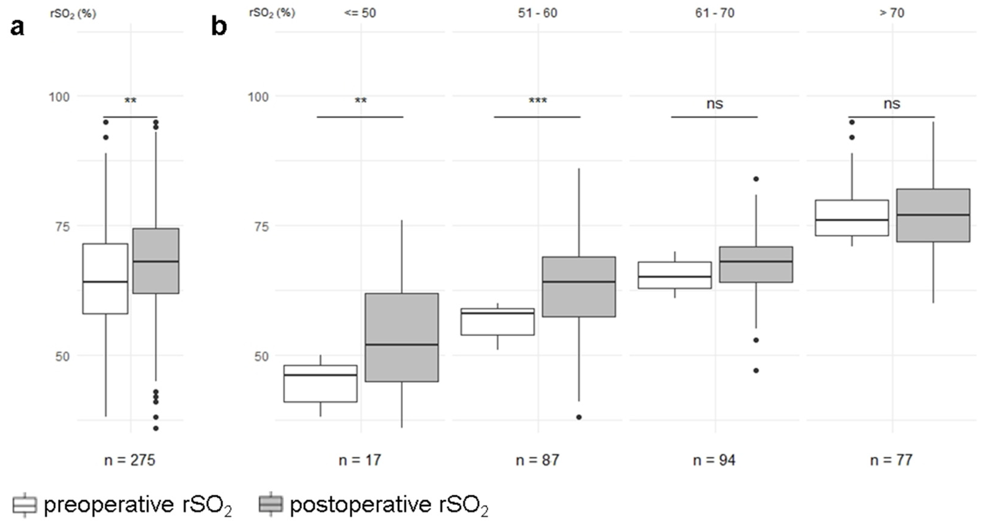
Overall, median alteration of rSO2 was +4.0 (−1.0–+8.0) %, and absolute values of rSO2 significantly increased from 64.6 ± 10% to 68.1 ± 10% following TAVI (p < 0.01; Figure 2a). Stratified by preoperative rSO2 values, cerebral oximetry was significantly increased in patients with a preoperative rSO2 that was lower than 50% (45.1 ± 3.7% vs. 53.6 ± 11.1%, p < 0.01) and between 51–60% (56.6 ± 3.0% vs. 63.2 ± 8.5%, p < 0.001). No significant changes were observed in patients with a preoperative rSO2 between 61–70% (65.7 ± 2.9% vs. 68.0 ± 6,4%, p = 0.16) and above 70% (76.8 ± 5.2% vs. 77.0 ± 7.22%), p = 1.0; Figure 2b).

3.4. Hemodynamic Confounder

The changes observed in hemodynamic parameters are illustrated in Table 3. Hb (11.6 ± 1.8 g/dL vs. 10.3 ± 1.7 g/dL, p < 0.01) and MAP (91 ± 17 mmHg vs. 76 ± 18 mmHg, p < 0.01) were found to significantly decrease postoperatively, while CVP increased during the procedure (11 ± 7 mmHg vs. 13 ± 7 mmHg, p < 0.01). PaCO2 also increased postoperatively (39 (36–43) mmHg vs. 42 (37–47) mmHg, p = 0.03), and this was accompanied by reciprocal changes in pH (7.41 (7.3–7.4) vs. 7.36 (7.3–7.4), p < 0.01). No significant changes were observed in PaO2 (128 (98–169) mmHg vs. 125 (90–160) mmHg, p = 0.49) and SaO2 (99 (98–99) % vs. 99 (97–99) %, p = 0.18).
To evaluate the influence of hemodynamic confounders on cerebral rSO2, differences in the pre- and post-operative variables were subsequently calculated and assessed against the changes in rSO2 using a multivariate linear regression model. The result of this analysis showed that only changes in Hb were independent predictors of altered cerebral rSO2 (p < 0.01; n = 60; adjusted R2 = 0.17, overall p = 0.029). According to the model, variations in MAP, CVP, pH, PaCO2, PaSO2 and SaO2 were of no predictive value for rSO2 in this study (Table 4).

3.5. Subgroup Analysis

rSO2 and catecholamine administration were comparable among patients with self-expanding or balloon-expandable valves and patients with POCD or POD following TAVI (Table 5). However, patients with a EuroScore II above 4% and those who had longer ICU or hospital stays had lower preoperative (63.5 ± 10.1% vs. 66.2 ± 9.7%, p = 0.03; 62.6 ± 10.0% vs. 66.2 ± 9.7%, p < 0.01; 61.2 ± 9.2% vs. 67.5 ± 9.8%, p < 0.01) and postoperative rSO2 (66.8 ± 10.9% vs. 69.8 ± 8.5%, p = 0.01; 66.5 ± 10.0% vs. 69.7 ± 9.9%, p < 0.01; 64.8 ± 10.4% vs. 70.9 ± 8.9%, p < 0.01), and had higher catecholamine requirements postoperatively (37% vs. 25%, p = 0.03; 40.4% vs. 24.5%, p < 0.01; 40.8% vs. 25.3%, p < 0.01). In contrast, preoperative catecholamine administration was comparable among these groups. Patients who experienced adverse events showed comparable preoperative rSO2 values (64.9 ± 10.1% vs. 60.9 ± 7.2%, p = 0.14), but had higher preoperative catecholamines requirements (26.7% vs. 7.7%, p = 0.03). Postoperative rSO2 values were lower (68.4 ± 10.1% vs. 62.5 ± 8.4%, p = 0.03), and were accompanied by a higher catecholamine requirement (29.2% vs. 86.7%, p < 0.01). In contrast, the numerical rise in rSO2 was comparable between all of the analyzed subgroups.

4. Discussion

A significant rise in cerebral rSO2 was observed in this retrospective cohort following TAVI. As the new competent aortic valve restores adequate cardiac output and oxygen delivery, this rise could be attributed to improved hemodynamic function. However, although significant, the actual numerical rise in rSO2 was small in our cohort, and it must therefore be considered that confounding factors contributed to these slightly elevated rSO2 values. To date, evidence on the course of cerebral NIRS following TAVI has focused mainly on the episode of RVP [25,26,27]. Consistent to the functionally circulatory arrest that is achieved during RVP, rSO2 was seen to fall significantly. Interestingly, a decrease of more than 20% resulted in a higher incidence of POD, and these patients showed also lower rSO2 baseline values [26]. Desaturation after RVP was also followed by transient hyperemia leading to elevated rSO2. However, rSO2 returned to its baseline value only a few minutes after RVP, and the rise in rSO2 observed after the procedure in this study should not therefore be attributed to recent RVP [27]. Thus, hyperemia could be attributed to acid metabolites accumulated during RVP leading to temporarily enhanced cerebral blood flow. Temporarily impaired cerebral autoregulation after prompt alteration of cardiac hemodynamic following TAVI may also be considered as explanation for the temporary rise in rSO2 in our study. In other clinical settings, such as in carotid endarterectomy procedures, cerebral hyperperfusion accompanied by neurological deficits is a well-known clinical syndrome that is associated with highly elevated rSO2 values [28]. It should be noted, however, that the magnitude of the rise in rSO2 observed in this study was much lower than in those patients. Furthermore, although values of baseline rSO2 were comparable, Suppan et al. observed improved rSO2 and cardiac output following cardiopulmonary exercise testing 5 days after TAVI [29]. Consequently, the rise in rSO2 observed in this study might not be a short-term change.
The increase in rSO2 in this study substantially differed between subgroups, depending on their preoperative cerebral oximetry values. The rise observed in patients with low preoperative rSO2 (<50%, 51–60%) was significantly greater than in patients with a preoperative rSO2 > 60%, whose values were unchanged postoperatively. This could suggest that patients with aortic valve stenosis and a preoperative rSO2 < 60% even suffer from hemodynamic restrictions at rest due to aortic valve stenosis, and this manifests not only as clinical symptoms, but also as imminent cardiac decompensation. In general, rSO2 values are subject to large variations between individuals [30]. In their study, Fanning et al. were unable to detect a difference between pre- and post-operative values in patients undergoing TAVI with general anesthesia, and similar results were found by Mayr et al., who compared rSO2 during conscious sedation with rSO2 during general anesthesia [27,31]. Although these results were not statistically investigated, a numerical increase in rSO2 was observed at the end of the TAVI procedure in both general anesthesia and conscious sedation cohorts. However, it should be noted that both trials utilized only small study cohorts, and the comparison of rSO2 during conscious sedation and general anesthesia may still be affected by anesthetic care. In our study, Hb and MAP were found to be significantly reduced, while CVP was raised postoperatively. However, these alterations were not found to be associated with an increase in rSO2: MAP remained within the autoregulatory range in our study cohort. The association between low Hb and lower rSO2 has been shown in cardiac surgery patients [32]. Thus, the reduction in Hb seen in this study results in a concordant reduction in oxygen supply, which would therefore reduce rSO2, rather than raise it [28,33]. Cerebral perfusion pressure and cerebral blood flow are reduced by higher CVP; hence increased venous filling or congestion illustrated by elevated CVP would be expected to lower rSO2, not raise it. However, PaCO2 was slightly, but still significantly, raised in our cohort, and consecutive alterations in pH were also detected. The numerical change in these values was lower than those observed by Mayr et al.; nevertheless, alterations in PaCO2 must be analyzed with particular care [25]. In contrast to other hemodynamic parameters, there is no cerebral autoregulation mechanism for PaCO2. This means that, within physiological ranges, cerebral blood flow enhances linearly with rising PaCO2 [34,35]. The relation of PaCO2 and rSO2 has been examined in various clinical settings using wide ranges of target PaCO2, from 35 mmHg to 55 mmHg [36,37,38,39]. rSO2 increases in association with PaCO2, but the magnitude of this increase is not fully understood. For example, during cardiopulmonary bypass, rSO2 increased from 55% to 64% after an increase in PaCO2 from 38 mmHg to 52 mmHg [39]. In another study by Sørensen et al. investigating changes in end tidal CO2 and rSO2 during reperfusion following aortic arch surgery, an increase of 3.75 mmHg was observed in etCO2, followed by a 2% increase in rSO2 [38]. In their study, Wong et al. randomly assigned patients undergoing major non-cardiac surgery to median target PaCO2 values of either 34.8 mmHg and 51.5 mmHg and detected a +19% increase in rSO2 in the hypercapnia group [37]. Compared to these trials, the rise observed in PaCO2 in this study from median 39 (36–43) mmHg to median 42 (37–47) mmHg was very low, but the rise in rSO2 was greater than that observed by Sørensen et al. [38]. These findings suggest that the observed rise in rSO2 in our cohort might be influenced by factors other than changes in PaCO2. This is supported by the result of our multivariate linear regression model, which found that altered Hb alone was a predictor of altered rSO2 in our cohort. It should be noted, that changes in PaCO2 are accompanied by corresponding changes in pH, which facilitates the oxygen release from hemoglobin via the Bohr effect. The increase in PaCO2 is likely to be influenced by the anesthetic procedure. Most of the patients in this study underwent TAVI with conscious sedation using propofol and remifentanil. In a randomized controlled trial by Mayr et al., patients under conscious sedation with propofol and opioid were shown to have significantly elevated levels of PaCO2 after induction and during valvuloplasty compared to patients under general anesthesia [25]. Hypoventilation is affected not only by the anesthetic procedure, but also by the choice of drugs used for sedation. Compared to propofol and opioids, conscious sedation with dexmedetomidine results in a lower incidence of hypercapnia and an overall reduction in PaCO2 [31]. However, as shown in this study, PaCO2 levels remained consistently within clinically safe ranges, regardless of the choice of sedative.
In our subgroup analysis, rSO2 and administration of catecholamines were comparable between patients treated with self-expandable and balloon-expanded valves, and these results are consistent with those of Eertmans et al. [40]. Comparable values of rSO2 were observed pre- and post-operatively, but intraoperative desaturation was more substantial during the implantation of balloon-expandable devices, which is due to the implantation technique. More substantial desaturations during RVP were observed in patients with a higher EuroScore, who also showed an overall reduction in rSO2 in our cohort [26]. Preoperative rSO2 was lower in patients with a higher EuroScore who underwent on-pump cardiac surgery, and these patients were found to have increased postoperative mortality [18,41]. Lower rSO2 was also shown to be an independent predictor of POD following on-pump cardiac surgery [41]. Whether this observation also applies following TAVI is still unclear, however, in our cohort, there was no difference between rSO2 and catecholamine requirement in patients with POD or POCD. This finding could be attributed to a shorter procedural duration, lower usage of general anesthesia, and reduced invasiveness of the procedure that avoided cardiopulmonary bypass, which is associated with a higher incidence of POD [42]. Nevertheless, patients with longer ICU and hospital stay were found to have lower preoperative rSO2 values in our cohort. This may be attributed to stronger restrictions of cardiopulmonary function and comorbidities. Interestingly, the intraoperative increase in rSO2 was comparable, but postoperative rSO2 remained significantly lower. Cerebral oximetry of these patients may therefore indicate a successful procedure, but oximetry values remained at an overall lower level than in patients who had shorter stays in the ICU and hospital. That a higher postoperative catecholamine requirement was observed both in patients with longer ICU and hospital stays and in those with a higher EuroScore II further suggests that these results indicate more difficulties in adapting to new cardiovascular conditions. Similarly, patients who experienced adverse events had lower postoperative rSO2 values and higher catecholamine requirements. However, despite comparable preoperative rSO2, their preoperative catecholamine requirements were already higher compared to those patients who did not experience adverse events. This suggests that intraoperative disturbances are not the only factors leading to severe circulatory restrictions represented by lower rSO2 values.
Of course, several limitations of our study must be acknowledged. The first is that retrospective data were used for analysis. For example, incidence of POD was assessed retrospectively using CAM-ICU criteria and therefore relevant under-detection, particularly of hypoactive delirium, cannot be ruled out. Second, problems with availability made it impossible to analyze cerebral oximetry data without the presence of additional oxygen administration. Cerebral oximetry during general anesthesia or conscious sedation was considered, and the measurements may therefore still have been affected by anesthetic care. Although cerebral oximetry values were measured continuously throughout the procedure, they were only partly documented, either at the anesthesiologist’s discretion, or automatically every 5 min. Additionally, it should be noted that the statistical power of some subgroup analysis may have been impaired due to small group sizes.

5. Conclusions

Our study shows a significant rise in rSO2 following TAVI. However, the actual numerical increase observed was small, and levels of PaCO2, a potential powerful confounder, also increased. Nevertheless, we showed that patients from clinically relevant subgroups with a higher EuroScore II, extended hospital or ICU stay, and adverse events had lower rSO2 levels and higher catecholamines requirements. Further prospective studies that eliminate differences in potential confounding variables are necessary to confirm the rise in rSO2 following TAVI. A synchronous comparison of different sedation regimes, including the use of dexmedetomidine, may be helpful to rule out the role of sedative-induced hypoventilation. Thus, future research may provide more information on the value of cerebral oximetry for identifying high-risk patients who will require further clinical interventions in the setting of the TAVI procedure.

Author Contributions

G.S., H.K., U.V.-R., M.S. and C.K. were involved in planning, data acquisition and writing the manuscript. E.S., F.E., H.N. and A.B. made substantial contributions to the conception and provided additional references, and statistical and clinical experience to the discussion. All authors have read and agreed to the published version of the manuscript.

Funding

This research received no external funding.

Institutional Review Board Statement

The study was conducted in accordance with the Declaration of Helsinki, and approved by the or Ethics Committee of the University of Giessen (AZ 243/20).

Informed Consent Statement

Patient consent was waived due to retrospective, anonymized data.

Data Availability Statement

The data presented in this study are available on request from the corresponding author.

Conflicts of Interest

The authors declare no conflict of interest.

References

  1. Carabello, B.A.; Paulus, W.J. Aortic stenosis. Lancet 2009, 373, 956–966. [Google Scholar] [CrossRef]
  2. Chiam, P.T.; Ewe, S.H. The expanding indications of transcatheter aortic valve implantation. Futur. Cardiol. 2016, 12, 209–219. [Google Scholar] [CrossRef] [PubMed]
  3. Marsh, K.; Hawken, N.; Brookes, E.; Kuehn, C.; Liden, B. Patient-centered benefit-risk analysis of transcatheter aortic valve replacement. F1000Research 2021, 8, 394. [Google Scholar] [CrossRef] [PubMed]
  4. Siemieniuk, R.A.; Agoritsas, T.; Manja, V.; Devji, T.; Chang, Y.; Bala, M.M.; Thabane, L.; Guyatt, G.H. Transcatheter versus surgical aortic valve replacement in patients with severe aortic stenosis at low and intermediate risk: Systematic review and meta-analysis. BMJ 2016, 354, i5130. [Google Scholar] [CrossRef] [PubMed] [Green Version]
  5. Baron, S.J.; Wang, K.; House, J.A.; Magnuson, E.A.; Reynolds, M.R.; Makkar, R.; Herrmann, H.C.; Kodali, S.; Thourani, V.H.; Kapadia, S.; et al. Cost-Effectiveness of Transcatheter Versus Surgical Aortic Valve Replacement in Patients With Severe Aortic Stenosis at Intermediate Risk. Circulation 2019, 139, 877–888. [Google Scholar] [CrossRef] [PubMed]
  6. Siontis, G.C.M.; Overtchouk, P.; Cahill, T.J.; Modine, T.; Prendergast, B.; Praz, F.; Pilgrim, T.; Petrinic, T.; Nikolakopoulou, A.; Salanti, G.; et al. Transcatheter aortic valve implantation vs. surgical aortic valve replacement for treatment of symptomatic severe aortic stenosis: An updated meta-analysis. Eur. Heart J. 2019, 40, 3143–3153. [Google Scholar] [CrossRef] [PubMed]
  7. Khan, S.U.; Lone, A.N.; Saleem, M.A.; Kaluski, E. Transcatheter vs surgical aortic-valve replacement in low-to intermediate-surgical-risk candidates: A meta-analysis and systematic review. Clin. Cardiol. 2017, 40, 974–981. [Google Scholar] [CrossRef] [PubMed]
  8. Kim, K.S.; Makhdoum, A.; Koziarz, A.; Gupta, S.; Alsagheir, A.; Pandey, A.; Reza, S.; Um, K.; Teoh, K.; Alhazzani, W.; et al. Outcomes of sutureless aortic valve replacement versus conventional aortic valve replacement and transcatheter aortic valve replacement, updated systematic review, and meta-analysis. J. Card. Surg. 2021, 36, 4734–4742. [Google Scholar] [CrossRef]
  9. Lou, Y.; Gao, Y.; Yu, Y.; Li, Y.; Xi, Z.; Swe, K.N.C.; Zhou, Y.; Nie, X.; Liu, W. Efficacy and Safety of Transcatheter vs. Surgical Aortic Valve Replacement in Low-to-Intermediate-Risk Patients: A Meta-Analysis. Front. Cardiovasc. Med. 2020, 7, 590975. [Google Scholar] [CrossRef]
  10. Vahanian, A.; Beyersdorf, F.; Praz, F.; Milojevic, M.; Baldus, S.; Bauersachs, J.; Capodanno, D.; Conradi, L.; De Bonis, M.; De Paulis, R.; et al. 2021 ESC/EACTS Guidelines for the management of valvular heart disease. Eur. Heart J. 2021, 43, 561–632. [Google Scholar] [CrossRef]
  11. Otto, C.M.; Nishimura, R.A.; Bonow, R.O.; Carabello, B.A.; Erwin, J.P.; Gentile, F.; Jneid, H.; Krieger, E.V.; Mack, M.; McLeod, C.; et al. 2020 ACC/AHA Guideline for the Management of Patients With Valvular Heart Disease: A Report of the American College of Cardiology/American Heart Association Joint Committee on Clinical Practice Guidelines. Circulation 2021, 143, e27–e227. [Google Scholar] [CrossRef]
  12. Lazkani, M.; Singh, N.; Howe, C.; Patel, N.; Colon, M.J.; Tasset, M.; Amabile, O.; Morris, M.; Fang, H.K.; Pershad, A. An updated meta-analysis of TAVR in patients at intermediate risk for SAVR. Cardiovasc. Revasc. Med. 2019, 20, 57–69. [Google Scholar] [CrossRef]
  13. McInerney, A.; Vera-Urquiza, R.; Tirado-Conte, G.; Marroquin, L.; Jimenez-Quevedo, P.; Nuñez-Gil, I.; Pozo, E.; Gonzalo, N.; de Agustín, J.A.; Escaned, J.; et al. Pre-dilation and Post-dilation in Transcatheter Aortic Valve Replacement: Indications, Benefits and Risks. Interv. Cardiol. Rev. Res. Resour. 2021, 16, e28. [Google Scholar] [CrossRef]
  14. Vigelius-Rauch, U.; Zajonz, T.; Sander, M. Anesthesiological implications of minimally invasive valve interventions: Transcatheter aortic valve implantation, clip reconstruction on the mitral and tricuspid valve. Der Anaesth. 2021, 70, 97–111. [Google Scholar] [CrossRef]
  15. Rivera-Lara, L.; Vaca, A.Z.; Geocadin, R.G.; Healy, R.J.; Ziai, W.; Mirski, M.A. Cerebral autoregulation-oriented therapy at the bedside a comprehensive review. Anesthesiology 2017, 126, 1187–1199. [Google Scholar] [CrossRef]
  16. Ferrari, M.; Mottola, L.; Quaresima, V. Principles, Techniques, and Limitations of Near Infrared Spectroscopy. Can. J. Appl. Physiol. 2004, 29, 463–487. [Google Scholar] [CrossRef] [Green Version]
  17. Lei, L.; Katznelson, R.; Fedorko, L.; Carroll, J.; Poonawala, H.; Machina, M.; Styra, R.; Rao, V.; Djaiani, G. Cerebral oximetry and postoperative delirium after cardiac surgery: A randomised, controlled trial. Anaesthesia 2017, 72, 1456–1466. [Google Scholar] [CrossRef] [Green Version]
  18. Heringlake, M.; Garbers, C.; Käbler, J.-H.; Anderson, I.; Heinze, H.; Schön, J.; Berger, K.-U.; Dibbelt, L.; Sievers, H.-H.; Hanke, T. Preoperative Cerebral Oxygen Saturation and Clinical Outcomes in Cardiac Surgery. Anesthesiology 2011, 114, 58–69. [Google Scholar] [CrossRef] [Green Version]
  19. Stannard, B.; Levin, M.A.; Lin, H.-M.; Weiner, M.M. Regional cerebral oximetry is consistent across self-reported racial groups and predicts 30-day mortality in cardiac surgery: A retrospective analysis. Int. J. Clin. Monit. Comput. 2021, 35, 413–421. [Google Scholar] [CrossRef]
  20. Ortega-Loubon, C.; Herrera-Gómez, F.; Bernuy-Guevara, C.; Jorge-Monjas, P.; Ochoa-Sangrador, C.; Bustamante-Munguira, J.; Tamayo, E.; Álvarez, F.J. Near-Infrared Spectroscopy Monitoring in Cardiac and Noncardiac Surgery: Pairwise and Network Meta-Analyses. J. Clin. Med. 2019, 8, 2208. [Google Scholar] [CrossRef] [Green Version]
  21. Mailhot, T.; Cossette, S.; Lambert, J.; Cournoyer, A.; Denault, A.Y. Cerebral oximetry as a biomarker of postoperative delirium in cardiac surgery patients. J. Crit. Care 2016, 34, 17–23. [Google Scholar] [CrossRef]
  22. Zorrilla-Vaca, A.; Healy, R.; Grant, M.; Joshi, B.; Rivera-Lara, L.; Brown, C.; Mirski, M.A. Intraoperative cerebral oximetry-based management for optimizing perioperative outcomes: A meta-analysis of randomized controlled trials. Can. J. Anaesth. 2018, 65, 529–542. [Google Scholar] [CrossRef] [Green Version]
  23. Gusmao-Flores, D.; Salluh, J.I.F.; Chalhub, R.Á.; Quarantini, L.C. The confusion assessment method for the intensive care unit (CAM-ICU) and intensive care delirium screening checklist (ICDSC) for the diagnosis of delirium: A systematic review and meta-analysis of clinical studies. Crit. Care 2012, 16, R115. [Google Scholar] [CrossRef] [Green Version]
  24. Nashef, S.A.; Roques, F.; Sharples, L.D.; Nilsson, J.; Smith, C.; Goldstone, A.R.; Lockowandt, U. EuroSCORE II. Eur. J. Cardio-Thorac. Surg. 2012, 41, 734–745. [Google Scholar] [CrossRef] [Green Version]
  25. Mayr, N.P.; Hapfelmeier, A.; Martin, K.; Kurz, A.; van der Starre, P.; Babik, B.; Mazzitelli, D.; Lange, R.; Wiesner, G.; Tassani-Prell, P. Comparison of sedation and general anaesthesia for transcatheter aortic valve implantation on cerebral oxygen saturation and neurocognitive outcome. Br. J. Anaesth. 2016, 116, 90–99. [Google Scholar] [CrossRef] [Green Version]
  26. Seppelt, P.C.; Mas-Peiro, S.; De Rosa, R.; Murray, I.M.; Arsalan, M.; Holzer, L.; Lotz, G.; Meybohm, P.; Zacharowski, K.; Walther, T.; et al. Dynamics of cerebral oxygenation during rapid ventricular pacing and its impact on outcome in transfemoral transcatheter aortic valve implantation. Catheter. Cardiovasc. Interv. 2021, 97, E146–E153. [Google Scholar] [CrossRef]
  27. Fanning, J.P.; Walters, D.L.; Wesley, A.J.; Anstey, C.; Huth, S.; Bellapart, J.; Collard, C.; Rapchuk, I.L.; Natani, S.; Savage, M.; et al. Intraoperative Cerebral Perfusion Disturbances During Transcatheter Aortic Valve Replacement. Ann. Thorac. Surg. 2017, 104, 1564–1568. [Google Scholar] [CrossRef] [Green Version]
  28. Pennekamp, C.; Bots, M.; Kappelle, L.; Moll, F.; de Borst, G. The Value of Near-Infrared Spectroscopy Measured Cerebral Oximetry During Carotid Endarterectomy in Perioperative Stroke Prevention. A Review. Eur. J. Vasc. Endovasc. Surg. 2009, 38, 539–545. [Google Scholar] [CrossRef] [Green Version]
  29. Suppan, M.; Barcelos, G.; Luise, S.; Diaper, J.; Frei, A.; Ellenberger, C.; Adamopoulos, D.; Noble, S.; Licker, M. Improved Exercise Tolerance, Oxygen Delivery, and Oxygen Utilization After Transcatheter Aortic Valve Implantation for Severe Aortic Stenosis. CJC Open 2020, 2, 490–496. [Google Scholar] [CrossRef]
  30. Chan, M.J.; Chung, T.; Glassford, N.; Bellomo, R. Near-Infrared Spectroscopy in Adult Cardiac Surgery Patients: A Systematic Review and Meta-Analysis. J. Cardiothorac. Vasc. Anesth. 2017, 31, 1155–1165. [Google Scholar] [CrossRef] [Green Version]
  31. Mayr, N.P.; Wiesner, G.; Van Der Starre, P.; Hapfelmeier, A.; Goppel, G.; Kasel, A.M.; Hengstenberg, C.; Husser, O.; Schunkert, H.; Tassani-Prell, P. Dexmedetomidine versus propofol-opioid for sedation in transcatheter aortic valve implantation patients: A retrospective analysis of periprocedural gas exchange and hemodynamic support. Can. J. Anaesth. 2018, 65, 647–657. [Google Scholar] [CrossRef] [PubMed] [Green Version]
  32. Kobayashi, K.; Kitamura, T.; Kohira, S.; Torii, S.; Horai, T.; Hirata, M.; Mishima, T.; Sughimoto, K.; Ohkubo, H.; Irisawa, Y.; et al. Factors associated with a low initial cerebral oxygen saturation value in patients undergoing cardiac surgery. J. Artif. Organs 2017, 20, 110–116. [Google Scholar] [CrossRef] [PubMed]
  33. Vretzakis, G.; Georgopoulou, S.; Stamoulis, K.; Stamatiou, G.; Tsakiridis, K.; Zarogoulidis, P.; Katsikogianis, N.; Kougioumtzi, I.; Machairiotis, N.; Tsiouda, T.; et al. Cerebral oximetry in cardiac anesthesia. J. Thorac. Dis. 2014, 6, S60–S69. [Google Scholar] [CrossRef] [PubMed]
  34. Battisti-Charbonney, A.; Fisher, J.; Duffin, J. The cerebrovascular response to carbon dioxide in humans. J. Physiol. 2011, 589, 3039–3048. [Google Scholar] [CrossRef]
  35. Madsen, P.L.; Secher, N.H. Near-infrared oximetry of the brain. Prog. Neurobiol. 1999, 58, 541–560. [Google Scholar] [CrossRef]
  36. Murphy, G.S.; Szokol, J.W.; Avram, M.J.; Greenberg, S.B.; Shear, T.D.; Vender, J.S.; Levin, S.D.; Koh, J.; Parikh, K.N.; Patel, S.S. Effect of ventilation on cerebral oxygenation in patients undergoing surgery in the beach chair position: A randomized controlled trial. Br. J. Anaesth. 2014, 113, 618–627. [Google Scholar] [CrossRef] [Green Version]
  37. Wong, C.; Churilov, L.; Cowie, D.; Tan, C.O.; Hu, R.; Tremewen, D.; Pearce, B.; Pillai, P.; Karalapillai, D.; Bellomo, R.; et al. Randomised controlled trial to investigate the relationship between mild hypercapnia and cerebral oxygen saturation in patients undergoing major surgery. BMJ Open 2020, 10, e029159. [Google Scholar] [CrossRef] [Green Version]
  38. Sørensen, H.; Nielsen, H.B.; Secher, N.H. Near-infrared spectroscopy assessed cerebral oxygenation during open abdominal aortic aneurysm repair: Relation to end-tidal CO2 tension. Int. J. Clin. Monit. Comput. 2015, 30, 409–415. [Google Scholar] [CrossRef]
  39. Akça, O.; Sessler, D.; Delong, D.; Keijner, R.; Ganzel, B.; Doufas, A. Tissue oxygenation response to mild hypercapnia during cardiopulmonary bypass with constant pump output. Br. J. Anaesth. 2006, 96, 708–714. [Google Scholar] [CrossRef] [Green Version]
  40. Eertmans, W.; Genbrugge, C.; Fret, T.; Beran, M.; Engelen, K.; Gutermann, H.; Laenen, M.V.; Boer, W.; Ferdinande, B.; Jans, F.; et al. Influence of continuously evolving transcatheter aortic valve implantation technology on cerebral oxygenation. Int. J. Clin. Monit. Comput. 2016, 31, 1133–1141. [Google Scholar] [CrossRef]
  41. Schoen, J.; Meyerrose, J.; Paarmann, H.; Heringlake, M.; Hueppe, M.; Berger, K.-U. Preoperative regional cerebral oxygen saturation is a predictor of postoperative delirium in on-pump cardiac surgery patients: A prospective observational trial. Crit. Care 2011, 15, R218. [Google Scholar] [CrossRef] [Green Version]
  42. Goudzwaard, J.A.; Ronde-Tillmans, M.J.A.G.D.; Jager, T.A.J.D.; Lenzen, M.J.; Nuis, R.-J.; van Mieghem, N.M.; Daemen, J.; de Jaegere, P.P.T.; Mattace-Raso, F.U.S. Incidence, determinants and consequences of delirium in older patients after transcatheter aortic valve implantation. Age Ageing 2020, 49, 389–394. [Google Scholar] [CrossRef]
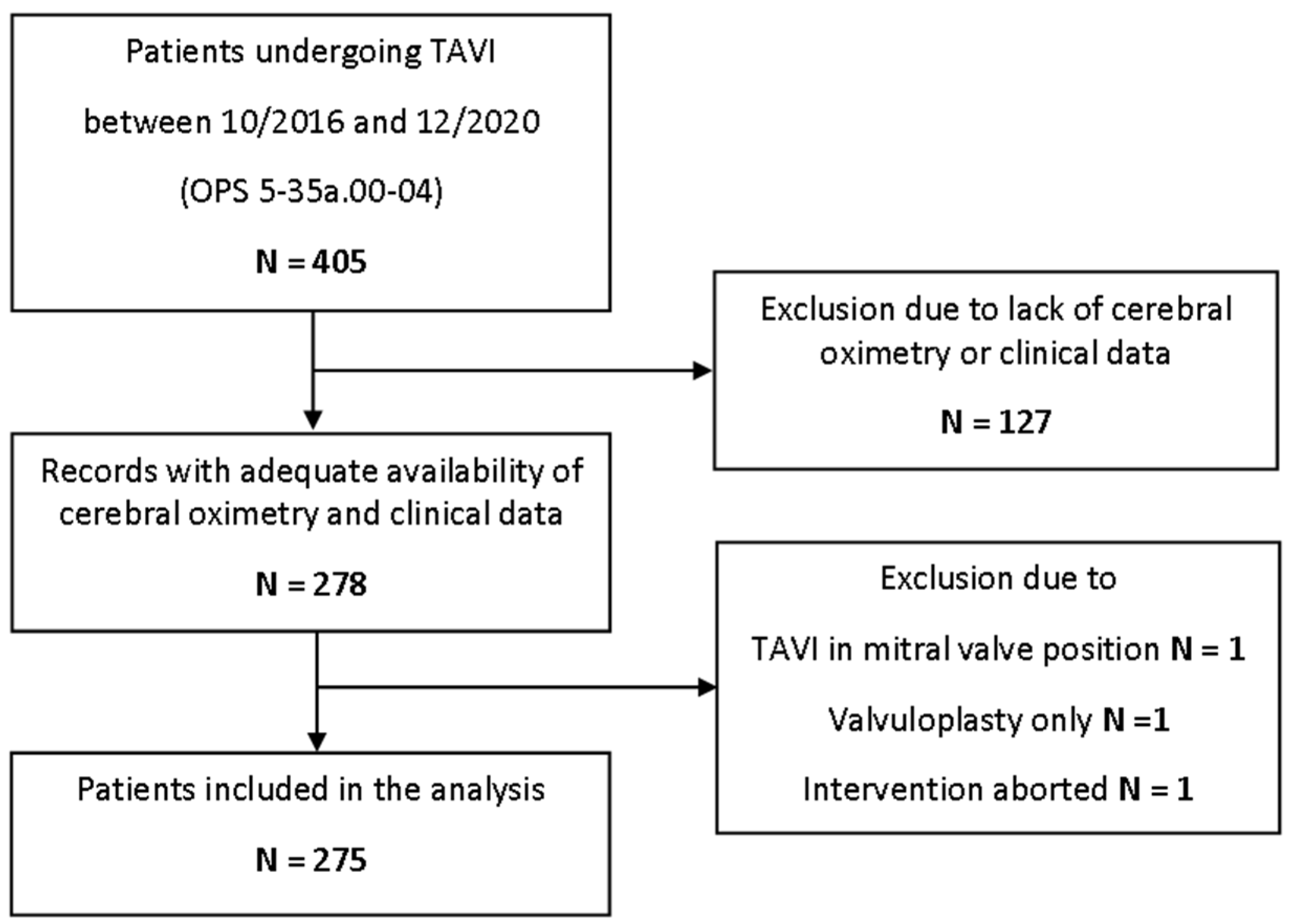
Figure 1. Study flowchart. TAVI = transcatheter aortic valve implantation.
Jcm 11 01930 g001
Figure 2. (a) Cerebral rSO2 was enhanced after TAVI. (b) Analysis stratified by preoperative values revealed a significant increase only in patients with preoperative rSO2 ≤ 50% and 51–60%. **: p < 0.01; ***: p < 0.001; ns: not significant; rSO2 = regional cerebral oxygen saturation.
Jcm 11 01930 g002
Table 1. Patient characteristics, anesthetic management, and procedural details.
Characteristicsn = 275
Patient characteristics
male-no. (%)139 (50.5)
median age (Q1–Q3)-years81 (77–84)
mean Body-Mass-Index (SD)-kg/m227 (±4)
arterial hypertension-no. (%)230 (83.6)
pulmonary hypertension-no. (%)14 (5.1)
coronary artery disease-no. (%)190 (69.1)
peripheral vessel disease-no. (%)34 (12.4)
carotid artery stenosis-no. (%)45 (16.4)
prior stroke or transient ischemic attack-no. (%)29 (10.5)
chronic kidney disease-no. (%)69 (25.1)
chronic obstructive pulmonary disease-no. (%)35 (12.7)
current smoker-no. (%)15 (5.5)
family history of CAD-no. (%)24 (8.7)
pacemaker-no. (%)31 (11.3)
pleural effusion present-no. (%)57 (22.2)
EuroScore II (SD)-%6.43 (±5.7)
Anesthetic management
conscious sedation-no. (%)239 (86.9)
general anesthesia-no. (%)22 (8.0)
switch to general anesthesia-no. (%)14 (5.1)
amount of crystalloids (Q1–Q3)-mL1000 (647–1071)
amount of colloids (Q1–Q3)-mL0 (0–0)
amount of red blood cell concentrate (Q1–Q3)-mL0 (0–0)
amount of fresh frozen plasma (Q1–Q3)-mL0 (0–0)
catecholamine administration-no. (%)206 (74.9)
cumulative dosage of noradrenaline (Q1–Q3)-µg59 (2–169)
cumulative dosage of adrenaline (Q1–Q3)-µg0 (0–0)
duration of anesthesia (SD)-min131 (±36)
Procedural details
duration of intervention (Q1–Q3)-min55 (45–69)
transfemoral approach-no. (%)267 (97.1)
transapical approach-no. (%)5 (1.8)
subclavian approach-no. (%)3 (1.1)
balloon-expandable valve-no. (%)52 (18.9)
self-expandable valve-no. (%)223 (81.1)
valvuloplasty-no. (%)196 (71.3)
postdilatation-no. (%)38 (13.8)
Second valve implantation-no. (%)4 (1.5)
number of rapid pacing (Q1–Q3)-no.1 (1–2)
number of fast pacing (Q1–Q3)-no.1 (0–1)
Cardioversion-no. (%)2 (0.7)
Table 2. Adverse events, postprocedural ECG and outcome.
Characteristicsn = 275
Postprocedural ECG
new high-grade AVB-no. (%)30 (10.9)
new LBBB-no. (%)45 (16.9)
new pacemaker dependency, permanent-no. (%)33 (12.0)
new pacemaker dependency, temporary-no. (%)10 (3.6)
Outcome
Any paravalvular leak-no. (%)113 (42.5)
Median length of ICU stay (Q1–Q3)-days4 (2.2–6.7)
Median length of hospital stay (Q1–Q3)-days11 (8.5–17.0)
POCD or delirium-no. (%)42 (15.3)
Adverse events
Conversion to open surgery-no. (%)0 (0.0)
CPR-no. (%)10 (3.6)
Intraoperative mortality-no. (%)0 (0.0)
In-hospital mortality-no. (%)9 (3.3)
Table 3. Overall rSO2, hemodynamic confounders and follow-up echocardiography.
CharacteristicsnPreoperativePostoperativenp
Cerebral oximetry
ΔrSO2 (Q1–Q3)-%2754.0 (−1.0–8.0)275
rSO2 (SD)-%27564.6 (±10)68.1 (±10)275<0.01
Confounding parameters
Hb (SD)-mg/dL26211.6 (±1.8)10.3 (±1.7)213<0.01
MAP (SD)-mmHg27591 (±17)76 (±18)275<0.01
CVP (SD)-mmHg20511 (±7)13 (±7)255<0.01
pH (Q1–Q3)-no.1017.41 (7.3–7.4)7.36 (7.3–7.4)121<0.01
PaCO2 (Q1–Q3)-mmHg10139 (36–43)42 (37–47)1210.03
PaO2 (Q1–Q3)-mmHg101128 (98–169)125 (90–160)1190.49
SaO2 (Q1–Q3)-%10199 (98–99)99 (97–99)1190.18
Echocardiography
LVEF (SD)-%23754 (±11)55 (±9)1760.38
AV-PPG (SD)-mmHg25566 (±22)16 (±8)237<0.01
AV-MPG (SD)-mmHg25940 (±14)9 (±4)225<0.01
rSO2 = regional cerebral oxygen saturation; Hb = hemoglobin; MAP = mean arterial pressure; CVP = central venous pressure; FiO2 = inspirational oxygen fraction; PaO2 = arterial partial pressure of oxygen; PaCO2 = arterial partial pressure of carbon dioxide; SaO2 = arterial oxygen saturation; SpO2 = partial oxygen saturation; LVEF = left ventricular ejection fraction; AV = aortic valve; PPG = peak pressure gradient; MPG = mean pressure gradient.
Table 4. Multivariate linear regression model of the alteration of rSO2 and differences in confounders only revealed Hemoglobin as predictor of altered rSO2.
VariableEstimateStandard Errorp
ΔrSO2
Intercerpt5.061.51<0.01
ΔHb4.211.12<0.01
ΔMAP−0.030.040.48
ΔCVP−0.060.150.70
ΔpH−23.9831.710.45
ΔPaCO20.100.240.66
ΔPaO20.000.020.78
ΔSaO20.270.350.44
n = 60Adjusted R2: 0.17; p = 0.029
rSO2 = regional cerebral oxygen saturation; Hb = hemoglobin; MAP = mean arterial pressure; CVP = central venous pressure; FiO2 = inspirational oxygen fraction; PaO2 = arterial partial pressure of oxygen; PaCO2 = arterial partial pressure of carbon dioxide; SaO2 = arterial oxygen saturation; SpO2 = partial oxygen saturation; LVEF = left ventricular ejection fraction.
Table 5. Subgroup analysis revealed lower rSO2 and higher need for catecholamines in patients with higher EuroScore II, longer ICU and hospital stay.
Characteristicsp
Valve TypeSelf-Expanding
n = 223
Balloon-Expandable
n = 52
ΔrSO2 (Q1–Q3)-%3.0 (−1.0–8.0)4.0 (−1.0–8.0)0.98
preoperative rSO2 (SD)-%64.7 (±10.3)64.2 (±8.8)0.71
catecholamine administration-no. (%)18 (8.1)6 (11.5)0.42
postoperative rSO2 (SD)-%68.2 (±10.2)67.7 (±9.8)0.73
catecholamine administration-no. (%)66 (29.6)23 (44.2)0.05
POCD or Deliriumno
n = 233
yes
n = 42
ΔrSO2 (Q1–Q3)-%3.0 (−1.0–8.0)4.0 (0–7.75)0.97
preoperative rSO2 (SD)-%64.9 (±10.3)63.2 (±8.0)0.33
catecholamine administration-no. (%)21 (9.0)3 (7.1)1.00
postoperative rSO2 (SD)-%68.3 (±10.5)66.7 (±7.5)0.34
catecholamine administration-no. (%)71 (30.5)18 (42.9)0.15
EuroScore II≤4%
n = 116
>4%
n = 151
ΔrSO2 (Q1–Q3)-%4.0 (−1.0–8.0)3.0 (−1.0–8.0)0.72
preoperative rSO2 (SD)-%66.2 (±9.7)63.5 (±10.1)0.03
catecholamine administration-no. (%)9 (7.8)15 (9.4)0.67
postoperative rSO2 (SD)-%69.8 (±8.5)66.8 (±10.9)0.01
catecholamine administration-no. (%)29 (25.0)60 (37.7)0.03
length of ICU stayshort (≤4 d)
n = 146
long (>4 d)
n = 129
ΔrSO2 (Q1–Q3)-%3.0 (−1.0–7.0)4.0 (−1.0–8.3)0.38
preoperative rSO2 (SD)-%66.2 (±9.7)62.6 (±10.0)<0.01
catecholamine administration-no. (%)9 (6.5)15 (11.0)0.20
postoperative rSO2 (SD)-%69.7 (±9.9)66.5 (±10.0)<0.01
catecholamine administration-no. (%)34 (24.5)55 (40.4)<0.01
length of hospital stayshort (≤11 d)
n = 150
long (>11 d)
n = 125
ΔrSO2 (Q1–Q3)-%3.5 (−1.0–7.75)4.0 (−1.0–8.0)0.77
preoperative rSO2 (SD)-%67.5 (±9.8)61.2 (±9.2)<0.01
catecholamine administration-no. (%)10 (6.7)14 (11.2)0.20
postoperative rSO2 (SD)-%70.9 (±8.9)64.8 (±10.4)<0.01
catecholamine administration-no. (%)38 (25.3)51 (40.8)<0.01
Adverse eventno
n = 260
yes
n = 15
ΔrSO2 (Q1–Q3)-%4 (−1.0–8.0)2 (−3.5–6.0)0.32
preoperative rSO2 (SD)-%64.9 (±10.1)60.9 (±7.2)0.14
catecholamine administration-no. (%)20 (7.7)4 (26.7)0.03
postoperative rSO2 (SD)-%68.4 (±10.1)62.5 (±8.4)0.03
catecholamine administration-no. (%)76 (29.2)13 (86.7)<0.01
Adverse event: in-hospital mortality, conversion to open surgery, CPR. Abbreviations: rSO2 = regional cerebral oxygen saturation; SD = standard deviation; POCD = postoperative cognitive disorder; ICU = intensive care unit; CPR = cardiopulmonary resuscitation.= regional cerebral oxygen saturation; Hb = hemoglobin; MAP = mean arterial pressure; CVP = central venous pressure; FiO2 = inspirational oxygen fraction; PaO2 = arterial partial pressure of oxygen; PaCO2 = arterial partial pressure of carbon dioxide; SaO2 = arterial oxygen saturation; SpO2 = partial oxygen saturation; LVEF = left ventricular ejection fraction; AV = aortic valve; PPG = peak pressure gradient; MPG = mean pressure gradient.
Publisher’s Note: MDPI stays neutral with regard to jurisdictional claims in published maps and institutional affiliations.