Article

Evaluation of FMR4, FMR5 and FMR6 Expression Levels as Non-Invasive Biomarkers for the Diagnosis of Fragile X-Associated Primary Ovarian Insufficiency (FXPOI)

1
Biochemistry and Molecular Genetics Department, Hospital Clinic of Barcelona and Institut d’Investigacions Biomèdiques August Pi i Sunyer (IDIBAPS), 08036 Barcelona, Spain
2
CIBER of Rare Diseases, Instituto de Salud Carlos III, 28029 Madrid, Spain
3
Clinical Institute of Gynecology, Obstetrics and Neonatology (ICGON), Hospital Clinic of Barcelona and Institut de Investigacions Biomèdiques August Pi iSunyer (IDIBAPS), 08036 Barcelona, Spain
*
Author to whom correspondence should be addressed.
These authors contributed equally to this work.
J. Clin. Med. 2022, 11(8), 2186; https://doi.org/10.3390/jcm11082186
Submission received: 17 February 2022 / Revised: 31 March 2022 / Accepted: 8 April 2022 / Published: 14 April 2022
(This article belongs to the Special Issue Update on Prenatal Diagnosis and Maternal Fetal Medicine)

Abstract

:
Female FMR1 (Fragile X mental retardation 1) premutation carriers are at risk for developing fragile X-associated primary ovarian insufficiency (FXPOI), a condition characterized by amenorrhea before age 40 years. Not all women with a FMR1 premutation suffer from primary ovarian insufficiency and nowadays there are no molecular or other biomarkers that can help predict the occurrence of FXPOI. Long non-coding RNAs (lncRNAs) comprise a group of regulatory transcripts which have versatile molecular functions, making them important regulators in all aspects of gene expression. In recent medical studies, lncRNAs have been described as potential diagnostic biomarkers in many diseases. The present study was designed to determine the expression profile of three lncRNAs derived from the FMR1 locus, FMR4, FMR5 and FMR6, in female FMR1 premutation carriers in order: (i) to determine a possible role in the pathogenesis of FXPOI and (ii) to investigate whether they could serve as a biomarker for the diagnosis of FXPOI. FMR4, FMR5 and FMR6 transcripts levels were evaluated in total RNA extracted from peripheral blood by digital droplet PCR and compared between FMR1 premutation carriers with FXPOI and without FXPOI. The diagnostic value of lncRNAs was evaluated by receiver operating characteristic (ROC) analysis. Results revealed a significant association between FXPOI and high expression levels of FMR4. No association was obtained for FMR5 or FMR6. ROC curve analysis revealed that FMR4 can distinguish FMR1 premutation carrier with FXPOI with a diagnostic power of 0.67. These findings suggest a potential role of FMR4 as a possible biomarker for FXPOI.

1. Introduction

The FMR1 (Fragile X mental retardation 1) gene (OMIM*309550) contains a CGG trinucleotide repeat in the 5′ untranslated region [1,2,3]. Among individuals in the general population, the number of CGG repeats is polymorphic and varies between 6 and 44. In this situation the FMR1 gene is active and there is synthesis of the Fragile Mental Retardation Protein (FMRP). Repeats in this size interval are stable when transmitted from generation to generation. In contrast, when the number of CGGs exceeds 200 repeats (full mutation) the FMR1 gene is silenced resulting in the absence of FMRP and the clinical manifestations of fragile X syndrome (FXS, OMIM#300624; ORPHA:908): intellectual disability and characteristic dysmorphic features [4]. Finally, individuals with alleles between 55 and 200 CGG repeats are the so-called FMR1 premutation carriers. In this situation, there is increased transcription of the FMR1 mRNA transcript, and slightly reduced synthesis of FMRP [5]. These repeat alleles are unstable and tend to increase in each cell division, conferring an increased risk of having FXS affected offspring.
FMR1 premutation occurs in the general population with an estimated frequency that range from 1 in 800 to 1200 males and from 1 in 250 to 400 females [6]. These individuals are at risk for fragile X-associated premature ovarian insufficiency (FXPOI) in females [7], and fragile X-associated tremor/ataxia syndrome (FXTAS) in both, males and females [8].
FXPOI symptoms include the cessation of ovarian function before the age of 40 years, ovarian dysfunction, and decreased fertility as evidenced by abnormal ovarian reserve biomarkers and reduced ovarian response to controlled ovarian hyperstimulation [9,10,11]. FXPOI occurs in ~20% of women with the FMR1 premutation, compared to only 1% of the general population [12]. Women with the FMR1 premutation not only struggle with the possibility of developing FXPOI but also with the risk of passing on the full mutation to their offspring.
Long non-coding RNAs (lncRNAs), defined as untranslated RNA molecules greater than 200 nucleotides in length, can be derived from sense or antisense strands within protein-coding genes, intergenic regions, or pseudogenes [13,14,15]. A lot of evidence has been accumulated showing that lncRNAs have versatile molecular functions, making them important regulators in all aspects of gene expression (reviewed in [16]). The biological mechanisms of lncRNAs are multiple. They can interact with DNA, RNA and protein as well as with other biological molecules, regulating the gene expression trough controlling processes such as protein synthesis, RNA maturation and transport, or the chromatin structure [17]. Moreover, lncRNAs participate in diverse biological processes, such as development, differentiation, energy metabolism, apoptosis and angiogenesis; controlling every level of gene expression pathway [18]. These particularities make them good candidate biomarkers for several diseases.
Several lncRNAs are derived from the FMR1 gene locus: FMR4, FMR6 and the ASFMR1 in the antisense direction and FMR5 in the sense direction (reviewed in [19]). Previous studies have described different expression levels of these lncRNAs among FMR1 permutation carriers, suggesting a functional association with fragile X-associated disorders [20,21,22,23,24]. Although the FMR1 premutation is the major risk factor for developing FXPOI, there are still some unknown genetic, epigenetic or environmental factors that might be affecting gene penetrance. On the basis of this observation, we aim to determine the expression profile of FMR4, FMR5 and FMR6 in female FMR1 premutation carriers in order to determine a possible role in the pathogenesis of FXPOI.

2. Material and Methods

2.1. Subjects

For this study, 36 female FMR1 premutation carriers registered in the FXS database from the Department of Biochemistry and Molecular Genetics, Hospital Clinic of Barcelona were selected. The cases met the following criteria: 20 women diagnosed with FXPOI (development of amenorrhea due to disruption of ovarian function before the age of 40 years) carrying the FMR1 premutation (CGG repeats between 55–200) and 16 non-FXPOI FMR1 premutation carriers with a reported normal ovarian function (regular cycles between 24–35 days) over the age of 40 years. This study was approved by the Institutional Ethical Review Board of Hospital Clinic, Barcelona. All patients that were included in this study signed a written informed consent.

2.2. RNA Extraction and cDNA Synthesis

Total RNA isolation was performed from blood using the PAXgene® Blood RNA Kit (Qiagen, Hilden, Germany) according to the manufacturer’s protocols. A Qubit RNA IQ assay (ThermoFisher Scientific, Waltham, MA, USA) was used to determine the RNA concentration. Its integrity was verified with the Bioanalyzer 2100 (Agilent, Santa Clara, CA, USA). The cDNA was synthesized from 350 ng RNA using the High-Capacity cDNA reverse transcription Kit (Applied Biosystems, Waltham, MA, USA), following the manufacturer’s instructions.

2.3. Digital Droplet Polymerase Chain Reaction (ddPCR)

LncRNA expression analysis was performed by ddPCR technology, which is an improvement of conventional PCR. Using a droplet generator, the sample is diluted and divided into multiple aliquots and subjected to endpoint PCR. Amplification occurs in each individual partition, which contains an individual nucleic acid molecule. A fluorescence signal is produced in each droplet with the target molecule. Quantification of cDNA molecules relies on the ability of the ddPCR system to determine the number of target molecules by the Poisson statistical analysis of “positive” (containing amplified target) and “negative” (no amplified target detected) droplets [25]. The assay was performed using the QX200™ ddPCR™ EvaGreen Supermix (Bio-Rad, Hercules, CA, USA), 100 nM specific primers targeted to FMR4 (f:5′-ACCAAACCAAACCAAACCAA-3′ and r:5′-GTGGGAAATCAAATGCATCC-3′), FMR5 (f:5′-AATGCTGGCAGTCGTTTCTT-3′ and r:5′-TTGACGGAGCATCTATCGTG-3′), FMR6 (f:5′-AGCACTTCAGGGCAGATTTT-3′ and r:5′-TGGTGAATGATCACCCAATG-3′) and the housekeeping gene GAPDH (f:5′-TCTCCTCTGACTTCAACAGCGAC-3′ and r:5′- CCCTGTTGCTGTAGCCAAATTC-3′) and 9 μL of the diluted cDNA sample. Droplet emulsion formation was performed by mixing 20 μL of reaction with 70 μL of droplet generation oil using a microfluidic droplet generation cartridge and QX200 Droplet Generator (Bio-Rad). End-point PCR amplification was performed using a C1000 Thermal Cycler (Bio-Rad). Primers for FMR5 and GAPDH were designed with Primer-3 (http://bioinfo.ut.ee/primer3-0.4.0/) (accessed on 10 May 2019). Primer sequences for FMR4 and FMR6 were extracted from Elizur and co-workers [24]. In order to normalize lncRNAs copies relative to nuclear DNA, the GAPDH gene was used as reference gene.
Results were analyzed with QuantasoftTM Software (Bio-Rad). Target RNA concentrations were calculated using the Poisson statistics and the expression of the lncRNA was reported as [copies/cell] corrected for the expression of the reference gene GAPDH. Expression levels are shown as transcripts per ten thousand cells.

2.4. FMR1 Molecular Parameters

CGG repeat analysis and X-chromosome inactivation (XCI) pattern were determined using the AmplideX® PCR/CE FMR1 and AmplideX® mPCR FMR1 kits, following manufacturer’s recommendations (Asuragen, Austin, TX, USA). Skewed XCI status was considered with a threshold value greater than 90% [26]. FMR1 mRNA quantification was performed as previously described [27].

2.5. Statistical Analysis

Statistical analysis was carried out using the IBM® SPSS® Statistics software version 25 (SPSS, Chicago, IL, USA) and the open-source computing environment R version 4.0.2 (R Foundation for Statistical Computing, Vienna, Austria). The pROC, ggplot2, MASS and caret R packages were used for assessment of the expression data. Results were expressed as mean ± standard deviation (SD). Statistical significance of differences between means or medians was examined using the parametric t-test or the non-parametric Mann–Whitney U test. The discriminatory ability of the lncRNAs to separate FXPOI from non-FXPOI women was assessed using receiver operating characteristic (ROC) curves and by calculating the area under the curve (AUC). Cut-off values were decided based on the optimal combined sensitivity and specificity threshold (Youden Index). The Odds Ratio (OR) was calculated to assess the association between the increased biomarker and FXPOI. Additionally, a multivariate logistic regression model was generated including FMR4 expression levels corrected by CGG repeats. Significance was accepted for p-value < 0.05.

3. Results

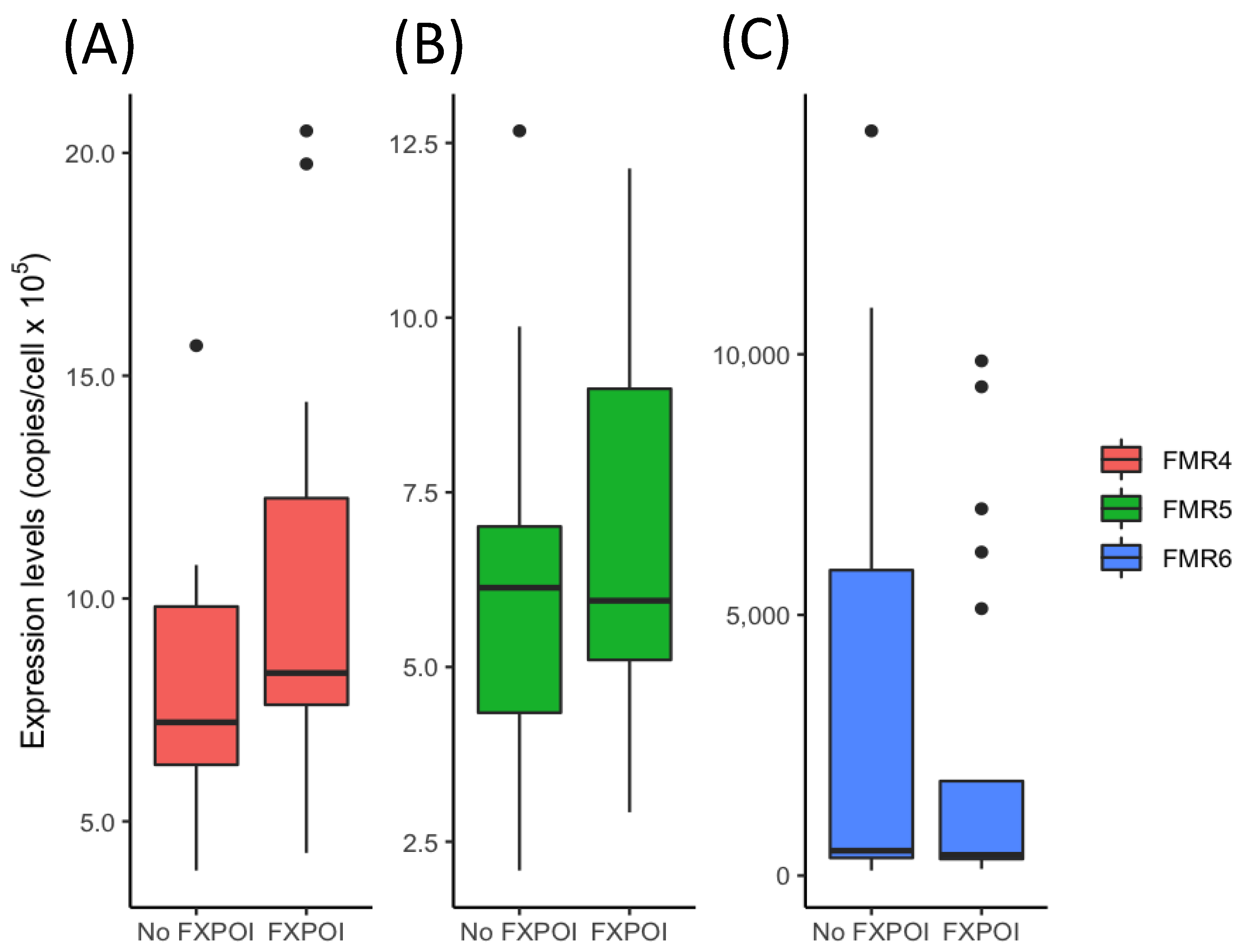
3.1. Expression Levels of FM4, FMR5 and FMR6 in FMR1 Premutation Carriers

Thirty-six FMR1 premutation female carriers were recruited; 20 with FXPOI and 16 without FXPOI. There were no differences between the two groups regarding patients’ CGG repeat expansion or FMR1 mRNA expression levels (Table 1). As for the patients’ age at time of analysis, statistically significant differences were obtained (p = 0.03). This difference can be attributed to a bias in the recruitment of women without FXPOI. Older women were included in this cohort in order to make sure that they did not have ovarian dysfunction. X-chromosome inactivation (XCI) pattern was also examined and compared between the FXPOI and non-FXPOI groups. Results evidenced that both groups showed a similar random XCI pattern (data not shown).
FMR4, FMR5 and FMR6 transcripts levels were evaluated in total RNA extracted from peripheral blood by ddPCR and compared between FMR1 premutation carriers with FXPOI and without FXPOI. Results showed no significant differences between groups for none of the lncRNAs analyzed (p = 0.09 for FMR4; p = 0.46 for FMR5 and p = 0.56 for FMR6) (Figure 1 and Table S1). Subsequently, and since the expression of the three lncRNAs varies over a wide range, we compared FMR1 premutation carriers with and without FXPOI among stratified subgroups based on expression levels. These subgroups consisted of low, medium and high expression levels for FMR4, FMR5 and FMR6. When comparing the distribution of lncRNAs expression levels (low, medium and high) between FMR1 premutation carrier groups (with and without FXPOI), significant results were obtained for FMR4 (p = 0.039). As shown in Table 2, while 30% (6/20) of FXPOI women showed high levels of FMR4 expression, only 10% (2/16) of women without FXPOI were found to have similar levels. Therefore, evidence was obtained for an association between FMR4 transcript levels and the development of FXPOI. A similar association was also found for FMR5, although not statistically significant. Contrary, for FMR6, no significant evidence of an association was obtained (Table 2).
In addition, the correlation between the expression levels of FMR4, FMR5, FMR6 and the FMR1 gene was appraised among the total number of female FMR1 premutation carriers as well as in the distinct groups with and without FXPOI (Figure 2). A high correlation was found between FMR5 and FMR6 expression levels while the correlation of these two lncRNAs with FMR4 was lower. All lncRNAs correlated negatively with respect to FMR1 expression. The correlation pattern did not change when FXPOI and non-FXPOI females were considered separately, suggesting that their relative expression behavior is not substantially affected by the presence of FXPOI.

3.2. Diagnostic Value of FMR4 for FXPOI

Since significant differences were obtained in the distribution of FMR4 expression levels between women with FXPOI and without FXPOI, the diagnostic value of FMR4 was assessed by establishing a ROC curve. As can be observed in Figure 3, the curve had an AUC value of 0.67 (95% CI 0.45–0.86). A sensitivity of 0.80 and specificity of 0.63 were achieved at the optimal threshold. These data indicate that FMR4 expression levels had a certain diagnostic value for FXPOI. FMR4 levels above the established threshold conferred a significantly increased risk of developing FXPOI (OR: 6.67 95% CI: 1.5–29.63, p = 0.016).

3.3. Association of lncRNAs Expression Levels with FMR1 CGG Repeat Size

We evaluated the correlation between the expression levels of the lncRNAs and the CGG repeat size (Figure 4). Expression of FMR4 and FMR5 showed a non-linear distribution, although the association was only statistically significant for FMR4 (p = 0.037). The highest levels of FMR4 were detected in women with 80–99 CGG repeats (Figure 4A). For FMR6, as the expression levels showed a wide distribution range, no evidence of an association was inferred (p = 0.9) (Figure 4C).
A multivariate logistic regression analysis was performed to explore whether the correction of FMR4 expression levels for the number of CGG repeats (as a curvilinear confounder) could improve the prediction of FXPOI. Although, the inclusion of both parameters also showed a similar 0.67 AUC, FMR4 expression did not show a significant adjusted odds ratio in the model (p = 0.15).

4. Discussion

Little is known about the disease pathology underlying FXPOI. Although the FMR1 premutation is the major risk factor, there are still some unknown genetic, epigenetic or environmental factors that might be affecting gene penetrance. Current research aims to identify molecular or other biomarkers that can predict the occurrence of FXPOI and help women with the FMR1 premutation to make decisions regarding their reproductive and family planning. LncRNAs have attracted a lot of attention in recent medical studies since emerging evidence suggests that they could be used as diagnostic biomarkers in many diseases such as cancer [28], cardiovascular or neurodegenerative disorders [29,30]. FMR4, FMR5 and FMR6 comprise a group of FMR1-derived lncRNAs which might partake to aspects of the clinical presentation of the Fragile X-associated disorders [21,22,23,24]. In fact, it has been described that the expression of these lncRNAs is different in both FXS patients and FMR1 premutation carriers. In brain tissue from FXS patients both, FMR4 and FMR6 expression have been found to be down-regulated [21,22]. On the other hand, in FMR1 premutation carriers whereas the FMR4 expression has been described up-regulated [22], the FMR6 expression has been found down-regulated [21].
On the basis of these observations, this study investigated FMR4, FMR5 and FMR6 expression levels in peripheral blood of female FMR1 premutation carriers with and without FXPOI in order to explore their feasibility as potential biomarkers of FXPOI. For this purpose, the ddPCR technique was used, as it is a highly sensitive and specific method for absolute quantification of transcript gene expression per cell.
The results showed a wide range of expression levels for all three lncRNAs analyzed, leading to a non-significant difference when comparing mean expression levels between female FMR1 premutation carriers with FXPOI and without FXPOI. We further assessed pairwise correlation between the expression of FMR4, FMR5, FMR6 and FMR1 mRNA transcripts. Again, no differences were observed when comparing women with FXPOI and without FXPOI; suggesting that pairwise correlations between expressions of these lncRNAs and the FMR1 gene might not be affected by the presence of FXPOI (Figure 2). However, a statistically significant difference was obtained for FMR4 (p = 0.039) when comparing both groups stratified by expression levels (low, medium or high), indicating a disequilibrium in the distribution. Among all the FMR1 premutation carriers presenting higher FMR4 expression levels, 86% (6/7) developed FXPOI whereas only 29% (1/6) did not. ROC curve analysis revealed that FMR4 can modestly distinguish female FMR1 premutation carriers with and without FXPOI with an AUC of 0.67. Although caution must be taken due to the limited sample size, our results showed an association between high FMR4 expression levels and FXPOI (OR 6.67, 95% CI = 1.5–29.63, p = 0.016), suggesting that this lncRNA could be a possible marker for FXPOI among female FMR1 premutation carriers.
To date, only CGG repeat size has been associated with the risk of developing FXPOI in a non-linear way [31,32,33]; leading to the highest risk for FXPOI in the mid-range of the repeat expansion (~80–99 CGG). Interestingly, our findings, although not statistically significant, also showed a non-linear correlation between the FMR4 and FMR5 expression levels and the FMR1 repeat size; yielding higher levels of both, FMR4 and FMR5 expression, in the mid-range. This correlation was not detected for FMR6 (Figure 4C). Nevertheless, since FMR6 showed the highest range of expression levels, it cannot be completely discarded. To our knowledge, only Elizur and co-workers [24] have previously analyzed the transcript levels of FMR4 and FMR6 in granulosa cells of FMR1 premutation carriers in order to determine a putative role in the pathogenesis of FXPOI. Contrary to our results, the authors did not observe any association neither between granulosa cells FMR4 expression levels nor between the number of CGG repeats in the FMR1 gene. On the other hand, whereas the FMR6 expression levels were not significantly different between FMR1 permutation carriers and controls, they reported a significant non-linear association between the number of CGG repeats and FMR6 levels in the granulosa cells [24]. Although these results are not in line with ours, it has to be taken into consideration that the methodology and tissue used to measure the lncRNAs levels were different. Whereas Elizur et al. [24] used qRT-PCR and granulosa cells, we used peripheral blood and ddPCR. Moreover, although the sample size was similar in both studies, they compared FMR1 premutation carriers against controls and we compared FMR1 premutation carriers with FXPOI against those without FXPOI. Finally, another important difference is that their cohort lack of FMR1 premutation carriers with FMR1 repeat size above 150 CGGs, which could somehow bias the results.
Our study, although exploratory, has two main limitations. First, the sample size, which is not large enough to provide reliable evidence for an association between FMR4, FMR5 and FMR6 expression levels and FXPOI. However, our study provides statistically significant results, highlighting a potential role of FMR4 in predicting FXPOI. Second, the age differences between groups and the fact that FMR1 premutation women with FXPOI had already developed ovarian dysfunction. Thus, it would be necessary to replicate our findings in other female FMR1 premutation cohorts and, ideally, in a longitudinal study, in order to make sure that age is not affecting lncRNAs expression levels. If validated in other populations, these results might provide evidence of a potential role of FMR4 as a possible biomarker for FXPOI.

Supplementary Materials

The following supporting information can be downloaded at: https://www.mdpi.com/article/10.3390/jcm11082186/s1, Table S1: Expression levels of FMR4, FMR5 and FMR6 measured in peripheral blood of FMR1 premutation carriers with and without FXPOI.

Author Contributions

M.I.A.-M.: substantial contributions to conception and design, acquisition of data, analysis and interpretation of data. Final approval of the version to be published. I.A.: substantial contributions to conception and design, acquisition of data, or analysis and interpretation of data. Final approval of the version to be published. R.W.: substantial contributions to conception and design, acquisition of data, or analysis and interpretation of data. Final approval of the version to be published. E.M.-B. and T.B.: acquisition, analysis and interpretation of data. Drafting the article and final approval of the version to be published. A.B., S.P., A.G. and M.G.: acquisition, analysis and interpretation of data. Final approval of the version to be published. D.M.: revising the work critically for important intellectual content and final approval of the version to be published. L.R.-R.: drafting the work or revising it critically for important intellectual content and final approval of the version to be published. All authors have read and agreed to the published version of the manuscript.

Funding

This study was supported by Fundación Merck Salud (19-FE-011) and the Instituto de Salud Carlos III (ISCIII) (through the project (PI21/01085), co-funded by the European Union, and AGAUR from the Autonomous Catalan Government (2017SGR1134).

Institutional Review Board Statement

The study was conducted in accordance with the Declaration of Helsinki, and approved by the Institutional Review Board (or Ethics Committee) of the Hospital Clinic Barcelona (protocol code HCB/2021/1030 at 21 January 2022).

Informed Consent Statement

Informed consent was obtained from all subjects involved in the study.

Data Availability Statement

The analyzed data sets generated during the study are available from the corresponding author on reasonable request.

Acknowledgments

We want to thank the Fragile X families, the “Associació Catalana Síndrome X fràgil”, and the Federación Española del Síndrome X Frágil for their cooperation. This study was supported by Fundación Merck Salud (19-FE-011), the Instituto de Salud Carlos III (ISCIII) (through the project (PI21/01085), co-funded by the European Union, and AGAUR from the Autonomous Catalan Government (2017SGR1134). The CIBER de Enfermedades Raras is an initiative of the Instituto de Salud Carlos III.

Conflicts of Interest

The authors have no conflict of interest.

References

  1. Verkerk, A.J.; Pieretti, M.; Sutcliffe, J.S.; Fu, Y.H.; Kuhl, D.P.; Pizzuti, A.; Reiner, O.; Richards, S.; Victoria, M.F.; Zhang, F.P.; et al. Identification of a gene (FMR-1) containing a CGG repeat coincident with a breakpoint cluster region exhibiting length variation in fragile X syndrome. Cell 1991, 65, 905–914. [Google Scholar] [CrossRef]
  2. Oberle, I.; Rousseau, F.; Heitz, D.; Kretz, C.; Devys, D.; Hanauer, A.; Boue, J.; Bertheas, M.F.; Mandel, J.L. Instability of a 550-base pair DNA segment and abnormal methylation in fragile X syndrome. Science 1991, 252, 1097–1102. [Google Scholar] [CrossRef] [PubMed]
  3. Yu, S.; Pritchard, M.; Kremer, E.; Lynch, M.; Nancarrow, J.; Baker, E.; Holman, K.; Mulley, J.C.; Warren, S.T.; Schlessinger, D.; et al. Fragile X genotype characterized by an unstable region of DNA. Science 1991, 252, 1179–1181. [Google Scholar] [CrossRef] [PubMed] [Green Version]
  4. Sutcliffe, J.S.; Nelson, D.L.; Zhang, F.; Pieretti, M.; Caskey, C.T.; Saxe, D.; Warren, S.T. DNA methylation represses FMR-1 transcription in fragile X syndrome. Hum. Mol. Genet. 1992, 1, 397–400. [Google Scholar] [CrossRef] [PubMed]
  5. Kenneson, A.; Zhang, F.; Hagedorn, C.H.; Warren, S.T. Reduced FMRP and increased FMR1 transcription is proportionally associated with CGG repeat number in intermediate-length and premutation carriers. Hum. Mol. Genet. 2001, 10, 1449–1454. [Google Scholar] [CrossRef] [PubMed] [Green Version]
  6. Tassone, F.; Iong, K.P.; Tong, T.H.; Lo, J.; Gane, L.W.; Berry-Kravis, E.; Nguyen, D.; Mu, L.Y.; Laffin, J.; Bailey, D.B.; et al. FMR1 CGG allele size and prevalence ascertained through newborn screening in the United States. Genome Med. 2012, 4, 100. [Google Scholar] [CrossRef] [Green Version]
  7. Sherman, S.L. Premature ovarian failure in the fragile X syndrome. Am. J. Med. Genet. 2000, 97, 189–194. [Google Scholar] [CrossRef]
  8. Hagerman, P.J.; Hagerman, R.J. The fragile-X premutation: A maturing perspective. Am. J. Hum. Genet. 2004, 74, 805–816. [Google Scholar] [CrossRef] [Green Version]
  9. Wheeler, A.C.; Raspa, M.; Green, A.; Bishop, E.; Bann, C.; Edwards, A.; Bailey, D.B., Jr. Health and reproductive experiences of women with an FMR1 premutation with and without fragile X premature ovarian insufficiency. Front. Genet. 2014, 5, 300. [Google Scholar] [CrossRef] [Green Version]
  10. Rohr, J.; Allen, E.G.; Charen, K.; Giles, J.; He, W.; Dominguez, C.; Sherman, S.L. Anti-Mullerian hormone indicates early ovarian decline in fragile X mental retardation (FMR1) premutation carriers: A preliminary study. Hum. Reprod. 2008, 23, 1220–1225. [Google Scholar] [CrossRef] [Green Version]
  11. Bibi, G.; Malcov, M.; Yuval, Y.; Reches, A.; Ben-Yosef, D.; Almog, B.; Amit, A.; Azem, F. The effect of CGG repeat number on ovarian response among fragile X premutation carriers undergoing preimplantation genetic diagnosis. Fertil. Steril. 2010, 94, 869–874. [Google Scholar] [CrossRef] [PubMed]
  12. Allingham-Hawkins, D.J.; Babul-Hirji, R.; Chitayat, D.; Holden, J.J.; Yang, K.T.; Lee, C.; Hudson, R.; Gorwill, H.; Nolin, S.L.; Glicksman, A.; et al. Fragile X premutation is a significant risk factor for premature ovarian failure: The International Collaborative POF in Fragile X study—Preliminary data. Am. J. Med. Genet. 1999, 83, 322–325. [Google Scholar] [CrossRef]
  13. Mercer, T.R.; Dinger, M.E.; Mattick, J.S. Long non-coding RNAs: Insights into functions. Nat. Rev. Genet. 2009, 10, 155–159. [Google Scholar] [CrossRef] [PubMed]
  14. Wilusz, J.E.; Sunwoo, H.; Spector, D.L. Long noncoding RNAs: Functional surprises from the RNA world. Genes. Dev. 2009, 23, 1494–1504. [Google Scholar] [CrossRef] [PubMed] [Green Version]
  15. Derrien, T.; Johnson, R.; Bussotti, G.; Tanzer, A.; Djebali, S.; Tilgner, H.; Guernec, G.; Martin, D.; Merkel, A.; Knowles, D.G.; et al. The GENCODE v7 catalog of human long noncoding RNAs: Analysis of their gene structure, evolution, and expression. Genome Res. 2012, 22, 1775–1789. [Google Scholar] [CrossRef] [Green Version]
  16. Yao, R.W.; Wang, Y.; Chen, L.L. Cellular functions of long noncoding RNAs. Nat. Cell Biol. 2019, 21, 542–551. [Google Scholar] [CrossRef]
  17. Wang, K.C.; Chang, H.Y. Molecular mechanisms of long noncoding RNAs. Mol. Cell 2011, 43, 904–914. [Google Scholar] [CrossRef] [Green Version]
  18. Wapinski, O.; Chang, H.Y. Long noncoding RNAs and human disease. Trends Cell Biol. 2011, 21, 354–361. [Google Scholar] [CrossRef]
  19. Huang, G.; Zhu, H.; Wu, S.; Cui, M.; Xu, T. Long Noncoding RNA Can Be a Probable Mechanism and a Novel Target for Diagnosis and Therapy in Fragile X Syndrome. Front. Genet. 2019, 10, 446. [Google Scholar] [CrossRef]
  20. Ladd, P.D.; Smith, L.E.; Rabaia, N.A.; Moore, J.M.; Georges, S.A.; Hansen, R.S.; Hagerman, R.J.; Tassone, F.; Tapscott, S.J.; Filippova, G.N. An antisense transcript spanning the CGG repeat region of FMR1 is upregulated in premutation carriers but silenced in full mutation individuals. Hum. Mol. Genet. 2007, 16, 3174–3187. [Google Scholar] [CrossRef] [Green Version]
  21. Khalil, A.M.; Faghihi, M.A.; Modarresi, F.; Brothers, S.P.; Wahlestedt, C. A novel RNA transcript with antiapoptotic function is silenced in fragile X syndrome. PLoS ONE 2008, 3, e1486. [Google Scholar] [CrossRef] [PubMed]
  22. Pastori, C.; Peschansky, V.J.; Barbouth, D.; Mehta, A.; Silva, J.P.; Wahlestedt, C. Comprehensive analysis of the transcriptional landscape of the human FMR1 gene reveals two new long noncoding RNAs differentially expressed in Fragile X syndrome and Fragile X-associated tremor/ataxia syndrome. Hum. Genet. 2014, 133, 59–67. [Google Scholar] [CrossRef] [PubMed] [Green Version]
  23. Peschansky, V.J.; Pastori, C.; Zeier, Z.; Motti, D.; Wentzel, K.; Velmeshev, D.; Magistri, M.; Bixby, J.L.; Lemmon, V.P.; Silva, J.P.; et al. Changes in expression of the long non-coding RNA FMR4 associate with altered gene expression during differentiation of human neural precursor cells. Front. Genet. 2015, 6, 263. [Google Scholar] [CrossRef] [PubMed] [Green Version]
  24. Elizur, S.E.; Dratviman-Storobinsky, O.; Derech-Haim, S.; Lebovitz, O.; Dor, J.; Orvieto, R.; Cohen, Y. FMR6 may play a role in the pathogenesis of fragile X-associated premature ovarian insufficiency. Gynecol. Endocrinol. 2016, 32, 334–337. [Google Scholar] [CrossRef] [PubMed]
  25. Zmienko, A.; Samelak-Czajka, A.; Goralski, M.; Sobieszczuk-Nowicka, E.; Kozlowski, P.; Figlerowicz, M. Selection of Reference Genes for qPCR- And ddPCR-Based Analyses of Gene Expression in Senescing Barley Leaves. PLoS ONE 2015, 10, e0118226. [Google Scholar] [CrossRef] [PubMed]
  26. Amos-Landgraf, J.M.; Cottle, A.; Plenge, R.M.; Friez, M.; Schwartz, C.E.; Longshore, J.; Willard, H.F. X chromosome-inactivation patterns of 1005 phenotypically unaffected females. Am. J. Hum. Genet. 2006, 79, 493–499. [Google Scholar] [CrossRef] [PubMed] [Green Version]
  27. Tassone, F.; Hagerman, R.J.; Taylor, A.K.; Gane, L.W.; Godfrey, T.E.; Hagerman, P.J. Elevated levels of FMR1 mRNA in carrier males: A new mechanism of involvement in the fragile-X syndrome. Am. J. Hum. Genet. 2000, 66, 6–15. [Google Scholar] [CrossRef] [Green Version]
  28. Yan, X.; Hu, Z.; Feng, Y.; Hu, X.; Yuan, J.; Zhao, S.D.; Zhang, Y.; Yang, L.; Shan, W.; He, Q.; et al. Comprehensive Genomic Characterization of Long Non-coding RNAs across Human Cancers. Cancer Cell 2015, 28, 529–540. [Google Scholar] [CrossRef] [Green Version]
  29. Haemmig, S.; Feinberg, M.W. Targeting LncRNAs in Cardiovascular Disease: Options and Expeditions. Circ. Res. 2017, 120, 620–623. [Google Scholar] [CrossRef] [Green Version]
  30. Zhang, M.; He, P.; Bian, Z. Long Noncoding RNAs in Neurodegenerative Diseases: Pathogenesis and Potential Implications as Clinical Biomarkers. Front. Mol. Neurosci. 2021, 14, 685143. [Google Scholar] [CrossRef]
  31. Sullivan, A.K.; Marcus, M.; Epstein, M.P.; Allen, E.G.; Anido, A.E.; Paquin, J.J.; Yadav-Shah, M.; Sherman, S.L. Association of FMR1 repeat size with ovarian dysfunction. Hum. Reprod. 2005, 20, 402–412. [Google Scholar] [CrossRef] [PubMed] [Green Version]
  32. Ennis, S.; Ward, D.; Murray, A. Nonlinear association between CGG repeat number and age of menopause in FMR1 premutation carriers. Eur. J. Hum. Genet. 2006, 14, 253–255. [Google Scholar] [CrossRef] [PubMed] [Green Version]
  33. Allen, E.G.; Sullivan, A.K.; Marcus, M.; Small, C.; Dominguez, C.; Epstein, M.P.; Charen, K.; He, W.; Taylor, K.C.; Sherman, S.L. Examination of reproductive aging milestones among women who carry the FMR1 premutation. Hum. Reprod. 2007, 22, 2142–2152. [Google Scholar] [CrossRef] [PubMed] [Green Version]
Figure 1. Expression levels of FMR4, FMR5 and FMR6 were compared between FMR1 premutation carriers with FXPOI and without FXPOI. (A) The expression level of FMR4, (B) The expression level of FMR5 and (C) The expression level of FMR6. None of the comparisons were statistically significant (p > 0.05).
Jcm 11 02186 g001
Figure 2. Correlation between expression levels of FMR4, FMR5 and FMR6 was evaluated among total FMR1 premutation carriers as well as groups of women with FXPOI and without FXPOI.
Jcm 11 02186 g002
Figure 3. ROC curve for assessment of diagnostic power of FMR4 among total FMR1 premutation female carriers. The area under the curve (AUC) was 0.67.
Jcm 11 02186 g003
Figure 4. lncRNAs expression level in peripheral blood of FMR1 premutation carriers according to the number of CGG repeats. (A) FMR4 expression levels, (B) FMR5 expression levels, and (C) FMR6 expression levels.
Jcm 11 02186 g004
Table 1. Clinical and molecular characteristics of individuals recruited in the study.
FMR1 Premutation with
FXPOI (n = 20)
FMR1 Premutation without
FXPOI (n = 16)
p-Value
Age (mean ± SD, years)41 ± 6.947 ± 8.30.03 *
CGG repeat (mean ± SD)100 ± 3588 ± 260.3
FMR1 mRNA (mean ± SD)1.5 ± 0.81.8 ± 1.30.3
Significance: * p < 0.05. The exact p-values were calculated with the U-Mann Whitney test.
Table 2. Distribution of FMR1 premutation carriers with and without FXPOI based on FMR4, FMR5 and FMR6 expression levels.
FMR4 expression levelp-Value
1–77–12>120.039 *
FXPOI (n = 20)2 (10%)12 (60%)6 (30%)
No FXPOI (n = 16)7(44%)8 (50%)1 (6%)
FMR5 expression level0.14
1–55–10>10
FXPOI (n = 20)5 (25%)10 (50%)5 (25%)
No FXPOI (n = 16)7 (44%)8 (50%)1 (6%)
FMR6 expression level0.556
<400400–1000>1000
FXPOI (n = 20)10 (50%)5 (25%)5 (25%)
No FXPOI (n = 16)6 (38%)5 (31%)5 (31%)
Significance: * p < 0.05. The exact p-values were calculated with the Fisher exact test.
Publisher’s Note: MDPI stays neutral with regard to jurisdictional claims in published maps and institutional affiliations.

