Systematic Review

The Effect of Bite Raise on AHI Values in Adult Patients Affected by OSA: A Systematic Review with Meta-Regression

by
Maria Lavinia Bartolucci
1,*,
Serena Incerti Parenti
1,
Francesco Bortolotti
1,
Giulia Corazza
1,
Livia Solidoro
1,
Corrado Paganelli
2 and
Giulio Alessandri-Bonetti
1
1
Section of Orthodontics and Dental Sleep Medicine, Department of Biomedical and Neuromotor Sciences, University of Bologna, via san Vitale 59, 40125 Bologna, Italy
2
Department of Medical and Surgical Specialties Radiological Sciences and Public Health, Dental School, University of Brescia, 25121 Brescia, Italy
*
Author to whom correspondence should be addressed.
J. Clin. Med. 2023, 12(11), 3619; https://doi.org/10.3390/jcm12113619
Submission received: 19 April 2023 / Revised: 21 May 2023 / Accepted: 22 May 2023 / Published: 23 May 2023
(This article belongs to the Section Dentistry, Oral Surgery and Oral Medicine)

Abstract

:
Obstructive sleep apnea (OSA) is a highly prevalent sleep breathing disorder characterized by the collapse of the pharyngeal walls that entails recurrent episodes of cessation of breathing or decrease in airflow while sleeping. This results in sleep fragmentation, decreased oxygen saturation and an increase in the partial pressure of carbon dioxide, causing excessive daytime sleepiness, hypertension and increased prevalence of cardiovascular morbidity and mortality. Mandibular advancement devices (MAD) represent a valid alternative therapy to Continuous Positive Airway Pressure, thrusting the mandible forward, increasing the lateral diameter of the pharynx and reducing the collapsibility of the airway. Several investigations have focused on the detection of the best mandibular advancement amount in terms of effectiveness and tolerance, but few and contrasting data are available on the role of occlusal bite raise in reducing the apnea/hypopnea index (AHI). The aim of this systematic review with meta-regression analysis was to investigate the effect of the bite raise of MAD on AHI values in adult patients affected by OSA. An electronic search was performed in MEDLINE, the Cochrane Database, Scopus, Web of Science and LILACS. Randomized controlled trials (RCT) investigating the effectiveness of MAD in OSA patients were included. The quality of evidence was evaluated with the Grading of Recommendations Assessment, Development and Evaluation (GRADE) and the risk of bias with the Cochrane risk-of-bias tool for randomized trials (RoB2). Six RCT were included. The success rate of each study was computed: (mean baseline AHI − mean post treatment AHI)/mean baseline AHI. The GRADE scores indicated that the quality of evidence was very low. The meta-regression analysis showed that there was no correlation between the occlusal bite raise and the AHI improvement.

1. Introduction

Obstructive sleep apnea (OSA) is a common sleep-related breathing disorder that affects 12.5% of men and 5.9% of women over 40 years old and is characterized by recurrent episodes of partial or complete collapse of the upper airway during sleep [1]. Repetitive obstructions may lead to fragmentation of the sleep pattern, a decrease in oxygen saturation and an increase in partial pressure of CO2, marked swings in intra-thoracic pressure and increased sympathetic activity [2]. Consequences of OSA are represented by excessive daytime sleepiness, loss of concentration, hypertension and, in more severe cases, stroke and heart failure, resulting in an increased prevalence of cardiovascular morbidity and mortality [3].
Mandibular advancement devices (MAD) have been reported to be an alternative treatment to Continuous Positive Airway Pressure (CPAP) in patients affected by mild to moderate OSA and in those with severe OSA who refuse CPAP treatment or surgery [2,4,5,6]. These appliances work by keeping the mandible in a protruded position during sleep, increasing the lateral diameter of the pharynx, stabilizing the hyoid bone and the soft palate, stretching the muscular bundles of the tongue and preventing the posterior rotation of the mandible, thus increasing the width of the airway and reducing its collapsibility [7,8]. However, the range of success of MAD presents high variability between individuals on the basis of peculiar pathophysiological traits presented by the single patient [9]. In fact, OSA is considered a heterogeneous disorder in terms of pathogenesis and clinical manifestations. Recently, this pathology has been related to phenotypic and endotypic traits [10]. OSA patients present some degree of anatomical compromise, that puts the upper airways at risk for collapse during sleep: the association of several phenotypic factors entail the severity of such predisposition (obesity, craniofacial morphology, etc.). In addition, four non-anatomical traits emerged as playing a crucial role in the onset of OSA viz., the pharyngeal airway collapsibility, the upper airway neuromuscular compensation, the ventilator control system or loop gain and the arousal threshold. Each trait differently contributes to the clinical manifestations of OSA, influencing the individual response to the therapies. Notably, a recent review suggested that patients with mild/moderate collapsibility and with a low loop gain could benefit from MAD therapy, underscoring that endotypic traits are just one aspect to consider before prescribing a MAD therapy [11].
Therefore, a correct diagnosis is crucial to achieving an improvement in OSA severity. Furthermore, among the selected patients, the results of the MAD therapy can be affected by the design features of these devices in terms of efficacy, tolerance and compliance [9,12,13,14,15]. A recent systematic review compared the efficacy of different types of customized MAD on reducing apnea/hypopnea index (AHI) values and improving oxygen saturation in patients affected by OSA [15]. As suggested by the authors, despite mono-bloc MAD having registered better success rate in improving the minimal oxygen saturation, the adjustability of duo-bloc MAD permits easier management of the therapy, which is reported to be better tolerated by the patients, achieving higher results in terms of compliance [15] (Figure 1).
The parameters to be followed in MAD manufacturing and the anatomical and/or dimensional changes obtained at the upper airway level are still a topic of debate. In terms of efficacy, two parameters emerged as playing a role, viz., the mandibular advancement amount and the vertical opening amount. Several studies have investigated what degree of mandibular advancement is most effective at reducing AHI value in patients affected by OSA, achieving conflicting results. Some authors suggest a mild initial protrusion [16,17], others support an initial approach with a high level of advancement [18,19,20], while stepwise mandibular advancements with progressive activations is supported by the evidence of increased compliance due to minimization of the side effects commonly brought about by MAD therapy [16,21].
A systematic review of randomized clinical trials concluded that the advancement amount does not seem to have an influence on the success rate of MAD therapy [22]. Despite this, 50% of the maximum mandibular protrusion is widely used as the minimum effective advancement to start MAD therapy, but this parameter has not been adequately addressed, since only two studies have investigated the efficacy of mandibular advancements lower than 50% [16,23]. Therefore, the most protruded mandibular position does not necessarily correspond to the best clinical outcome, and considering the amount of mandibular protrusion to be the only aspect influencing the effectiveness of MAD would be simplistic. In fact, other factors have been described as concurrently modulating the individual response to the treatment [22]. The role of the vertical increase provided by the MAD in terms of effectiveness has been little investigated. The occlusal vertical dimension is the distance measured between two points of reference when the dental arches or the upper and lower jaws are in contact. In order to determine this parameter, several methods have been proposed that can be divided as pre-extraction and post-extraction measurements. Among the pre-extraction methods, that are applicable only if the patients is dentate and with a stable occlusion, the intraoral dimensions taken clinically and on dental casts, the profile tracing, the cephalometric tracing and the phonetics are the most used. In edentulous patients, the occlusal vertical dimension can be determined by using the rest position of the subject who is asked to pronounce specific sounds, to swallow and/or to keep the mandible in a comfortable position. Other post-extraction methods provide for the evaluation of the facial esthetic appearance, of craniofacial landmarks and of phonetics. A recent review examines and discusses these different techniques, concluding that the determination of occlusal vertical dimension cannot be precise, for every available method comes along with limitations that should be taken into account by clinicians. The choice should be based on the patient’s needs and the operator’s experience [24].
A systematic review published by Ahrens et al. that investigated the efficacy of different designs of MAD emphasized the importance of considering the potential role of the amount of vertical opening in increasing the patients’ improvement [4].
Some authors have supposed that a higher vertical dimension could cause a posterior rotation of the mandibular angle and a restriction of the pharyngeal lumen [25]. Fluoroscopic recordings suggest that bite opening should be kept to a minimum because, in awake OSAS patients, it results in a posterior movement of both tongue and soft palate, with consequent narrowing of the oropharyngeal airway [26]. Similarly, Meurice et al. demonstrated in healthy subjects that mouth opening at an interincisal distance of 15 mm during sleep was associated with an increased collapsibility of the upper airway [27]. Moreover, the retracted position of the mandible caused by the rise of the vertical dimension could decrease the range of mandibular advancement; thus the efficacy of the therapy with MAD could be reduced [28].
However, Lowe at al. hypothesized that the mandibular protrusion, displacing the tongue away from the posterior wall of the upper airway, combined with an increased vertical dimension of occlusion, should hinder the pharyngeal closure induced by the increased vertical opening alone [29]. In addition, Pitsis et al. compared two intra-oral devices with the same degree of mandibular advancement, but with a different inter-incisal opening (4 mm vs. 14 mm); significant reductions in the AHI and in other polysomnographic variables were achieved with both oral appliances, with no statistically significant differences between the two devices, thus suggesting that the amount of the vertical opening induced by the appliances did not have a direct impact on treatment efficacy to any great extent [30].
Thus, given the wide heterogeneity of data present to date in the literature, the aim of this systematic review with meta-regression analysis is to investigate the effect of the bite raise of MAD on AHI values in adult patients affected by OSA.

2. Materials and Methods

The present systematic review was conducted according to the Preferred Reporting Items for Systematic Reviews and Meta-Analysis (PRISMA) system [31].

2.1. Search Strategy

To identify the studies to be considered for inclusion, an electronic database search of the literature was performed in MEDLINE, Cochrane Database of Systematic Reviews, Google Scholar Beta, Scopus, LILACS and Web of Sciences. Studies published before September 2022 were analyzed, and only studies written in English were included. The search strategy used for MEDLINE, including the MeSH and text words, was: ((apnea, obstructive sleep) OR (obstructive sleep apnea syndrome) OR (obstructive sleep apnea)) AND ((occlusal splint) OR (mandibular advancement device) OR (oral appliance)) AND (randomized controlled trials). Further studies were identified through hand searching the reference lists of all relevant studies.

2.2. Inclusion Criteria

The study selection included randomized controlled trials (RCTs) involving adult patients affected by OSA and treated with customized MAD. To be considered for evaluation, each study must report a detailed description of the appliance design, specifically the amount of occlusal bite opening, and the baseline and control PSG or sleep polygraphic parameters. Two researchers independently selected the articles. Intra-examiner conflicts were solved by discussion of each article to reach a consensus.

2.3. Data Items and Collection

The following data items were collected from each study: study design, sample size, mean age, mean BMI, MAD design, mean vertical opening, AHI values at baseline and after therapy, follow-up and author’s main conclusions.

2.4. Risk of Bias in Individual Studies and across the Studies

To document the methodological soundness of each article, the revised Cochrane risk-of-bias tool for randomized trials (RoB2) [32] was used. To evaluate the quality of body evidence, the Grading of Recommendation Assessment, Development and Evaluation (GRADE) was performed [33].
Two assessors independently performed the RoB2 analysis and the other two researchers performed the GRADE assessment; when in disagreement, a conjunct evaluation was performed to reach a consensus.
The risk of bias across the studies was evaluated by means of Egger’s test and Funnel plot. Statistical tests of heterogeneity were performed to assess whether the variability in study results (effect sizes) was greater than expected to occur by chance. The heterogeneity among studies was assessed using a χ2−-based Q statistic; only an I2 index greater than 50 percent was considered and associated with a substantial heterogeneity among the studies. Moreover, the tau2 was calculated for the heterogeneity assessment.

2.5. Methodology of Synthesis of the Results of the Individual Studies

A fixed effect model was used if homogeneity across studies was proved (p value greater than 0.10); if homogeneity was rejected (p-value less than 0.10), a random effects model was used to better aggregate the data [34]. The success rate and 95 percent confidence intervals between baseline and follow-up were computed for each treatment group within the studies. A meta-analysis was conducted to analyze the effect of occlusal bite rise on success rate; the analysis was performed using Comprehensive Meta-Analysis Software v. 2.2.064 (Biostat Inc., Englewood, NJ, USA).

3. Results

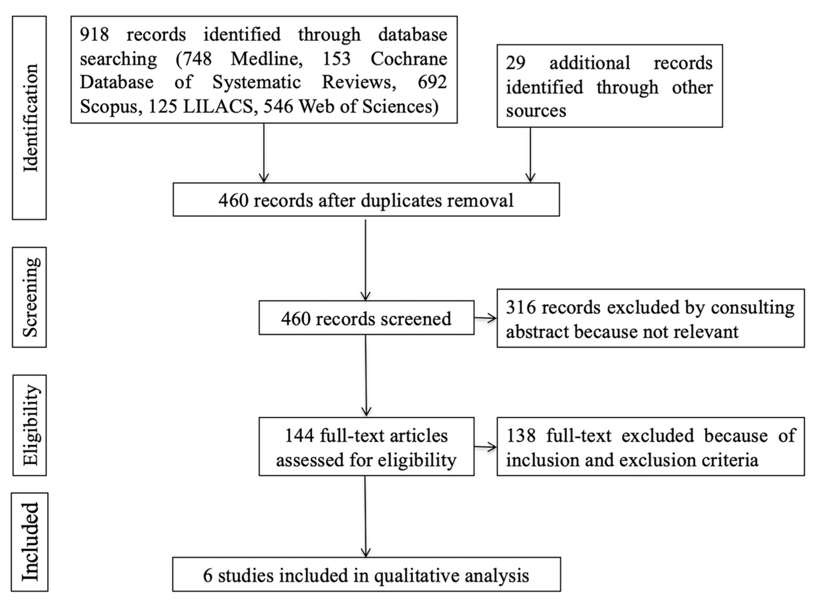
Figure 2 presents the PRISMA flow diagram describing the selection process. Six studies fulfilled the inclusion criteria and were selected for the present systematic review; their main characteristics are described in Table 1.
Since the main purpose of this systematic review was to verify the mean percentage change in AHI values before and after the raise of occlusal bite (defined as “success rate”, or the difference between mean AHI at baseline and mean AHI after treatment/mean AHI at baseline), studies that did not explicitly report these values were excluded [16,40,41,42,43,44,45,46,47].
Nikolopoulou investigated the effect of raising the bite between two groups (splint in situ versus without splint) [39] and measured the vertical bite opening considering the thickness of the acrylic splint at the level of the first molar in millimeters. Johnston and coworkers [37] used the same method to measure the bite raise caused by the MAD. In four studies the MAD was compared with a placebo device [8,36,37,38], while Blanco evaluated a single MAD with or without mandibular advancement [35]. Treatments were performed with different types of MAD and occlusal splints. In the papers by Mehta [8], Gotsopoulos [36] and Naismith [38], the occlusal vertical opening was evaluated measuring the average thickness of the acrylic appliances in millimeters. Blanco and coworkers performed measurement in the same way using a soft elastic silicone positioner.

3.1. Quality Analysis and Risk of Bias in Individual Studies

Table 2 shows that the risk of bias was low in three studies [35,36,39], with some concerns in two studies [37,38] and high in one studies [8]. The main reason for lowering the study quality was the missing outcome data, that is, the high drop-out rate without any analysis that estimated the effect of the deviation to the intervention.
The GRADE score for the quality of evidence was very low, as shown in Table 3. The reasons for lowering the quality of evidence were the inconsistency, the indirectness, that is the fact that most studies did not directly evaluate the bite-raising efficacy in reducing AHI values, and the risk of bias.

3.2. Results of Individual Studies

Table 1 reports the main results of individual studies.
Nikolopoulou et al. assessed the influence of increasing the vertical dimension of occlusion on AHI values of patients affected by OSAS: in this study, the use of stabilization splints provoked a small but significant aggravation of AHI values [39].
Blanco et al. compared the effects on AHI values using a MAD with and without mandibular advancement, obtaining a significant decrease in the mean values in both groups, with the same follow-up period, after treatment [35].
Four studies evaluated the effects of the MAD compared to a placebo device that provided only a bite raise in the control group. In two of these studies [8,37], the control group did not register significant improvements of AHI compared to baseline values, while in the others [36,38], AHI showed a reduction, albeit lower than the one obtained with the mandibular protrusion induced by MAD.

3.3. Synthesis of Results

Concerning the heterogeneity of the obtained results, a random effects model was used, resulting in I2 = 50.54 and p = 0.072. Figure 3 shows the success rate and the confidence interval of each group included in the quantitative analysis: the overall success rate was −0.048 [−0.193–0.098] and p was 0.518. The effectiveness of the MAD in reducing AHI emerging from the meta-analysis of the included studies was not statistically significant.
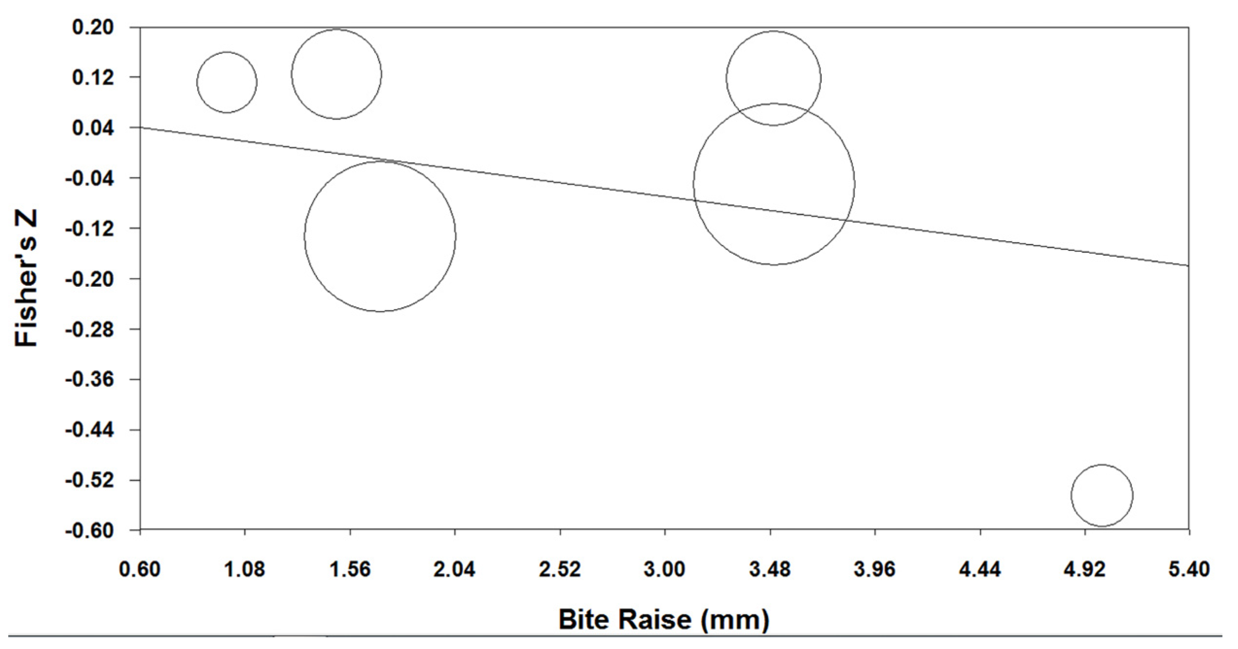
The meta-regression analysis demonstrated the absence of a significant correlation between the occlusal bite raise (indicated in millimeters) and the success rate (Q = 1.090, p = 0.296), as shown in Figure 4.

3.4. Risk of Bias across Studies

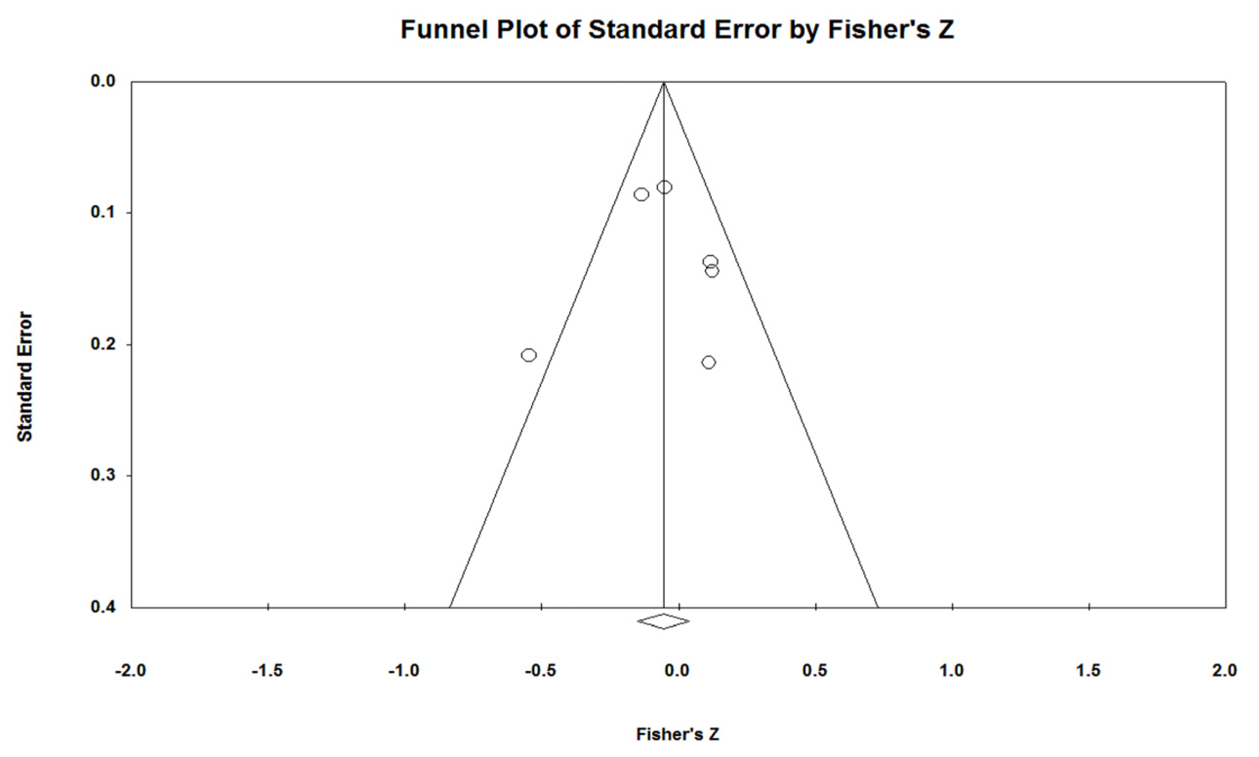
Concerning publication bias, the Egger’s regression test (intercept of 0.056, p = 0.977) demonstrated the absence of a significant deviation of the intercept from the symmetry; this means that studies with greater sample sizes were distributed close to the average, while studies with smaller sample sizes appeared to be more widely spread (Figure 5).

4. Discussion

The aim of the present systematic review with meta-analysis was to assess the effect of the occlusal bite raise on AHI values in adult patients affected by OSA. Only RCT were considered, and after a thorough full text analysis, only six resulted eligible for this investigation. The main outcome is represented by the absence of a significant correlation between the occlusal bite rise and the AHI improvement.
The qualitative analysis showed that the risk of bias was low in two included studies, with some concerns in two studies and high in two studies. The main reason for lowering the quality of evidence was the missing outcome data, that is, the high drop-out rate without any analysis that estimated the effect of the deviation to the intervention.
The GRADE results indicated that the quality of the body evidence was very low for each comparison; the reasons for lowering the quality of evidence were the inconsistency, namely the heterogeneity, of the results among the studies included, and the indirectness resulting from the extent to which the studies evaluated did not directly investigate the outcome of interest of the meta-analysis, viz., the efficacy of different degrees of vertical increase in reducing AHI values.
The meta-analysis was conducted to evaluate the success rate of the therapies investigated by the selected studies, defined as the percentage of improvement of AHI post-treatment values, based on baseline values. The overall score corresponded to a reduction of 4.8%, with a confidence interval from −19.3% to +9.8% (Figure 2).
Several studies investigated the effectiveness of MAD therapy in treating OSA patients, but the literature does not provide detailed guidelines for their appropriate use, nor regarding the design of the appliance (manufacturing characteristics) that provides the best AHI improvement. Therefore, different outcomes in terms of success rate often correspond to different characteristics of the MAD used since most of the studies focused on the more effective or appropriate mandibular advancement amount to improve AHI values and clinical symptoms of patients, rarely providing explanations regarding the rationale of the protrusion rate adopted [22].
A recent systematic review evaluated the effect of MAD design on AHI reduction, sleepiness improvement, compliance, patient preferences and side effects [48]. Thirteen randomized controlled trials from 2000 to 2020 were included, and showed that MAD design features, such as the vertical dimension, need to be considered during device selection. Sari and coworkers [49] concluded that a higher vertical dimension could be associated with more TMD-related pain, while other authors indicated that a higher vertical dimension may be an important factor in increasing the airway lumen, as it causes a more pronounced stretching of the pharyngeal wall [50,51,52]. On the contrary, another study showed that a higher vertical dimension could possibly result in a reduction of the maximum protrusive position and in an increase in the posterior position or backward rotation of the mandible [28].
Pitsis et al. precisely assessed the influence of the bite opening induced by a MAD on the efficacy and side effects during OSAS treatment [30]. However, the authors did not report the posterior vertical dimension but only the interincisal one and concluded that the vertical opening had no influence on treatment efficacy [30].
The mean bite raise value of the studies evaluated in the present systematic review was 3 mm, and the meta-regression analysis showed a not-significant correlation between the bite raise (indicated in millimeters) and the success rate of MAD therapy. The regression line of the bubble chart appeared mostly flat, with no up and down trend, indicating the absence of a positive correlation between the bite raise and the success rate obtained (Figure 3). This result is in contrast with the conclusion of Gagnon et al. [53] and Nikolopoulou et al. [46], that is, occlusal stabilization splints, usually utilized for treatment of bruxism and temporo-mandibular disorders, could worsen the breathing disorders of patients affected by OSAS, since these devices could alter the upper airway patency during sleep and modify the space between dental arches.
A recent study compared three different mandibular positions using 3D volume rendering reconstructions of the pharynx in OSAS wake patients [54]. The authors noticed an increase in upper airway volume, mainly at the velopharynx region, with great variability between patients. Specifically, these study findings suggested that a minimal bite opening degree of the MAD resulted in be more effective in increasing the airway volume and inspiratory gradients compared to a larger bite-raising (15 mm) [54]. It appeared that MAD could generate a fairly significant stable pressure gradient, able to expand the pharynx volume and theoretically decrease its collapsibility. These results are in accordance with the present outcomes, as studies with low bite raise register higher improvement [8,37,39]. However, according to Op De Beeck and coworkers, in some patients, a reduction in AHI values was also achieved without evident enlargements of the pharynx, and vice versa [55]. They concluded that the patency of the airway was reduced in OSAS patients during sleep, but the treatment response to MAD was variably different among patients as the sites of collapse could be widely heterogeneous [55]. Furthermore, a recent review indicated the absence of a positive correlation between the degree of mandibular advancement and the success rate of therapy [22].
The present study was conducted with a strict methodology, and despite the analysis not showing a significant correlation among the parameters investigated, the results were equally remarkable, taking into consideration that the Egger’s test together with the funnel plot showed a low degree of publication bias, which is a form of research misconduct.
From the analysis of the available data, it emerged that only a small number of studies, with limited methodological soundness, investigated the influence of occlusal bite raise on the AHI improvement. Specifically, patients’ overbite was not related to the bite opening provided by the MAD, and no absolute values were reported, representing a limitation of the present investigation. The authors used different points of reference on the casts to measure the bite raise and also different materials in MAD manufacturing. Further improvements in the measurement of mandibular repositioning caused by the MAD are desirable with higher attention to standardize the research methodologies and to recruit homogeneous samples allowing to have comparable results and draw reliable conclusions.
Therefore, further and more methodologically sound clinical trials would be desirable to investigate the effect of occlusal bite raise on AHI values in adult patients affected by OSAS. Following the latest evidence, it emerged that many other factors could influence the individual response of patients to the treatment due to the multifactorial nature of this syndrome [9]. Therefore, the design of oral appliances should be evaluated along with other personal variables, in order to provide patients with a customized therapy, aimed at obtaining the maximum success rate with minimum side effects.

5. Conclusions

To date, the evidence regarding predictive factors for the individual response to MAD treatments are few and inconclusive [56]; moreover, there is low-quality evidence suggesting that the occlusal bite raise is not significantly correlated with a worsening of AHI values in OSA patients.
Thus, further well-conducted randomized clinical trials, controlling all the variables related to the design of the devices used for OSA therapy, are needed. Studies should report, in millimeters, the degree of the occlusal bite raise and the mandibular advancement, in order to overcome the results obtained from this meta-analysis and improve the evidence related to the treatment strategies for OSA.

Author Contributions

Conceptualization, F.B. and G.A.-B.; methodology, G.C. and S.I.P.; formal analysis, F.B.; investigation, G.C. and L.S.; data curation, G.C.; writing—original draft preparation, M.L.B.; writing—review and editing, S.I.P.; supervision, G.A.-B. and C.P. All authors have read and agreed to the published version of the manuscript.

Funding

This research received no external funding.

Institutional Review Board Statement

Not applicable.

Informed Consent Statement

Not applicable.

Data Availability Statement

The data presented in this study are available on request from the corresponding author.

Conflicts of Interest

The authors declare no conflict of interest.

References

  1. Heinzer, R.; Marti-Soler, H.; Haba-Rubio, J. Prevalence of Sleep Apnoea Syndrome in the Middle to Old Age General Population. Lancet Respir. Med. 2016, 4, e5–e6. [Google Scholar] [CrossRef] [PubMed]
  2. Epstein, L.J.; Kristo, D.; Strollo, P.J.; Friedman, N.; Malhotra, A.; Patil, S.P.; Ramar, K.; Rogers, R.; Schwab, R.J.; Weaver, E.M.; et al. Clinical Guideline for the Evaluation, Management and Long-Term Care of Obstructive Sleep Apnea in Adults. J. Clin. Sleep Med. 2009, 5, 263–276. [Google Scholar] [PubMed]
  3. Shawon, M.S.R.; Perret, J.L.; Senaratna, C.V.; Lodge, C.; Hamilton, G.S.; Dharmage, S.C. Current Evidence on Prevalence and Clinical Outcomes of Co-Morbid Obstructive Sleep Apnea and Chronic Obstructive Pulmonary Disease: A Systematic Review. Sleep Med. Rev. 2017, 32, 58–68. [Google Scholar] [CrossRef] [PubMed]
  4. Ahrens, A.; McGrath, C.; Hägg, U. A Systematic Review of the Efficacy of Oral Appliance Design in the Management of Obstructive Sleep Apnoea. Eur. J. Orthod. 2011, 33, 318–324. [Google Scholar] [CrossRef]
  5. Lim, J.; Lasserson, T.J.; Fleetham, J.; Wright, J.J. Oral Appliances for Obstructive Sleep Apnoea. Cochrane Database Syst. Rev. 2006, 2006, CD004435. [Google Scholar] [CrossRef]
  6. Levrini, L.; Sacchi, F.; Milano, F.; Polimeni, A.; Cozza, P.; Bernkopf, E.; Segù, M. Italian recommendations on dental support in the treatment of adult obstructive sleep apnea syndrome (OSAS). Ann. Stomatol. 2015, 6, 81–86. [Google Scholar] [CrossRef]
  7. Chan, A.S.L.; Sutherland, K.; Schwab, R.J.; Zeng, B.; Petocz, P.; Lee, R.W.W.; Darendeliler, M.A.; Cistulli, P.A. The Effect of Mandibular Advancement on Upper Airway Structure in Obstructive Sleep Apnoea. Thorax 2010, 65, 726–732. [Google Scholar] [CrossRef]
  8. Mehta, A.; Qian, J.; Petocz, P.; Ali Darendeliler, M.; Cistulli, P.A. A Randomized, Controlled Study of a Mandibular Advancement Splint for Obstructive Sleep Apnea. Am. J. Respir. Crit. Care Med. 2001, 163, 1457–1461. [Google Scholar] [CrossRef]
  9. Bosi, M.; De Vito, A.; Kotecha, B.; Viglietta, L.; Braghiroli, A.; Steier, J.; Pengo, M.; Sorrenti, G.; Gobbi, R.; Vicini, C.; et al. Phenotyping the Pathophysiology of Obstructive Sleep Apnea Using Polygraphy/Polysomnography: A Review of the Literature. Sleep Breath. 2018, 22, 579–592. [Google Scholar] [CrossRef]
  10. Redline, S.; Sands, S.A.; Owens, R.L.; Edwards, B.A. More Than the Sum of the Respiratory Events: Personalized Medicine Approaches for Obstructive Sleep Apnea. Am. J. Respir. Crit. Care Med. 2019, 200, 691–703. [Google Scholar] [CrossRef]
  11. Bosi, M.; Incerti Parenti, S.; Sanna, A.; Plazzi, G.; De Vito, A.; Alessandri-Bonetti, G. Non-Continuous Positive Airway Pressure Treatment Options in Obstructive Sleep Apnoea: A Pathophysiological Perspective. Sleep Med. Rev. 2021, 60, 101521. [Google Scholar] [CrossRef] [PubMed]
  12. Fransson, A.M.C.; Tegelberg, Å.; Johansson, A.; Wenneberg, B. Influence on the Masticatory System in Treatment of Obstructive Sleep Apnea and Snoring with a Mandibular Protruding Device: A 2-Year Follow-Up. Am. J. Orthod. Dentofac. Orthop. 2004, 126, 687–693. [Google Scholar] [CrossRef] [PubMed]
  13. Long, J.; Ogawa, T.; Ito, T.; Matsuda, M.; Li, W.; Yu, H.; Sasaki, K. Effect of Bite Openings and Mandibular Protrusion on Genioglossus Muscle Activity in Healthy Adults with Oral Appliance. Odontology 2018, 106, 90–95. [Google Scholar] [CrossRef] [PubMed]
  14. Iftikhar, I.H.; Cistulli, P.A.; Jahrami, H.; Alamoud, K.A.; Saeed, M.; Soulimiotis, A.P.; BaHammam, A.S. Comparative Efficacy of Mandibular Advancement Devices in Obstructive Sleep Apnea: A Network Meta-Analysis. Sleep Breath. 2022. [Google Scholar] [CrossRef]
  15. Bartolucci, M.L.; Bortolotti, F.; Corazza, G.; Incerti Parenti, S.; Paganelli, C.; Alessandri Bonetti, G. Effectiveness of Different Mandibular Advancement Device Designs in Obstructive Sleep Apnoea Therapy: A Systematic Review of Randomised Controlled Trials with Meta-Analysis. J. Oral Rehabil. 2021, 48, 469–486. [Google Scholar] [CrossRef]
  16. Aarab, G.; Lobbezoo, F.; Hamburger, H.L.; Naeije, M. Effects of an Oral Appliance with Different Mandibular Protrusion Positions at a Constant Vertical Dimension on Obstructive Sleep Apnea. Clin. Oral Investig. 2010, 14, 339–345. [Google Scholar] [CrossRef]
  17. Tegelberg, Å.; Walker-Engström, M.L.; Vesding, O.; Wilhelmsson, B. Two Different Degrees of Mandibular Advancement with a Dental Appliance in Treatment of Patients with Mild to Moderate Obstructive Sleep Apnea. Acta Odontol. Scand. 2003, 61, 356–362. [Google Scholar] [CrossRef]
  18. Walker-Engström, M.L.; Ringqvist, I.; Vestling, O.; Wilhelmsson, B.; Tegelberg, A. A Prospective Randomized Study Comparing Two Different Degrees of Mandibular Advancement with a Dental Appliance in Treatment of Severe Obstructive Sleep Apnea. Sleep Breath. 2003, 7, 119–130. [Google Scholar] [CrossRef]
  19. Gindre, L.; Gagnadoux, F.; Meslier, N.; Gustin, J.M.; Racineux, J.L. Mandibular Advancement for Obstructive Sleep Apnea: Dose Effect on Apnea, Long-Term Use and Tolerance. Respiration 2008, 76, 386–392. [Google Scholar] [CrossRef]
  20. Gagnadoux, F.; Fleury, B.; Vielle, B.; Pételle, B.; Meslier, N.; N’Guyen, X.L.; Trzepizur, W.; Racineux, J.L. Titrated Mandibular Advancement versus Positive Airway Pressure for Sleep Apnoea. Eur. Respir. J. 2009, 34, 914–920. [Google Scholar] [CrossRef]
  21. Campbell, A.J.; Reynolds, G.; Trengrove, H.; Neill, A.M. Mandibular Advancement Splint Titration in Obstructive Sleep Apnoea. Sleep Breath. 2009, 13, 157–162. [Google Scholar] [CrossRef]
  22. Bartolucci, M.L.; Bortolotti, F.; Raffaelli, E.; D’Antò, V.; Michelotti, A.; Alessandri Bonetti, G. The Effectiveness of Different Mandibular Advancement Amounts in OSA Patients: A Systematic Review and Meta-Regression Analysis. Sleep Breath. 2016, 20, 911–919. [Google Scholar] [CrossRef]
  23. Kato, J.; Isono, S.; Tanaka, A.; Watanabe, T.; Araki, D.; Tanzawa, H.; Nishino, T. Dose-Dependent Effects of Mandibular Advancement on Pharyngeal Mechanics and Nocturnal Oxygenation in Patients with Sleep-Disordered Breathing. Chest 2000, 117, 1065–1072. [Google Scholar] [CrossRef]
  24. Alhajj, M.N.; Khalifa, N.; Abduo, J.; Amran, A.G.; Ismail, I.A. Determination of Occlusal Vertical Dimension for Complete Dentures Patients: An Updated Review. J. Oral Rehabil. 2017, 44, 896–907. [Google Scholar] [CrossRef]
  25. Kuna, S.T.; Remmers, J.E. Neural and Anatomic Factors Related to Upper Airway Occlusion during Sleep. Med. Clin. North Am. 1985, 69, 1221–1242. [Google Scholar] [CrossRef]
  26. L’Estrange, P.R.; Battagel, J.M.; Harkness, B.; Spratley, M.H.; Nolan, P.J.; Jorgensen, G.I. A Method of Studying Adaptive Changes of the Oropharynx to Variation in Mandibular Position in Patients with Obstructive Sleep Apnoea. J. Oral Rehabil. 1996, 23, 699–711. [Google Scholar] [CrossRef]
  27. Meurice, J.C.; Marc, I.; Carrier, G.; Sériès, F. Effects of Mouth Opening on Upper Airway Collapsibility in Normal Sleeping Subjects. Am. J. Respir. Crit. Care Med. 1996, 153, 255–259. [Google Scholar] [CrossRef]
  28. Mayoral, P.; Lagravère, M.O.; Míguez-Contreras, M.; Garcia, M. Antero-Posterior Mandibular Position at Different Vertical Levels for Mandibular Advancing Device Design. BMC Oral Health 2019, 19, 85. [Google Scholar] [CrossRef]
  29. Lowe, A. Principles and Practice of Sleep Medicine, 3rd ed.; Kryger, M., Roth, T., Dement, W., Eds.; Saunders: Philadelphia, PA, USA, 2000. [Google Scholar]
  30. Pitsis, A.J.; Ali Darendeliler, M.; Gotsopoulos, H.; Petocz, P.; Cistulli, P.A. Effect of Vertical Dimension on Efficacy of Oral Appliance Therapy in Obstructive Sleep Apnea. Am. J. Respir. Crit. Care Med. 2002, 166, 860–864. [Google Scholar] [CrossRef]
  31. Moher, D.; Liberati, A.; Tetzlaff, J.; Altman, D.G. Preferred Reporting Items for Systematic Reviews and Meta-Analyses: The PRISMA Statement. J. Clin. Epidemiol. 2009, 62, 1006–1012. [Google Scholar] [CrossRef]
  32. Sterne, J.A.C.; Savović, J.; Page, M.J.; Elbers, R.G.; Blencowe, N.S.; Boutron, I.; Cates, C.J.; Cheng, H.Y.; Corbett, M.S.; Eldridge, S.M.; et al. RoB 2: A Revised Tool for Assessing Risk of Bias in Randomised Trials. BMJ 2019, 366, l4898. [Google Scholar] [CrossRef]
  33. Guyatt, G.; Oxman, A.D.; Akl, E.A.; Kunz, R.; Vist, G.; Brozek, J.; Norris, S.; Falck-Ytter, Y.; Glasziou, P.; Debeer, H.; et al. GRADE Guidelines: 1. Introduction—GRADE Evidence Profiles and Summary of Findings Tables. J. Clin. Epidemiol. 2011, 64, 383–394. [Google Scholar] [CrossRef] [PubMed]
  34. Borenstein, M.; Hedges, L.; Higgins, J.; Rothstein, H. Introduction to Meta-Analysis; John Wiley & Sons: Hoboken, NJ, USA, 2009. [Google Scholar]
  35. Blanco, J.; Zamarrón, C.; Abeleira Pazos, M.T.; Lamela, C.; Suarez Quintanilla, D. Prospective Evaluation of an Oral Appliance in the Treatment of Obstructive Sleep Apnea Syndrome. Sleep Breath. 2005, 9, 20–25. [Google Scholar] [CrossRef]
  36. Gotsopoulos, H.; Kelly, J.J.; Cistulli, P.A. Oral Appliance Therapy Reduces Blood Pressure in Obstructive Sleep Apnea: A Randomized, Controlled Trial. Sleep 2004, 27, 934–941. [Google Scholar] [CrossRef]
  37. Johnston, C.D.; Gleadhill, I.C.; Cinnamond, M.J.; Gabbey, J.; Burden, D.J. Mandibular Advancement Appliances and Obstructive Sleep Apnoea: A Randomized Clinical Trial. Eur. J. Orthod. 2002, 24, 251–262. [Google Scholar] [CrossRef]
  38. Naismith, S.L.; Winter, V.R.; Hickie, I.B.; Cistulli, P.A. Effect of Oral Appliance Therapy on Neurobehavioral Functioning in Obstructive Sleep Apnea: A Randomized Controlled Trial. J. Clin. Sleep Med. 2005, 1, 374–380. [Google Scholar] [CrossRef]
  39. Nikolopoulou, M.; Ahlberg, J.; Visscher, C.; Hamburger, H.; Naeije, M.; Lobbezoo, F. Effects of Occlusal Stabilization Splints on Obstructive Sleep Apnea: A Randomized Controlled Trial. J. Orofac. Pain 2013, 27, 199–205. [Google Scholar] [CrossRef]
  40. Aarab, G.; Lobbezoo, F.; Heymans, M.W.; Hamburger, H.L.; Naeije, M. Long-Term Follow-up of a Randomized Controlled Trial of Oral Appliance Therapy in Obstructive Sleep Apnea. Respiration 2011, 82, 162–168. [Google Scholar] [CrossRef]
  41. Petri, N.; Svanholt, P.; Solow, B.; Wildschiødtz, G.; Winkel, P. Mandibular Advancement Appliance for Obstructive Sleep Apnoea: Results of a Randomised Placebo Controlled Trial Using Parallel Group Design. J. Sleep Res. 2008, 17, 221–229. [Google Scholar] [CrossRef]
  42. Andrén, A.; Hedberg, P.; Walker-Engström, M.L.; Wahlén, P.; Tegelberg, Å. Effects of Treatment with Oral Appliance on 24-h Blood Pressure in Patients with Obstructive Sleep Apnea and Hypertension: A Randomized Clinical Trial. Sleep Breath. 2013, 17, 705–712. [Google Scholar] [CrossRef]
  43. Durán-Cantolla, J.; Crovetto-Martínez, R.; Alkhraisat, M.H.; Crovetto, M.; Municio, A.; Kutz, R.; Aizpuru, F.; Miranda, E.; Anitua, E. Efficacy of Mandibular Advancement Device in the Treatment of Obstructive Sleep Apnea Syndrome: A Randomized Controlled Crossover Clinical Trial. Med. Oral Patol. Oral Cir. Bucal 2015, 20, e605–e615. [Google Scholar] [CrossRef] [PubMed]
  44. Gagnadoux, F.; Pépin, J.L.; Vielle, B.; Bironneau, V.; Chouet-Girard, F.; Launois, S.; Meslier, N.; Meurice, J.C.; Nguyen, X.L.; Paris, A.; et al. Impact of Mandibular Advancement Therapy on Endothelial Function in Severe Obstructive Sleep Apnea. Am. J. Respir. Crit. Care Med. 2017, 195, 1244–1252. [Google Scholar] [CrossRef] [PubMed]
  45. Recoquillon, S.; Pépin, J.L.; Vielle, B.; Andriantsitohaina, R.; Bironneau, V.; Chouet-Girard, F.; Fleury, B.; Goupil, F.; Launois, S.; Martinez, M.C.; et al. Effect of Mandibular Advancement Therapy on Inflammatory and Metabolic Biomarkers in Patients with Severe Obstructive Sleep Apnoea: A Randomised Controlled Trial. Thorax 2019, 74, 496–499. [Google Scholar] [CrossRef]
  46. Nikolopoulou, M.; Naeije, M.; Aarab, G.; Hamburger, H.L.; Visscher, C.M.; Lobbezoo, F. The Effect of Raising the Bite without Mandibular Protrusion on Obstructive Sleep Apnoea. J. Oral Rehabil. 2011, 38, 643–647. [Google Scholar] [CrossRef]
  47. Nikolopoulou, M.; Aarab, G.; Ahlberg, J.; Hamburger, H.L.; de Lange, J.; Lobbezoo, F. Oral Appliance Therapy versus Nasal Continuous Positive Airway Pressure in Obstructive Sleep Apnea: A Randomized, Placebo-Controlled Trial on Temporomandibular Side-Effects. Clin. Exp. Dent. Res. 2020, 6, 400–406. [Google Scholar] [CrossRef]
  48. Venema, J.A.M.U.; Rosenmöller, B.R.A.M.; de Vries, N.; de Lange, J.; Aarab, G.; Lobbezoo, F.; Hoekema, A. Mandibular Advancement Device Design: A Systematic Review on Outcomes in Obstructive Sleep Apnea Treatment. Sleep Med. Rev. 2021, 60, 101557. [Google Scholar] [CrossRef] [PubMed]
  49. Sari, E.; Menillo, S. Comparison of Titratable Oral Appliance and Mandibular Advancement Splint in the Treatment of Patients with Obstructive Sleep Apnea. ISRN Dent. 2011, 2011, 581692. [Google Scholar] [CrossRef]
  50. Rose, E.; Staats, R.; Virchow, C.; Jonas, I.E. A Comparative Study of Two Mandibular Advancement Appliances for the Treatment of Obstructive Sleep Apnoea. Eur. J. Orthod. 2002, 24, 191–198. [Google Scholar] [CrossRef]
  51. Zhou, J.; Liu, Y.H. A Randomised Titrated Crossover Study Comparing Two Oral Appliances in the Treatment for Mild to Moderate Obstructive Sleep Apnoea/Hypopnoea Syndrome. J. Oral Rehabil. 2012, 39, 914–922. [Google Scholar] [CrossRef]
  52. Isacsson, G.; Fodor, C.; Sturebrand, M. Obstructive Sleep Apnea Treated with Custom-Made Bibloc and Monobloc Oral Appliances: A Retrospective Comparative Study. Sleep Breath. 2017, 21, 93–100. [Google Scholar] [CrossRef]
  53. Gagnon, Y.; Mayer, P.; Morisson, F.; Rompré, P.H.; Lavigne, G.J. Aggravation of Respiratory Disturbances by the Use of an Occlusal Splint in Apneic Patients: A Pilot Study. Int. J. Prosthdontics 2004, 17, 447–453. [Google Scholar]
  54. Barbero, M.; Flores-Mir, C.; Blanco, J.C.; Nuño, V.C.; Casellas, J.B.; Girado, J.L.C.; Amezaga, J.A.; Carlos, F. De Tridimensional Upper Airway Assessment in Male Patients with OSA Using Oral Advancement Devices Modifying Their Vertical Dimension. J. Clin. Sleep Med. 2020, 6, 1721–1729. [Google Scholar] [CrossRef]
  55. Op de Beeck, S.; Dieltjens, M.; Verbruggen, A.E.; Vroegop, A.V.; Wouters, K.; Hamans, E.; Willemen, M.; Verbraecken, J.; De Backer, W.A.; Van De Heyning, P.H.; et al. Phenotypic Labelling Using Drug-Induced Sleep Endoscopy Improves Patient Selection for Mandibular Advancement Device Outcome: A Prospective Study. J. Clin. Sleep Med. 2019, 15, 1089–1099. [Google Scholar] [CrossRef]
  56. Saffer, F.; Lubianca, J.F.L.; Rösing, C.; Dias, C.; Closs, L. Predictors of Success in the Treatment of Obstructive Sleep Apnea Syndrome with Mandibular Repositioning Appliance: A Systematic Review. Int. Arch. Otorhinolaryngol. 2013, 19, 80–85. [Google Scholar] [CrossRef] [PubMed]
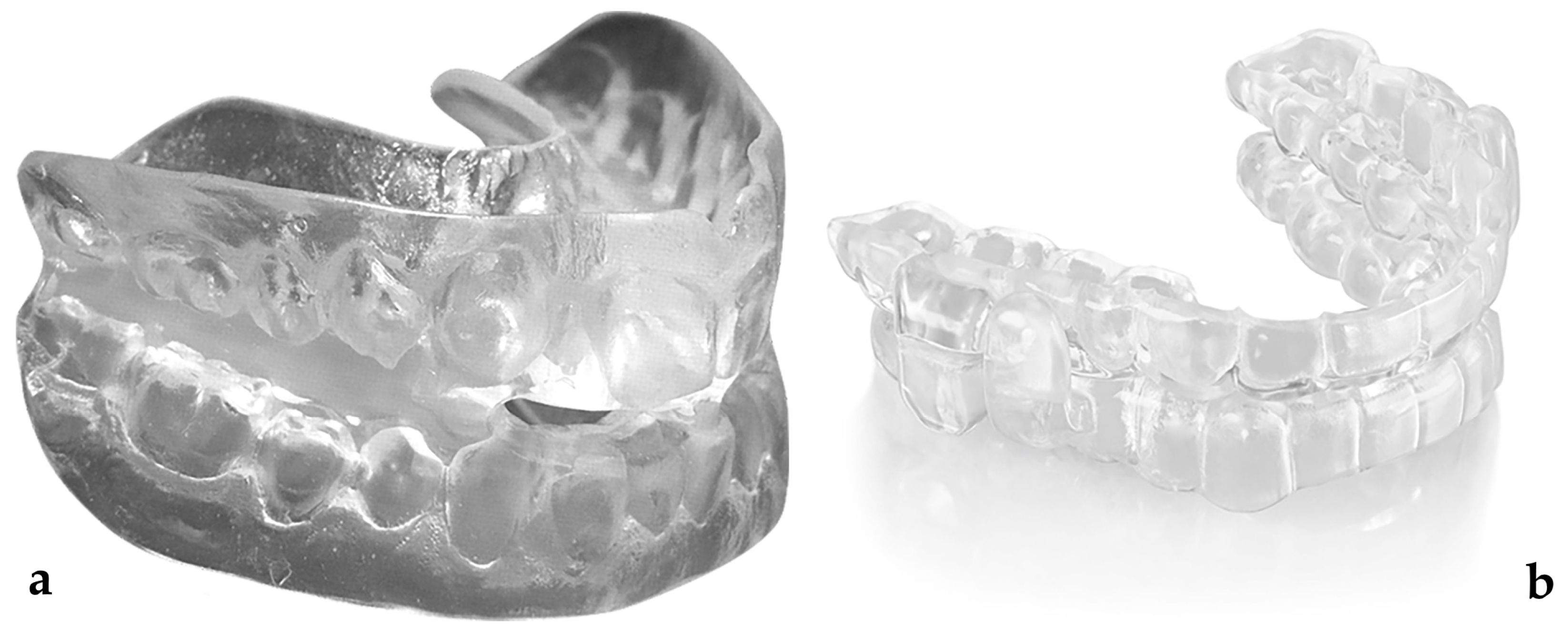
Figure 1. Mono-bloc MAD (a) and duo-bloc MAD (b).
Jcm 12 03619 g001
Figure 2. PRISMA flow diagram of the studies included in the systematic review.
Jcm 12 03619 g002
Figure 3. Forrest plots of success rate (correlation = success rate) [8,35,36,37,38,39].
Jcm 12 03619 g003
Figure 4. Bubble chart of the ratio between the success rate (reported as Fisher’s Z) and the amount of bite raise (size of bubbles is proportional to the weight of studies in the meta-regression).
Jcm 12 03619 g004
Figure 5. Funnel plot of the size effect of the included studies vs. standard error. Each bubble represents one study.
Jcm 12 03619 g005
Table 1. Description of the studies included in the systematic review.
StudyStudy DesignControlSample SizeAge
(Mean ± SD)
BMI
(Mean ± SD)
Degree of Vertical OpeningAHI Pre
(Mean ± SD)
AHI Post
(Mean ± SD)
Follow-UpAuthor’s Main Conclusions
Blanco
2005 [35]
RCTMAD with advancement vs. without advancement753.0 ± 12.728.4 ± 4.25 mm24.0 ± 12.211.7 ± 7.93 monthsMean AHI significantly decreases in both groups.
Gotsopoulos 2004 [36]RCT
cross-over
MAD vs. Placebo67 (53 M;14 F)48 ± 1129.2 ± 4.83.5 mm27 ± 1524 ± 21 monthA reduction of 50% in mean AHI value was obtained in MAD group, while no significant reduction was seen in Placebo group (p = 0.18).
Johnston 2002 [37]RCT
cross-over
MAD vs. Placebo20 (16 M;4 F)55.10 ± 6.8731.63 ± 5.941.5 mm31.93 ± 21.1837.68 ± 24.684–6 weeksMAD is significantly more efficient in improving AHI values compared to the placebo, with a success rate of 33%.
Mehta 2001 [8]RCT
cross-over
MAD vs. Placebo24 (19 M;5 F)48 ± 929.4 ± 3.13.5 mm27.0 ± 1730.0 ± 25 weeksThe Placebo device has no effects on AHI values.
Naismith 2005 [38]RCT
cross-over
MAD vs. Placebo73 (59 M;14 F)48.4 ± 11.0029.0 ± 5.03.5 mm26.9 ± 15.425.4 ± 14.52 monthsThe mean AHI value is reduced of 50% in MAD group; the reduction is not significant in Placebo group (p = 0.18).
Nikolopoulou 2013 [39]RCT
cross-over
MAD in situ vs. non in situ10 (3 M;7 F)47.3 ± 11.7<401 mm15.9 ± 6.417.4 ± 7.03 nightsA small but significant increase in AHI values is noted in OSA patients with MAD in situ.
Table 2. Risk of bias in randomized trials (Cochrane Collaboration’s RoB 2.0 tool).
StudyBias Arising from the Randomisation ProcessBias Due to Deviations from Intended InterventionsBias Due to Missing Outcome DataBias in Measurement of the OutcomeBias in Selection of Reported ResultResponse
Blanco 2005 [35]Some concernsSome concernsHigh riskLow riskLow riskHigh risk
Gotsopoulos 2004 [36]Low riskLow riskLow riskLow riskLow riskLow risk
Johnston 2002 [37]Some concernsSome concernsLow riskLow riskLow riskSome concerns
Mehta 2001 [8]Some concernsSome concernsHigh riskLow riskLow riskHigh risk
Naismith 2005 [38]Low riskSome concernsLow riskLow riskLow riskSome concerns
Nikolopoulou 2013 [39]Low riskLow riskLow riskLow riskLow riskLow risk
Table 3. GRADE summary of findings for success rate.
Success Rate
Quality AssessmentSummary of Findings
Number of SubgroupsRisk of BiasInconsistencyIndirectnessImprecisionPublication Bias (Egger’s Test)Number of PatientsSuccess Rate (95% CI)Quality
6SeriousSerious (I2 = 50.54, p = 0.072)SeriousNot seriousNot serious (p = 0.977)195−0.054 [−0.054 − 0.040]Very Low
Disclaimer/Publisher’s Note: The statements, opinions and data contained in all publications are solely those of the individual author(s) and contributor(s) and not of MDPI and/or the editor(s). MDPI and/or the editor(s) disclaim responsibility for any injury to people or property resulting from any ideas, methods, instructions or products referred to in the content.