Article

Attachment of Respiratory Pathogens and Candida to Denture Base Materials—A Pilot Study

1
Department of Prosthetic Dentistry, Regensburg University Medical Center, 93042 Regensburg, Germany
2
Institute for Medical Microbiology and Virology, Leipzig University Clinics, 04103 Leipzig, Germany
3
Department of Prosthetic Dentistry and Dental Materials Science, Leipzig University, 04103 Leipzig, Germany
*
Author to whom correspondence should be addressed.
J. Clin. Med. 2023, 12(19), 6127; https://doi.org/10.3390/jcm12196127
Submission received: 22 August 2023 / Revised: 19 September 2023 / Accepted: 20 September 2023 / Published: 22 September 2023

Abstract

:
Denture prostheses are an ideal and extensive reservoir for microorganisms to attach to their surfaces. The aim of the study was to elucidate interactions between materials for the fabrication of denture bases and the attachment of microorganisms, focusing on respiratory pathogens and Candida species. Specimens (6 mm × 1 mm) with a standardized surface roughness (Sa = 0.1 µm) were prepared from heat-pressed polymethyl methacrylate (PMMA), CAD/CAM-processed PMMA, and CAD/CAM-processed polyether ether ketone (PEEK). The specimens were randomly placed in the vestibular areas of complete upper dentures in seven patients and were removed either after 24 h without any oral hygiene measures or after a period of four weeks. The microorganisms adherent to the surface of the specimens were cultivated and subsequently analyzed using mass spectrometry (MALDI-TOF). The means and standard deviations were calculated, and the data were analyzed using a two-way analysis of variance (ANOVA) and Tukey post-hoc test where appropriate (α = 0.05). There was a significant increase (p ≤ 0.004) in the total bacterial counts (CFU/mL) between the first (24 h) and the second (four weeks) measurements. Regarding quantitative microbiological analyses, no significant differences between the various materials were identified. Respiratory microorganisms were detected in all samples at both measurement time points, with a large variance between different patients. Only after four weeks, Candida species were identified on all materials but not in all participants. Candida species and respiratory microorganisms accumulate on various denture base resins. While no significant differences were identified between the materials, there was a tendency towards a more pronounced accumulation of microorganisms on conventionally processed PMMA.

Graphical Abstract

1. Introduction

Data gathered from a cross-sectional study in France indicate that almost 14% of the adult population is supplied with removable dental prostheses (RDPs) [1]. Especially in the older age groups, edentulism and complete dentures are still very common [2]. Due to current demographic developments, the number of people over the age of 60 is expected to increase to two billion by 2050 [3]. Thus, despite of the implementation of frequent and effective preventative dental check-ups, dental prophylaxis, and an associated decline in edentulism [4], it can be expected that complete dentures will continue to be a relevant treatment option in prosthetic dentistry.
Complete dentures are in close contact with the oral mucosa and feature an extensive interface. This circumstance makes complete dentures an ideal reservoir for oral biofilms, which have been defined as a “complex, functional community of one or more species of microbes, encased in an extracellular polysaccharide matrix and attached to one another or to a solid surface” [5]. During the formation of biofilms, a salivary pellicle consisting of proteins, lipids, and carbohydrates initially forms on the previously cleaned surface within hours as a result of electrostatic interactions. The components of the pellicle act as receptors for the attachment of microorganisms, which are referred to as initial or pioneer colonizers. These mainly include Gram-positive streptococci (e.g., Streptococcus mitis/oralis/sanguinis) and rods. During the maturation process of the biofilm, further bacteria—so-called late colonizers—attach themselves to the biofilm via co-adhesion. Subsequently, there is a progressive development of the biofilm, which finally consists of facultatively and obligatory anaerobic organisms, Gram-negative cocci and rods, fusobacteria, spirochetes, and actinobacteria [6,7]. So far, about 700 bacterial [8] and up to 100 fungal species [9] have been identified in the oral microbiome. The human body and the microbiome form a symbiotic community called holobiont [10]. Environmental factors influence this dynamic relationship, which can lead to changes in the oral microbiome. Disturbances in the balance between microorganisms and the immune system may affect oral health [10]. The conventional wisdom is that oral biofilms can cause and promote local diseases such as gingivitis, periodontitis, oral candidiasis, dental caries, and endodontic infections, as well as systemic diseases such as aspiration pneumoniae and blood-borne infections.
With regard to these considerations, it has been highlighted that between 11% and 70% of all patients wearing RDPs suffer from denture stomatitis [11]. Candida albicans has been identified as the major causative microbial agent in denture stomatitis [12] and can be found in different morphological forms: blastophores, hyphae, and pseudohyphae. The maturation of oral biofilms causes an increased prevalence of the hyphae form, which is associated with increased virulence and pathogenicity [13,14].
In addition to that, it was reported that wearing RDPs is a risk predictor for an increased incidence of pneumonia [15]. It was shown that in older people wearing complete dentures during sleep, the risk for pneumonia increases by a factor of 2.3 [16]. The main reason for this phenomenon is the accumulation of biofilms on the RDP, which may contain respiratory pathogens. While S. pneumonie is regarded as the most common causative agent of pneumonia [17], other potential respiratory pathogens such as S. aureus, P. aeruginosa, H. influenza serotype B, S. pyogenes, and M. catarrhalis have also been identified in healthy denture wearers [18]. The accumulation of biofilm is promoted by inadequate or lacking oral hygiene [19]. Dysphagia and chronic obstructive pulmonary disease may further increase the risk for aspiration pneumonia [20].
A simple way to prevent the onset of these diseases is regular and sufficient denture cleaning. A mechanical cleaning of RDPs can be performed with brushes, microwave irradiation, and ultrasonic devices. Chemical cleaning agents include products based on sodium hypochlorite (NaOCl), peroxides, neutral peroxides with enzymes, enzymes, or acids. Combinations of mechanical and chemical protocols can also be employed, and are superior to the application of sole mechanical or chemical protocols [21]. However, tactility and visual ability usually decrease with increasing age. Thus, a sufficient cleaning of RDPs may be difficult for impaired patients and, in some cases, may also not be performed without help. In nursing homes, the staff are often restricted with time, which is why denture hygiene is regularly not adequately performed [22].
For the fabrication of RDPs, several materials are available on the dental market, including polymethyl methacrylate (PMMA) and polyether ether ketone (PEEK). In the dental laboratory, these materials can be processed by heat pressing (PMMA) or with computer-aided design/computer-aided manufacturing (CAD/CAM) techniques by milling from pre-polymerized blanks (both PMMA and PEEK). Pre-polymerization under industrial conditions produces materials with improved stability and mechanical behavior as well as less accumulation of biofilms [23]. With regard to microbial adherence, various surface properties of dental materials may have an impact, including surface roughness, surface free energy, and—to a minor extent—surface charge. With regard to surface roughness, the conventional wisdom is that rough surfaces favor the accumulation of microorganisms as they provide larger surface areas that are available for microbial attachment than smooth surfaces [24]. Moreover, rough surfaces protect microorganisms from oral shear forces during their initial attachment and may also impair the removal of organized microbial biofilms [25]. The interactions between surface free energy and the attachment of microorganisms are complex, and simple correlations have not yet been established [25]. However, it was summarized from the existing evidence that bacteria preferentially attach to surfaces with a high surface free energy [26,27]. Schmalz and Cieplik [28] and Teughels et al. [29] have provided comprehensive overviews dealing about the explanatory models.
Since the biofilms on RDPs may affect systemic health, it is worth developing materials that prevent the attachment of microorganisms such as respiratory pathogens and Candida albicans [30]. As no clinical data are currently available regarding the impact of the denture base material on the attachment and proliferation of respiratory microorganisms, the aim of the current study was to elucidate potential interactions between various denture base materials and the attachment of several microorganisms associated with respiratory diseases as well Candida albicans. It was hypothesized that resin-based materials polymerized under industrial conditions show less accumulation of these particular microorganisms and Candida species than conventional resin-based materials for the fabrication of RDPs.

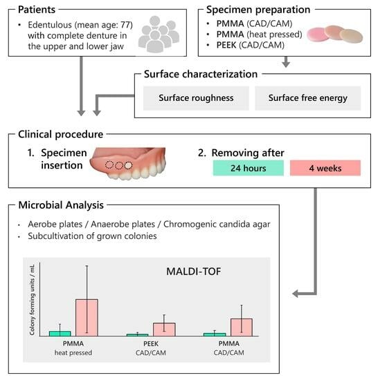
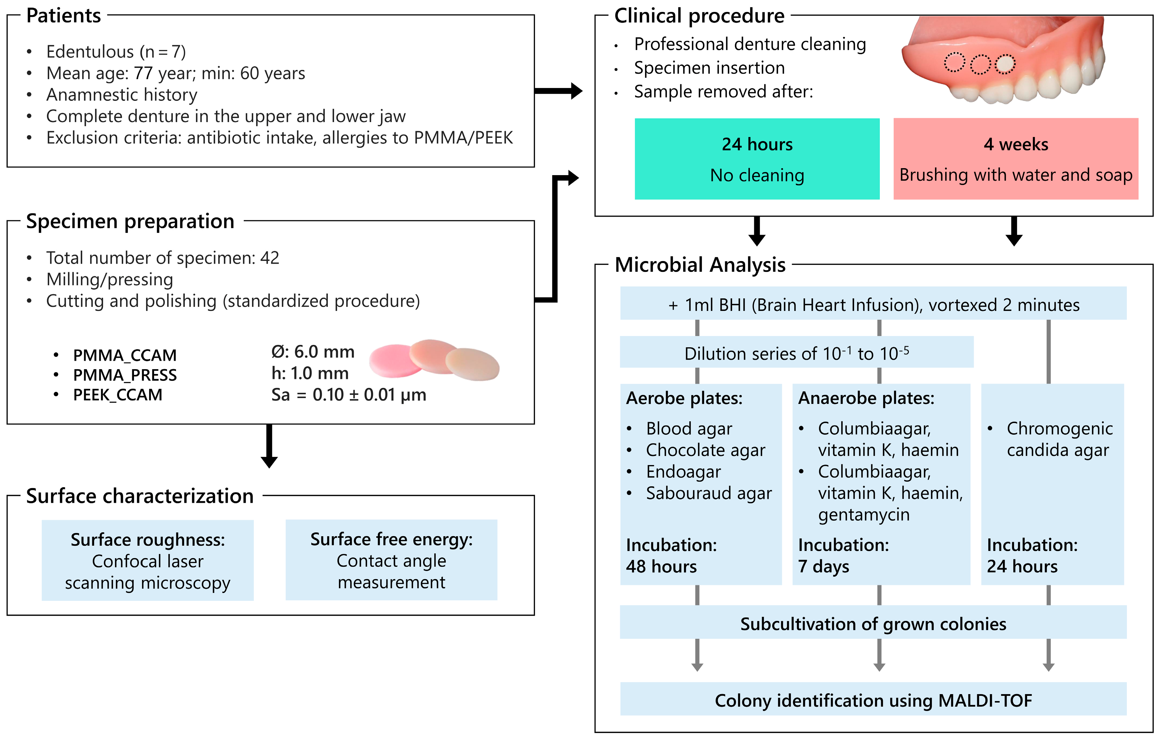
2. Materials and Methods

The design of the study is displayed in Figure 1.

2.1. Study Population

Seven edentulous patients (57.1% female) with a minimum age of 60 years (mean age 77.3 years ± 7.1 years; 72–92 years) supplied with complete dentures fabricated from polymethyl methacrylate in the upper and lower jaw were included in this observational study, which was performed between March 2020 and March 2022 at the Department of Prosthetic Dentistry and Dental Materials Science of the Leipzig University. All patients were able to carry out oral hygiene measures on their own accord; only patients with adequate oral hygiene were included. The grading of prosthetic hygiene took place by visual assessment. Care was taken to ensure that no plaque was visible on the dentures; only little amounts of stain were tolerated. The exclusion criteria included the intake of antibiotics in the last six months as well as allergies to PMMA or PEEK. Information on diseases such as bronchitis, pneumonia, and nasogastric/tracheal intubation within the last six months and pulmonary diseases such as chronic obstructive pulmonary diseases as well as an anamnestic history of tuberculosis (ever) were recorded. The patients were informed in advance about the investigation and consented in writing to participate in the study that had been approved by the local Ethical Committee (071/19-ek).

2.2. Specimen Preparation

Forty-two cylindrical specimens (Ø: 6 mm, h: 1 mm) were prepared from polymethyl methacrylate either through heat pressing (Ivobase Hybrid, Ivoclar Vivadent, Schaan, Liechtenstein) (PMMA_press) or CAD/CAM (Vita Vionic Base, Vita, Bad Säckingen, Germany) (PMMA_CCAM), and polyether ether ketone was prepared through CAD/CAM (Juvora, Invibio Ltd., Thornton-Cleveleys, UK) (PEEK_CCAM). The surfaces of the specimens were cut (IsoMet 4000, Buehler, Lake Bluff, IL, USA) and ground/polished using a semi-automatic grinding machine (Pedemin-2/DAP-V, Struers, Willich, Germany) with silicon carbide paper (up to P2000) and a composite disc (MD-Largo, Struers, Willich, Germany) with diamond polishing suspension (grain size: 15 μm). Consistent surface roughness was verified through confocal laser scanning microscopy. Subsequently, all specimens were stored under light-proof conditions in distilled water for six days at 37 ± 1 °C to minimize the impact of residual monomer leakage on cell viability. The specimens were cleaned with ethanol (70%) and applicator brush tips prior to being mounted onto the dentures.

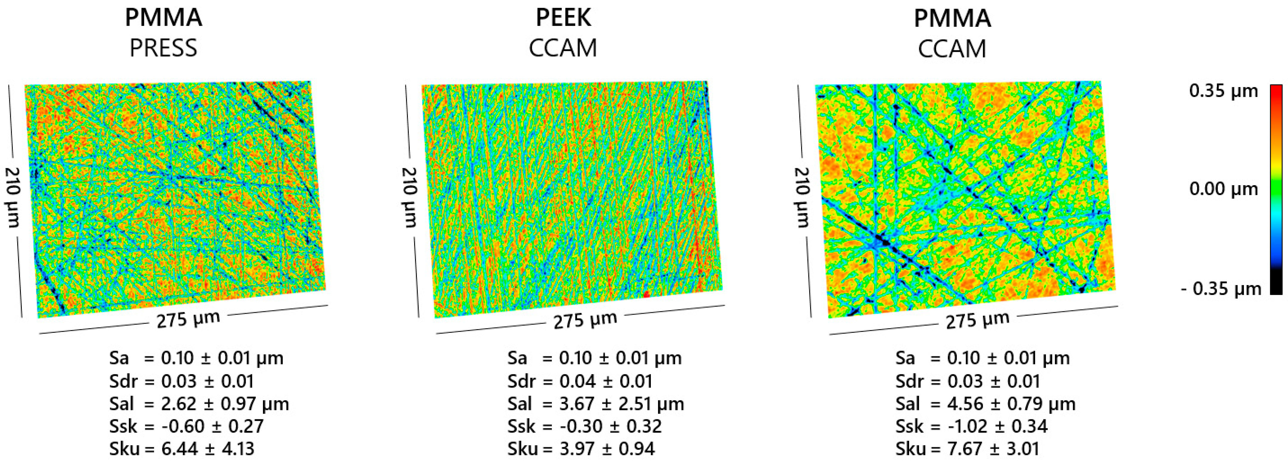
2.3. Surface Characterization

Sample surfaces were validated with confocal laser scanning microscopy (Keyence VK-X1000/1050, Keyence, Osaka, Japan) with a 50× objective (Nikon CF IC EPI Plan 50×; NA: 0.5; NIKON, Osaka, Japan) and a red laser (λ  =  661 nm). Five areas per sample were analyzed using the software “MultiFileAnalyzer” 2.1.3.89 (Keyence, Osaka, Japan), according to ISO 25178-2:2012 and appropriate filtering (S-Filter: 0.5 µm; F-Filter: 0.1 mm; Filter type: spline; end effect correction). For an analysis of the surface texture, the parameters of the arithmetical mean height (Sa), developed interfacial area ratio (Sdr), the auto-correlation length (Sal), and the skewness (Ssk) and the kurtosis (Sku) of the surface distribution were further surveyed.
The analysis of the surface free energy was performed on 15 samples via contact angle measurements. For this purpose, 0.5 µL each of purified water and diiodomethane was applied to the surface using a “DSA25S” with “Liquid Needle DO3252” and measured using “ADVANCE 1.11” (all KRÜSS, Hamburg, Germany) after a delay of 30 s (fitting method: ellipse). Following the advanced contact angle measurements, another 0.5 µL was added to the drops and measured again until a total volume of 2 µL was reached [31]. The total surface free energy as well as the polar and dispersive parts were calculated according to Owens and Wendt [32].

2.4. Specimen Insertion

One specimen of each material was randomly placed in the vestibular area of the upper denture molars according to a previously established randomization list to counteract known and unknown confounding variables. The specimens were fixed with a soft denture liner (Mucopren soft, Kettenbach, Eschenburg, Germany) in a small cavity (Ø: 6.5 mm, h: 1 mm) that had previously been inserted using a trepan drill. Prior to mounting the specimens, the upper and lower RDPs were forwarded for professional cleaning and polishing to remove adherent biofilms.

2.5. Study Protocol

First study part: The patients wore the RDPs equipped with the various specimens permanently for 24 h without any hygiene interventions. The specimens were then carefully removed, placed in an Eppendorf tube with 1 mL of brain heart infusion (BHI) bouillon, and immediately forwarded for microbiological analysis. The RDPs were professionally cleaned and polished.
Second study part: The RDPs were equipped with a new set of specimens. The patients wore the RDPs for 24 h/day and were instructed to clean the dentures daily by brushing with water and neutral soap as demonstrated. For this purpose, a denture brush was moistened with neutral soap, and the denture was then cleaned schematically from the denture base to the occlusal surface. The patients were instructed to refrain from the application of chemical detergents as well as antiseptic agents. After a period of four weeks, the specimens were carefully removed and forwarded for microbial analysis accordingly.
Group one (24 h—no cleaning) was introduced to produce biofilms on the specimens undisturbed from hygiene measure. The period of 24 h may represent a reasonable point between the initial adhesion and the maturation of biofilm and was chosen according to previous studies [33,34,35]. A period of four weeks (Group two) was introduced to produce biofilms on the surface of the specimens that occur during continuous and regular hygiene regimens.

2.6. Microbial Analysis

Specimens immersed in BHI (brain heart infusion) broth were vortexed for 2 min (IKA VF2 Vortex Mixer, IKA®-Werke, Staufen, Germany); subsequently, a dilution series of 10−1 to 10−5 was prepared. A volume of 0.1 mL of each dilution was inoculated onto seven different culture media (Figure 1) using a pipette (Multipette® (4780); Eppendorf Combitips advanced®, Eppendorf, Hamburg, Germany) and sterile disposable spatulas in a microbiological safety workbench (HeraSafe KS Class II Workbench, Thermo Fisher Scientific, Dreieich, Germany). The media blood agar, chocolate agar, Endoagar, and Sabouraud agar were aerobically incubated for 48 h at 37 °C with approximately 5% CO2 in a CO2-incubator (Heracell 150i CO2-Incubator, Thermo Fisher Scientific, Dreieich, Germany). The chromogenic Candida agar plates were incubated aerobically at 36 °C for 20 h. The Columbia blood agar plates were cultivated for seven days in an anaerobic workstation (Whitley MG 1000, anaerobic workstation, Meintrup Laborgeräte, Lähden, Germany) at 37 °C. All different colonies were visually described, counted, and subcultivated. After re-incubation (aerobe: 24 h; anaerobe: 4–7 d), colonies were identified using matrix-assisted laser desorption ionization time-of-flight mass spectrometry (MALDI-TOF; VITEK® MS, bioMérieux, Lyon, France), employing the commercially available V2.0 knowledge database for clinical use. Chromogenic Candida agar plates were used in addition to the Sabouraud agar plates for the easier identification of fungal species. All microbiological analyses were performed by the same examiner.

2.7. Statistical Analysis
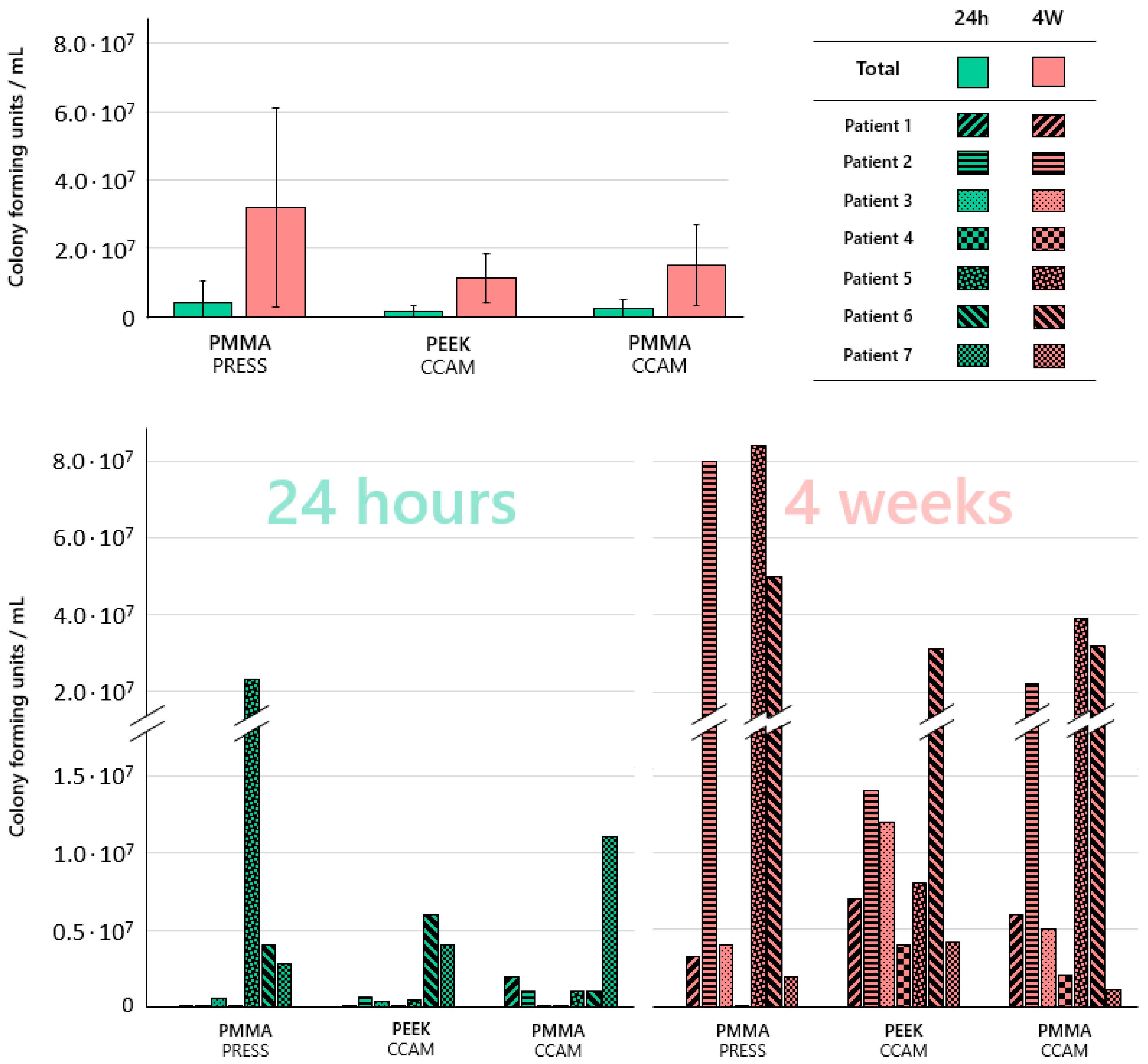
The means and standard deviations for colony forming units per mL (CFU/mL) were calculated. The normal distribution of the data was assessed using the Shapiro–Wilk test and the equality of variance was evaluated using Levene’s tests. A two-way analysis of variance (ANOVA) and the Tukey post-hoc test were applied where appropriate. The level of significance (α) was set to 0.05. All analyses were performed with IBM SPSS Statistics (Version 28.0).

3. Results

After the preparation of the specimens, all the groups resulted in an Sa of 0.10 ± 0.01 µm and an Sdr of 0.03 ± 0.01 to 0.04 ± 0.01 (Figure 2). Based on the three-dimensional surface renderings, the specimens of the PMMA_CCAM group showed a coarser ground pattern with fewer and broader as well as deeper valleys (Figure 2). Correspondingly, a higher Sal with 4.56 ± 0.79 µm and a reduced Ssk with −1.02 ± 0.34 of the surface distribution can be observed for the PMMA_CCAM samples. The Sku is greater than three for all sample groups (3.97 ± 0.94 to 7.67 ± 3.01) but varies in both mean and standard deviation.
Considering the standard deviation, the measurement of the contact angles resulted in almost identical values for the total surface free energy for all groups of specimens with 46.45 ± 2.20 mN/m to 48.17 ± 3.65 mN/m (Table 1). However, the distribution of the polar and dispersive fractions was different, with the former increasing from PEEK_CCAM (11.39 ± 1.92 mN/m) and PMMA_PRESS (8.09 ± 0.94 mN/m) to PMMA_CCAM (13.42 ± 1.57 mN/m) and the latter decreasing from PEEK_CCAM (38.36 ± 1.26 mN/m) and PMMA_PRESS (36.78 ± 1.73 mN/m) to PMMA_CCAM (33.25 ± 1.36 mN/m).
There was significantly more microbial growth on all specimens after four weeks than after 24 h (p ≤ 0.004). Higher total bacterial counts as well as larger standard deviations were identified on the specimens fabricated from conventionally processed PMMA than on the other two materials, but no statistically significant differences (p ≥ 0.211) between the three materials were determined (Figure 3).
The microbial growth on the different materials in the various patients showed a high variability. Bacteria associated with aspiration pneumonia were identified on the various denture base materials, yet large differences were observed (Table 2 and Table 3). Candida species were identified on all materials after four weeks but not after 24 h and not in all participants.

4. Discussion

The results of the current study suggest a rejection of the research hypothesis, as no significant differences in microbial counts were identified in the biofilms grown on the three different resin-based materials for the fabrication of RDPs. Nevertheless, less accumulation of microorganisms on the materials polymerized under industrial conditions than on conventionally processed PMMA was observed.
The data have to be interpreted within the limitations of the study. These include the design as a pilot study with a limited number of participants, which was due to the corona pandemic that made it particularly difficult to acquire voluntary participants and also to conduct the study in a university hospital setting. Despite the strict inclusion and exclusion criteria, there are still differences between the study participants that can hardly be controlled or standardized. Each patient features a very individual oral microbiome with differences in microbial species as well as microbial load, which might serve as an explanation for the high interindividual variability observed in the current study. The specimens were mounted onto the vestibular areas of the upper denture molars, yet, for higher clinical relevance, it might have been useful to insert the specimens into the basal areas of complete dentures as these are in contact with the oral mucosa. These areas are frequently not polished during the fabrication of the denture and, later, not regularly adequately cleaned. However, the shape of the specimens (flat discs) required an area without pronounced curvatures, and the patients were instructed to use the RDPs as in their common routine, which is why the authors tried to minimize the discomfort associated with the study. Since the fixed specimens were visible to the patients, they were aware of their location. Therefore, an improvement in the thoroughness of the oral hygiene measures during the study cannot be excluded. However, as the improvement in oral hygiene affected all sample bodies, this does not limit the comparability of the results within the study.
Although there were no statistically significant differences between the three materials regarding the total number of microorganisms identified by colonization, the absolute values as well as the standard deviations were higher for conventionally processed PMMA. This observation suggests that the manufacturing method employed for processing the denture base resin may in fact have an effect on biofilm formation. One reason for this phenomenon might be that the industrially standardized production of the materials to be used in a CAD/CAM process generates more homogeneous materials with less porosity and residual monomers in comparison to the conventionally processed denture base resins [36]. With regard to this aspect, it was highlighted that porosities can promote microbial adhesion [37]. While in the current study all specimens were subjected to a standardized polishing protocol prior to being mounted on the RDPs, surface porosities were not investigated, and it might be useful to elucidate a potential correlation between surface porosity and microbial adhesion in further studies. The conventional wisdom is that surface roughness is the most important material-associated predictor for microbial adherence. In order to eliminate the influence of surface roughness on the adherence of microorganisms to the various materials investigated in the current study, all specimens were polished to a Sa value of 0.1 μm, which is far below the commonly accepted threshold of 0.2 μm relevant for microbial adhesion [38].
Regarding the amount and effect of the residual monomers eluted from CAD/CAM dental polymers and conventional polymers, conflicting results have been reported. Wei et al. (2022) [39] reported that CAD/CAM dental polymers feature less residual monomer elution and, as a result, higher cell proliferation in cytotoxicity assays than conventional polymers. These results suggest that microbial adhesion to these materials might be enhanced, too [40]. In the current study, all specimens were stored in distilled water prior to the experiments in order to minimize the potential effects of the residual monomer elution. Moreover, it has also been reported that the elution of a residual monomer is more related to the material itself rather than the mode of fabrication [41].
The surface texture of the specimens had the same arithmetical mean height (Sa) due to the targeted polishing in order to minimize deviations and their effect on microbial adhesion. Even though primarily the arithmetical mean height was recommended in previous studies for the characterization of biomaterial surfaces [38,42,43], a visible difference in texture could already be observed based on the 3D surface renderings of this study, according to which CAD/CAM-processed PMMA exhibited a more coarse grinding pattern. Specifically, a lower number as well as a greater depth and width of the valleys created by the grinding/polishing process was observed for CAD/CAM-processed PMMA. These features result in a greater auto-correlation length (Sal) as well as in a lower skewness (Ssk) of the surface roughness. The specific surface area of the samples was similar for all samples. The same microbial adhesion on the CAD/CAM-processed PMMA and CAD/CAM-processed PEEK specimens despite the mentioned differences in the surface texture parameters Ssk and Sku in this pilot study confirms the findings of Schubert et al. [43] and Etxeberria et al. [42] that Sa is suitable as a surface characterization of dental biomaterials. Nevertheless, in subsequent studies, a differently pronounced surface texture with the same Sa should be assumed in order to take this into account with respect to any differences that may occur with respect to microbial adhesion. With regard to surface free energy, the results of the current study do not suggest a simple correlation with biofilm formation, which corroborates previously published data from the literature [25]. However, it has to be taken into consideration that the surface free energies between the materials investigated in the current study were rather similar and their relations to biofilm formation might be more pronounced between materials with more distinct surface free energy.
In the current study, Candida species were identified in the biofilms after four weeks but not after 24 h. This result is not surprising as Candida species do not belong to the early-colonizing microorganisms in oral biofilms [7]. In addition to these results, the data of the current study emphasize the relevance of RDPs as a potential reservoir of pneumonia-associated microorganisms, as reported by O’Donnell et al. (2016) [18]. Despite the pronounced interindividual variability and strict hygiene interventions during the study, several microorganisms associated with pneumonia were identified in the biofilm on the various materials. The presence of these potential pathogens appears to be independent of the denture base resin. Nevertheless, further studies focusing on potentially pathogenic microorganisms and their presence in biofilms on RDPs are necessary to corroborate the results of this pilot study.
As a result, the outcome of this study emphasizes that sufficient daily cleaning of RDPs is necessary to avoid microbial colonization. A recently published systematic review from our research group indicates that the combined use of chemical and mechanical denture hygiene interventions is more effective than single cleaning approaches [21]. However, it should be noted that cleaning procedures may cause damage to the denture surface, which may increase surface roughness and affect microbial adhesion. This phenomenon might particularly occur in materials with low surface hardness [44]. Furthermore, the enzymes and acid secreted by microorganisms can also lead to a roughening of the material surfaces, which in turn promotes microbial attachment [45,46]. However, the influence of these aging factors can be considered minor or negligible in the present investigation, as the study period was limited to a period of four weeks.

5. Conclusions

The surfaces of RDPs are an ideal reservoir for the accumulation of biofilms. The present study did not find any significant differences in the total bacterial counts on different polymer materials that can be used for the fabrication of RDPs. While no significant differences were identified between the materials, there was a tendency towards a more pronounced accumulation of microorganisms on conventionally processed PMMA. It was shown that the total bacterial counts increase with time. Potential respiratory pathogens as well as Candida species were identified on all three examined materials, although there was a large interindividual variability.

Author Contributions

Conceptualization, S.H., E.G. and F.F.; methodology, all authors; software, A.R.; validation, R.L.; formal analysis, S.H., A.S. and F.F.; investigation, A.S.; resources, C.S.S., S.H. and A.K.; data curation, A.S. and A.R.; writing—original draft preparation, A.S. and S.H.; writing—review and editing, all authors.; visualization, F.F.; supervision, C.S.S., F.F and S.H.; project administration, S.H. All authors have read and agreed to the published version of the manuscript.

Funding

This research received no external funding.

Institutional Review Board Statement

The study was conducted in accordance with the Declaration of Helsinki, and approved by the Ethics Committee of the Medical Faculty of the University of Leipzig (protocol code: 071/19-ek; date of approval: 26 January 2022).

Informed Consent Statement

Informed consent was obtained from all subjects involved in the study.

Data Availability Statement

The datasets generated and/or analyzed during the current study are available from the corresponding author upon reasonable request.

Acknowledgments

We would like to thank Annett Hennig-Rolle for their laboratory support.

Conflicts of Interest

The authors declare no conflict of interest.

References

  1. Fauroux, M.-A.; Germa, A.; Tramini, P.; Nabet, C. Prosthetic treatment in the adult French population: Prevalence and relation with demographic, socioeconomic and medical characteristics. Rev. Epidemiol. Sante Publique 2019, 67, 223–231. [Google Scholar] [CrossRef] [PubMed]
  2. Nitschke, I.; Stark, H. Krankheits- und Versorgungsprävalenzen bei Älteren Senioren (75- bis 100- Jährige). In Fünfte Deutsche Mundgesundheitsstudie (DMS V); Jordan, A.R., Micheelis, W., Eds.; Deutscher Zahnärzte Verlag DÄV: Köln, Germany, 2016; pp. 517–548. ISBN 978-3-7691-0020-4. [Google Scholar]
  3. Guzmán, J.M.; Pawliczko, A.; Beales, S.; Till, C.; Voelcker, I. Ageing in the Twenty-First Century: Celabration and a Challenge; United Nations Population Fund; HelpAge International: New York, NY, USA; London, UK, 2012; ISBN 9780897149815. [Google Scholar]
  4. Schwendicke, F.; Nitschke, I.; Stark, H.; Micheelis, W.; Jordan, R.A. Epidemiological trends, predictive factors, and projection of tooth loss in Germany 1997–2030: Part II. Edentulism in seniors. Clin. Oral Investig. 2020, 24, 3997–4003. [Google Scholar] [CrossRef] [PubMed]
  5. Do, T.; Devine, D.; Marsh, P.D. Oral biofilms: Molecular analysis, challenges, and future prospects in dental diagnostics. Clin. Cosmet. Investig. Dent. 2013, 5, 11–19. [Google Scholar] [CrossRef] [PubMed]
  6. Samaranayake, L.; Bandara, N.; Pesee, S. Oral Biofilms: What Are They? In Oral Biofilms and Modern Dental Materials: Advances Toward Bioactivity; Ionescu, A.C., Hahnel, S., Eds.; Springer International Publishing: Cham, Germany, 2021; pp. 1–7. ISBN 9783030673888. [Google Scholar]
  7. Günther, E.; Kommerein, N.; Hahnel, S. Biofilms on polymeric materials for the fabrication of removable dentures. DZZ Int. 2020, 2, 142–145. [Google Scholar] [CrossRef]
  8. Voorhis, A.; Miranda-Sanchez, F.; Dewhirst, F.E.; Mark Welch, J.; Kauffman, K.; Viala, S.; Yost, S.; Chen, T.; Wade, W.G. HOMD: Human Oral Microbiome Database. Available online: https://www.homd.org/ (accessed on 11 July 2023).
  9. Ghannoum, M.A.; Jurevic, R.J.; Mukherjee, P.K.; Cui, F.; Sikaroodi, M.; Naqvi, A.; Gillevet, P.M. Characterization of the oral fungal microbiome (mycobiome) in healthy individuals. PLoS Pathog. 2010, 6, e1000713. [Google Scholar] [CrossRef]
  10. Kilian, M.; Chapple, I.L.C.; Hannig, M.; Marsh, P.D.; Meuric, V.; Pedersen, A.M.L.; Tonetti, M.S.; Wade, W.G.; Zaura, E. The oral microbiome-an update for oral healthcare professionals. Br. Dent. J. 2016, 221, 657–666. [Google Scholar] [CrossRef]
  11. Gendreau, L.; Loewy, Z.G. Epidemiology and etiology of denture stomatitis. J. Prosthodont. 2011, 20, 251–260. [Google Scholar] [CrossRef]
  12. Budtz-Jörgensen, E. The significance of Candida albicans in denture stomatitis. Scand. J. Dent. Res. 1974, 82, 151–190. [Google Scholar] [CrossRef]
  13. Susewind, S.; Lang, R.; Hahnel, S. Biofilm formation and Candida albicans morphology on the surface of denture base materials. Mycoses 2015, 58, 719–727. [Google Scholar] [CrossRef]
  14. Bilhan, H.; Sulun, T.; Erkose, G.; Kurt, H.; Erturan, Z.; Kutay, O.; Bilgin, T. The role of Candida albicans hyphae and Lactobacillus in denture-related stomatitis. Clin. Oral Investig. 2009, 13, 363–368. [Google Scholar] [CrossRef]
  15. Alzamil, H.; Wu, T.T.; van Wijngaarden, E.; Mendoza, M.; Malmstrom, H.; Fiscella, K.; Kopycka-Kedzierawski, D.T.; Billings, R.J.; Xiao, J. Removable Denture Wearing as a Risk Predictor for Pneumonia Incidence and Time to Event in Older Adults. JDR Clin. Trans. Res. 2021, 8, 23800844211049406. [Google Scholar] [CrossRef] [PubMed]
  16. Iinuma, T.; Arai, Y.; Abe, Y.; Takayama, M.; Fukumoto, M.; Fukui, Y.; Iwase, T.; Takebayashi, T.; Hirose, N.; Gionhaku, N.; et al. Denture wearing during sleep doubles the risk of pneumonia in the very elderly. J. Dent. Res. 2015, 94, 28S–36S. [Google Scholar] [CrossRef] [PubMed]
  17. Kim, G.-L.; Seon, S.-H.; Rhee, D.-K. Pneumonia and Streptococcus pneumoniae vaccine. Arch. Pharm. Res. 2017, 40, 885–893. [Google Scholar] [CrossRef]
  18. O’Donnell, L.E.; Smith, K.; Williams, C.; Nile, C.J.; Lappin, D.F.; Bradshaw, D.; Lambert, M.; Robertson, D.P.; Bagg, J.; Hannah, V.; et al. Dentures are a Reservoir for Respiratory Pathogens. J. Prosthodont. 2016, 25, 99–104. [Google Scholar] [CrossRef] [PubMed]
  19. Kusama, T.; Aida, J.; Yamamoto, T.; Kondo, K.; Osaka, K. Infrequent Denture Cleaning Increased the Risk of Pneumonia among Community-dwelling Older Adults: A Population-based Cross-sectional Study. Sci. Rep. 2019, 9, 13734. [Google Scholar] [CrossRef]
  20. Marik, P.E.; Kaplan, D. Aspiration pneumonia and dysphagia in the elderly. Chest 2003, 124, 328–336. [Google Scholar] [CrossRef]
  21. Schmutzler, A.; Rauch, A.; Nitschke, I.; Lethaus, B.; Hahnel, S. Cleaning of removable dental prostheses-a systematic review. J. Evid. Based Dent. Pract. 2021, 21, 101644. [Google Scholar] [CrossRef]
  22. El-Solh, A.A. Association between pneumonia and oral care in nursing home residents. Lung 2011, 189, 173–180. [Google Scholar] [CrossRef]
  23. Stawarczyk, B.; Eichberger, M.; Uhrenbacher, J.; Wimmer, T.; Edelhoff, D.; Schmidlin, P.R. Three-unit reinforced polyetheretherketone composite FDPs: Influence of fabrication method on load-bearing capacity and failure types. Dent. Mater. J. 2015, 34, 7–12. [Google Scholar] [CrossRef]
  24. Quirynen, M. The clinical meaning of the surface roughness and the surface free energy of intra-oral hard substrata on the microbiology of the supra- and subgingival plaque: Results of in vitro and in vivo experiments. J. Dent. 1994, 22 (Suppl. S1), S13–S16. [Google Scholar] [CrossRef]
  25. Bürgers, R.; Krohn, S.; Wassmann, T. Surface Properties of Dental Materials and Biofilm Formation. In Oral Biofilms and Modern Dental Materials: Advances toward Bioactivity; Ionescu, A.C., Hahnel, S., Eds.; Springer International Publishing: Cham, Germany, 2021; pp. 55–69. ISBN 9783030673888. [Google Scholar]
  26. Hannig, C.; Hannig, M. The oral cavity—A key system to understand substratum-dependent bioadhesion on solid surfaces in man. Clin. Oral Investig. 2009, 13, 123–139. [Google Scholar] [CrossRef] [PubMed]
  27. Sterzenbach, T.; Helbig, R.; Hannig, C.; Hannig, M. Bioadhesion in the oral cavity and approaches for biofilm management by surface modifications. Clin. Oral Investig. 2020, 24, 4237–4260. [Google Scholar] [CrossRef]
  28. Schmalz, G.; Cieplik, F. Biofilms on Restorative Materials. Monogr. Oral Sci. 2021, 29, 155–194. [Google Scholar] [CrossRef] [PubMed]
  29. Teughels, W.; van Assche, N.; Sliepen, I.; Quirynen, M. Effect of material characteristics and/or surface topography on biofilm development. Clin. Oral Implant. Res. 2006, 17 (Suppl. S2), 68–81. [Google Scholar] [CrossRef] [PubMed]
  30. Koch, C.; Bürgers, R.; Hahnel, S. Candida albicans adherence and proliferation on the surface of denture base materials. Gerodontology 2013, 30, 309–313. [Google Scholar] [CrossRef] [PubMed]
  31. Huhtamäki, T.; Tian, X.; Korhonen, J.T.; Ras, R.H.A. Surface-wetting characterization using contact-angle measurements. Nat. Protoc. 2018, 13, 1521–1538. [Google Scholar] [CrossRef] [PubMed]
  32. Owens, D.K.; Wendt, R.C. Estimation of the surface free energy of polymers. J. Appl. Polym. Sci. 1969, 13, 1741–1747. [Google Scholar] [CrossRef]
  33. de Castro, D.T.; do Nascimento, C.; Alves, O.L.; de Souza Santos, E.; Agnelli, J.A.M.; Dos Reis, A.C. Analysis of the oral microbiome on the surface of modified dental polymers. Arch. Oral Biol. 2018, 93, 107–114. [Google Scholar] [CrossRef]
  34. Keijser, B.J.F.; van den Broek, T.J.; Slot, D.E.; van Twillert, L.; Kool, J.; Thabuis, C.; Ossendrijver, M.; van der Weijden, F.A.; Montijn, R.C. The Impact of Maltitol-Sweetened Chewing Gum on the Dental Plaque Biofilm Microbiota Composition. Front. Microbiol. 2018, 9, 381. [Google Scholar] [CrossRef]
  35. Schmidt, K.E.; Auschill, T.M.; Heumann, C.; Frankenberger, R.; Eick, S.; Sculean, A.; Arweiler, N.B. Clinical and laboratory evaluation of the effects of different treatment modalities on titanium healing caps: A randomized, controlled clinical trial. Clin. Oral Investig. 2018, 22, 2149–2160. [Google Scholar] [CrossRef]
  36. Murat, S.; Alp, G.; Alatalı, C.; Uzun, M. In Vitro Evaluation of Adhesion of Candida albicans on CAD/CAM PMMA-Based Polymers. J. Prosthodont. 2019, 28, e873–e879. [Google Scholar] [CrossRef] [PubMed]
  37. An, J.; Ding, N.; Zhang, Z. Mechanical and antibacterial properties of polymethyl methacrylate modified with zinc dimethacrylate. J. Prosthet. Dent. 2022, 128, 100.e1–100.e8. [Google Scholar] [CrossRef] [PubMed]
  38. Bollen, C.M.; Lambrechts, P.; Quirynen, M. Comparison of surface roughness of oral hard materials to the threshold surface roughness for bacterial plaque retention: A review of the literature. Dent. Mater. 1997, 13, 258–269. [Google Scholar] [CrossRef] [PubMed]
  39. Wei, X.; Pan, Y.; Wang, M.; Wang, Y.; Lin, H.; Jiang, L.; Lin, D.; Cheng, H. Comparative analysis of leaching residual monomer and biological effects of four types of conventional and CAD/CAM dental polymers: An in vitro study. Clin. Oral Investig. 2022, 26, 2887–2898. [Google Scholar] [CrossRef] [PubMed]
  40. Waltimo, T.; Vallittu, P.; Haapasalo, M. Adherence of Candida species to newly polymerized and water-stored denture base polymers. Int. J. Prosthodont. 2001, 14, 457–460. [Google Scholar] [PubMed]
  41. Engler, M.L.P.D.; Güth, J.-F.; Keul, C.; Erdelt, K.; Edelhoff, D.; Liebermann, A. Residual monomer elution from different conventional and CAD/CAM dental polymers during artificial aging. Clin. Oral Investig. 2020, 24, 277–284. [Google Scholar] [CrossRef]
  42. Etxeberria, M.; Escuin, T.; Vinas, M.; Ascaso, C. Useful surface parameters for biomaterial discrimination. Scanning 2015, 37, 429–437. [Google Scholar] [CrossRef]
  43. Schubert, A.; Wassmann, T.; Holtappels, M.; Kurbad, O.; Krohn, S.; Bürgers, R. Predictability of Microbial Adhesion to Dental Materials by Roughness Parameters. Coatings 2019, 9, 456. [Google Scholar] [CrossRef]
  44. Prpić, V.; Schauperl, Z.; Ćatić, A.; Dulčić, N.; Čimić, S. Comparison of Mechanical Properties of 3D-Printed, CAD/CAM, and Conventional Denture Base Materials. J. Prosthodont. 2020, 29, 524–528. [Google Scholar] [CrossRef]
  45. Nedeljkovic, I.; de Munck, J.; Ungureanu, A.-A.; Slomka, V.; Bartic, C.; Vananroye, A.; Clasen, C.; Teughels, W.; van Meerbeek, B.; van Landuyt, K.L. Biofilm-induced changes to the composite surface. J. Dent. 2017, 63, 36–43. [Google Scholar] [CrossRef]
  46. Matsuo, H.; Suenaga, H.; Takahashi, M.; Suzuki, O.; Sasaki, K.; Takahashi, N. Deterioration of polymethyl methacrylate dentures in the oral cavity. Dent. Mater. J. 2015, 34, 234–239. [Google Scholar] [CrossRef] [PubMed]
Figure 1. Study design; CAD/CAM-processed polymethyl methacrylate (PMMA_CCAM); heat-pressed polymethyl methacrylate (PMMA_PRESS); CAD/CAM-processed polyether ether ketone (PEEK_CCAM); number (n); diameter (ø); height (h); arithmetical mean height of surface roughness (Sa); matrix-assisted laser desorption ionization time-of-flight mass spectrometry (MALDI-TOF).
Jcm 12 06127 g001
Figure 2. Exemplary three-dimensional surface renderings of the used materials with the corresponding surface texture parameters of the respective sample group; CAD/CAM-processed polymethyl methacrylate (PMMA_CCAM); heat-pressed polymethyl methacrylate (PMMA_PRESS); CAD/CAM-processed polyether ether ketone (PEEK_CCAM); arithmetical mean height (Sa); developed interfacial area ratio (Sdr); auto-correlation length (Sal); skewness (Ssk); kurtosis (Sku).
Jcm 12 06127 g002
Figure 3. Colony forming units identified on the different materials/in the different participants; CAD/CAM-processed polymethyl methacrylate (PMMA_CCAM); heat-pressed polymethyl methacrylate (PMMA_PRESS); CAD/CAM-processed polyether ether ketone (PEEK_CCAM).
Jcm 12 06127 g003
Table 1. Measured contact angles and calculated surface free energy of the materials used in the study; CAD/CAM-processed polymethyl methacrylate (PMMA_CCAM); heat-pressed polymethyl methacrylate (PMMA_PRESS); CAD/CAM-processed polyether ether ketone (PEEK_CCAM).
PMMA_PRESS PEEK_CCAM PMMA_CCAM
Contact angle in °
 Water63.89 ± 3.3769.36 ± 1.8162.71 ± 2.54
 Diiodo methane45.42 ± 3.2242.44 ± 2.4351.83 ± 2.40
Surface free energy in mN/m
 Total48.17 ± 3.6546.45 ± 2.20 46.67 ± 2.93
 Polar part11.39 ± 1.92 8.09 ± 0.9413.42 ± 1.57
 Dispersive part36.78 ± 1.73 38.36 ± 1.26 33.25 ± 1.36
Table 2. Isolated microorganisms after 24 h; female (f); male (m); CAD/CAM-processed polymethyl methacrylate (PMMA_CCAM); heat-pressed polymethyl methacrylate (PMMA_PRESS); CAD/CAM-processed polyether ether ketone (PEEK_CCAM); colony forming units (CFU).
After 24 hPatient
1 (f)2 (f)3 (m)4 (f)5 (m)6 (m)7 (f)
Number of different isolated microorganisms per sample
PMMA_press166121213612
PEEK_CCAM147131114913
PMMA_CCAM115111113915
Isolated respiratory bacteria (CFU/mL)
Escherichia coli
PMMA_press3000000000
PEEK_CCAM00000030,000
PMMA_CCAM0000000
Enterobacter species
PMMA_press0000000
PEEK_CCAM1000000000
PMMA_CCAM0000000
Haemophilus influenzae
PMMA_press00001,200,00000
PEEK_CCAM00050040,00000
PMMA_CCAM000060,00000
Klebsiella species
PMMA_press00001,400,000100,0000
PEEK_CCAM00000400,0000
PMMA_CCAM00000300,0000
Staphylococcus aureus
PMMA_press0000700000
PEEK_CCAM0000000
PMMA_CCAM0000000
Streptococcus pneumoniae
PMMA_press400000000100,000
PEEK_CCAM50,00000100050,00000
PMMA_CCAM120,000000000
Candida species (CFU/mL)
PMMA_press0000000
PEEK_CCAM0000000
PMMA_CCAM0000000
Table 3. Isolated microorganisms after four weeks; female (f); male (m); CAD/CAM-processed polymethyl methacrylate (PMMA_CCAM); heat-pressed polymethyl methacrylate (PMMA_PRESS); CAD/CAM-processed polyether ether ketone (PEEK_CCAM); colony forming units (CFU).
After Four WeeksPatient
1 (f)2 (f)3 (m)4 (f)5 (m)6 (m)7 (f)
Number of different isolated microorganisms per sample
PMMA_press1512174171616
PEEK_CCAM15131216131213
PMMA_CCAM14191116171215
Isolated respiratory bacteria (CFU/mL)
Escherichia coli
PMMA_press00001,000,00002000
PEEK_CCAM0002000000
PMMA_CCAM00010,000110,00000
Enterobacter species
PMMA_press00000020,000
PEEK_CCAM0100,000010,000000
PMMA_CCAM00030,0001,100,00000
Haemophilus influenzae
PMMA_press010,00000000
PEEK_CCAM0000000
PMMA_CCAM0000150,00000
Klebsiella species
PMMA_press0000000
PEEK_CCAM0000000
PMMA_CCAM000600,000000
Staphylococcus aureus
PMMA_press0000000
PEEK_CCAM0000000
PMMA_CCAM0000000
Streptococcus pneumoniae
PMMA_press100,000000000
PEEK_CCAM0000000
PMMA_CCAM0000000
Candida species (CFU/mL)
PMMA_press02,006,00031,0000500,0003,509,0000
PEEK_CCAM02,020,00000010,100,0000
PMMA_CCAM1003,140,0003000004,700,0000
Disclaimer/Publisher’s Note: The statements, opinions and data contained in all publications are solely those of the individual author(s) and contributor(s) and not of MDPI and/or the editor(s). MDPI and/or the editor(s) disclaim responsibility for any injury to people or property resulting from any ideas, methods, instructions or products referred to in the content.

