
Clinical Outcomes of Conservative Surgery for Diffuse Uterine Leiomyomatosis: Preliminary Experience of 17 Cases in a Single Center

Abstract
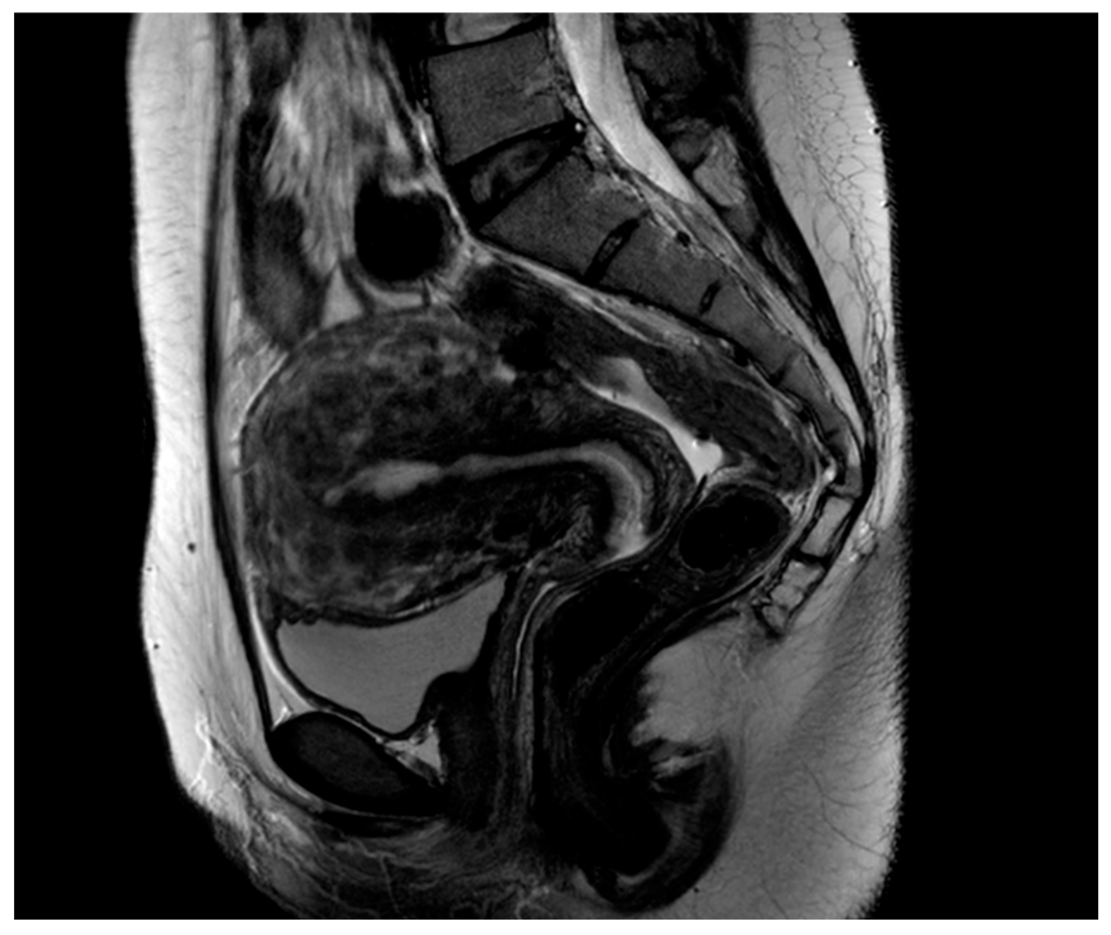
:1. Introduction
2. Materials and Methods
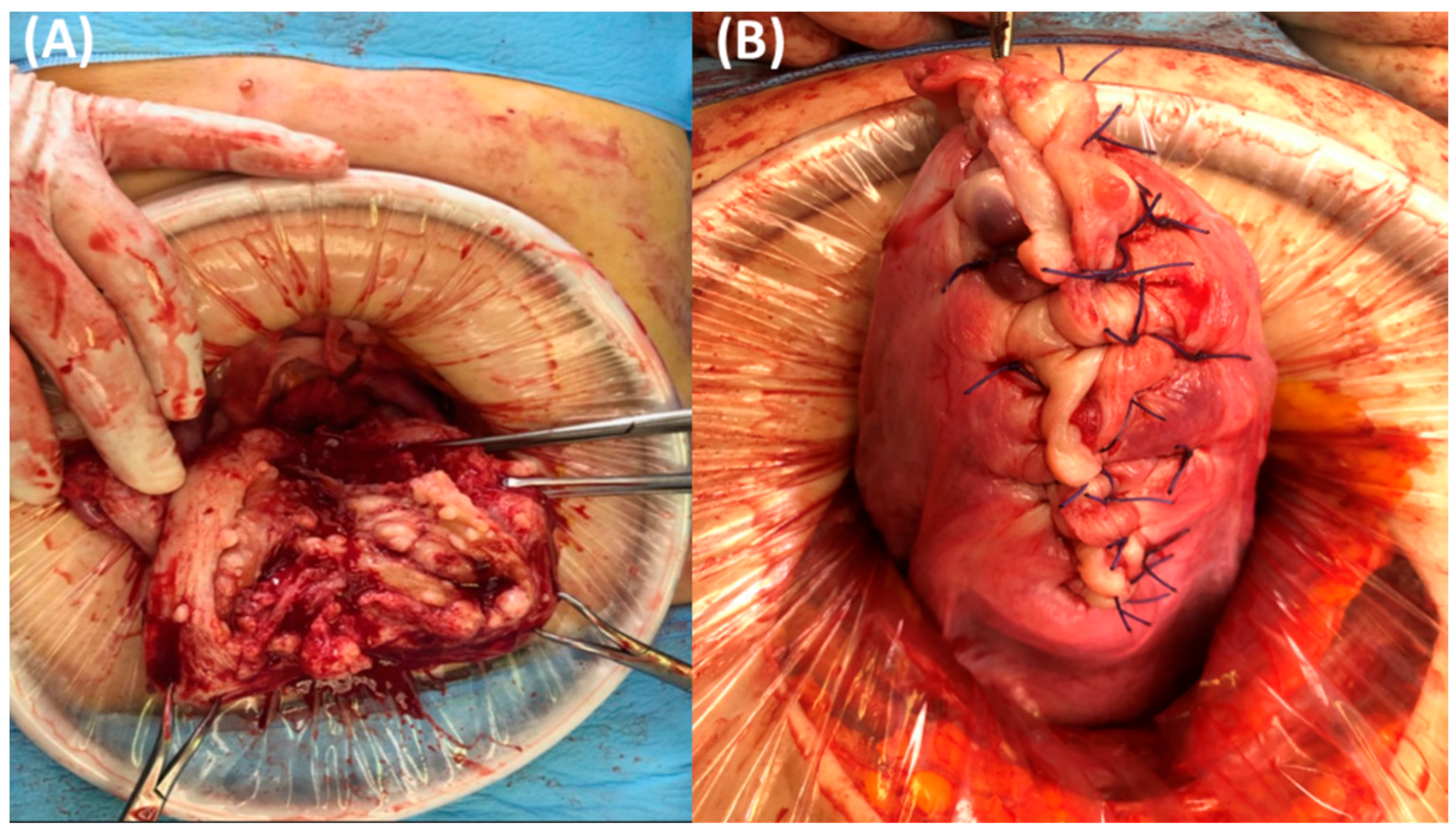
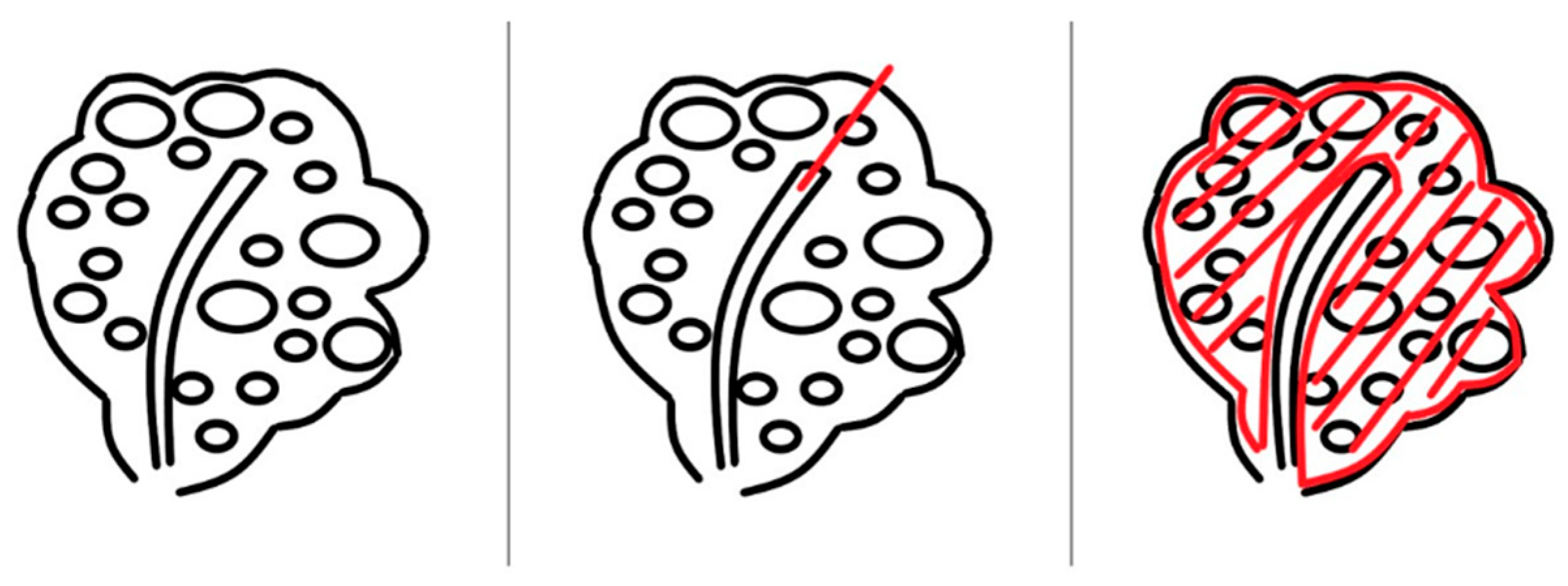
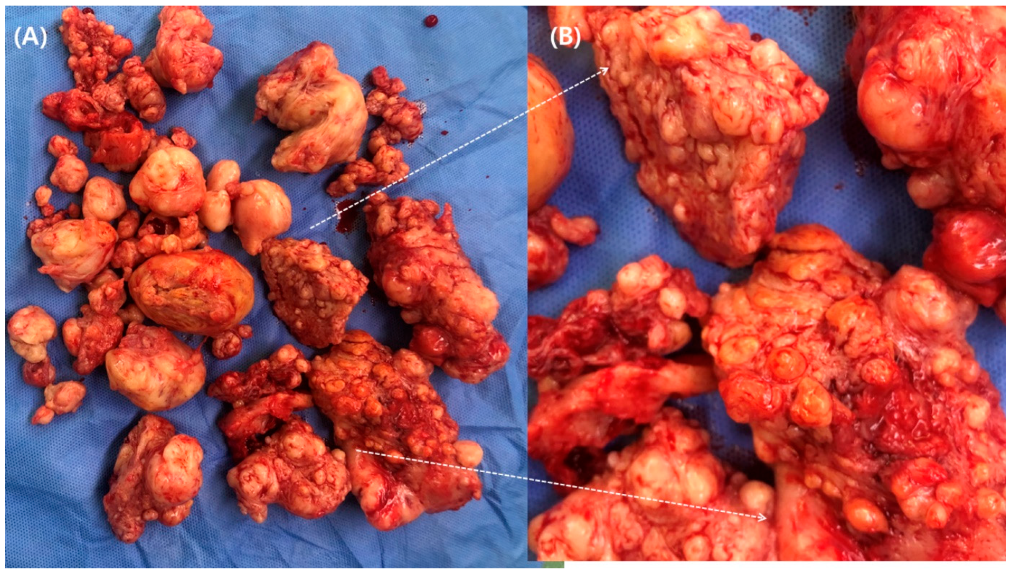
Surgical Technique
3. Results
4. Discussion
5. Conclusions
Author Contributions
Funding
Institutional Review Board Statement
Informed Consent Statement
Data Availability Statement
Conflicts of Interest
References
- Kido, A.; Togashi, K.; Koyama, T.; Yamaoka, T.; Fujiwara, T.; Fujii, S. Diffusely enlarged uterus: Evaluation with MR imaging. Radiographics 2003, 23, 1423–1439. [Google Scholar] [CrossRef]
- Baschinsky, D.Y.; Isa, A.; Niemann, T.H.; Prior, T.W.; Lucas, J.G.; Frankel, W.L. Diffuse leiomyomatosis of the uterus: A case report with clonality analysis. Hum. Pathol. 2000, 31, 1429–1432. [Google Scholar] [CrossRef] [PubMed]
- Stewart, E.A. Uterine fibroids. Lancet 2001, 357, 293–298. [Google Scholar] [CrossRef]
- Coskun, A.; Ozdemir, O.; Vardar, M.A.; Kiran, G.; Arikan, D.; Ersoz, C. A case with diffuse uterine leiomyomatosis and review of the literature. Clin. Exp. Obstet. Gynecol. 2008, 35, 227–230. [Google Scholar] [PubMed]
- Otsubo, Y.; Nishida, M.; Arai, Y.; Ichikawa, R.; Sakanaka, M. Diffuse uterine leiomyomatosis in patient with successful pregnancy following new surgical management. Arch. Gynecol. Obstet. 2014, 290, 815–818. [Google Scholar] [CrossRef] [PubMed]
- Morgan, E.D.; Kahiye, M.; Kule, I.; Yahaya, J.J.; Othieno, E. Disseminated peritoneal leiomyomatosis as an incidental finding: A case report. Clin. Case Rep. 2022, 10, e05541. [Google Scholar] [CrossRef] [PubMed]
- Kubik-Huch, R.A.; Weston, M.; Nougaret, S.; Leonhardt, H.; Thomassin-Naggara, I.; Horta, M.; Cunha, T.M.; Maciel, C.; Rockall, A.; Forstner, R. European Society of Urogenital Radiology (ESUR) Guidelines: MR Imaging of Leiomyomas. Eur. Radiol. 2018, 28, 3125–3137. [Google Scholar] [CrossRef]
- Clement, P.B.; Young, R.H. Diffuse leiomyomatosis of the uterus: A report of four cases. Int. J. Gynecol. Pathol. 1987, 6, 322–330. [Google Scholar] [CrossRef]
- Fedele, L.; Zamberletti, D.; Carinelli, S.; Motta, T.; Candiani, G.B. Diffuse uterine leiomyomatosis. Acta Eur. Fertil. 1982, 13, 125–131. [Google Scholar]
- Prasad, I.; Sinha, S.; Sinha, U.; Kumar, T.; Singh, J. Diffuse Leiomyomatosis of the Uterus: A Diagnostic Enigma. Cureus 2022, 14, e29595. [Google Scholar] [CrossRef]
- Ueda, H.; Togashi, K.; Konishi, I.; Kataoka, M.L.; Koyama, T.; Fujiwara, T.; Cunha, T.M.; Maciel, C.; Rockall, A.; Forstner, R. Unusual appearances of uterine leiomyomas: MR imaging findings and their histopathologic backgrounds. Radiographics 1999, 19, 3125–3137. [Google Scholar] [CrossRef] [PubMed]
- Andreyko, J.L.; Blumenfeld, Z.; Marshall, L.A.; Monroe, S.E.; Hricak, H.; Jaffe, R.B. Use of an agonistic analog of gonadotropin-releasing hormone (nafarelin) to treat leiomyomas: Assessment by magnetic resonance imaging. Am. J. Obstet. Gynecol. 1988, 158, 903–910. [Google Scholar] [CrossRef] [PubMed]
- Ren, H.M.; Wang, Q.Z.; Wang, J.N.; Hong, G.J.; Zhou, S.; Zhu, J.Y.; Li, S.-J. Diffuse uterine leiomyomatosis: A case report and review of literature. World J. Clin. Cases 2022, 10, 8797–8804. [Google Scholar] [CrossRef]
- Suminaga, Y.; Taki, M.; Okamoto, H.; Kawamura, Y.; Sagae, Y.; Sunada, M.; Chigusa, Y.; Horie, A.; Mandai, M.; Mogami, H. A Case of a Patient with Adhesive Small Bowel Obstruction in Pregnancy after Extensive Myomectomy for Diffuse Uterine Leiomyomatosis. Case Rep. Obstet. Gynecol. 2022, 2022, 3601945. [Google Scholar] [CrossRef] [PubMed]
- Dai, Y.X.; Feng, F.Z.; Leng, J.H.; Shi, H.H.; Cheng, N.H.; Wan, X.R.; Zhu, L. Imaging features and clinical analysis of diffuse uterine leiomyomatosis cases. Zhonghua Yi Xue Za Zhi 2020, 100, 2263–2267. [Google Scholar] [PubMed]
- Farrar, J.T.; Young, J.P.; Jr LaMoreaux, L.; Werth, J.L.; Poole, M.R. Clinical importance of changes in chronic pain intensity measured on an 11-point numerical pain rating scale. Pain 2001, 94, 149–158. [Google Scholar] [CrossRef] [PubMed]
- Mansfield, P.K.; Voda, A.; Allison, G. Validating a pencil-and-paper measure of perimenopausal menstrual blood loss. Women’s Health Issues 2004, 14, 242–247. [Google Scholar] [CrossRef]
- Scheurig, C.; Islam, T.; Zimmermann, E.; Hamm, B.; Kroencke, T.J. Uterine artery embolization in patients with symptomatic diffuse leiomyomatosis of the uterus. J. Vasc. Interv. Radiol. 2008, 19 Pt 1, 279–284. [Google Scholar] [CrossRef]
- Nishida, M.; Ichikawa, R.; Arai, Y.; Sakanaka, M.; Otsubo, Y. New myomectomy technique for diffuse uterine leiomyomatosis. J. Obstet. Gynaecol. Res. 2014, 40, 1689–1694. [Google Scholar] [CrossRef]
- Koh, J.; Kim, M.D.; Jung, D.C.; Lee, M.; Lee, M.S.; Won, J.Y.; Lee, D.Y.; Park, S.I.; Lee, K.H. Uterine artery embolization (UAE) for diffuse leiomyomatosis of the uterus: Clinical and imaging results. Eur. J. Radiol. 2012, 81, 2726–2729. [Google Scholar] [CrossRef]
- McLucas, B.; Voorhees, W.D., 3rd; Elliott, S. Fertility after uterine artery embolization: A review. Minim. Invasive Ther. Allied Technol. 2016, 25, 1–7. [Google Scholar] [CrossRef] [PubMed]
- Teo, U.L.; Kopeika, J.; Pundir, J.; El-Toukhy, T. Peri-operative morbidity and fertility outcome after repeat abdominal myomectomy for large fibroid uterus. J. Obstet. Gynaecol. 2020, 40, 673–677. [Google Scholar] [CrossRef] [PubMed]
- Hehenkamp, W.J.; Volkers, N.A.; Broekmans, F.J.; de Jong, F.H.; Themmen, A.P.; Birnie, E.; Reekers, J.A.; Ankum, W.M. Loss of ovarian reserve after uterine artery embolization: A randomized comparison with hysterectomy. Hum. Reprod. 2007, 22, 1996–2005. [Google Scholar] [CrossRef] [PubMed]
- Berkane, N.; Moutafoff-Borie, C. Impact of previous uterine artery embolization on fertility. Curr. Opin. Obstet. Gynecol. 2010, 22, 242–247. [Google Scholar] [CrossRef] [PubMed]
- Spies, J.B.; Roth, A.R.; Gonsalves, S.M.; Murphy-Skrzyniarz, K.M. Ovarian function after uterine artery embolization for leiomyomata: Assessment with use of serum follicle stimulating hormone assay. J. Vasc. Interv. Radiol. 2001, 12, 437–442. [Google Scholar] [CrossRef]
- Mara, M.; Fucikova, Z.; Maskova, J.; Kuzel, D.; Haakova, L. Uterine fibroid embolization versus myomectomy in women wishing to preserve fertility: Preliminary results of a randomized controlled trial. Eur. J. Obstet. Gynecol. Reprod. Biol. 2006, 126, 226–233. [Google Scholar] [CrossRef] [PubMed]
- Tropeano, G.; Di Stasi, C.; Litwicka, K.; Romano, D.; Draisci, G.; Mancuso, S. Uterine artery embolization for fibroids does not have adverse effects on ovarian reserve in regularly cycling women younger than 40 years. Fertil. Steril. 2004, 81, 1055–1061. [Google Scholar] [CrossRef]
- Ciebiera, M.; Madueke-Laveaux, O.S.; Feduniw, S.; Ulin, M.; Spaczynski, R.; Zgliczynska, M.; Bączkowska, M.; Zarychta, E.; Łoziński, T.; Ali, M.; et al. GnRH agonists and antagonists in therapy of symptomatic uterine fibroids-current roles and future perspectives. Expert. Opin. Pharmacother. 2023, 24, 1799–1809. [Google Scholar] [CrossRef]
- Dubuisson, J. The current place of mini-invasive surgery in uterine leiomyoma management. J. Gynecol. Obstet. Hum. Reprod. 2019, 48, 77–81. [Google Scholar] [CrossRef]
- Kwon, Y.S.; Roh, H.J.; Ahn, J.W.; Lee, S.H.; Im, K.S. Conservative adenomyomectomy with transient occlusion of uterine arteries for diffuse uterine adenomyosis. J. Obstet. Gynaecol. Res. 2015, 41, 938–945. [Google Scholar] [CrossRef]
- Chan, R.; Cuthbertson, D.; Jeng, Z.; Hsieh, G.; Antosh, D. Intraoperative ear bleeding with bilateral otorrhagia during laparoscopic sacrocolpopexy. Female Pelvic Med. Reconstr. Surg. 2015, 21, e6–e7. [Google Scholar] [CrossRef] [PubMed]
- Honda, I.; Sato, T.; Adachi, H.; Kobayashi, Y.; Shimada, K.; Watanabe, H.; Okada, Y.; Inoue, M. Uterine artery embolization for leiomyoma: Complications and effects on fertility. Nihon Igaku Hoshasen Gakkai Zasshi 2003, 63, 294–302. [Google Scholar] [PubMed]
- Chen, L.; Xiao, X.; Wang, Q.; Wu, C.; Zou, M.; Xiong, Y. High-intensity focused ultrasound ablation for diffuse uterine leiomyomatosis: A case report. Ultrason Sonochem. 2015, 27, 717–721. [Google Scholar] [CrossRef] [PubMed]
- Zhang, X.; Tian, T.; Zhu, J.; Lin, B.; Feng, X.; Zhang, L.; Yang, X.; Aili, A. High intensity focused ultrasound treatment for diffuse uterine leiomyomatosis: A feasibility study. Int. J. Hyperthermia 2020, 37, 1060–1065. [Google Scholar] [CrossRef] [PubMed]
- Fedele, L.; Bianchi, S.; Zanconato, G.; Carinelli, S.; Berlanda, N. Conservative treatment of diffuse uterine leiomyomatosis. Fertil. Steril. 2004, 82, 450–453. [Google Scholar] [CrossRef] [PubMed]
- Kido, A.; Monma, C.; Togashi, K.; Ueda, H.; Itoh, K.; Fujii, S.; Konishi, J. Uterine arterial embolization for the treatment of diffuse leiomyomatosis. J. Vasc. Interv. Radiol. 2003, 14, 643–647. [Google Scholar] [CrossRef] [PubMed]
- Kunchala, S.; Dumoff, K.; Shafique, K.; Kinson, M.S.; Shlansky-Goldberg, R.D. Expulsion of Diffuse Adenomyosis following Uterine Artery Embolization. J. Vasc. Interv. Radiol. 2020, 31, 1908–1911. [Google Scholar] [CrossRef]
- Hiramatsu, Y. Myomectomy for Multiple or Giant Uterine Fibroids. Surg. J. 2020, 6 (Suppl. 1), S22–S34. [Google Scholar] [CrossRef]
- Konishi, I. Diffuse Leiomyomatosis: Complete Myomectomy for Innumerable Small Nodules to Achieve Fertility Sparing and Childbearing. Surg. J. 2020, 6 (Suppl. 1), S50–S57. [Google Scholar] [CrossRef]

| Mean age (years) | 36.12 (29–48, SD = 5.4) |
| Previous myomectomy | |
| Laparotomic | 6 (35.3%) |
| Laparoscopic | 6 (35.3%) |
| Hysteroscopic | 2 (11.8%) |
| None | 3 (17.6%) |
| Marital status | |
| No | 8 (47.1%) |
| Yes | 9 (52.9%) |
| Abortion | |
| No | 4/9 (44.4%) |
| Yes | 5/9 (55.6%) |
| Main symptom | |
| Menorrhagia a | 16 (94.1%) |
| Dysmenorrhea b | 0 |
| Combined | 1 (5.9%) |
| Operation time (minute) | 97.06 (70–160, SD = 22.71) |
| EBL (mL) | 283.53 (20–1000, SD = 273.72) |
| Weight of excised specimen (gram) | 345.41 (89–910, SD = 240.65) |
| Intraoperative transfusion (N = 17) | |
| No | 16 (94.1%) |
| Yes | 1 (5.9%) |
| Hb 2 week before operation | 12.32 (10–14.9, SD = 1.57) |
| Hb 1 day after operation | 9.64 (7.2–13.1, SD = 1.85) |
| Hospital stay (day) | 6.47 (6–8, SD = 0.62) |
| Mean follow-up duration (weeks) | 116.41 (32–216, SD = 50.88) |
| Relief of symptoms recurrence a | 100% |
| No | 12 (70.6%) |
| Yes | 5 (29.4%) |
| Recurrence-free interval (weeks) | 50.6 (27–87, SD = 23.71) |
| Medication for recurrence (N = 5) | |
| No | 4 (80%) |
| Yes | 1 (20%) |
| Reoperation | 0 (0%) |
| Case | Reference | Study | Enrolled Number | Symptoms | Treatment | Outcome |
|---|---|---|---|---|---|---|
| 1 | Jieun Koh et al. [20] | Retrospective | 7 | Menorrhagia | Uterine artery embolization | Symptom relief |
| 2 | Li Chen et al. [33] | Case report | 1 | Menorrhagia Dysmenorrhea | High intensity focused ultrasound | Symptom control |
| 3 | Xiaofei Zhang et al. [34] | Research article | 8 | Menorrhagia Dysmenorrhea | High intensity focused ultrasound | Uterine volume shrinkage |
| 4 | Luigi Fedele et al. [35] | Descriptive study | 3 | Menorrhagia | Myomectomy | Menstrual pattern, pregnancy |
| 5 | Yasuo Otsubo et al. [5] | Case report | 1 | infertility | Myomectomy | Pregnancy |
| 6 | Masato Nishida et al. [19] | Original article | 7 | Menorrhagia | Myomectomy | Anemia improved. Pregnancy |
| 7 | Christian Scheurig et al. [18] | Case report | 6 | Menorrhagia Dysmenorrhea | Uterine artery embolization | Alleviation of symptoms |
| 8 | Aki Kido et al. [36] | Case report | 1 | Hypermenorrhea Dysmenorrhea | Uterine artery embolization | Reduced volume |
| 9 | Sudhir Kunchala et al. [37] | Case report | 1 | Menorrhagia Dysmenorrhea | Uterine artery embolization | Resolution of symptoms |
| 10 | Yuji Hiramatsu et al. [38] | Case report | 3 | Menorrhagia | Myomectomy | |
| 11 | Ikuo Konishi et al. [39] | Case report | 1 | Menorrhagia Dysmenorrhea Infertility | Myomectomy | Pregnancy |
| 12 | Sojung Kweon et al. (this work) | Original article | 17 | Menorrhagia Dysmenorrhea | Myomectomy | Symptoms relief Reduced volume |
Disclaimer/Publisher’s Note: The statements, opinions and data contained in all publications are solely those of the individual author(s) and contributor(s) and not of MDPI and/or the editor(s). MDPI and/or the editor(s) disclaim responsibility for any injury to people or property resulting from any ideas, methods, instructions or products referred to in the content. |
© 2023 by the authors. Licensee MDPI, Basel, Switzerland. This article is an open access article distributed under the terms and conditions of the Creative Commons Attribution (CC BY) license (https://creativecommons.org/licenses/by/4.0/).
Share and Cite
Kweon, S.; Park, J.; Sim, Y.; Kwack, J.Y.; Kwon, Y.-S. Clinical Outcomes of Conservative Surgery for Diffuse Uterine Leiomyomatosis: Preliminary Experience of 17 Cases in a Single Center. J. Clin. Med. 2023, 12, 7638. https://doi.org/10.3390/jcm12247638
Kweon S, Park J, Sim Y, Kwack JY, Kwon Y-S. Clinical Outcomes of Conservative Surgery for Diffuse Uterine Leiomyomatosis: Preliminary Experience of 17 Cases in a Single Center. Journal of Clinical Medicine. 2023; 12(24):7638. https://doi.org/10.3390/jcm12247638
Chicago/Turabian StyleKweon, Sojung, Joowon Park, Youngseo Sim, Jae Young Kwack, and Yong-Soon Kwon. 2023. "Clinical Outcomes of Conservative Surgery for Diffuse Uterine Leiomyomatosis: Preliminary Experience of 17 Cases in a Single Center" Journal of Clinical Medicine 12, no. 24: 7638. https://doi.org/10.3390/jcm12247638
APA StyleKweon, S., Park, J., Sim, Y., Kwack, J. Y., & Kwon, Y.-S. (2023). Clinical Outcomes of Conservative Surgery for Diffuse Uterine Leiomyomatosis: Preliminary Experience of 17 Cases in a Single Center. Journal of Clinical Medicine, 12(24), 7638. https://doi.org/10.3390/jcm12247638