Article

Prospective Assessment of Cerebral Microbleeds with Low-Field Magnetic Resonance Imaging (0.55 Tesla MRI)

1
Department of Radiology, Clinic of Radiology & Nuclear Medicine, University Hospital of Basel, University of Basel, 4001 Basel, Switzerland
2
Imamed Radiologie Nordwest AG, 4051 Basel, Switzerland
3
Department of Radiology, Gesundheitszentrum Fricktal, 4310 Rheinfelden, Switzerland
4
Department of Neurology, University Hospital of Basel, University of Basel, 4001 Basel, Switzerland
5
Department of Diagnostic and Interventional Neuroradiology, University Medical Center Hamburg-Eppendorf, 20251 Hamburg, Germany
6
Department of Radiology and Neuroradiology, Stadtspital Zürich, 8063 Zürich, Switzerland
*
Author to whom correspondence should be addressed.
These authors contributed equally to this work.
J. Clin. Med. 2023, 12(3), 1179; https://doi.org/10.3390/jcm12031179
Submission received: 19 December 2022 / Revised: 22 January 2023 / Accepted: 31 January 2023 / Published: 2 February 2023
(This article belongs to the Special Issue New Advances in Diagnostic Radiology of Ischemic Stroke)

Abstract

:
Purpose: Accurate detection of cerebral microbleeds (CMBs) on susceptibility-weighted (SWI) magnetic resonance imaging (MRI) is crucial for the characterization of many neurological diseases. Low-field MRI offers greater access at lower costs and lower infrastructural requirements, but also reduced susceptibility artifacts. We therefore evaluated the diagnostic performance for the detection of CMBs of a whole-body low-field MRI in a prospective cohort of suspected stroke patients compared to an established 1.5 T MRI. Methods: A prospective scanner comparison was performed including 27 patients, of whom 3 patients were excluded because the time interval was >1 h between acquisition of the 1.5 T and 0.55 T MRI. All SWI sequences were assessed for the presence, number, and localization of CMBs by two neuroradiologists and additionally underwent a Likert rating with respect to image impression, resolution, noise, contrast, and diagnostic quality. Results: A total of 24 patients with a mean age of 74 years were included (11 female). Both readers detected the same number and localization of microbleeds in all 24 datasets (sensitivity and specificity 100%; interreader reliability ϰ = 1), with CMBs only being observed in 12 patients. Likert ratings of the sequences at both field strengths regarding overall image quality and diagnostic quality did not reveal significant differences between the 0.55 T and 1.5 T sequences (p = 0.942; p = 0.672). For resolution and contrast, the 0.55 T sequences were even significantly superior (p < 0.0001; p < 0.0003), whereas the 1.5 T sequences were significantly superior (p < 0.0001) regarding noise. Conclusion: Low-field MRI at 0.55 T may have similar accuracy as 1.5 T scanners for the detection of microbleeds and thus may have great potential as a resource-efficient alternative in the near future.

1. Introduction

Cerebral microbleeds (CMBs) are small (2–10 mm diameter), round, or ovoid hypointense foci with associated blooming with enhanced visibility on MRI sequences sensitive to susceptibility artifacts [1,2]. They can be observed in patients with cognitive complaints and stroke but also in healthy individuals. Technically, local magnetic field inhomogeneities caused by paramagnetic iron in CMBs result in signal loss on MRI sequences, such as T2*-weighted gradient echo sequences or susceptibility-weighted imaging (SWI). SWI is derived from gradient echo sequences with additional post-processing to improve contrast resolution and is usually acquired in three dimensions to increase spatial resolution with flow compensation in all three planes to reduce artifacts. SWI has increased sensitivity and reliability for CMBs compared with T2*-weighted gradient echo but requires a longer acquisition time [3]. Further, technical aspects such as a low flip angle, long echo time, and long repetition time increase the sensitivity to susceptibility effects [2]. Moreover, the susceptibility effect and signal-to-noise ratio have been described to increase with higher magnetic field strength [4].
On the other hand, MRI accessibility is low and extremely inhomogeneous around the world, because MRI installations require expensive infrastructure (e.g., site preparation to host the large magnets, magnetic/radiofrequency shielding, and emergency helium exhaust conduit), high maintenance costs (i.e., for helium refill), have high operational costs for specialized radiographic technicians, and require huge amounts of electricity and water leaving a large ecological footprint [5,6]. Thus, the distribution of MRI scanners is concentrated mainly within high-income countries, and ~70% of the world’s population has little to no access to MRI (OECD (2022), magnetic resonance imaging (MRI) units (indicator); DOI: 10.1787/1a72e7d1-en). Moreover, even in high-income countries, clinical MRI scanners are mostly located in highly specialized radiology departments, large and centralized imaging centers, and housed on the ground floors of hospitals and clinics, excluding easy access to neurology clinics, trauma centers, surgical suites, neonatal/pediatric centers, and community clinics [5]. With this in mind, low-field MRI is increasingly coming into focus, offering MR imaging at a much lower cost, offering a considerable energy-saving potential but also reducing possible complications with metallic implants. Therefore, the aim of our study was to evaluate the performance of a 0.55 T low-field MRI in a prospective cohort of suspected stroke patients and to directly compare the diagnostic value for the detection of CMBs to a standard 1.5 T MRI.

2. Materials and Methods

This prospective study was reviewed and approved by the cantonal (Basel-Stadt, Switzerland) ethics committee (BASEC2021-00166). All included patients signed an informed consent form.

2.1. Data Acquisition

Data acquisition was performed from 1 May 2021 to 30 June 2021 at the Division of Neuroradiology, Clinic of Radiology and Nuclear Medicine, University Hospital Basel, Switzerland. All patients who presented to the emergency room with suspected ischemic stroke or transient ischemic attack (TIA) and who underwent MRI using a 1.5 T scanner (Siemens MAGNETOM Avanto FIT 1.5 T; Siemens Healthineers; Erlangen, Germany) as part of the diagnostic stroke workup were included. Immediately afterwards, patients were examined using a 0.55 T scanner (Siemens MAGNETOM FreeMax 0.55 T; Siemens Healthineers; Erlangen, Germany). The 1.5 T scanning protocol was in accordance with the hospital’s internal standard protocol for emergency stroked diagnostics including SWI sequences (Table 1). The 0.55 T SWI protocol was adapted to the 1.5 T SWI protocol as far as technically possible (same slice thickness (ST) and slice spacing (SP); comparable in-plane resolution) to ensure the most objective scanner comparison. After subsequent verification with respect to data completeness (scan protocols with complete image acquisition) and image quality (artifacts, image contrast), the datasets were transferred to the Picture Archiving and Communication System (PACS; General Electric (GE); Waukesha, WI, USA) for further analysis.

2.2. Data Analysis

Data analysis was performed in a two-step procedure: First, the 0.55 T and 1.5 T SWI datasets were evaluated using Likert rating. Second, a reading study was performed regarding identification, localization, and number of CMBs.

2.3. Likert Rating

Likert rating was performed by a neuroradiologist and a neuroradiologist in training with an experience of 9 and 5 years. Each acquired 0.55 T or 1.5 T SWI dataset was rated with respect to the following criteria with a numerical value between 1 and 10 (1 worst, 10 best):
(a)
Overall image quality;
(b)
Resolution;
(c)
Noise;
(d)
Contrast;
(e)
Diagnostic quality.
Sample SWI sequences from a 3 T scanner (Siemens MAGNETOM Skyra 3 T; Siemens Healthineers; Erlangen, Germany) were set as the gold standard (numerical value = 10). Dataset assessment was PACS-based using a standardized bookmark. Both readers were blinded to the results of the other reader.

2.4. Reading Study

Reading of 0.55 T and 1.5 T datasets was performed PACS-based and blinded (no clinical information, no image information) by two neuroradiologists with 8 and 13 years of professional experience. PACS-based post-processing as part of image analysis was allowed. For each dataset, number of SWI lesions (0, 1, 2–10, >10) and SWI lesion localization were analyzed.
The final neuroradiological report was defined as the underlying gold standard for the accuracy of the reading study.

3. Statistical Analysis

For statistical evaluation of Likert rating, a mean of the ratings of Readers 1 and 2 was first calculated for each 0.55 T and 1.5 T patient dataset and evaluation points (a)–(e). Subsequently, a Wilcoxon signed-rank test was used to evaluate significant or non-significant differences in Likert rating between the 0.55 T and 1.5 T sequences. Then interreader comparisons were performed to determine intraclass correlation coefficients (ICC). Calculation of sensitivity and specificity of Readers 1 and 2 in the reading study was performed in relation to the gold standard.

4. Results

A total of 27 patients with complete and artifact-free datasets were prospectively included in this study, of whom 3 patients were excluded because the time interval between the 1.5 T scan and 0.55 T scan was >1 h. The mean age of the remaining 24 patients was 74 years (standard deviation 14 years), and 11 patients were female (46%). The mean time interval between the 1.5 T scans and 0.55 T scans was 36 ± 14 min. Baseline characteristics of the patient cohort are presented in Table 2.
Both readers detected the same number and localization of microbleeds in all 24 0.55 T and 1.5 T datasets (sensitivity and specificity 100%; interreader reliability ϰ = 1). Ten 1.5 T datasets did not contain any microbleeds by analysis of the more experienced neuroradiologist and were therefore defined as the control group. No false-positive findings were observed by assessment of these images by Reader 2 (positive predictive value and negative predictive value 100%).
Likert ratings of the sequences with both field strengths regarding overall image quality (a) and diagnostic quality (e) did not reveal significant differences between the 0.55 T and 1.5 T sequences (p = 0.942 and p = 0.672, respectively; see Figure 1). Regarding the subjective evaluation of the spatial resolution (b) and contrast (d), the 0.55 T sequences were rated to be significantly superior: (b) p < 0.0001; (d) p < 0.0003. In contrast, the 1.5 T sequences were superior (p < 0.0001) regarding noise (c). Interreader comparisons showed moderate to high levels of agreement between Reader 1 and Reader 2 for the Likert ratings (ICC: (a) 0.91; (b) 0.93; (c) 0.60; (d) 0.87; (e) 0.88).

5. Discussion

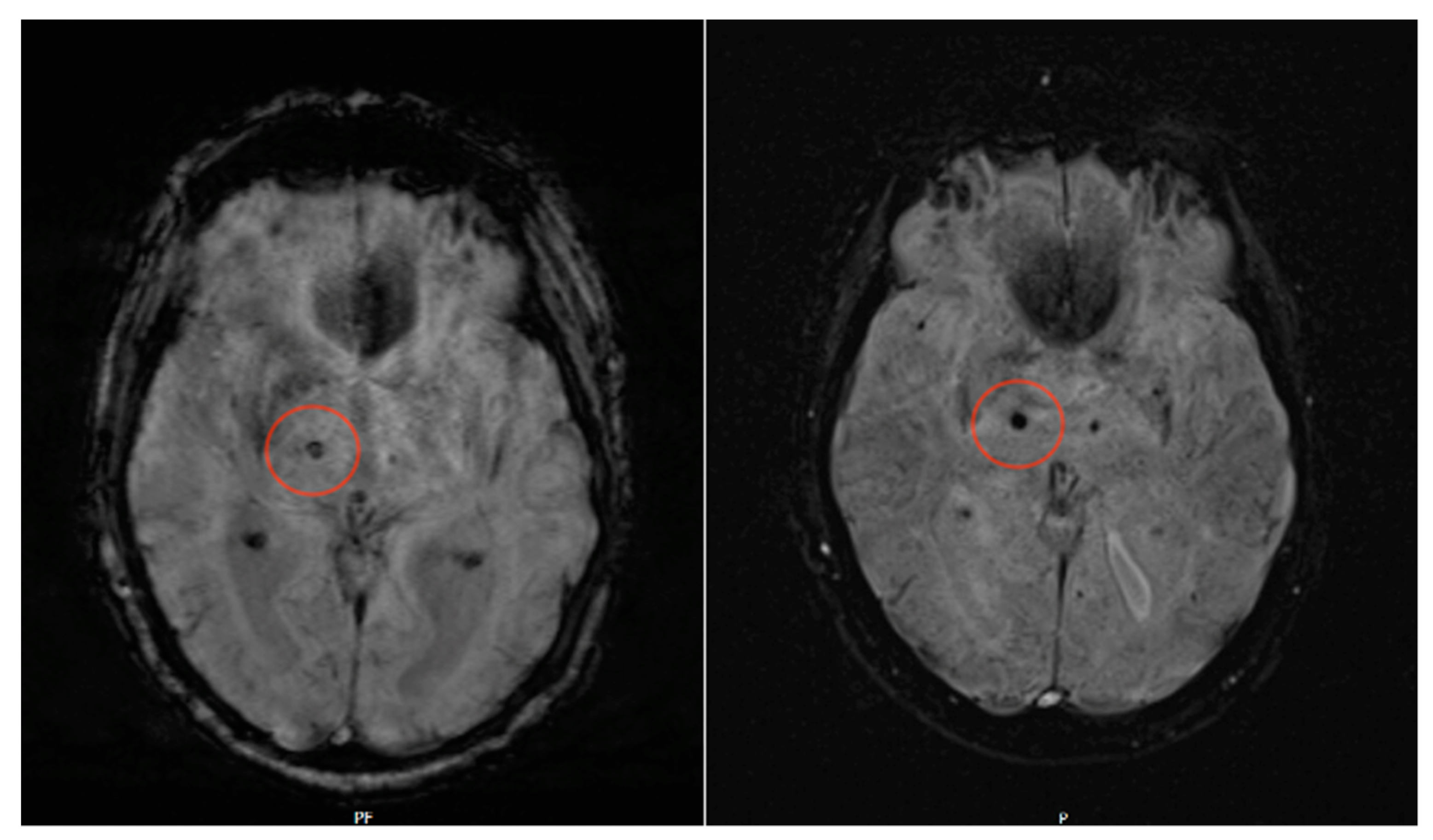
Our study shows that detection of microbleeds in SWI MRI sequences at a field strength of 0.55 T is possible with the same specificity and sensitivity compared to conventional 1.5 T MRI (for sample sequences, see Figure 2). Moreover, Likert ratings for the subjective evaluation of spatial resolution and contrast resolution were not significantly different for both 0.55 T and 1.5 T sequences, whereas for SWI noise, 0.55 T sequences were even superior.
To the best of our knowledge, no comparable study has performed a 0.55 T versus 1.5 T scanner comparison regarding the detection of microbleeds before. In a previous study, we showed that low-field MRI is not inferior to scanners with higher field strength for the detection of small infarcts in DWI and FLAIR sequences; however, in this study, there were minor limitations in the detection of very small infarcts [7]. Another group evaluated the performance of a modern 0.55 T MRI in the diagnosis of intracranial aneurysms in comparison to the gold standard digital subtraction angiography (DSA) [8]. This study included a total of 19 aneurysms in 16 patients, which were identified in both 0.55 T magnetic resonance angiography and DSA. Moreover, measurements of the two readers showed no significant differences between 0.55 T TOF MRA and DSA in the overall aneurysm size (calculated as the mean from height/width/neck), as well as in the mean width and neck values. The mean height was significantly larger in 0.55 T TOF MRA in comparison to DSA, whereas intermodality (1.5 T and 3 T TOF MRA) and interrater agreement were excellent (ICC > 0.94). Thus, the authors concluded that TOF MRA acquired with a modern 0.55 T MRI is a reliable tool for the detection and initial assessment of intracranial aneurysms. Moreover, in another study, we showed that patients perceived 0.55 T new-generation low-field MRI to be more comfortable than conventional 1.5 T MRI, given its larger bore opening and reduced noise levels during image acquisition, and concluded that new concepts regarding bore design and noise level reduction of MR scanner systems may help to reduce patient anxiety and improve well-being when undergoing MR imaging [9]. For microbleeds, the diagnostic accuracy of SWI sequences of the 0.55 T Magnetom FreeMax in our study seemed to be even higher, offering great potential for the characterization of associated diseases such as diffuse axonal injury or cerebral amyloid angiopathies [8].
In principle, the use of low-field MRI in clinical routine has several advantages. First, accessibility is low and extremely inhomogeneous around the world because MRI installations require expensive infrastructure, so the distribution of MRI scanners is concentrated mainly within high-income countries, and ~70% of the world’s population has little to no access to MRI [5]. Moreover, even in high-income countries, clinical MRI scanners are mostly located in highly specialized radiology departments, large and centralized imaging centers, and in those are housed on the ground floors of hospitals [5]. In a recent analysis by our group, we could show that in terms of purchase price, the savings potential of a 0.55 T MRI compared to a 1.5 T MRI system is about 40–50% [10]. The 25% lower weight of the system additionally reduces the transportation costs incurred, and the smaller size of the unit allows for installation by a remotely controlled mobile robotic system without opening the exterior façade, if the operating site is at ground level. Together with the lack of need to install a quench pipe, this reduces the total cost of installation by up to 70%. The maintenance cost of a 0.55 T MRI is approximately 45% less than that of a 1.5 T unit with a comparable service contract. Further cost reductions result from the smaller room size and potentially lower energy consumption for examinations and cooling. In conclusion, the use of lower-field-strength MRI systems offers enormous economic and environmental potential for both hospitals and practice operators, as well as for the healthcare system as a whole. In this context, offering MR imaging at a lower cost and with fewer infrastructural requirements will be key to increasing access to MRI for many patients. In developed countries, the energy-saving potential [6], and the possibility to reduce complications and artifacts caused by metallic implants will be the major arguments for implementing low-field MR imaging. The increasing exploration of the potential and limitations of low-field MRI is crucial to guide this implementation at a larger scale without harming patients.

Limitations

There are several limitations that need to be addressed. First, a 1.5 T device of routine clinical use is in fact not the “gold standard” for the detection of CMBs. Various studies have shown that higher field strengths (3 T and 7 T) have the highest sensitivity for the detection of CMBs [2,4,11,12,13,14,15,16,17]. For example, Conijn et al. [4] showed that the detection of CMBs is more reliable at 7 T compared to 1.5 T. Stehling et al. [18] had similar results when comparing 1.5 T versus 3 T. Greenberg et al. [2] also described better CMB detection at higher field strengths (3 T or higher), as CMBs are better visible in this case due to stronger susceptibility artifacts and thus stronger blooming artifacts. These results were most recently supported by data from Bian et al. [14], who also confirmed a higher sensitivity in the diagnosis of radiotherapy-induced CMBs at 7 T compared to 3 T sequences. For this reason, the detection of CBMs with lower field strengths (0.55 T and 1.5 T) is already a priori suboptimal, and thus the definition of a 1.5 T scanner as a comparison scanner and “gold standard” is afflicted with deficiencies. Thus, future studies should compare 0.55 T versus 3 T or 7 T. Second, both scanners differ with respect to their gradient and coil system as well as the field strength. Third, the study cohort (only 12 patients with CMBs), nonetheless prospective, is still relatively small. Larger-scale studies to further define indications for the detection of microbleeds at 0.55 T are needed and should assess whether scanner choice has an impact on patient outcomes. Fourth, results may have been different if the study cohort would have been selected from a population where a high CMB burden is already likely due to expected or diagnosed underlying diseases (cerebral amyloid angiopathy (CAA) or Alzheimer’s disease). Thus, more CMBs would be detectable and comparable in the collective as a whole.

6. Conclusions

Low-field MRI at 0.55 Tesla may have the same accuracy as 1.5 T MRI for the detection of microbleeds and thus may have great potential as a low-cost alternative in the near future.

Author Contributions

Conceptualization, T.R. and P.B.S.; Data curation, T.R., H.-C.B., M.B. and J.W.; Formal analysis, T.R., P.B.S., H.-C.B., J.G., S.M. and J.M.L.; Funding acquisition, T.R. and P.B.S.; Investigation, T.R.; Methodology, T.R., M.B. and P.B.S.; Project administration, T.R. and P.B.S.; Resources, T.R. and P.B.S.; Software, T.R. and J.W.; Supervision, T.R. and P.B.S.; Validation, T.R. and P.B.S.; Visualization, T.R. and P.B.S.; Writing—original draft, T.R.; Writing—review and editing, T.R., H.-C.B., M.B., G.M.D.M., M.P. and P.B.S. All authors have read and agreed to the published version of the manuscript.

Funding

This research received no external funding.

Institutional Review Board Statement

The study was approved under the provisions of the local ethics committee (BASEC2021-00166) in accordance with the Declaration of Helsinki.

Informed Consent Statement

Informed consent was obtained from all subjects involved in the study.

Data Availability Statement

The data presented in this study are available on request from the corresponding author.

Conflicts of Interest

The authors declare no conflict of interest.

Abbreviations

ADCapparent diffusion coefficient
bSSFPbalanced steady-state free precession
BWbandwidth
CAAcerebral amyloid angiopathy
CMBcerebral microbleed
CNRcontrast-to-noise ratio
CTcomputed tomography
DWIdiffusion-weighted imaging
FLAIRfluid-attenuated inversion recovery
ICCintraclass correlation coefficient
MRImagnetic resonance imaging
NIHSSNational Institutes of Health Stroke Scale
PACSPicture Archiving and Communication System
QALYquality-adjusted life-years
SNRsignal-to-noise ratio
SPslice spacing
STslice thickness
SWIsusceptibility-weighted imaging
TIAtransient ischemic attack

References

  1. Fazekas, F.; Kleinert, R.; Roob, G.; Kapeller, P.; Schmidt, R.; Hartung, H.P. Histopathologic analysis of foci of signal loss on gradient-echo T2*-weighted MR images in patients with spontaneous intracerebral hemorrhage: Evidence of microangiopathy-related microbleeds. Am. J. Neuroradiol. 1999, 20, 637–642. [Google Scholar] [PubMed]
  2. Greenberg, S.M.; Vernooij, M.W.; Cordonnier, C.; Viswanathan, A.; Al-Shahi Salman, R.; Warach, S.; Launer, L.J.; Van Buchem, M.A.; Breteler, M.M.; Microbleed Study Group. Cerebral microbleeds: A guide to detection and interpretation. Lancet Neurol. 2009, 8, 165–174. [Google Scholar] [CrossRef] [PubMed]
  3. Shams, S.; Martola, J.; Cavallin, L.; Granberg, T.; Shams, M.; Aspelin, P.; Wahlund, L.O.; Kristoffersen-Wiberg, M. SWI or T2*: Which MRI sequence to use in the detection of cerebral microbleeds? The Karolinska Imaging Dementia Study. Am. J. Neuroradiol. 2015, 36, 1089–1095. [Google Scholar] [CrossRef] [PubMed]
  4. Conijn, M.M.A.; Geerlings, M.I.; Biessels, G.J.; Takahara, T.; Witkamp, T.D.; Zwanenburg, J.J.M.; Luijten, P.R.; Hendrikse, J. Cerebral microbleeds on MR imaging: Comparison between 1.5 and 7T. Am. J. Neuroradiol. 2011, 32, 1043–1049. [Google Scholar] [CrossRef]
  5. Liu, Y.; Leong, A.T.L.; Zhao, Y.; Xiao, L.; Mak, H.K.F.; Tsang, A.C.O.; Lau, G.K.K.; Leung, G.K.K.; Wu, E.X. A low-cost and shielding-free ultra-low-field brain MRI scanner. Nat. Commun. 2021, 12, 7238. [Google Scholar] [CrossRef] [PubMed]
  6. Heye, T.; Knoerl, R.; Wehrle, T.; Mangold, D.; Cerminara, A.; Loser, M.; Plumeyer, M.; Degen, M.; Lüthy, R.; Brodbeck, D.; et al. The energy consumption of Radiology: Energy- And cost-saving opportunities for CT and MRI operation. Radiology 2020, 295, 593–605. [Google Scholar] [CrossRef] [PubMed]
  7. Rusche, T.; Breit, H.C.; Bach, M.; Wasserthal, J.; Gehweiler, J.; Manneck, S.; Lieb, J.M.; De Marchis, G.M.; Psychogios, M.N.; Sporns, P.B. Potential of Stroke Imaging Using a New Prototype of Low-Field MRI: A Prospective Direct 0.55 T/1.5 T Scanner Comparison. J. Clin. Med. 2022, 11, 2798. [Google Scholar] [CrossRef] [PubMed]
  8. Osmanodja, F.; Rösch, J.; Knott, M.; Doerfler, A.; Grodzki, D.; Uder, M.; Heiss, R. Diagnostic Performance of 0.55 T MRI for Intracranial Aneurysm Detection. Investig. Radiol. 2023, 58, 121–125. [Google Scholar] [CrossRef] [PubMed]
  9. Rusche, T.; Vosshenrich, J.; Winkel, D.J.; Donners, R.; Segeroth, M.; Bach, M.; Merkle, E.M.; Breit, H.-C. More Space, Less Noise-New-generation Low-Field Magnetic Resonance Imaging Systems Can Improve Patient Comfort: A Prospective 0.55 T–1.5 T-Scanner Comparison. J. Clin. Med. 2022, 11, 6705. [Google Scholar] [CrossRef] [PubMed]
  10. Vosshenrich, J.; Breit, H.C.; Bach, M.; Merkle, E.M. Economic aspects of low-field magnetic resonance imaging: Acquisition, installation, and maintenance costs of 0.55 T systems. Radiologe 2022, 62, 400–404. [Google Scholar] [CrossRef] [PubMed]
  11. Charidimou, A.; Shams, S.; Romero, J.R.; Ding, J.; Veltkamp, R.; Horstmann, S.; Eiriksdottir, G.; van Buchem, M.A.; Gudnason, V.; Himali, J.J.; et al. Clinical significance of cerebral microbleeds on MRI: A comprehensive meta-analysis of risk of intracerebral hemorrhage, ischemic stroke, mortality, and dementia in cohort studies (v1). Int. J. Stroke 2018, 13, 454–468. [Google Scholar] [CrossRef] [PubMed]
  12. Charidimou, A.; Imaizumi, T.; Moulinm, S.; Biffi, A.; Samarasekera, N.; Yakushiji, Y.; Peeters, A.; Vandermeeren, Y.; Laloux, P.; Baron, J.C.; et al. Brain hemorrhage recurrence, small vessel disease type, and cerebral microbleeds: A meta-analysis. Neurology 2017, 89, 820–829. [Google Scholar] [CrossRef] [PubMed]
  13. Charidimou, A.; Werring, D.J. Cerebral microbleeds and cognition in cerebrovascular disease: An update. J. Neurol. Sci. 2012, 322, 50–55. [Google Scholar] [CrossRef] [PubMed]
  14. Bian, W.; Hess, C.P.; Chang, S.M.; Nelson, S.J.; Lupo, J.M. Susceptibility-weighted MR imaging of radiation therapy-induced cerebral microbleeds in patients with glioma: A comparison between 3 T and 7 T. Neuroradiology 2014, 56, 91–96. [Google Scholar] [CrossRef]
  15. Hütter, B.O.; Altmeppen, J.; Kraff, O.; Maderwa, S.; Theysohn, J.M.; Ringelstein, A.; Wrede, K.H.; Dammann, P.; Quick, H.H.; Schlamann, M.; et al. Higher sensitivity for traumatic cerebral microbleeds at 7 T ultra-high field MRI: Is it clinically significant for the acute state of the patients and later quality of life? Ther. Adv. Neurol. Disord. 2020, 13, 1756286420911295. [Google Scholar] [CrossRef] [PubMed]
  16. Conijn, M.M.; Hoogduin, J.M.; van der Graaf, Y.; Hendrikse, J.; Luijten, P.R.; Geerlings, M.I. Microbleeds, lacunar infarcts, white matter lesions and cerebrovascular reactivity—A 7 T study. Neuroimage 2012, 59, 950–956. [Google Scholar] [CrossRef]
  17. Theysohn, J.M.; Kraff, O.; Maderwald, S.; Barth, M.; Ladd, S.C.; Forsting, M.; Ladd, M.E.; Gizewski, E.R. 7 tesla MRI of microbleeds and white matter lesions as seen in vascular dementia. J. Magn. Reson. Imaging 2011, 33, 782–791. [Google Scholar] [CrossRef] [PubMed]
  18. Stehling, C.; Wersching, H.; Kloska, S.P.; Kirchhof, P.; Ring, J.; Nassenstein, I.; Allkemper, T.; Knecht, S.; Bachmann, R.; Heindel, W. Detection of asymptomatic cerebral microbleeds: A comparative study at 1.5 and 3.0 T. Acad. Radiol. 2008, 15, 895–900. [Google Scholar] [CrossRef] [PubMed]
Figure 1. Average Likert scoring of Readers 1 and 2 SWI sequences.
Jcm 12 01179 g001
Figure 2. SWI lesion of the right thalamus. In both images (left, axial 1.5 T SWI sequence; right, axial 0.55 T SWI sequence), this lesion is clearly detectable, although it is even better delineated in the 0.55 T dataset.
Jcm 12 01179 g002
Table 1. Scan protocols Siemens MAGNETOM Avanto FIT 1.5 T and Siemens MAGNETOM FreeMax 0.55 T.
Siemens MAGNETOM FreeMax 0.55 TSiemens MAGNETOM
Avanto Fit 1.5 T
FLAIR tra
Field strength in T0.551.5
Field of view (FOV) in mm2209 × 230187 × 230
Slice thickness (ST) in mm33
Slice spacing (SS)3.63.6
Number of slices4040
Pixel spacing (PS) in mm21.28 × 1.030.9 × 0.9
Repetition time (TR) in msec77808510
Echo time (TE) in msec96112
Inversion delay (TI) in msec2368.82460
Turbo factor1519
Time of acquisition (TA) in min05:2803:26
BW ((BW))150130
3D SWI tra
Field strength in T0.551.5
Sequence typeMulti-shot 3D EPI3D FLASH
Field of view (FOV) in mm2201 × 230194 × 230
Slice thickness (ST) in mm33
Number of slices4048
Pixel spacing (PS) in mm20.94 × 0.81.12 × 0.9
Repetition time (TR) in msec17248
Echo time (TE) in msec10040
Parallel imaging-GRAPPA factor 2
Time of acquisition (TA) in min02:2302:17
BW ((BW))27680
Single-shot diffusion EPI tra
Field strength in T0.551.5
Field of view (FOV) in mm2220 × 220230 × 230
Slice thickness (ST) in mm33
Slice spacing (SS)3.63.6
Number of slices4040
Pixel spacing (PS) in mm21.67 × 1.671.44 × 1.44
b-values in s/mm20, 10000, 1000
Repetition time (TR) in msec74006200
Echo time (TE) in msec102103
Parallel imagingGRAPPA factor 2GRAPPA factor 2
Time of acquisition (TA) in min04:3502:04
BW ((BW))8421490
Table 2. Detailed patient data.
PatientPatient AgeCMB Yes/NoNumber of CMB(Main)-Localization of CMBTime Gap between Scans in min
Patient 187Yes1Left occipital46
Patient 273Yes3Right frontal/periventricular25
Patient 388Yes4Right temporal/parietal37
Patient 429No0-33
Patient 582No0-93
Patient 670No0-44
Patient 787Yes2Left occipital32
Patient 874No0-25
Patient 960No0-21
Patient 1044No0-49
Patient 1184Yes1Left occipital33
Patient 1258No0-40
Patient 1380No0-35
Patient 1465Yes1Left Putamen20
Patient 1565Yes2Left frontal24
Patient 1675No0-22
Patient 1784No0-48
Patient 1882Yes4Left frontal32
Patient 1979Yes1Left occipital42
Patient 2084Yes1Left periventricular32
Patient 2186Yes>10Bilateral Thalamus25
Patient 2283No0-31
Patient 2389No0-38
Patient 2469Yes3Left periventricular42
Patient 2553Excluded--916
Patient 2659Excluded--2936
Patient 2746Excluded--2812
Disclaimer/Publisher’s Note: The statements, opinions and data contained in all publications are solely those of the individual author(s) and contributor(s) and not of MDPI and/or the editor(s). MDPI and/or the editor(s) disclaim responsibility for any injury to people or property resulting from any ideas, methods, instructions or products referred to in the content.