Case Report

Severe Microcytic Anemia Caused by Complex Hereditary Spherocytosis and X-Linked Sideroblastic Anemia with Mutations in SPTB and ALAS2 Genes

Department of Hematology, Institute of Hematology, West China Hospital of Sichuan University, Chengdu 610041, China
*
Author to whom correspondence should be addressed.
J. Clin. Med. 2023, 12(5), 1990; https://doi.org/10.3390/jcm12051990
Submission received: 7 January 2023 / Revised: 17 February 2023 / Accepted: 27 February 2023 / Published: 2 March 2023
(This article belongs to the Section Hematology)

Abstract

:
We report a case of severe anemia caused by complex hereditary spherocytosis (HS) and X-linked sideroblastic anemia (XLSA) with two mutations in the spectrin beta (SPTB) and 5-aminolevulinic acid synthase (ALAS2) genes. The proband was a 16-year-old male with severe jaundice and microcytic hypochromic anemia since his childhood. He had more severe anemia requiring erythrocyte transfusion, and had no response to vitamin B6 treatment. Next-generation sequencing (NGS) revealed double heterozygous mutations, one in exon 19 (c.3936G > A:p.W1312X) of the SPTB gene and another in exon 2 (c.37A > G:p.K13E) of the ALAS2 gene, and confirmed again by Sanger sequencing. The mutation of ALAS2 (c.37A > G) is inherited from his asymptomatic heterozygous mother, causing amino acid p.K13E, and the mutation has not yet been reported. The mutation of SPTB (c.3936G > A) is a nonsense mutation, leading to a premature termination codon in exon 19, and the mutation in the SPTB gene is not found in any of his relatives, which indicates a de novo monoallelic mutation. Conclusions: The double heterozygous mutations in the SPTB and ALAS2 genes lead to the joint occurrence of HS and XLSA in this patient, and are implicated in the more severe clinical phenotypes.

1. Background

Hereditary spherocytosis (HS) is a common inherited hemolytic anemia caused by mutations in genes coding erythrocyte membrane proteins, which leads to defects in the membrane proteins, including ankyrin, band 3, β-spectrin, α-spectrin, or protein 4.2. Erythrocyte membrane defects reduce deformability, and the abnormal red blood cells (RBCs) are trapped and destroyed in the spleen, which result in hemolysis, anemia, jaundice, splenomegaly, and gallstones. The clinical severity of the disease depends on the extent of the surface area loss and ranges from asymptomatic forms to severe anemia cases requiring erythrocyte transfusion [1].
X-linked sideroblastic anemia (XLSA) is the most common hereditary sideroblastic anemia and is caused by mutations in the erythroid-specific 5-aminolevulinic acid synthase (ALAS) gene, which is located on chromosome Xp11.21 [2]. ALAS is the first enzyme involved in heme biosynthesis in erythroid cells, and ALAS2 gene mutation leads to ALAS function deficiency, heme synthesis anomaly, iron overload, and anemia. XLSA occurs preferentially in males and usually presents as asymptomatic or mild microcytic anemia. However, approximately one-quarter of the affected XLSA probands are females, which is mostly associated with a skewed X-chromosome inactivation [3]. Pyridoxal phosphate (PLP) as a cofactor can enhance the activity of ALAS; therefore, vitamin B6 can improve anemia in most XLSA patients [4].
Here, we report a male patient with severe transfusion-dependent anemia. Next-generation sequencing (NGS) revealed double heterozygous mutations in the SPTB gene and ALAS2 gene, characterized with two disease entities, HS and XLSA. In contrast to the human gene bank databases, the ALAS2 mutation is a novel pathogenic mutation that has not yet been reported. Written informed consent was obtained from the patient and his parents for the publication of this case report and any accompanying images.

2. Case Presentation

A 16-year-old male was admitted to the West China Hospital due to jaundice and anemia that had been present since his childhood. His parents and brother were asymptomatic with normal blood test results. On physical examination, the patient had pale skin, icteric sclera, and hepatosplenomegaly (liver 2 cm below costal; spleen 9 cm below costal). Blood tests showed microcytic hypochromic anemia: hemoglobin 70 g/L (ref value: 130–175 g/L); mean corpuscular volume 77 fL (ref value: 82–100 fL); mean corpuscular hemoglobin concentration 303 g/L (ref value: 316–354 g/L), and the reticulocyte percentage (Ret%) and immature reticulocytes fraction (IRF) were 38.2% (ref value: 0.5–1.5%) and 10.3%, respectively, with a normal white blood cell count (6.14 × 10⁹/L) (ref value: 3.5–9.5 × 10⁹/L) and platelet count (169 × 109/L) (ref value: 100–300 × 109/L). Peripheral blood smear showed erythrocyte morphological abnormalities, including anisocytosis, spherocytes and elliptocytes (shown in Figure 1). His total bilirubin was 83.9 µmol/L (ref value: 5.0–28.0 µmol/L), indirect bilirubin was 78.7 µmol/L (ref value: <20 µmol/L), and lactate dehydrogenase was 319 IU/L (ref value: 120–250 IU/L). For differential diagnosis tests, Coomb’s test was negative, hemoglobin electrophoresis was normal, and no mutation of the α- and β-globin genes was detected by the copy number variation of DNA sequences (CNVplex®) and single nucleotide polymorphisms assay (SNapShot). The bone marrow smear showed erythroid hyperplasia with a 44% proportion of total sideroblasts, and the proportion of ring sideroblasts was 30%. Serum ferritin, transferrin, and transferrin receptor (sTfR) were 260 ng/mL (ref value: 24–336 ng/mL), 1.80 g/L (ref value: 2.5–4.3 g/L), and 9.21 mg/L (ref value: 0.76–1.76 mg/L), respectively. All of these results indicated the diagnosis of hemolytic anemia and sideroblastic anemia.
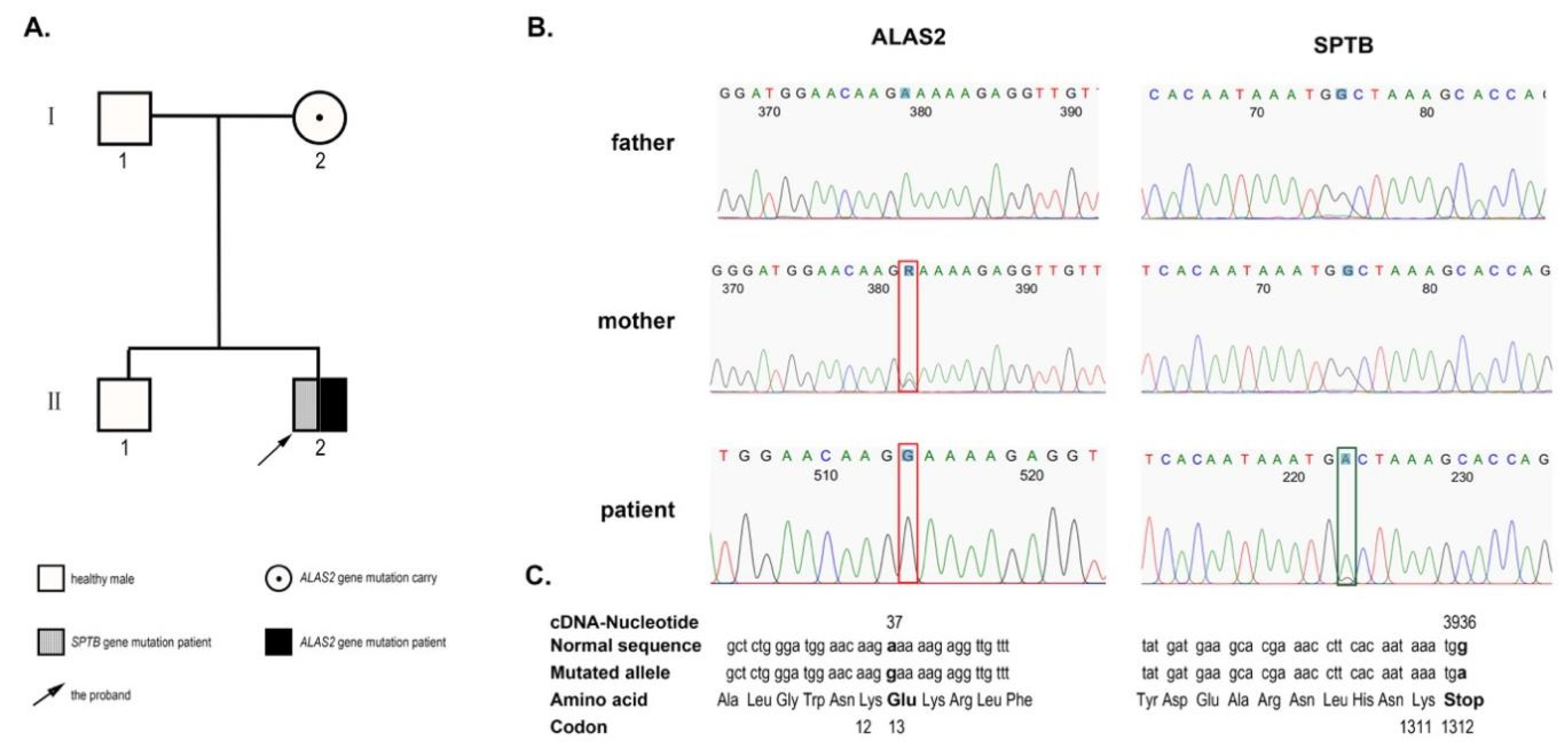
To confirm the patient’s diagnosis, heparinized anticoagulated blood was obtained from the patient. DNA samples were extracted using a QIAamp DNA Blood MiniKit (Qiagen, Valencia, CA, USA). Then, the DNA sample was subjected to NGS by Kindstar Global Esoteric Test & Services Co., Ltd. (Beijing, China) in a panel of 700 genes for red blood diseases. The primer design, synthesis, and NGS tests were performed in accordance with standard laboratory procedures. The sequences were then aligned to human genome version 19 (HG19) using information from databases, including the NCBI dbSNP, HapMap, and the 1000 Genomes Project human dataset. The sequencing analysis identified double heterozygous mutations, a heterozygous mutation in exon 19 (c.3936G > A:p.W1312X) of the SPTB gene, and a hemizygous mutation in exon 2 (c.37A > G:p.K13E) of the ALAS2 gene. These mutations were confirmed again by Sanger sequencing. Further pedigree investigations were finished for his parents and brother with their informed consent. The results showed that the mutation in exon 19 (c.3936G > A) of the SPTB gene identified in the proband was a de novo monoallelic variant because his parents showed a wild type in Sanger sequencing. The mutation in exon 2 (c.37A > G) of the ALAS2 gene was inherited from his asymptomatic heterozygous mother. The patient’s father and brother were not found to have the two mutations (shown in Figure 2). To our knowledge, the ALAS2 mutation has not yet been reported.
The patient was diagnosed with hemolytic anemia caused by hereditary spherocytosis and X-linked sideroblastic anemia. He was treated with folic acid and vitamin B6. After 6 months of treatment, he showed little symptomatic improvement, his blood tests indicated severe anemia (Hb 60 g/L; MCV 77.4 fL) with recitulocytosis (12.12%), and his white blood cells and platelet counts were normal.

3. Discussion and Conclusions

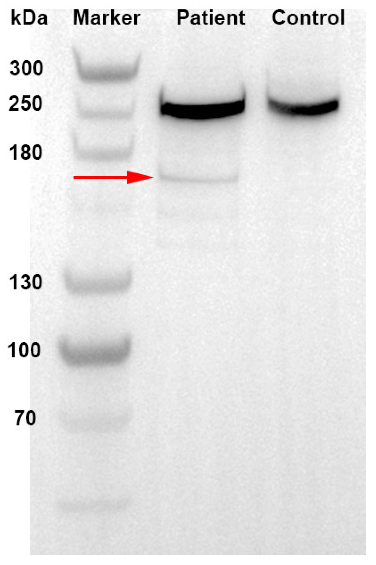
Spectrins are the major constituent of the erythrocyte cytoskeletal network. Hereditary spherocytosis can be caused by α-spectrin (SPTA1 gene) or β-spectrin (SPTB gene) mutations [5]. In healthy erythroid cells, the production of α-spectrin chains is three-fold to four-fold greater than β-spectrin production. Thus, a mutation of one β-spectrin allele is sufficient to cause spherocytosis, whereas both α-spectrin alleles have to be affected for the disease to arise [6]. The SPTB gene codes β-spectrin, along with α-spectrin, to form spectrin heterodimers and finally to form tetramers (α2β2) by a head-to-head connection [7]. The interaction of α2β2 tetramers with other proteins (such as ankyrin, band 3, etc.) forms the biconcave shape of human erythrocytes, which is critical for erythrocyte deformability and stability. The mutation in exon 19 (c.3936G > A) of the SPTB gene was found and submitted to the ClinVar database by the researchers from the UOS Fisiopatologia delle Anemie and the Mayo Clinic in 2019, and was published firstly by Fermo E et al. in 2021 [8]. It is a nonsense mutation, which caused tryptophan at position 1312 of β-spectrin chain to change to a termination codon and led to a truncated spectrin-β comprising 1311 amino acids instead of 2328 amino acids in the wild-type counterpart (shown in Figure 3). The c.3936G > A mutation of the patient is located in the SPTB anchoring area. The nonsense mutation in the SPTB gene may lead to the loss of the ankyrin-binding domain and tetramerization domain of β-spectrin, resulting in the loss of erythrocyte membrane cohesion and lipid vesicle formation. These are responsible for the loss of the cell surface area, increased cell sphericity, and reduced cellular deformability. Abnormal spherocytes are trapped and destroyed in the spleen, resulting in hemolysis, anemia, and jaundice.
The SPTB gene mutations were classified into autosomal dominant (AD) inheritance. However, with rare exceptions, mutations of the β-spectrin gene are isolated and might be associated with monoallelic expression, suggesting that null mutations are common [9]. In addition, in our case, the mutation c.3936G > A on exon 19 of SPTB led to a termination codon resulting in a truncated β-spectrin, and was only identified in the proband, and not found in any of his family members. That is a de novo monoallelic pathogenic mutation. To date, several de novo SPTB mutations have been reported [10,11]. Tole S et al. confirmed nine cases (17%) with de novo monoallelic mutations in 52 patients with SPTB-HS [12]. Peng GX et al. reported five de novo monoallelic variants of SPTB gene in 14 Chinese patients with SPTB-HS [13]. A study from South Korea also identified four de novo SPTB monoallelic mutations in 12 SPTB-HS patients [14]. These results suggest that the de novo SPTB monoallelic mutations are not rare in patients with SPTB-HS.
The ALAS2 gene is located on chromosome X p11.21 and encodes 5-aminolevulinate synthase (ALAS). ALAS is a key rate-controlling enzyme of the heme biosynthetic pathway. In the mitochondria, ALAS catalyzes the formation of 5-aminolevulinate (ALA) from glycine and succinyl CoA. In this reaction, as a cofactor, pyridoxal phosphate (PLP) can enhance the activity of ALAS [15]. Missense mutations in the ALAS2 gene cause ALAS dysfunction, which hinders the synthesis of ALA and then affects the synthesis of heme, further resulting in iron overload and anemia. Due to an X-linked recessive pattern of inheritance, XLSA typically affects younger males. Approximately two-thirds of cases present in males during early childhood or adolescence and a third in young to middle-aged females [16]. However, as a result of familial-skewed inactivation of the normal X chromosome, females with ALAS2 mutations may have a late-onset clinical phenotype [3,17]. In our proband, the ALAS2 gene revealed a missense mutation at c.37A > G of exon 2. The mutation resulted in a change from lysine to glutamate at position 13 of the ALAS2 protein. His mother was an asymptomatic heterozygous carrier.
The human ALAS2 gene consists of 10 coding exons and a 5′untranslated exon. For the first translated exon (exon 2), the ALAS2 gene primarily encodes the signal sequence required for targeting the precursor protein to the mitochondrion. The sequence encoded by exons 3 and 4, together with the remainder of the exon 2-encoded sequence that is predicted to not be part of the proteolytically removed mitochondrial targeting sequence, constitutes a 93 amino acid stretch at the N-terminus of the ALAS protein. Exons 5–11 encode the catalytic domain, and most of the identified ALAS2 point mutations associated with XLSA were located in these exons. Missense mutations of exons 5–11 on ALAS2 can result in a decreased affinity of PLP with ALAS or can change the stability of the enzyme, so the administration of pyridoxine can restore the enzymatic activity and alleviate anemia [18]. Shoolingin-Jordan et al. found that mutations located in the vicinity of the pyridoxal 5′-phosphate binding site, such as M426V, G291S, T388S and K299Q, exhibit a nearly complete response to pyridoxine therapy [15]. However, XLSA patients with mutations in the promoter region and enhancer region of the first intron, or the creation of premature stop codons were refractory to pyridoxin [19]. In our case, the missense mutation of ALAS2 was detected in exon 2. This mutation may have little influence on the binding between pyridoxine and ALAS, and on the activity of the catalytic domain, so the therapeutic effect of vitamin B6 is not obvious.
In general, regarding the clinical phenotype, patients with β-spectrin deficiency typically have mild to moderate disease manifestation and do not need transfusion. Splenectomy can improve symptoms in most patients, including the alleviation of anemia and hyperbilirubinemia and reduction in the reticulocyte count. XLSA usually presents as asymptomatic or mild microcytic anemia. This proband had more severe anemia requiring erythrocyte transfusion due to the complex hereditary spherocytosis and X-linked sideroblastic anemia with two mutations in the SPTB and ALAS2 genes. After treatment with folic acid and vitamin B6, the anemia symptoms of the patient had little improvement, because the mutation of ALAS2 was not located in exons 5–11 encoding the catalytic domain and not in the vicinity of the pyridoxal 5′-phosphate binding site.
In summary, we reported a severe anemia case caused by the complex hereditary spherocytosis and X-linked sideroblastic anemia with two mutations in the SPTB and ALAS2 genes, a de novo nonsense mutation in the SPTB gene (c.3936G > A:p.W1312X) and a missense mutation in the ALAS2 gene (c.37A > G:p.K13E). The patient did not respond to vitamin B6 treatment, possibly because the mutation was not located in the binding site of the encoding catalytic domain of ALAS. The optimal treatment for this patient needs to be pursued.

Author Contributions

J.Z. and T.L. made the diagnosis and treated this patient. J.Z., H.Z. and Y.Q. collected the clinical data. J.Z. and T.L. analyzed the data and wrote the paper. All authors have read and agreed to the published version of the manuscript.

Funding

This research received no external funding.

Institutional Review Board Statement

Not applicable.

Informed Consent Statement

Written informed consent was obtained from the patient and his parents for publication of this case report and any accompanying images.

Data Availability Statement

The data used to support the findings of this case report are included within the article.

Acknowledgments

The authors are grateful to the patient and his family members.

Conflicts of Interest

The authors declare no conflict of interest.

Abbreviations

ADautosomal dominant
ALAS5-aminolevulinic acid synthase
SPTAspectrin, alpha
HShereditary spherocytosis
NGSNext-generation sequencing
PLPpyridoxal phosphate
RBCsred blood cells
SPTBspectrin, beta
sTfRserum transferring receptor
XLSAX-linked sideroblastic anemia

References

  1. Costa, L.D.; Galimand, J.; Fenneteau, O.; Mohandas, N. Hereditary spherocytosis, elliptocytosis, and other red cell membrane disorders. Blood Rev. 2013, 27, 167–178. [Google Scholar] [CrossRef] [PubMed]
  2. Ducamp, S.; Fleming, M.D. The molecular genetics of sideroblastic anemia. Blood 2019, 133, 59–69. [Google Scholar] [CrossRef] [PubMed] [Green Version]
  3. Cazzola, M.; May, A.; Bergamaschi, G.; Cerani, P.; Rosti, V.; Bishop, D.F. Familial-skewed X-chromosome inactivation as a predisposing factor for late-onset X-linked sideroblastic anemia in carrier females. Blood 2000, 96, 4363–4365. [Google Scholar] [CrossRef] [PubMed]
  4. May, A.; Bishop, D.F. The molecular biology and pyridoxine responsiveness of X-linked sideroblastic anaemia. Haematologica 1998, 83, 56–70. [Google Scholar] [PubMed]
  5. Shohet, S.B. Spectrin and spherocytosis. N. Engl. J. Med. 1982, 306, 1170–1171. [Google Scholar] [CrossRef] [PubMed]
  6. Perrotta, S.; Gallagher, P.G.; Mohandas, N. Hereditary spherocytosis. Lancet 2008, 372, 1411–1426. [Google Scholar] [CrossRef] [PubMed]
  7. Bennett, V.; Healy, J. Membrane domains based on ankyrin and spectrin associated with cell-cell interactions. Cold Spring Harb. Perspect. Biol. 2009, 1, a003012. [Google Scholar] [CrossRef] [PubMed] [Green Version]
  8. Fermo, E.; Vercellati, C.; Marcello, A.P.; Keskin, E.Y.; Perrotta, S.; Zaninoni, A.; Brancaleoni, V.; Zanella, A.; Giannotta, J.A.; Barcellini, W.; et al. Targeted Next Generation Sequencing and Diagnosis of Congenital Hemolytic Anemias: A Three Years Experience Monocentric Study. Front. Physiol. 2021, 12, 684569. [Google Scholar] [CrossRef] [PubMed]
  9. Hassoun, H.; Vassiliadis, J.N.; Murray, J.; Njolstad, P.R.; Rogus, J.J.; Ballas, S.K.; Schaffer, F.; Jarolim, P.; Brabec, V.; Palek, J. Characterization of the underlying molecular defect in hereditary spherocytosis associated with spectrin deficiency. Blood 1997, 90, 398–406. [Google Scholar] [PubMed]
  10. Miraglia del Giudice, E.; Lombardi, C.; Francese, M.; Nobili, B.; Conte, M.L.; Amendola, G. Frequent de novo monoallelic expression of beta-spectrin gene (SPTB) in children with hereditary spherocytosis and isolated spectrin deficiency. Br. J. Haematol. 1998, 101, 251–254. [Google Scholar] [CrossRef] [PubMed]
  11. Zhang, Y.M.; Shao, S.M.; Liu, J.; Zeng, C.M.; Han, Y.; Zhang, X.R. Neonatal hereditary spherocytosis caused by a de novo frameshift mutation of the SPTB gene characterized by hydrops fetalis. Medicine 2021, 100, e24804. [Google Scholar] [CrossRef] [PubMed]
  12. Tole, S.; Dhir, P.; Pugi, J.; Drury, L.J.; Butchart, S.; Fantauzzi, M.; Langer, J.C.; Baker, J.M.; Blanchette, V.S.; Kirby-Allen, M.; et al. Genotype–phenotype correlation in children with hereditary spherocytosis. Br. J. Haematol. 2020, 191, 486–496. [Google Scholar] [CrossRef] [PubMed]
  13. Peng, G.X.; Yang, W.R.; Zhao, X.; Jin, L.P.; Zhang, L.; Zhou, K.; Li, Y.; Ye, L.; Li, J.P.; Fan, H.H.; et al. The characteristic of hereditary spherocytosis related gene mutation in 37 Chinese hereditary spherocytisis patients. Zhonghua Xue Ye Xue Za Zhi 2018, 39, 898–903. [Google Scholar] [PubMed]
  14. Park, J.; Jeong, D.C.; Yoo, J.; Jang, W.; Chae, H.; Kim, J.; Kwon, A.; Choi, H.; Lee, J.; Chung, N.-G.; et al. Mutational characteristics of ANK1 and SPTB genes in hereditary spherocytosis. Clin. Genet. 2016, 90, 69–78. [Google Scholar] [CrossRef] [PubMed]
  15. Shoolingin-Jordan, P.M.; Al-Daihan, S.; Alexeev, D.; Baxter, R.L.; Bottomley, S.S.; Kahari, I.D.; Roy, I.; Sarwar, M.; Sawyer, L.; Wang, S.-F. 5-Aminolevulinic acid synthase: Mechanism, mutations and medicine. Biochim. Biophys. Acta (BBA)—Proteins Proteom. 2003, 1647, 361–366. [Google Scholar] [CrossRef]
  16. Ducamp, S.; Kannengiesser, C.; Touati, M.; Garcon, L.; Guerci-Bresler, A.; Guichard, J.F.-o.; Vermylen, C.; Dochir, J.; Poirel, H.A.; Fouyssac, F.; et al. Sideroblastic anemia: Molecular analysis of the ALAS2 gene in a series of 29 probands and functional studies of 10 missense mutations. Hum. Mutat. 2011, 32, 590–597. [Google Scholar] [CrossRef] [PubMed] [Green Version]
  17. Aivado, M.; Gattermann, N.; Rong, A.; Giagounidis, A.A.; Prall, W.C.; Czibere, A.; Hildebrandt, B.; Haas, R.; Bottomley, S.S. X-linked sideroblastic anemia associated with a novel ALAS2 mutation and unfortunate skewed X-chromosome inactivation patterns. Blood Cells Mol. Dis. 2006, 37, 40–45. [Google Scholar] [CrossRef] [PubMed]
  18. Cox, T.C.; Sadlon, T.J.; Schwarz, Q.P.; Matthews, C.S.; Wise, P.D.; Cox, L.L.; Bottomley, S.S.; May, B.K. The major splice variant of human 5-aminolevulinate synthase-2 contributes significantly to erythroid heme biosynthesis. Int. J. Biochem. Cell. Biol. 2004, 36, 281–295. [Google Scholar] [CrossRef] [PubMed]
  19. Harigae, H.; Furuyama, K. Hereditary sideroblastic anemia: Pathophysiology and gene mutations. Int. J. Hematol. 2010, 92, 425–431. [Google Scholar] [CrossRef] [PubMed]
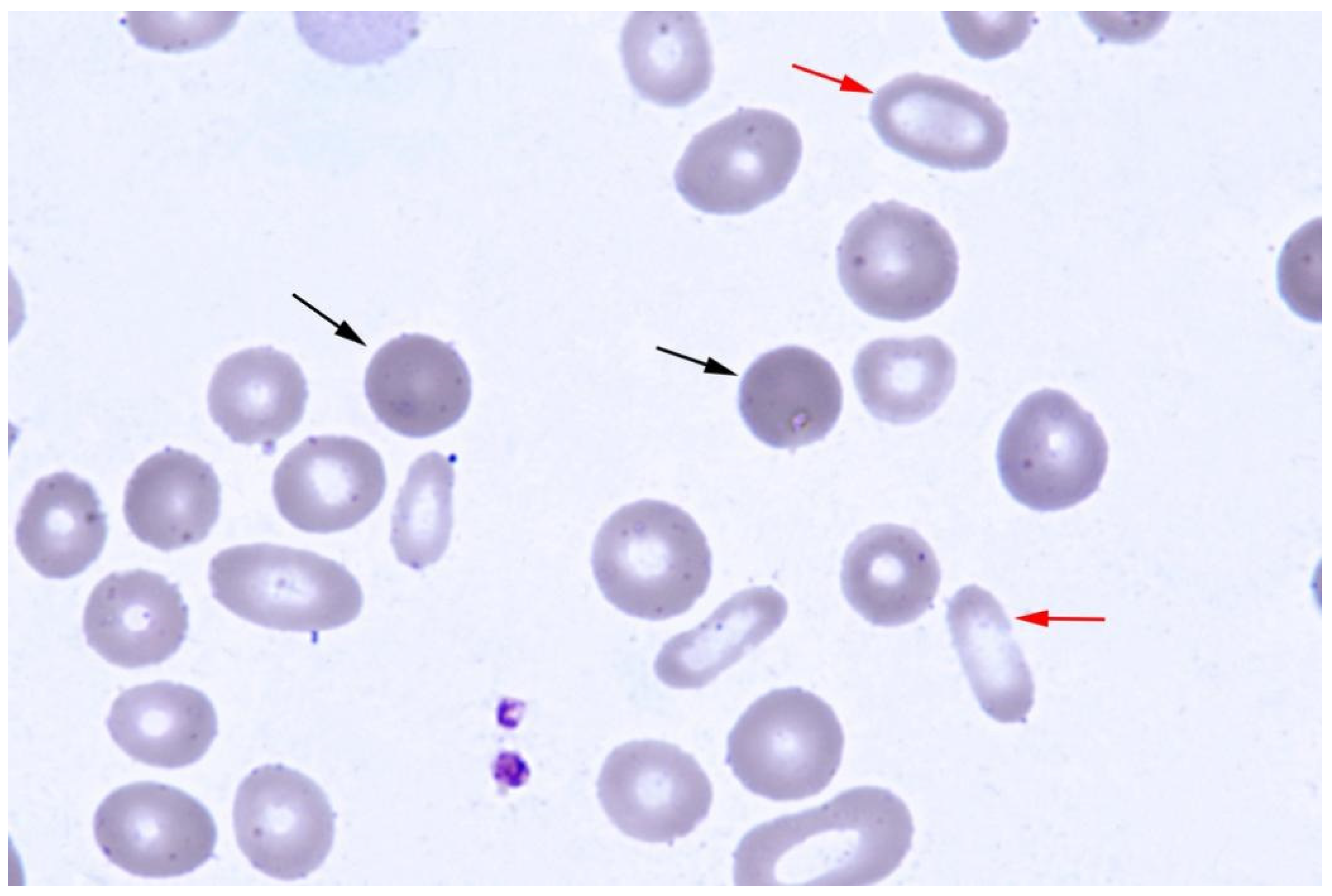
Figure 1. Peripheral blood smear shows anisocytosis, and spherocytes (black arrow) and elliptocytes (red arrow) could be seen easily (Wright’s stain).
Jcm 12 01990 g001
Figure 2. Schematic of the inheritance pattern in the family and the results of NGS. (A) Pedigree of the family with hereditary spherocytosis (HS) and X-linked sideroblastic anemia (XLSA). (B)The patient harbors a heterozygous mutation c.3936G > A in the SPTB gene and a hemizygous mutation c.37A > G in the ALAS2 gene identified by NGS. The mutation of ALAS2 is inherited from his asymptomatic heterozygous mother, and the mutation of SPTB may be a de novo monoallelic variant. (C) ALAS2 mutation results in amino acid switching (p.K13E), and SPTB mutation led to the formation of truncated β-spectrin (p.W1312X).
Jcm 12 01990 g002
Figure 3. Western blot-based detection of β-spectrin: red arrow showed the truncated β-spectrin.
Jcm 12 01990 g003
Disclaimer/Publisher’s Note: The statements, opinions and data contained in all publications are solely those of the individual author(s) and contributor(s) and not of MDPI and/or the editor(s). MDPI and/or the editor(s) disclaim responsibility for any injury to people or property resulting from any ideas, methods, instructions or products referred to in the content.