
A Cone Beam CT Study on the Correlation between Crestal Bone Loss and Periapical Disease

 , , ,
, , ,  and
and
Abstract
1. Introduction
2. Materials and Methods
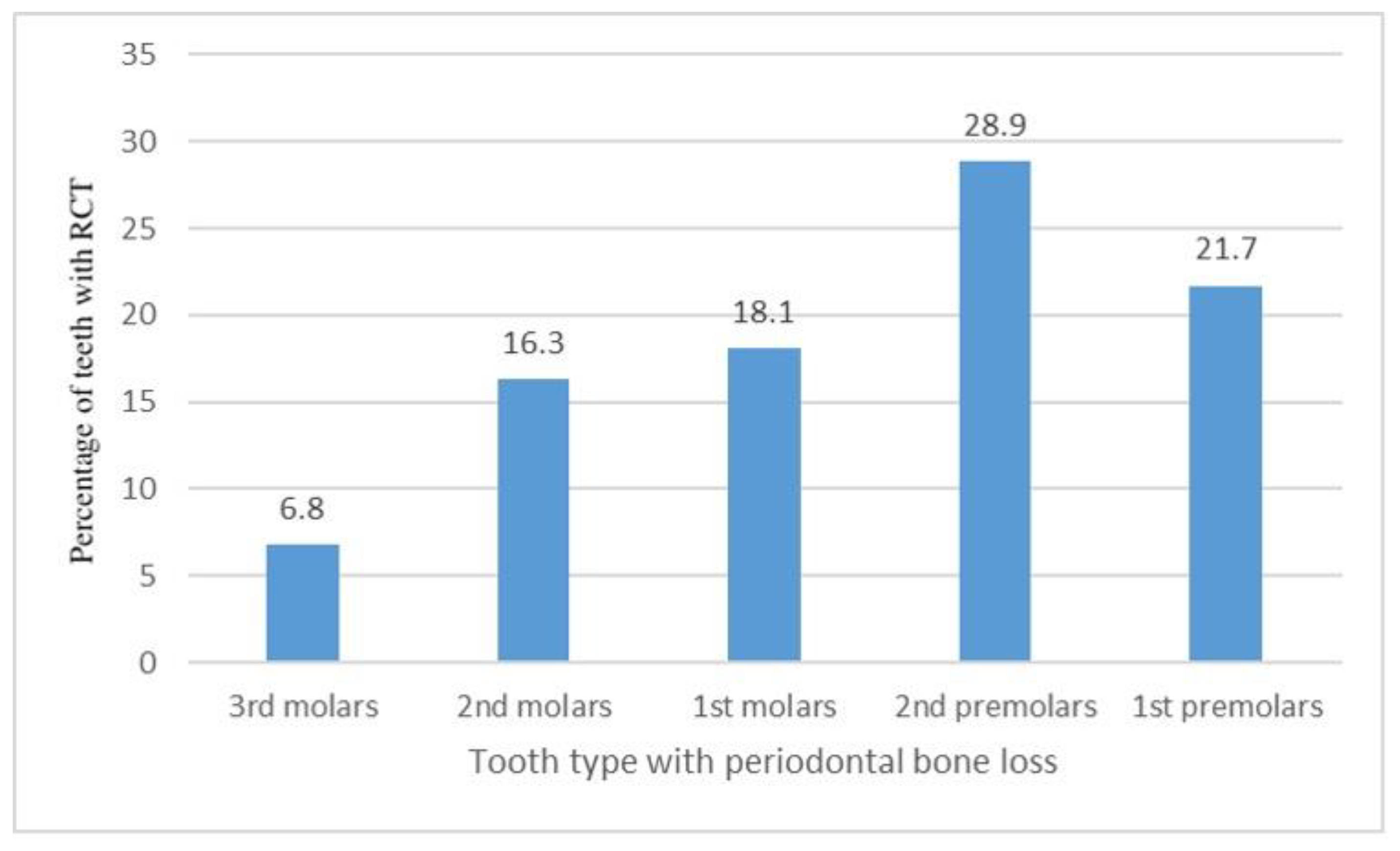
3. Results
4. Discussion
5. Conclusions
Author Contributions
Funding
Institutional Review Board Statement
Informed Consent Statement
Data Availability Statement
Conflicts of Interest
References
- Simring, M.; Goldberg, M. The pulpal pocket approach: Retrograde periodontitis. J. Periodontol. 1964, 35, 22–48. [Google Scholar] [CrossRef]
- Rotstein, I.; Simon, J.H. Diagnosis, prognosis and decision-making in the treatment of combined periodontal-endodontic lesions. Periodontol. 2000 2004, 34, 165–203. [Google Scholar] [CrossRef] [PubMed]
- Ray, H.; Trope, M. Periapical status of endodontically treated teeth in relation to the technical quality of the root filling and the coronal restoration. Int. Endod. J. 1995, 28, 12–18. [Google Scholar] [CrossRef] [PubMed]
- Tronstad, L.; Asbjørnsen, K.; Døving, L.; Pedersen, I.; Eriksen, H. Influence of coronal restorations on the periapical health of endodontically treated teeth. Dent. Traumatol. 2000, 16, 218–221. [Google Scholar] [CrossRef] [PubMed]
- De Deus, Q.D. Frequency, location, and direction of the lateral, secondary, and accessory canals. J. Endod. 1975, 1, 361–366. [Google Scholar] [CrossRef]
- Kim, S. Color Atlas of Microsurgery in Endodontics; WB Saunders Company: Philadelphia, PA, USA, 2001. [Google Scholar]
- Simon, J.H.; Glick, D.H.; Frank, A.L. The relationship of endodontic-periodontic lesions. J. Periodontol. 1972, 43, 202–208. [Google Scholar] [CrossRef]
- Papapanou, P.N.; Sanz, M.; Buduneli, N.; Dietrich, T.; Feres, M.; Fine, D.H.; Flemmig, T.F.; Garcia, R.; Giannobile, W.V.; Graziani, F. Periodontitis: Consensus report of workgroup 2 of the 2017 World Workshop on the Classification of Periodontal and Peri-Implant Diseases and Conditions. J. Periodontol. 2018, 89, S173–S182. [Google Scholar] [CrossRef]
- Nardi, C.; Calistri, L.; Pradella, S.; Desideri, I.; Lorini, C.; Colagrande, S. Accuracy of orthopantomography for apical periodontitis without endodontic treatment. J. Endod. 2017, 43, 1640–1646. [Google Scholar] [CrossRef]
- Izzetti, R.; Nisi, M.; Aringhieri, G.; Crocetti, L.; Graziani, F.; Nardi, C. Basic knowledge and new advances in panoramic radiography imaging techniques: A narrative review on what dentists and radiologists should know. Appl. Sci. 2021, 11, 7858. [Google Scholar] [CrossRef]
- Bender, I.; Seltzer, S. Roentgenographic and direct observation of experimental lesions in bone: I. J. Endod. 2003, 29, 702–706. [Google Scholar] [CrossRef]
- Dutra, K.L.; Haas, L.; Porporatti, A.L.; Flores-Mir, C.; Santos, J.N.; Mezzomo, L.A.; Correa, M.; Canto, G.D.L. Diagnostic accuracy of cone-beam computed tomography and conventional radiography on apical periodontitis: A systematic review and meta-analysis. J. Endod. 2016, 42, 356–364. [Google Scholar] [CrossRef] [PubMed]
- Abdinian, M.; Yaghini, J.; Jazi, L. Comparison of intraoral digital radiography and cone-beam computed tomography in the measurement of periodontal bone defects. Dent. Med. Probl. 2020, 57, 269–273. [Google Scholar] [CrossRef] [PubMed]
- Woelber, J.P.; Fleiner, J.; Rau, J.; Ratka-Krüger, P.; Hannig, C. Accuracy and Usefulness of CBCT in Periodontology: A Systematic Review of the Literature. Int. J. Periodontics Restor. Dent. 2018, 38, 289–297. [Google Scholar] [CrossRef] [PubMed]
- Nardi, C.; Molteni, R.; Lorini, C.; Taliani, G.G.; Matteuzzi, B.; Mazzoni, E.; Colagrande, S. Motion artefacts in cone beam CT: An in vitro study about the effects on the images. Br. J. Radiol. 2016, 89, 20150687. [Google Scholar] [CrossRef] [PubMed]
- Mol, A.; Balasundaram, A. In vitro cone beam computed tomography imaging of periodontal bone. Dentomaxillofac. Radiol. 2008, 37, 319–324. [Google Scholar] [CrossRef]
- Nardi, C.; Calistri, L.; Grazzini, G.; Desideri, I.; Lorini, C.; Occhipinti, M.; Mungai, F.; Colagrande, S. Is panoramic radiography an accurate imaging technique for the detection of endodontically treated asymptomatic apical periodontitis? J. Endod. 2018, 44, 1500–1508. [Google Scholar] [CrossRef]
- Sullivan, M.; Gallagher, G.; Noonan, V. The root of the problem: Occurrence of typical and atypical periapical pathoses. J. Am. Dent. Assoc. 2016, 147, 646–649. [Google Scholar] [CrossRef]
- Kontogiannis, T.; Tosios, K.; Kerezoudis, N.; Krithinakis, S.; Christopoulos, P.; Sklavounou, A. Periapical lesions are not always a sequelae of pulpal necrosis: A retrospective study of 1521 biopsies. Int. Endod. J. 2015, 48, 68–73. [Google Scholar] [CrossRef]
- Stodle, I.H.; Verket, A.; Hovik, H.; Sen, A.; Koldsland, O.C. Prevalence of periodontitis based on the 2017 classification in a Norwegian population: The HUNT study. J. Clin. Periodontol. 2021, 48, 1189–1199. [Google Scholar] [CrossRef]
- Catunda, R.Q.; Levin, L.; Kornerup, I.; Gibson, M.P. Prevalence of Periodontitis in Young Populations: A Systematic Review. Oral Health Prev. Dent. 2019, 17, 195–202. [Google Scholar] [CrossRef]
- Eke, P.I.; Dye, B.A.; Wei, L.; Thornton-Evans, G.O.; Genco, R.J.; Cdc Periodontal Disease Surveillance workgroup: James Beck, G.D.R.P. Prevalence of periodontitis in adults in the United States: 2009 and 2010. J. Dent. Res. 2012, 91, 914–920. [Google Scholar] [CrossRef] [PubMed]
- Abbott, P.V.; Salgado, J.C. Strategies for the endodontic management of concurrent endodontic and periodontal diseases. Aust. Dent. J. 2009, 54 (Suppl. S1), S70–S85. [Google Scholar] [CrossRef] [PubMed]
- Torabinejad, M.; Rotstein, I. Endodontic-periodontic interrelationship. In Endodontics: Principles and Practice; Torabinejad, M., Walton, R.E., Fouad, A.F., Eds.; Saunders: St. Louis, MO, USA, 2014; pp. 106–120. [Google Scholar]
- Neuvald, L.; Consolaro, A. Cementoenamel junction: Microscopic analysis and external cervical resorption. J. Endod. 2000, 26, 503–508. [Google Scholar] [CrossRef]
- Roa, I.; del Sol, M.; Cuevas, J. Morphology of the Cement-Enamel Junction (CEJ), Clinical Correlations. Int. J. Morphol. 2013, 31. [Google Scholar] [CrossRef]
- Zhao, H.; Wang, H.; Pan, Y.; Pan, C.; Jin, X. The relationship between root concavities in first premolars and chronic periodontitis. J. Periodontal Res. 2014, 49, 213–219. [Google Scholar] [CrossRef] [PubMed]
- Kartal, N.; Ozcelik, B.; Cimilli, H. Root canal morphology of maxillary premolars. J. Endod. 1998, 24, 417–419. [Google Scholar] [CrossRef]
- Gutmann, J.L. Prevalence, location, and patency of accessory canals in the furcation region of permanent molars. J. Periodontol. 1978, 49, 21–26. [Google Scholar] [CrossRef]
- Nair, P.R. Pathogenesis of apical periodontitis and the causes of endodontic failures. Crit. Rev. Oral Biol. Med. 2004, 15, 348–381. [Google Scholar] [CrossRef]
- Ng, Y.L.; Mann, V.; Gulabivala, K. Outcome of secondary root canal treatment: A systematic review of the literature. Int. Endod. J. 2008, 41, 1026–1046. [Google Scholar] [CrossRef]
- Bürklein, S.; Schäfer, E.; Jöhren, H.-P.; Donnermeyer, D. Quality of root canal fillings and prevalence of apical radiolucencies in a German population: A CBCT analysis. Clin. Oral Investig. 2020, 24, 1217–1227. [Google Scholar] [CrossRef]
- Köse, T.; Günaçar, D.; Çene, E.; Arıcıoğlu, B. Evaluation of endodontically treated teeth and related apical periodontitis using periapical and endodontic status scale: Retrospective cone-beam computed tomography study. Aust. Endod. J. 2022, 48, 431–443. [Google Scholar]
- Rodriguez, F.-R.; Paganoni, N.; Eickholz, P.; Weiger, R.; Walter, C. Presence of root canal treatment has no influence on periodontal bone loss. Clin. Oral Investig. 2017, 21, 2741–2748. [Google Scholar] [CrossRef] [PubMed]
- Ng, Y.L.; Mann, V.; Gulabivala, K. A prospective study of the factors affecting outcomes of nonsurgical root canal treatment: Part 1: Periapical health. Int. Endod. J. 2011, 44, 583–609. [Google Scholar] [CrossRef] [PubMed]
- Mandelaris, G.A.; Scheyer, E.T.; Evans, M.; Kim, D.; McAllister, B.; Nevins, M.L.; Rios, H.F.; Sarment, D. American Academy of Periodontology best evidence consensus statement on selected oral applications for cone-beam computed tomography. J. Periodontol. 2017, 88, 939–945. [Google Scholar] [CrossRef]
- Radiology MSC-oAfO-EIiC, Safety MSCoOR. Implementation of the Principle of as Low as Reasonably Achievable (ALARA) for Medical and Dental Personnel: Recommendations of the National Council on Radiation Protection and Measurements: National Council on Radiation; 1990. Available online: https://ncrponline.org/shop/reports/report-no-107-implementation-of-the-principle-of-as-low-as-reasonably-achievable-alara-for-medical-and-dental-personnel-1990/ (accessed on 19 February 2023).
- Eshraghi, V.T.; Malloy, K.A.; Tahmasbi, M. Role of cone-beam computed tomography in the management of periodontal disease. Dent. J. 2019, 7, 57. [Google Scholar] [CrossRef]
- Assiri, H.; Dawasaz, A.A.; Alahmari, A.; Asiri, Z. Cone beam computed tomography (CBCT) in periodontal diseases: A systematic review based on the efficacy model. BMC Oral Health 2020, 20, 191. [Google Scholar] [CrossRef]


| Endo-Periodontal with Root Damage | Endo-Periodontal without Root Damage | |||||||
|---|---|---|---|---|---|---|---|---|
| Root fracture | Perforations | External root resorption | Periodontitis Patient | Non-Periodontitis Patient | ||||
| Grade 1: narrow, deep periodontal pocket in one tooth surface | Grade 2: wide, deep periodontal pocket in one tooth surface | Grade 3: narrow, deep periodontal pocket in more than one tooth surface | Grade 1: narrow, deep periodontal pocket in one tooth surface | Grade 2: wide, deep periodontal pocket in one tooth surface | Grade 3: narrow, deep periodontal pocket in more than one tooth surface | |||
Disclaimer/Publisher’s Note: The statements, opinions and data contained in all publications are solely those of the individual author(s) and contributor(s) and not of MDPI and/or the editor(s). MDPI and/or the editor(s) disclaim responsibility for any injury to people or property resulting from any ideas, methods, instructions or products referred to in the content. |
© 2023 by the authors. Licensee MDPI, Basel, Switzerland. This article is an open access article distributed under the terms and conditions of the Creative Commons Attribution (CC BY) license (https://creativecommons.org/licenses/by/4.0/).
Share and Cite
Mahasneh, S.A.; Al-Hadidi, A.; Kadim Wahab, F.; Sawair, F.A.; AL-Rabab’ah, M.A.; Al-Nazer, S.; Bakain, Y.; Nardi, C.; Cunliffe, J. A Cone Beam CT Study on the Correlation between Crestal Bone Loss and Periapical Disease. J. Clin. Med. 2023, 12, 2423. https://doi.org/10.3390/jcm12062423
Mahasneh SA, Al-Hadidi A, Kadim Wahab F, Sawair FA, AL-Rabab’ah MA, Al-Nazer S, Bakain Y, Nardi C, Cunliffe J. A Cone Beam CT Study on the Correlation between Crestal Bone Loss and Periapical Disease. Journal of Clinical Medicine. 2023; 12(6):2423. https://doi.org/10.3390/jcm12062423
Chicago/Turabian StyleMahasneh, Sari A., Abeer Al-Hadidi, Fouad Kadim Wahab, Faleh A. Sawair, Mohammad Abdalla AL-Rabab’ah, Sarah Al-Nazer, Yara Bakain, Cosimo Nardi, and Joanne Cunliffe. 2023. "A Cone Beam CT Study on the Correlation between Crestal Bone Loss and Periapical Disease" Journal of Clinical Medicine 12, no. 6: 2423. https://doi.org/10.3390/jcm12062423
APA StyleMahasneh, S. A., Al-Hadidi, A., Kadim Wahab, F., Sawair, F. A., AL-Rabab’ah, M. A., Al-Nazer, S., Bakain, Y., Nardi, C., & Cunliffe, J. (2023). A Cone Beam CT Study on the Correlation between Crestal Bone Loss and Periapical Disease. Journal of Clinical Medicine, 12(6), 2423. https://doi.org/10.3390/jcm12062423