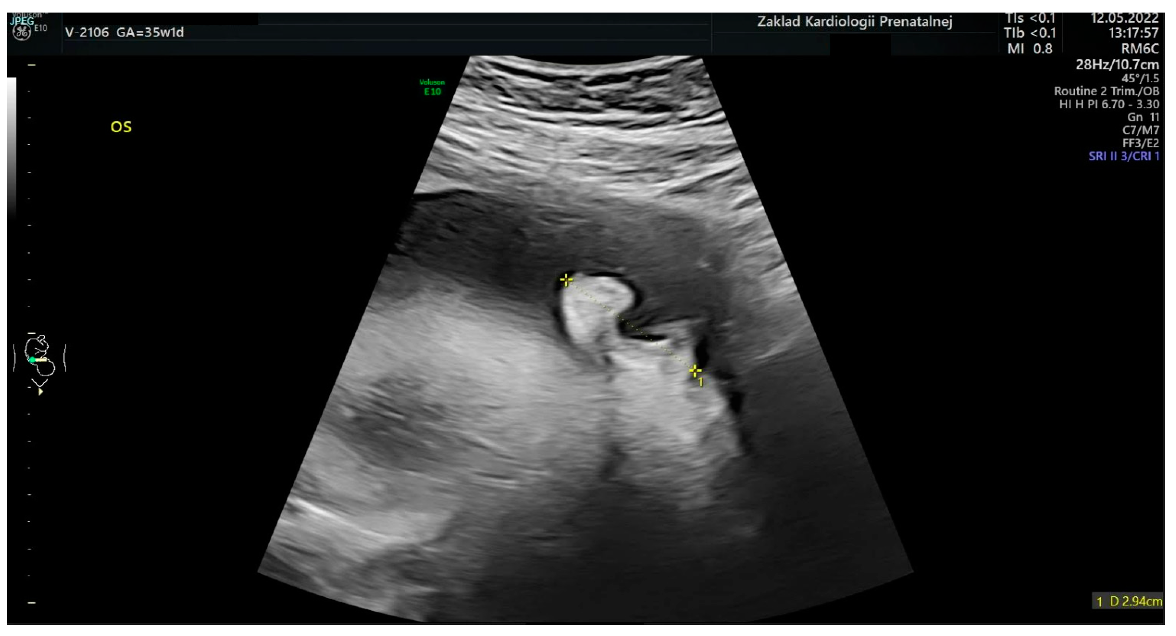
Measurement of the Fetal Ear Length Has No Clinical Value
Abstract
:1. Introduction
2. Materials and Methods
2.1. Exclusions
2.2. Fetal Ultrasound Examinations
2.3. Statistical Analysis
3. Results
4. Discussion
5. Conclusions
Author Contributions
Funding
Institutional Review Board Statement
Informed Consent Statement
Data Availability Statement
Conflicts of Interest
References
- Rosa, R.C.M.; Rosa, R.F.M.; Zen, P.R.G.; Paskulin, G.A. Cardiopatias congênitas e malformações extracardíacas. Rev. Paul. Pediatr. 2013, 31, 243–251. [Google Scholar] [CrossRef] [PubMed]
- Agathokleous, M.; Chaveeva, P.; Poon, L.C.; Kosinski, P.; Nicolaides, K. Meta-analysis of second-trimester markers for trisomy. Ultrasound Obstet. Gynecol. 2013, 41, 247–261. [Google Scholar] [CrossRef]
- Chitkara, U.; Lee, L.; Oehlert, J.W.; Bloch, D.A.; Holbrook, R.H.; El-Sayed, Y.Y.; Druzin, M.L. Fetal ear length measurement: A useful predictor of aneuploidy? Ultrasound Obstet. Gynecol. 2002, 19, 131–135. [Google Scholar] [CrossRef] [PubMed]
- Sadler, T.W. Embriología Médica; Walters Kluwer, Lippincott Williams & Wilkins: Barcelona, España, 2010; pp. 321–327. [Google Scholar]
- Hunter, A.G.; Yotsuyanagi, T. The external ear: More attention to detail may aid syndrome diagnosis and contribute answers to embryological questions. Am. J. Med. Genet. Part A 2005, 135, 237–250. [Google Scholar] [CrossRef]
- Idigo, F.; Ajibo, K.; Anakwue, A.-M.; Nwogu, U.; Robinson, E. Sonographic measurement of ear length among normal fetuses of pregnant Igbo women in port Harcourt, Nigeria. Afr. Health Sci. 2021, 21, 338–348. [Google Scholar] [CrossRef]
- Hatanaka, A.R.; Rolo, L.C.; Mattar, R.; Júnior, E.A.; Nardozza, L.M.M.; Moron, A.F. Reference intervals for fetal ear length between 19 and 24 weeks of pregnancy on 3-dimensional sonography. J. Ultrasound Med. 2011, 30, 1185–1190. [Google Scholar] [CrossRef]
- Chitkara, U.; Lee, L.; El-Sayed, Y.Y.; Holbrook, R.H.J.; Bloch, D.A.; Oehlert, J.W.; Druzin, M.L. Ultrasonographic ear length measurement in normal second- and third-trimester fetuses. Am. J. Obstet. Gynecol. 2000, 183, 230–234. [Google Scholar] [CrossRef]
- Dudarewicz, L.; Kałuzewski, B. Prenatal screening for fetal chromosomal abnormalities using ear length and shape as an ultrasound marker. Experiment 2001, 6, 801–806. [Google Scholar]
- Zieliński, R.; Respondek-Liberska, M. Craniofacial malformations in prenatal ultrasound evaluation. Literature review. Ginekol. Pol. 2013, 84, 801–806. [Google Scholar] [CrossRef]
- Wei, J.; Ran, S.; Yang, Z.; Lin, Y.; Tang, J.; Ran, H. Prenatal Ultrasound Screening for External Ear Abnormality in the Fetuses. BioMed Res. Int. 2014, 2014, 357564. [Google Scholar] [CrossRef]
- Ozdemir, M.E.; Uzun, I.; Karahasanoglu, A.; Aygun, M.; Akin, H.; Yazicioglu, F. Sonographic Measurement of Fetal Ear Length in Turkish Women with a Normal Pregnancy. Balk. Med. J. 2015, 31, 302–306. [Google Scholar] [CrossRef] [PubMed]
- Joshi, K.S.; Chawla, C.D.; Karki, S.; Shrestha, N.C. Sonographic Measurement of Fetal Pinna Length in Normal Pregnancies. Kathmandu Univ. Med. J. 2012, 9, 49–53. [Google Scholar] [CrossRef] [PubMed]
- Lettieri, L.; Rodis, J.F.; Vintzileos, A.M.; Feeney, L.; Ciarleglio, L.; Craffey, A. Ear length in second-trimester aneuploid fetuses. Obstet Gynecol. 1993, 81, 57–60. [Google Scholar] [CrossRef] [PubMed]
- Shimizu, T.; Salvador, L.; Hughes-Benzie, R.; Dawson, L.; Nimrod, C.; Allanson, J. THE ROLE OF REDUCED EAR SIZE IN THE PRENATAL DETECTION OF CHROMOSOMAL ABNORMALITIES. Prenat. Diagn. 1997, 17, 545–549. [Google Scholar] [CrossRef]
- Summers, A. Prenatal diagnosis for paediatricians. Paediatr. Child Health 2003, 8, 25–29. [Google Scholar] [CrossRef] [PubMed]
- Chang, C.-H.C.; Chang, F.-M.; Yu, C.-H.; Liang, R.-I.; Ko, H.-C.; Chen, H.-Y. Fetal ear assessment and prenatal detection of aneuploidy by the quantitative three-dimensional ultrasonography. Ultrasound Med. Biol. 2000, 26, 743–749. [Google Scholar] [CrossRef] [PubMed]
- Ginsberg, N.A.; Cohen, L.; Dungan, J.S.; Concialdi, S.; Mangers, K.; Shulman, L.P. 3-D ultrasound of the fetal ear and fetal autosomal trisomies: A pilot study of a new screening protocol. Prenat. Diagn. 2011, 31, 311–314. [Google Scholar] [CrossRef]
- Sacchini, C.; El-Sheikhah, A.; Cicero, S.; Rembouskos, G.; Nicolaides, K.H. Ear length in trisomy 21 fetuses at 11-14 weeks of gestation. Ultrasound Obstet. Gynecol. 2003, 22, 460–463. [Google Scholar] [CrossRef]
- Gonzalez, C.H.; Sommer, A.; Meisner, L.F.; Elejalde, B.R.; Opitz, J.M.; Francke, U. The trisomy 4p syndrome: Case report and review. Am. J. Med. Genet. 1977, 1, 137–156. [Google Scholar] [CrossRef]
- Liu, X.; Sun, W.; Wang, J.; Chu, G.; He, R.; Zhang, B.; Zhao, Y. Prenatal diagnosis of auriculocondylar syndrome with a novel missense variant of GNAI3: A case report. BMC Pregnancy Childbirth 2021, 21, 780. [Google Scholar] [CrossRef] [PubMed]
- Slavotinek, A. Fryns Syndrome. In GeneReviews® [Internet]; Adam, M.P., Mirzaa, G.M., Pagon, R.A., Wallace, S.E., Bean, L.J.H., Gripp, K.W., Amemiya, A., Eds.; University of Washington: Seattle, WA, USA, 1993. [Google Scholar]
- Beger, O.; Koç, T.; Beger, B.; Özalp, H.; Hamzaoğlu, V.; Vayisoğlu, Y.; Talas, D.; Olgunus, Z.K. Multiple muscular abnormalities in a fetal cadaver with CHARGE syndrome. Surg. Radiol. Anat. 2018, 41, 601–605. [Google Scholar] [CrossRef] [PubMed]
- Traisrisilp, K.; Chankhunaphas, W.; Sittiwangkul, R.; Phokaew, C.; Shotelersuk, V.; Tongsong, T. Prenatal Sonographic Features of CHARGE Syndrome. Diagnostics 2021, 11, 415. [Google Scholar] [CrossRef] [PubMed]
- Gabrielli, L.; Bonasoni, M.P.; Santini, D.; Piccirilli, G.; Chiereghin, A.; Guerra, B.; Landini, M.P.; Capretti, M.G.; Lanari, M.; Lazzarotto, T. Human fetal inner ear involvement in congenital cytomegalovirus infection. Acta Neuropathol. Commun. 2013, 1, 63. [Google Scholar] [CrossRef] [PubMed]
- Witkowski, S.; Żalinska, A.; Słodki, M.; Respondek-Liberska, M. Normograms in prenatal life of stomach and urinary bladder in the second and third trimesters of pregnancy. J. Ultrason. 2022, 22, 161–167. [Google Scholar] [CrossRef] [PubMed]


| Case No. | Fetal Age According to LMP [Weeks] | Fetal Ear Length [mm] | Extracardiac Anomalies and Genetic or Non-Genetic Disorders | Interpretation of Fetal Ear |
|---|---|---|---|---|
| 1 | 36.3 | 35.7 | Mother with hearing deficiency, father with ear agenesis | Around 90th percentile |
| 2 | 33.5 | 30.0 | Multicystic dysplastic kidney | Above 50th percentile |
| 3 | 34.6 | 17.0 | Microcephaly | Around 10th percentile |
| 4 | 36.4 | 22.0 | Hydrocephalus | Below 50th percentile |
| 5 | 28.2 | 25.0 | Craniorachischisis, cleft palate with cleft lip, esophageal atresia | Above 50th percentile |
| 6 | 31.3 | 27.0 | Dandy–Walker syndrome | Above 50th percentile |
| 7 | 33.0 | 21.0 | Down syndrome | Around 10th percentile |
| 8 | 40.0 | 29.0 | Down syndrome | Below 50th percentile |
| 9 | 25.1 | 17.0 | Down syndrome | Below 50th percentile |
| 10 | 26.5 | 20.0 | Down syndrome | Around 50th percentile |
| 11 | 33.6 | 25.0 | Down syndrome | Below 50th percentile |
| 12 | 30.1 | 20.0 | Down syndrome | Around 10th percentile |
| 13 | 24.3 | 16.0 | Down syndrome | Below 50th percentile |
| 14 | 24.4 | 16.7 | Down syndrome | Below 50th percentile |
| 15 | 37.2 | 22.0 | Edwards’ syndrome | Below 10th percentile |
Disclaimer/Publisher’s Note: The statements, opinions and data contained in all publications are solely those of the individual author(s) and contributor(s) and not of MDPI and/or the editor(s). MDPI and/or the editor(s) disclaim responsibility for any injury to people or property resulting from any ideas, methods, instructions or products referred to in the content. |
© 2023 by the authors. Licensee MDPI, Basel, Switzerland. This article is an open access article distributed under the terms and conditions of the Creative Commons Attribution (CC BY) license (https://creativecommons.org/licenses/by/4.0/).
Share and Cite
Witkowski, S.; Respondek-Liberska, M.; Zieliński, R.; Strzelecka, I. Measurement of the Fetal Ear Length Has No Clinical Value. J. Clin. Med. 2023, 12, 3084. https://doi.org/10.3390/jcm12093084
Witkowski S, Respondek-Liberska M, Zieliński R, Strzelecka I. Measurement of the Fetal Ear Length Has No Clinical Value. Journal of Clinical Medicine. 2023; 12(9):3084. https://doi.org/10.3390/jcm12093084
Chicago/Turabian StyleWitkowski, Sławomir, Maria Respondek-Liberska, Rafał Zieliński, and Iwona Strzelecka. 2023. "Measurement of the Fetal Ear Length Has No Clinical Value" Journal of Clinical Medicine 12, no. 9: 3084. https://doi.org/10.3390/jcm12093084
APA StyleWitkowski, S., Respondek-Liberska, M., Zieliński, R., & Strzelecka, I. (2023). Measurement of the Fetal Ear Length Has No Clinical Value. Journal of Clinical Medicine, 12(9), 3084. https://doi.org/10.3390/jcm12093084


