
Diagnostic Challenges in Hemophagocytic Lymphohistiocytosis, a Rare, Potentially Fatal Disease: Two Case Studies
 , and
, and
Abstract
:1. Introduction
2. Literature Review
2.1. Epidemiology and Incidence Trends in HLH
2.2. The Genetic Background of HLH
2.3. Immunological Mechanisms in HLH
2.4. Diagnosis
2.5. Advances in the Treatment of HLH

3. Case Presentations
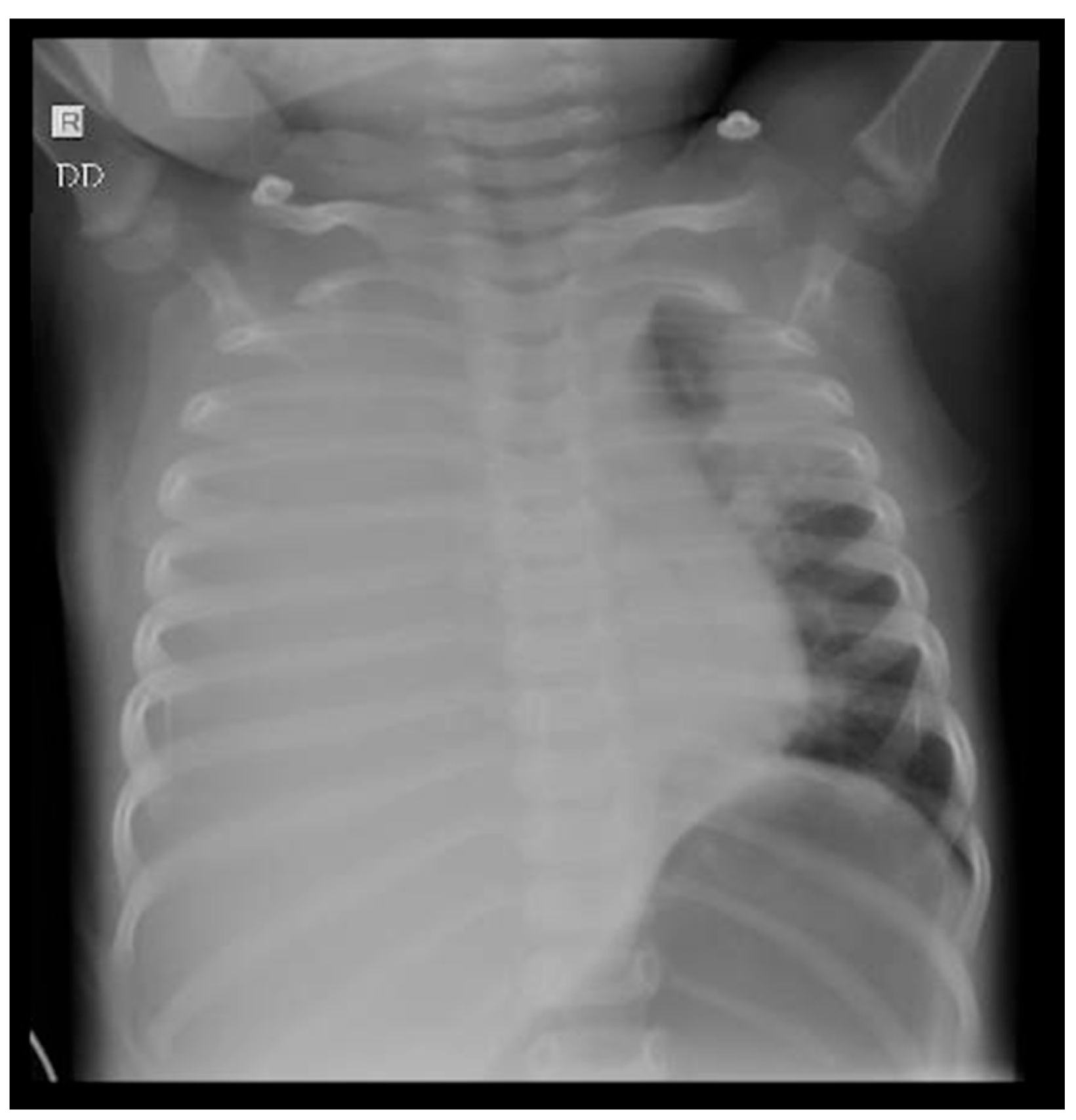
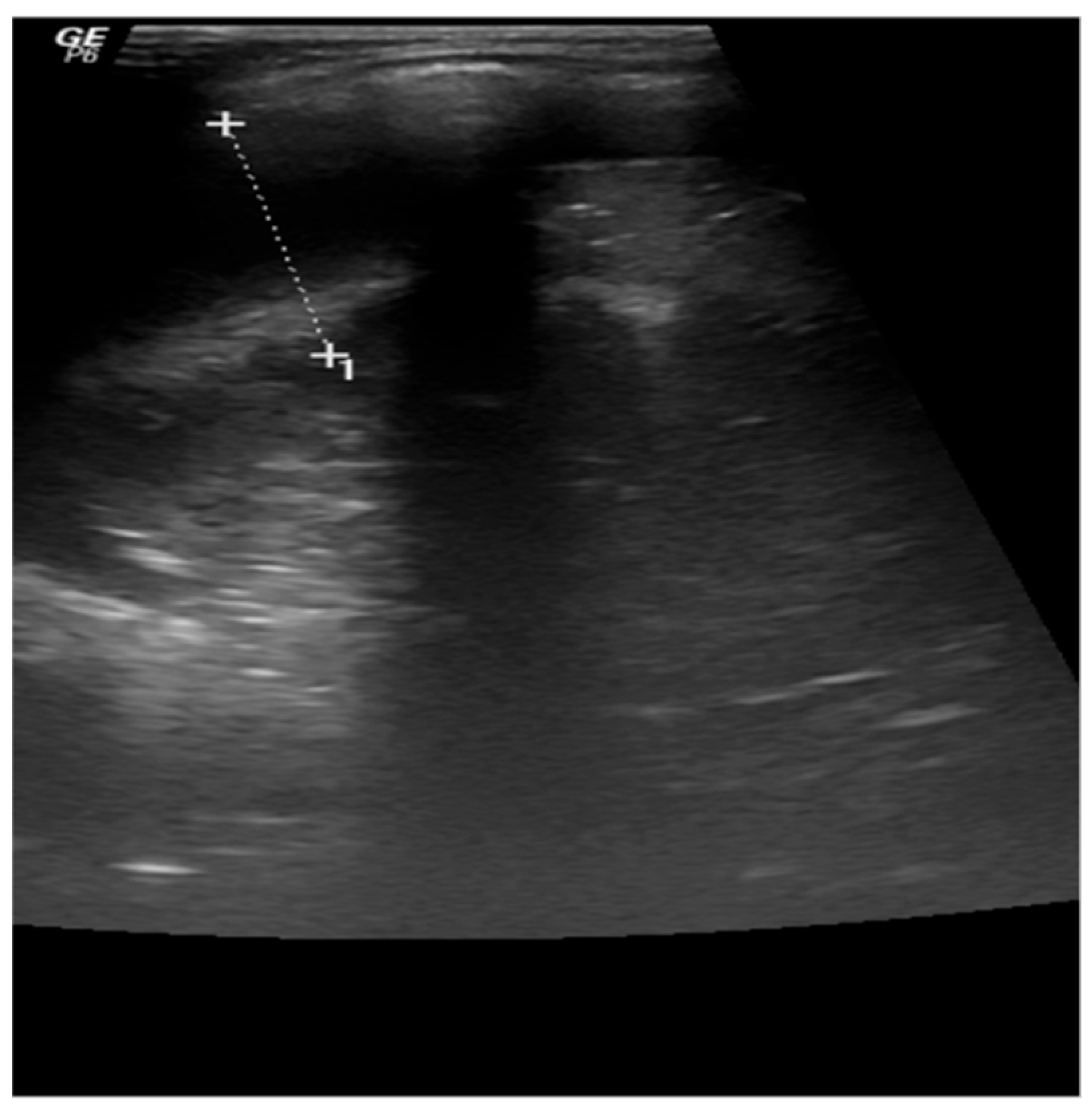
3.1. Case 1
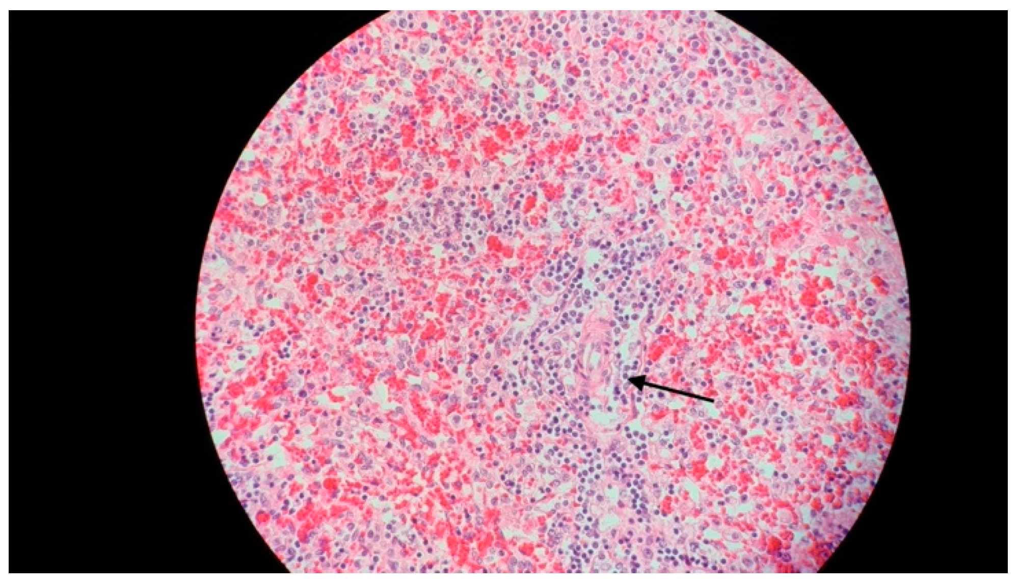
3.2. Case 2
4. Discussion
5. Conclusions
Author Contributions
Funding
Institutional Review Board Statement
Informed Consent Statement
Data Availability Statement
Conflicts of Interest
References
- Tang, Y.; Xu, X.; Song, H.; Yang, S.; Shi, S.; Wei, J.; Pan, B.; Zhao, F.; Liao, C.; Luo, C. Early Diagnostic and Prognostic Significance of a Specific Th1/Th2 Cytokine Pattern in Children with Haemophagocytic Syndrome. Br. J. Haematol. 2008, 143, 84–91. [Google Scholar] [CrossRef]
- Mazodier, K.; Marin, V.; Novick, D.; Farnarier, C.; Robitail, S.; Schleinitz, N.; Veit, V.; Paul, P.; Rubinstein, M.; Dinarello, C.A.; et al. Severe Imbalance of IL-18/IL-18BP in Patients with Secondary Hemophagocytic Syndrome. Blood 2005, 106, 3483–3489. [Google Scholar] [CrossRef]
- Ionescu, M.D.; Taras, R.; Dombici, B.; Balgradean, M.; Berghea, E.C.; Nicolescu, A. The Challenging Diagnosis of Pediatric Multisystem Inflammatory Syndrome Associated with SARS-CoV-2 Infection-Two Case Reports and Literature Review. J. Pers. Med. 2021, 11, 318. [Google Scholar] [CrossRef]
- Jordan, M.B.; Allen, C.E.; Greenberg, J.; Henry, M.; Hermiston, M.L.; Kumar, A.; Hines, M.; Eckstein, O.; Ladisch, S.; Nichols, K.E.; et al. Challenges in the Diagnosis of Hemophagocytic Lymphohistiocytosis: Recommendations from the North American Consortium for Histiocytosis (NACHO). Pediatr. Blood Cancer 2019, 66, e27929. [Google Scholar] [CrossRef]
- Henter, J.I.; Elinder, G.; Söder, O.; Ost, A. Incidence in Sweden and Clinical Features of Familial Hemophagocytic Lymphohistiocytosis. Acta Paediatr. Scand. 1991, 80, 428–435. [Google Scholar] [CrossRef]
- Ramos-Casals, M.; Brito-Zerón, P.; López-Guillermo, A.; Khamashta, M.A.; Bosch, X. Adult Haemophagocytic Syndrome. Lancet 2014, 383, 1503–1516. [Google Scholar] [CrossRef]
- Nagafuji, K.; Nonami, A.; Kumano, T.; Kikushige, Y.; Yoshimoto, G.; Takenaka, K.; Shimoda, K.; Ohga, S.; Yasukawa, M.; Horiuchi, H.; et al. Perforin Gene Mutations in Adult-Onset Hemophagocytic Lymphohistiocytosis. Haematologica 2007, 92, 978–981. [Google Scholar] [CrossRef]
- Chandrakasan, S.; Filipovich, A.H. Hemophagocytic Lymphohistiocytosis: Advances in Pathophysiology, Diagnosis, and Treatment. J. Pediatr. 2013, 163, 1253–1259. [Google Scholar] [CrossRef]
- Ishii, E.; Ueda, I.; Shirakawa, R.; Yamamoto, K.; Horiuchi, H.; Ohga, S.; Furuno, K.; Morimoto, A.; Imayoshi, M.; Ogata, Y.; et al. Genetic Subtypes of Familial Hemophagocytic Lymphohistiocytosis: Correlations with Clinical Features and Cytotoxic T Lymphocyte/Natural Killer Cell Functions. Blood 2005, 105, 3442–3448. [Google Scholar] [CrossRef]
- Janka, G.E. Familial and Acquired Hemophagocytic Lymphohistiocytosis. Annu. Rev. Med. 2012, 63, 233–246. [Google Scholar] [CrossRef]
- Lykens, J.E.; Terrell, C.E.; Zoller, E.E.; Risma, K.; Jordan, M.B. Perforin Is a Critical Physiologic Regulator of T-Cell Activation. Blood 2011, 118, 618–626. [Google Scholar] [CrossRef]
- Khare, N.; Jinkala, S.R.; Kanungo, S. Performance of HScore in Reactive Hemophagocytic Lymphohistiocytosis. Indian. J. Hematol. Blood Transfus. 2021, 37, 256–263. [Google Scholar] [CrossRef]
- Risma, K.A.; Marsh, R.A. Hemophagocytic Lymphohistiocytosis: Clinical Presentations and Diagnosis. J. Allergy Clin. Immunol. Pract. 2019, 7, 824–832. [Google Scholar] [CrossRef]
- Kaufman, K.M.; Linghu, B.; Szustakowski, J.D.; Husami, A.; Yang, F.; Zhang, K.; Filipovich, A.H.; Fall, N.; Harley, J.B.; Nirmala, N.R.; et al. Whole-Exome Sequencing Reveals Overlap Between Macrophage Activation Syndrome in Systemic Juvenile Idiopathic Arthritis and Familial Hemophagocytic Lymphohistiocytosis. Arthritis Rheumatol. 2014, 66, 3486–3495. [Google Scholar] [CrossRef]
- Diamantidis, M.D.; Palioura, A.; Ioannou, M.; Tsangalas, E.; Karakousis, K. Hemophagocytic Lymphohistiocytosis as a Manifestation of Underlying Visceral Leishmaniasis. Cureus 2020, 12, e11911. [Google Scholar] [CrossRef]
- zur Stadt, U.; Schmidt, S.; Kasper, B.; Beutel, K.; Diler, A.S.; Henter, J.-I.; Kabisch, H.; Schneppenheim, R.; Nürnberg, P.; Janka, G.; et al. Linkage of Familial Hemophagocytic Lymphohistiocytosis (FHL) Type-4 to Chromosome 6q24 and Identification of Mutations in Syntaxin 11. Hum. Mol. Genet. 2005, 14, 827–834. [Google Scholar] [CrossRef]
- Sieni, E.; Cetica, V.; Santoro, A.; Beutel, K.; Mastrodicasa, E.; Meeths, M.; Ciambotti, B.; Brugnolo, F.; Stadt, U.; Pende, D.; et al. Genotype-Phenotype Study of Familial Haemophagocytic Lymphohistiocytosis Type 3. J. Med. Genet. 2011, 48, 343–352. [Google Scholar] [CrossRef]
- zur Stadt, U.; Rohr, J.; Seifert, W.; Koch, F.; Grieve, S.; Pagel, J.; Strauß, J.; Kasper, B.; Nürnberg, G.; Becker, C.; et al. Familial Hemophagocytic Lymphohistiocytosis Type 5 (FHL-5) Is Caused by Mutations in Munc18-2 and Impaired Binding to Syntaxin 11. Am. J. Hum. Genet. 2009, 85, 482–492. [Google Scholar] [CrossRef]
- Stepp, S.E.; Dufourcq-Lagelouse, R.; Le Deist, F.; Bhawan, S.; Certain, S.; Mathew, P.A.; Henter, J.I.; Bennett, M.; Fischer, A.; de Saint Basile, G.; et al. Perforin Gene Defects in Familial Hemophagocytic Lymphohistiocytosis. Science 1999, 286, 1957–1959. [Google Scholar] [CrossRef]
- Ueda, I.; Morimoto, A.; Inaba, T.; Yagi, T.; Hibi, S.; Sugimoto, T.; Sako, M.; Yanai, F.; Fukushima, T.; Nakayama, M.; et al. Characteristic Perforin Gene Mutations of Haemophagocytic Lymphohistiocytosis Patients in Japan. Br. J. Haematol. 2003, 121, 503–510. [Google Scholar] [CrossRef]
- Aricò, M.; Danesino, C.; Pende, D.; Moretta, L. Pathogenesis of Haemophagocytic Lymphohistiocytosis. Br. J. Haematol. 2001, 114, 761–769. [Google Scholar] [CrossRef]
- Cetica, V.; Sieni, E.; Pende, D.; Danesino, C.; De Fusco, C.; Locatelli, F.; Micalizzi, C.; Putti, M.C.; Biondi, A.; Fagioli, F.; et al. Genetic Predisposition to Hemophagocytic Lymphohistiocytosis: Report on 500 Patients from the Italian Registry. J. Allergy Clin. Immunol. 2016, 137, 188–196.e4. [Google Scholar] [CrossRef]
- Filipovich, A.; McClain, K.; Grom, A. Histiocytic Disorders: Recent Insights into Pathophysiology and Practical Guidelines. Biol. Blood Marrow Transpl. 2010, 16, S82–S89. [Google Scholar] [CrossRef]
- Risma, K.; Jordan, M.B. Hemophagocytic Lymphohistiocytosis: Updates and Evolving Concepts. Curr. Opin. Pediatr. 2012, 24, 9–15. [Google Scholar] [CrossRef]
- Egeler, R.M.; Shapiro, R.; Loechelt, B.; Filipovich, A. Characteristic Immune Abnormalities in Hemophagocytic Lymphohistiocytosis. J. Pediatr. Hematol. Oncol. 1996, 18, 340–345. [Google Scholar] [CrossRef]
- Pachlopnik Schmid, J.; Côte, M.; Ménager, M.M.; Burgess, A.; Nehme, N.; Ménasché, G.; Fischer, A.; de Saint Basile, G. Inherited Defects in Lymphocyte Cytotoxic Activity. Immunol. Rev. 2010, 235, 10–23. [Google Scholar] [CrossRef]
- Eife, R.; Janka, G.E.; Belohradsky, B.H.; Holtmann, H. Natural Killer Cell Function and Interferon Production in Familial Hemophagocytic Lymphohistiocytosis. Pediatr. Hematol. Oncol. 1989, 6, 265–272. [Google Scholar] [CrossRef]
- Bracaglia, C.; de Graaf, K.; Pires Marafon, D.; Guilhot, F.; Ferlin, W.; Prencipe, G.; Caiello, I.; Davì, S.; Schulert, G.; Ravelli, A.; et al. Elevated Circulating Levels of Interferon-γ and Interferon-γ-Induced Chemokines Characterise Patients with Macrophage Activation Syndrome Complicating Systemic Juvenile Idiopathic Arthritis. Ann. Rheum. Dis. 2017, 76, 166–172. [Google Scholar] [CrossRef]
- Weiss, E.S.; Girard-Guyonvarc’h, C.; Holzinger, D.; de Jesus, A.A.; Tariq, Z.; Picarsic, J.; Schiffrin, E.J.; Foell, D.; Grom, A.A.; Ammann, S.; et al. Interleukin-18 Diagnostically Distinguishes and Pathogenically Promotes Human and Murine Macrophage Activation Syndrome. Blood 2018, 131, 1442–1455. [Google Scholar] [CrossRef]
- Search-UpToDate. Available online: https://www.uptodate.com/contents/search (accessed on 9 November 2023).
- Horne, A.; Wickström, R.; Jordan, M.B.; Yeh, E.A.; Naqvi, A.; Henter, J.-I.; Janka, G. How to Treat Involvement of the Central Nervous System in Hemophagocytic Lymphohistiocytosis? Curr. Treat. Options Neurol. 2017, 19, 3. [Google Scholar] [CrossRef]
- Jordan, M.B.; Allen, C.E.; Weitzman, S.; Filipovich, A.H.; McClain, K.L. How I Treat Hemophagocytic Lymphohistiocytosis. Blood 2011, 118, 4041–4052. [Google Scholar] [CrossRef] [PubMed]
- Bergsten, E.; Horne, A.; Aricó, M.; Astigarraga, I.; Egeler, R.M.; Filipovich, A.H.; Ishii, E.; Janka, G.; Ladisch, S.; Lehmberg, K.; et al. Confirmed Efficacy of Etoposide and Dexamethasone in HLH Treatment: Long-Term Results of the Cooperative HLH-2004 Study. Blood 2017, 130, 2728–2738. [Google Scholar] [CrossRef] [PubMed]
- Canna, S.W.; Marsh, R.A. Pediatric Hemophagocytic Lymphohistiocytosis. Blood 2020, 135, 1332–1343. [Google Scholar] [CrossRef] [PubMed]
- Stapp, J.; Wilkerson, S.; Stewart, D.; Coventry, S.; Mo, J.Q.; Bove, K.E. Fulminant Neonatal Liver Failure in Siblings: Probable Congenital Hemophagocytic Lymphohistiocytosis. Pediatr. Dev. Pathol. 2006, 9, 239–244. [Google Scholar] [CrossRef] [PubMed]
- Schram, A.M.; Berliner, N. How I Treat Hemophagocytic Lymphohistiocytosis in the Adult Patient. Blood 2015, 125, 2908–2914. [Google Scholar] [CrossRef] [PubMed]
- Henter, J.-I.; Horne, A.; Aricó, M.; Egeler, R.M.; Filipovich, A.H.; Imashuku, S.; Ladisch, S.; McClain, K.; Webb, D.; Winiarski, J.; et al. HLH-2004: Diagnostic and Therapeutic Guidelines for Hemophagocytic Lymphohistiocytosis. Pediatr. Blood Cancer 2007, 48, 124–131. [Google Scholar] [CrossRef] [PubMed]
- Ammann, S.; Lehmberg, K.; zur Stadt, U.; Klemann, C.; Bode, S.F.N.; Speckmann, C.; Janka, G.; Wustrau, K.; Rakhmanov, M.; Fuchs, I.; et al. Effective Immunological Guidance of Genetic Analyses Including Exome Sequencing in Patients Evaluated for Hemophagocytic Lymphohistiocytosis. J. Clin. Immunol. 2017, 37, 770–780. [Google Scholar] [CrossRef]
- Fardet, L.; Galicier, L.; Lambotte, O.; Marzac, C.; Aumont, C.; Chahwan, D.; Coppo, P.; Hejblum, G. Development and Validation of the HScore, a Score for the Diagnosis of Reactive Hemophagocytic Syndrome. Arthritis Rheumatol. 2014, 66, 2613–2620. [Google Scholar] [CrossRef]
- Benevenuta, C.; Mussinatto, I.; Orsi, C.; Timeus, F.S. Secondary Hemophagocytic Lymphohistiocytosis in Children (Review). Exp. Ther. Med. 2023, 26, 423. [Google Scholar] [CrossRef]
- Fatma, A.; Raida, B.S.; Mourad, C.; Ikram, D.; Zouheir, B.; Henda, E. Performances of the H-Score and the HLH-2004 Score in the Positive Diagnosis of Secondary Hemophagocytic Lymphohistiocytosis. Curr. Res. Transl. Med. 2024, 72, 103430. [Google Scholar] [CrossRef]
- Ravelli, A.; Minoia, F.; Davì, S.; Horne, A.; Bovis, F.; Pistorio, A.; Aricò, M.; Avcin, T.; Behrens, E.M.; De Benedetti, F.; et al. 2016 Classification Criteria for Macrophage Activation Syndrome Complicating Systemic Juvenile Idiopathic Arthritis: A European League Against Rheumatism/American College of Rheumatology/Paediatric Rheumatology International Trials Organisation Collaborative Initiative. Arthritis Rheumatol. 2016, 68, 566–576. [Google Scholar] [CrossRef]
- Henter, J.-I. Treatment of Hemophagocytic Lymphohistiocytosis with HLH-94 Immunochemotherapy and Bone Marrow Transplantation. Blood 2002, 100, 2367–2373. [Google Scholar] [CrossRef] [PubMed]
- Mahlaoui, N.; Ouachée-Chardin, M.; de Saint Basile, G.; Neven, B.; Picard, C.; Blanche, S.; Fischer, A. Immunotherapy of Familial Hemophagocytic Lymphohistiocytosis with Antithymocyte Globulins: A Single-Center Retrospective Report of 38 Patients. Pediatrics 2007, 120, e622–e628. [Google Scholar] [CrossRef] [PubMed]
- Lovisari, F.; Terzi, V.; Lippi, M.G.; Brioschi, P.R.; Fumagalli, R. Hemophagocytic Lymphohistiocytosis Complicated by Multiorgan Failure. Medicine 2017, 96, e9198. [Google Scholar] [CrossRef] [PubMed]
- Henderson, L.A.; Cron, R.Q. Macrophage Activation Syndrome and Secondary Hemophagocytic Lymphohistiocytosis in Childhood Inflammatory Disorders: Diagnosis and Management. Paediatr. Drugs 2020, 22, 29–44. [Google Scholar] [CrossRef] [PubMed]
- Park, S.Y.; Lee, J.M. Hemophagocytic Lymphohistiocytosis. Clin. Pediatr. Hematol.-Oncol. 2017, 24, 11–20. [Google Scholar] [CrossRef]
- Vlacha, V.; Feketea, G. Atypical Familial Hemophagocytic Lymphohistiocytosis Responding to Non-Cytotoxic Therapy. Pediatr. Blood Cancer 2007, 48, 118. [Google Scholar] [CrossRef]
- Giri, P.P.; Biswas, N.; Chakravarty, S. Familial Hemophagocytic Lymphohistiocytosis Due to Mutation of UNC13D Gene. Indian J. Hematol. Blood Transfus. 2016, 32, 344–346. [Google Scholar] [CrossRef] [PubMed]
- Feldmann, J.; Callebaut, I.; Raposo, G.; Certain, S.; Bacq, D.; Dumont, C.; Lambert, N.; Ouachée-Chardin, M.; Chedeville, G.; Tamary, H.; et al. Munc13-4 Is Essential for Cytolytic Granules Fusion and Is Mutated in a Form of Familial Hemophagocytic Lymphohistiocytosis (FHL3). Cell 2003, 115, 461–473. [Google Scholar] [CrossRef] [PubMed]
- Gurgey, A.; Aytac, S.; Balta, G.; Oguz, K.K.; Gumruk, F. Central Nervous System Involvement in Turkish Children with Primary Hemophagocytic Lymphohistiocytosis. J. Child Neurol. 2008, 23, 1293–1299. [Google Scholar] [CrossRef]
- Janka, G. Hemophagocytic Lymphohistiocytosis: When the Immune System Runs Amok. Klin. Pädiatrie 2009, 221, 278–285. [Google Scholar] [CrossRef]
- Marsh, R.A.; Vaughn, G.; Kim, M.-O.; Li, D.; Jodele, S.; Joshi, S.; Mehta, P.A.; Davies, S.M.; Jordan, M.B.; Bleesing, J.J.; et al. Reduced-Intensity Conditioning Significantly Improves Survival of Patients with Hemophagocytic Lymphohistiocytosis Undergoing Allogeneic Hematopoietic Cell Transplantation. Blood 2010, 116, 5824–5831. [Google Scholar] [CrossRef]
- François, B.; Trimoreau, F.; Vignon, P.; Fixe, P.; Praloran, V.; Gastinne, H. Thrombocytopenia in the Sepsis Syndrome: Role of Hemophagocytosis and Macrophage Colony-Stimulating Factor. Am. J. Med. 1997, 103, 114–120. [Google Scholar] [CrossRef]


|
|
|
|
|
|
|
|
|
| Parameter | Points |
|---|---|
| History of immunosuppression * | No—0 points Yes—18 points |
| Temperature | <38.4 °C—0 points 38.4–39.4 °C—33 points >39.4 °C—49 points |
| Organomegaly | None—0 points Hepatomegaly or splenomegaly—23 points Hepatomegaly and splenomegaly—38 points |
| Cytopenia ** | 1 lineage—0 points 2 lineages—24 points 3 lineages—34 points |
| Ferritin | <2000 ng/mL—0 points 2000–6000 ng/mL—35 points >6000 ng/mL—50 points |
| Triglyceride | <132 mg/dL—0 points 132–350 mg/dL—44 points >350 mg/dL—64 points |
| Fibrinogen | >250 mg/dL—0 points ≤250 mg/dL—30 points |
| Alanine aminotransferase | <30 UI/L—0 points ≥30 UI/L—19 points |
| Hemophagocytosis on bone marrow aspirate | No—0 points Yes—35 points |
| HScore | HLH Probability % |
|---|---|
| 250 | >99 |
| 240 | 99 |
| 230 | 98 |
| 220 | 96 |
| 210 | 93 |
| 200 | 88 |
| 190 | 80 |
| 180 | 70 |
| 170 | 54 |
| 160 | 40 |
| 150 | 25 |
| 140 | 16 |
| 130 | 9 |
| 120 | 5 |
| 110 | 3 |
| 100 | 1 |
| 90 | <1 |
| Fever |
| SJIA suspected/proved |
| Hyperferritinemia > 684 ng/mL |
| AND ANY 2 OF THE FOLLOWING: |
| Thrombocytopenia < 181,000/mm3 |
| Aspartate aminotransferase > 48 UI/L |
| Triglycerides > 156 mg/dL |
| Fibrinogen ≤ 360 mg/dL |
| On Admission | Normal Range | |
|---|---|---|
| Complete blood count | ||
| Leucocytes | 3640/μL | 5.50–15.50 × 10³/μL |
| Lymphocytes | 2290/μL | 2–8 × 10³/μL |
| Neutrophils | 840/μL | 1.5–8.5 103/μL |
| Platelets | 68,000/μL | 150,000–450,000/μL |
| Hemoglobin | 5.6 g/dL | 11–14 g/dL |
| Inflammatory markers | ||
| CRP | 9.75 mg/L | 0–5 mg/L |
| Procalcitonin | 0.256 ng/mL | <0.05 ng/mL |
| IL-6 | 16 pg/mL | <7 pg/mL |
| LDH | 402 U/L | 120–300 U/L |
| Ferritin | 1342 μg/L | 4–67 μg/L |
| Coagulation | ||
| PT | 16.2 s | 11.3–15.6 s |
| APTT | 35.2 s | 24–37 s |
| INR | 1.24 | 0.84–1.2 |
| Fibrinogen | 77 mg/dL | 160–390 mg/dL |
| D-dimers | 1.83 μg/mL | 0–0.5 μg/mL |
| Liver function | ||
| AST | 33.2 U/L | 2–48 U/L |
| ALT | 15.3 U/L | 2–29 U/L |
| Kidney function | ||
| Creatinine | 0.12 mg/dL | <0.47 mg/dL |
| BUN | 12.2 mg/dL | <39 mg/dL |
| Triglycerides | 360 mg/dL | 40–150 mg/dL |
| Patient’s Results | Normal Range | |
|---|---|---|
| IgG CMV | 16.6 U/mL | <0.5 U/mL—negative >1 U/mL—positive |
| IgM CMV | 0.35 U/mL | <0.7 U/mL—negative >1 U/mL—positive |
| HIV1 + 2 antibody/antigen combo | 0.15 U/mL | <0.9 U/mL—negative >1 U/mL—positive |
| IgG Parvovirus B19 | 0.2 U/mL | <2 U/mL—negative >3 U/mL—positive |
| IgM Parvovirus B19 | <0.1 U/mL | <20 U/mL—negative >25 U/mL—positive |
| VCA-IgG EBV | 7.93 U/mL | <0.75 U/mL—negative >1 U/mL—positive |
| VCA-IgM EBV | 0.01 U/mL | <0.5 U/mL—negative >1 U/mL—positive |
| IgG Measles | 641.7 U/mL | <200 U/mL—negative >250 U/mL—positive |
| IgM Measles | <1.9 U/mL | <20 U/mL—negative >25 U/mL—positive |
| IgM Rubella | 0.26 U/mL | <0.8 U/mL—negative >1 U/mL—positive |
| PCR SARS-CoV-2 | Negative | |
| PCR EBV | Negative | |
| Adenovirus fecal antigen | Negative | |
| Rotavirus fecal antigen | Negative | |
| Blood culture | Negative | |
| Stool culture | Negative | |
| Urine culture | Negative | |
| Pharyngeal exudate | Negative |
| On Admission | 4 H after Admission | Normal Range | |
|---|---|---|---|
| Complete blood count | |||
| Leucocytes | 1820/μL | 6890/μL | 5000–20,000/μL |
| Lymphocytes | 170/μL | 1000/μL | 4000–10,500/μL |
| Neutrophils | 1530/μL | 5620/μL | 1500–8500/μL |
| Hemoglobin | 8.8 g/dL | 6.6 g/dL | 11.3–14.1/μL |
| Platelets | 20,000/μL | 29,000/μL | 150,000–450,000/μL |
| Inflammatory markers | |||
| CRP | 247 mg/L | <5 mg/dL | |
| Procalcitonin | 10 ng/mL | <0.05 ng/mL | |
| ESR | 6 mm/h | <10 mm/h | |
| Ferritin | 4596 μg/L | 6–67 μg/L | |
| Coagulation | |||
| Fibrinogen | 291.6 mg/dL | 194–374 mg/dL | |
| APTT | 80 s | 21.1–28.7 s | |
| PT | 16.2 s | 8.7–12.2 s | |
| INR | 1.55 | 0.84–1.2 | |
| Biochemistry | |||
| ALT | 37 U/L | <29 U/L | |
| AST | 303 U/L | <59 U/L | |
| Creatinine | 1.48 mg/dL | <0.41 mg/dL | |
| Blood urea nitrogen | 78 mg/dL | <18 mg/dL | |
| Tryglicerides | 162 mg/dL | <159 mg/dL | |
| Creatine-kinase | 1314 U/L | <192 U/L | |
| Creatine-kinase MB | 188 U/L | <24 U/L | |
| LDH | 2765 U/L | 225–600 U/L | |
| Sodium | 127.5 mmol/L | 128.4 mmol/L | 135–148 mmol/L |
| Potassium | 3.75 mmol/L | 5.59 mmol/L | 3.5–4.5 mmol/L |
| pH | 7.32 | 7.16 | 7.31–7.41 |
| HCO3 | 16.3 mmol/L | 14.7 mmol/L | 22–26 mmol/L |
| Immunogram | |||
| IgA | 36.5 mg/dL | 20–100 mg/dL | |
| IgG | 422.9 mg/dL | 453–916 mg/dL | |
| IgM | 61.6 mg/dL | 19–146 mg/dL | |
Disclaimer/Publisher’s Note: The statements, opinions and data contained in all publications are solely those of the individual author(s) and contributor(s) and not of MDPI and/or the editor(s). MDPI and/or the editor(s) disclaim responsibility for any injury to people or property resulting from any ideas, methods, instructions or products referred to in the content. |
© 2024 by the authors. Licensee MDPI, Basel, Switzerland. This article is an open access article distributed under the terms and conditions of the Creative Commons Attribution (CC BY) license (https://creativecommons.org/licenses/by/4.0/).
Share and Cite
Ionescu, M.D.; Prajescu, B.; Taras, R.; Popescu, N.; Vidlescu, R.; Smarandoiu, M.; Rosca, L.-E.; Enculescu, A.; Berghea, E.C.; Toma, C.L. Diagnostic Challenges in Hemophagocytic Lymphohistiocytosis, a Rare, Potentially Fatal Disease: Two Case Studies. J. Clin. Med. 2024, 13, 1643. https://doi.org/10.3390/jcm13061643
Ionescu MD, Prajescu B, Taras R, Popescu N, Vidlescu R, Smarandoiu M, Rosca L-E, Enculescu A, Berghea EC, Toma CL. Diagnostic Challenges in Hemophagocytic Lymphohistiocytosis, a Rare, Potentially Fatal Disease: Two Case Studies. Journal of Clinical Medicine. 2024; 13(6):1643. https://doi.org/10.3390/jcm13061643
Chicago/Turabian StyleIonescu, Marcela Daniela, Bianca Prajescu, Roxana Taras, Nicoleta Popescu, Ruxandra Vidlescu, Mihaela Smarandoiu, Loredana-Elena Rosca, Augustina Enculescu, Elena Camelia Berghea, and Claudia Lucia Toma. 2024. "Diagnostic Challenges in Hemophagocytic Lymphohistiocytosis, a Rare, Potentially Fatal Disease: Two Case Studies" Journal of Clinical Medicine 13, no. 6: 1643. https://doi.org/10.3390/jcm13061643