

Management of a Rare Case of Multiple Coronary Artery Fistulas Associated with Ascending Aortic and Root Aneurysm: Case Report and Review of Literature
 , and
, and 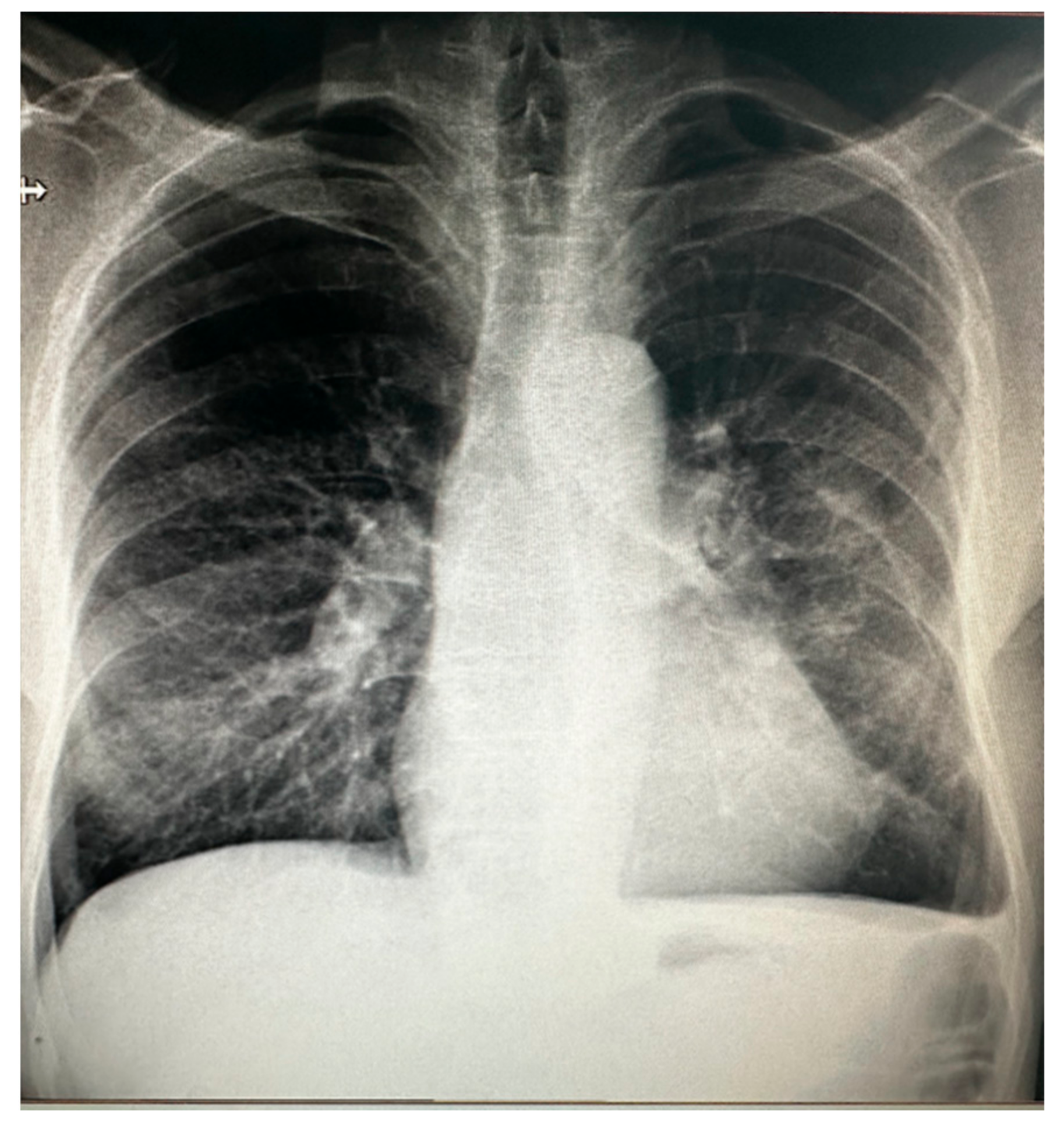
Abstract
:1. Introduction
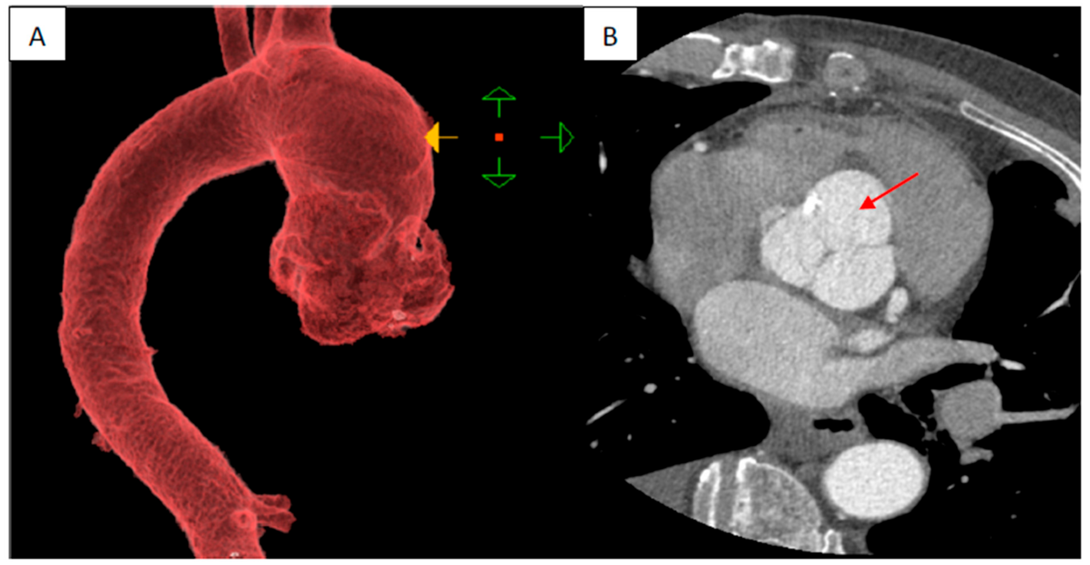
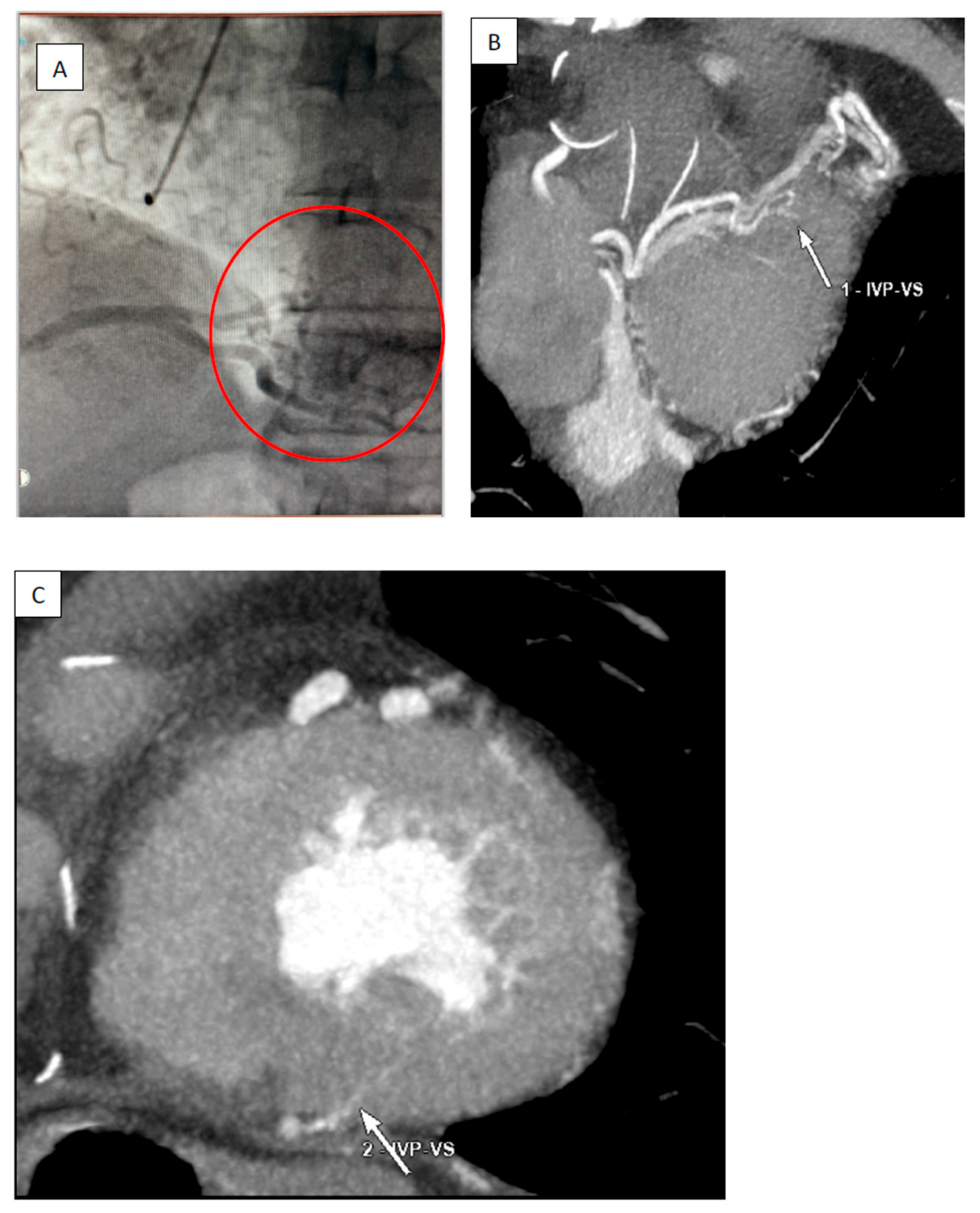
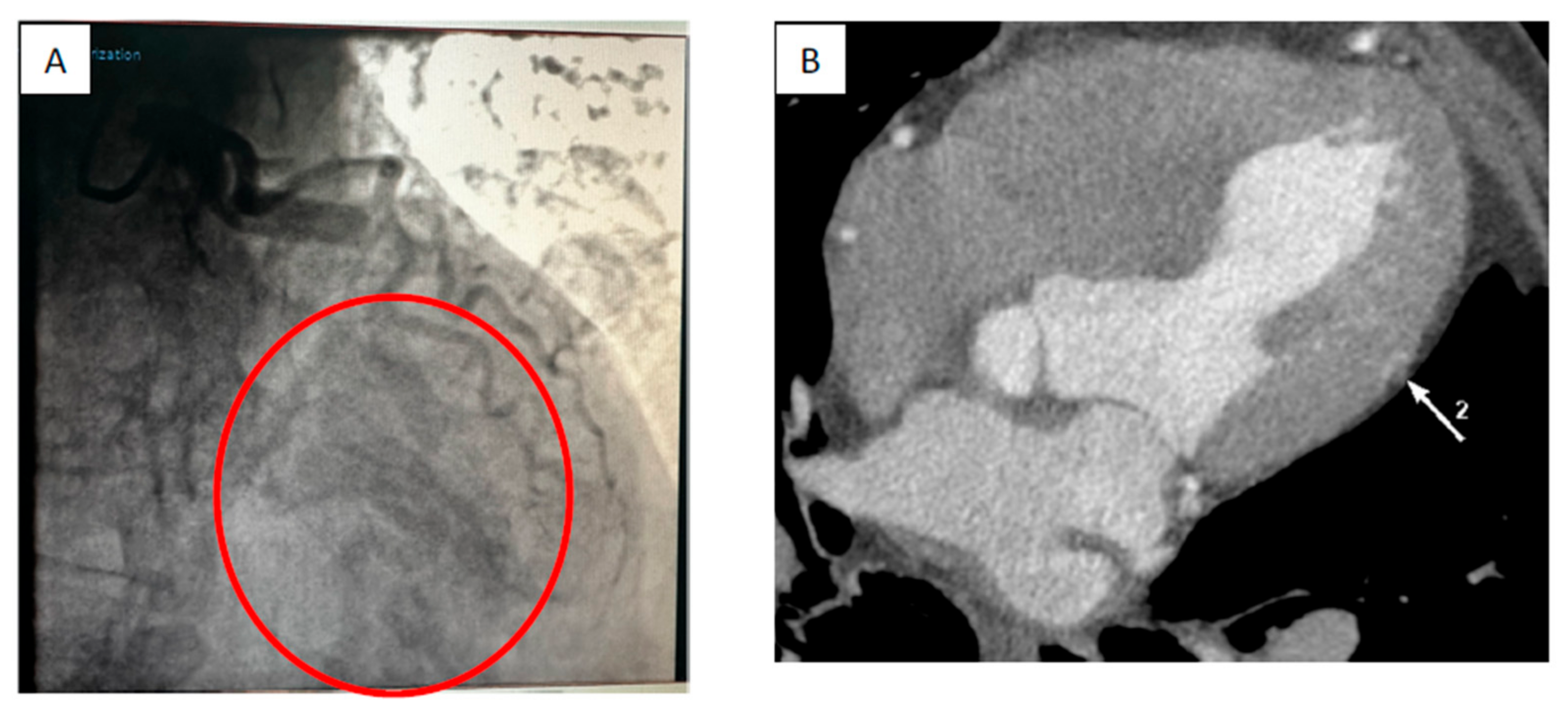
2. Case Report
Surgical Technique
3. Discussion
4. Review of Literature
5. Conclusions
Author Contributions
Funding
Institutional Review Board Statement
Informed Consent Statement
Data Availability Statement
Conflicts of Interest
References
- Kumar, R.; Kumar, J.; O’Connor, C.; Ullah, I.; Tyrell, B.; Pearson, I.; Matiullah, S.; Bainey, K. Coronary Artery Fistula: A Diagnostic Dilemma. Interv. Cardiol. 2023, 18, e25. [Google Scholar] [CrossRef] [PubMed]
- Stout, K.K.; Daniels, C.J.; Aboulhosn, J.A.; Bozkurt, B.; Broberg, C.S.; Colman, J.M.; Crumb, S.R.; Dearani, J.A.; Fuller, S.; Gurvitz, M.; et al. 2018 AHA/ACC Guideline for the Management of Adults with Congenital Heart Disease: Executive Summary: A Report of the American College of Cardiology/American Heart Association Task Force on Clinical Practice Guidelines. J. Am. Coll. Cardiol. 2019, 73, 1494–1563, Erratum in J. Am. Coll. Cardiol. 2019, 73, 2361. [Google Scholar] [CrossRef] [PubMed]
- Gowda, R.M.; Vasavada, B.C.; Khan, I.A. Coronary artery fistulas: Clinical and therapeutic considerations. Int. J. Cardiol. 2006, 107, 7–10. [Google Scholar] [CrossRef] [PubMed]
- Gowda, S.T.; Latson, L.A.; Kutty, S.; Prieto, L.R. Intermediate to long-term outcome following congenital coronary artery fistulae closure with focus on thrombus formation. Am. J. Cardiol. 2011, 107, 302–308. [Google Scholar] [CrossRef] [PubMed]
- Ata, Y.; Turk, T.; Bicer, M.; Yalcin, M.; Ata, F.; Yavuz, S. Coronary arteriovenous fistulas in the adults: Natural history and management strategies. J. Cardiothorac. Surg. 2009, 4, 62. [Google Scholar] [CrossRef] [PubMed]
- Agostini, M.; Ribichini, F.; Portolan, M.; Ugliengo, G.; Iacovoni, A.; Grossi, C. Giant coronary artery fistula connecting the left coronary sinus with the superior atriocaval junction. Ital. Heart J. 2004, 5, 483–485. [Google Scholar] [PubMed]
- Toledo, I.C.; Braile, V.; Leal, J.C.; Braile, D.M. Fístula entre artéria coronária interventricular anterior e o tronco arterial pulmonar: Tratamentocirúrgico de cinco pacientes [Fistula between anterior intraventricular coronary artery and the pulmonary artery trunk: Five operated patients]. Rev. Bras. Cir. Cardiovasc. 2007, 22, 241–244. [Google Scholar] [CrossRef]
- Said, S.A. Current characteristics of congenital coronary artery fistulas in adults: A decade of global experience. World J. Cardiol. 2011, 3, 267–277. [Google Scholar] [CrossRef] [PubMed]
- Fernandes, E.D.; Kadivar, H.; Hallman, G.L.; Reul, G.J.; Ott, D.A.; Cooley, D.A. Congenital malformations of the coronary arteries: The Texas Heart Institute experience. Ann. Thorac. Surg. 1992, 54, 732–740. [Google Scholar] [CrossRef]
- Warnes, C.A.; Williams, R.G.; Bashore, T.M.; Child, J.S.; Connolly, H.M.; Dearani, J.A.; Del Nido, P.; Fasules, J.W.; Graham, T.P., Jr.; Hijazi, Z.M.; et al. ACC/AHA 2008 guidelines for the management of adults with congenital heart disease: A report of the American College of Cardiology/American Heart Association Task Force on Practice Guidelines (Writing Committee to Develop Guidelines on the Management of Adults with Congenital Heart Disease). Developed in Collaboration with the American Society of Echocardiography, Heart Rhythm Society, International Society for Adult Congenital Heart Disease, Society for Cardiovascular Angiography and Interventions, and Society of Thoracic Surgeons. J. Am. Coll. Cardiol. 2008, 52, e143–e263. [Google Scholar] [CrossRef] [PubMed]
- Kim, K.; Ball, C.; Grady, P.; Mick, S. Use of del Nido Cardioplegia for Adult Cardiac Surgery at the Cleveland Clinic: Perfusion Implications. J. Extra Corpor. Technol. 2014, 46, 317–323. [Google Scholar] [CrossRef] [PubMed]
- Behera, S.K.; Danon, S.; Levi, D.S.; Moore, J.W. Transcatheter closure of coronary artery fistulae using the Amplatzer Duct Occluder. Catheter. Cardiovasc. Interv. 2006, 68, 242–248. [Google Scholar] [CrossRef] [PubMed]
- Ascoop, A.K.; Budts, W. Percutaneous closure of a congenital coronary artery fistula complicated by an acute myocardial infarction. Acta Cardiol. 2004, 59, 67–69. [Google Scholar] [CrossRef] [PubMed]
- Meerkin, D.; Balkin, J.; Klutstein, M. Rapid transcatheter occlusion of a coronary cameral fistula using a three-lobed vascular occlusion plug. J. Invasive Cardiol. 2009, 21, E151–E153. [Google Scholar] [PubMed]
- Mullasari, A.S.; Umesan, C.V.; Kumar, K.J. Transcatheter closure of coronary artery to pulmonary artery fistula using covered stents. Heart. 2002, 87, 60. [Google Scholar] [CrossRef] [PubMed]
- Hirose, H.; Amano, A.; Yoshida, S.; Nagao, T.; Sunami, H.; Takahashi, A.; Nagano, N. Coronary artery aneurysm associated with fistula in adults: Collective review and a case report. Ann. Thorac. Cardiovasc. Surg. 1999, 5, 258–264. [Google Scholar]
- Erdem, K.; Ozbay, Y. Prevalence and Characteristics of Coronary Artery Anomalies Using Invasive Coronary Angiography in 6237 Consecutive Patients in a Single Center in Turkey. Arch. Iran. Med. 2018, 21, 240–245. [Google Scholar] [PubMed]
- Yildiz, A.; Okcun, B.; Peker, T.; Arslan, C.; Olcay, A.; Bulent Vatan, M. Prevalence of coronary artery anomalies in 12,457 adult patients who underwent coronary angiography. Clin. Cardiol. 2010, 33, E60–E64. [Google Scholar] [CrossRef] [PubMed]
- Vitarelli, A.; De Curtis, G.; Conde, Y.; Colantonio, M.; Di Benedetto, G.; Pecce, P.; De Nardo, L.; Squillaci, E. Assessment of congenital coronary artery fistulas by transesophageal color Doppler echocardiography. Am. J. Med. 2002, 113, 127–133. [Google Scholar] [CrossRef]
- Bazzocchi, G.; Romagnoli, A.; Sperandio, M.; Simonetti, G. Evaluation with 64-slice CT of the prevalence of coronary artery variants and congenital anomalies: A retrospective study of 3,236 patients. Radiol. Med. 2011, 116, 675–689. [Google Scholar] [CrossRef]
- Lee, S.K.; Jung, J.I.; O, J.H.; Kim, H.W.; Youn, H.J. Coronary-to-pulmonary artery fistula in adults: Evaluation with thallium-201 myocardial perfusion SPECT. PLoS ONE 2017, 12, e0189269. [Google Scholar] [CrossRef] [PubMed]
- Angelini, P. Coronary artery anomalies-current clinical issues: Definitions, classification, incidence, clinical relevance, and treatment guidelines. Tex. Heart Inst. J. 2002, 29, 271–278. [Google Scholar] [PubMed]
- Kirklin, J.W.; Barratt-Boyes, B.G. Congenital anomalies of the coronary arteries. In Cardiac Surgery, 2nd ed.; Kirklin, J.W., Barratt-Boyes, B.G., Eds.; Churchill-Livingstone: New York, NY, USA, 1993; pp. 945–955. [Google Scholar]
- Balanescu, S.; Sangiorgi, G.; Castelvecchio, S.; Medda, M.; Inglese, L. Coronary artery fistulas: Clinical consequences and methods of closure. A literature review. Ital. Heart J. 2001, 2, 669–676. [Google Scholar] [PubMed]
- Al-Hijji, M.; El Sabbagh, A.; El Hajj, S.; AlKhouli, M.; El Sabawi, B.; Cabalka, A.; Miranda, W.R.; Holmes, D.R.; Rihal, C.S. Coronary Artery Fistulas: Indications, Techniques, Outcomes, and Complications of Transcatheter Fistula Closure. JACC Cardiovasc. Interv. 2021, 14, 1393–1406. [Google Scholar] [CrossRef]
- Moldovan, H.; Antoniac, I.; Gheorghiță, D.; Safta, M.S.; Preda, S.; Broască, M.; Badilă, E.; Fronea, O.; Scafa-Udrişte, A.; Cacoveanu, M.; et al. Biomaterials as Haemostatic Agents in Cardiovascular Surgery: Review of Current Situation and Future Trends. Polymers 2022, 14, 1189. [Google Scholar] [CrossRef] [PubMed]
- Filip, C.; Cirstoveanu, C.; Bizubac, M.; Berghea, E.C.; Căpitănescu, A.; Bălgrădean, M.; Pavelescu, C.; Nicolescu, A.; Ionescu, M.D. Pulse Wave Velocity as a Marker of Vascular Dysfunction and Its Correlation with Cardiac Disease in Children with End-Stage Renal Disease (ESRD). Diagnostics 2021, 12, 71. [Google Scholar] [CrossRef] [PubMed]
- Mishra, P.; Jadhav, R.B.; Mohapatra, C.K.; Khandekar, J.; Raut, C.; Ammannaya, G.K.; Seth, H.S.; Singh, J.; Shah, V. Comparison of del Nido cardioplegia and St. Thomas Hospital solution—Two types of cardioplegia in adult cardiac surgery. Kardiochir. Torakochirurgia Pol. 2016, 13, 295–299. [Google Scholar] [CrossRef] [PubMed]
- Choudhary, S.K.; Airan, B. Comparison of del Nido and St Thomas Cardioplegia Solutions in Pediatric Patients: A Prospective Randomized Clinical Trial. Semin. Thorac. Cardiovasc. Surg. 2017, 29, 366–374. [Google Scholar] [CrossRef]
- Cheung, D.L.; Au, W.K.; Cheung, H.H.; Chiu, C.S.; Lee, W.T. Coronary artery fistulas: Long-term results of surgical correction. Ann. Thorac. Surg. 2001, 71, 190–195. [Google Scholar] [CrossRef] [PubMed]


Disclaimer/Publisher’s Note: The statements, opinions and data contained in all publications are solely those of the individual author(s) and contributor(s) and not of MDPI and/or the editor(s). MDPI and/or the editor(s) disclaim responsibility for any injury to people or property resulting from any ideas, methods, instructions or products referred to in the content. |
© 2024 by the authors. Licensee MDPI, Basel, Switzerland. This article is an open access article distributed under the terms and conditions of the Creative Commons Attribution (CC BY) license (https://creativecommons.org/licenses/by/4.0/).
Share and Cite
Robu, M.; Radulescu, B.; Nayyerani, R.; Enache, R.; Stiru, O.; Iosifescu, A.; Olaru, G.; Ciomag, R.; Iliescu, V.A.; Moldovan, H. Management of a Rare Case of Multiple Coronary Artery Fistulas Associated with Ascending Aortic and Root Aneurysm: Case Report and Review of Literature. J. Clin. Med. 2024, 13, 2297. https://doi.org/10.3390/jcm13082297
Robu M, Radulescu B, Nayyerani R, Enache R, Stiru O, Iosifescu A, Olaru G, Ciomag R, Iliescu VA, Moldovan H. Management of a Rare Case of Multiple Coronary Artery Fistulas Associated with Ascending Aortic and Root Aneurysm: Case Report and Review of Literature. Journal of Clinical Medicine. 2024; 13(8):2297. https://doi.org/10.3390/jcm13082297
Chicago/Turabian StyleRobu, Mircea, Bogdan Radulescu, Reza Nayyerani, Robert Enache, Ovidiu Stiru, Andrei Iosifescu, Georgiana Olaru, Raluca Ciomag (Ianula), Vlad Anton Iliescu, and Horatiu Moldovan. 2024. "Management of a Rare Case of Multiple Coronary Artery Fistulas Associated with Ascending Aortic and Root Aneurysm: Case Report and Review of Literature" Journal of Clinical Medicine 13, no. 8: 2297. https://doi.org/10.3390/jcm13082297