
Free Flap Reconstruction of Traumatic Skin Defects of the Entire Hand Dorsum


Abstract
:1. Introduction
2. Materials and Methods
3. Results
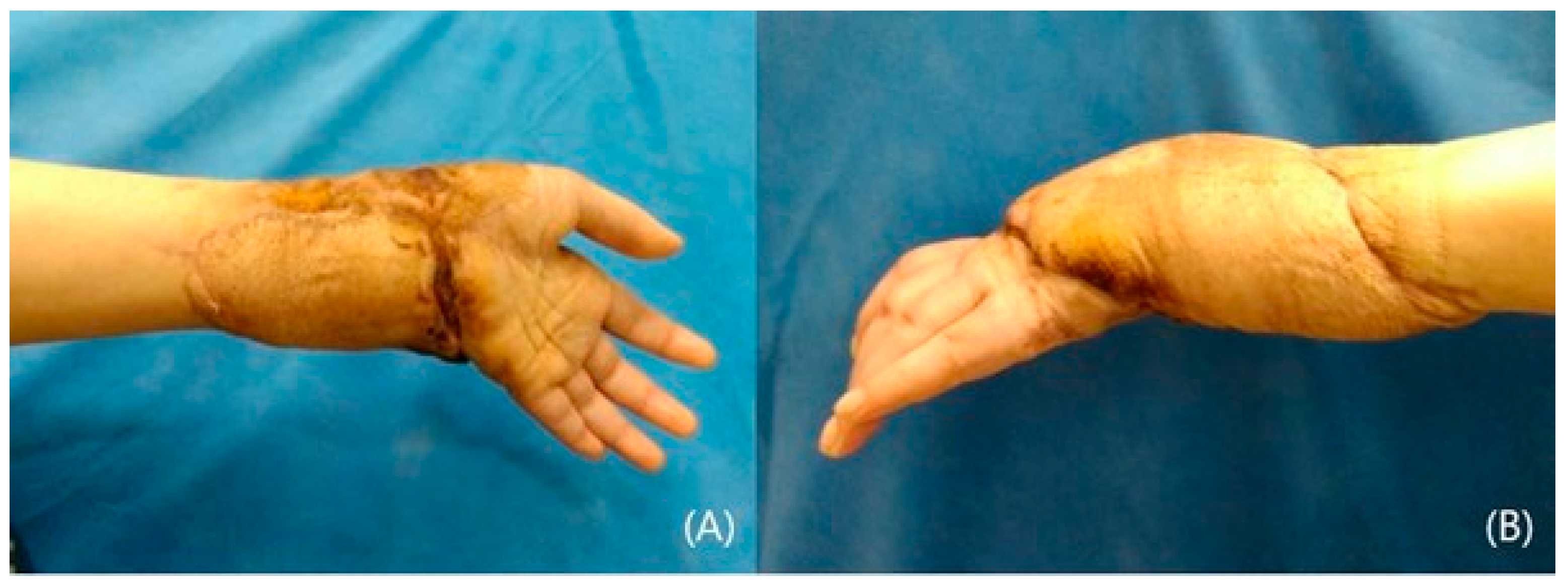
3.1. Case Series
3.1.1. Patient 1
3.1.2. Patient 2
3.1.3. Patient 3
4. Discussion
5. Conclusions
Author Contributions
Funding
Institutional Review Board Statement
Informed Consent Statement
Data Availability Statement
Conflicts of Interest
Abbreviations
| ALT | Anterolateral thigh |
| SCIP | Superficial circumflex iliac artery perforator |
References
- Hocaoğlu, E.; Arıncı, A.; Berköz, Ö.; Özkan, T. Free pre-expanded lateral circumflex femoral artery perforator flap for extensive resurfacing and reconstruction of the hand. J. Plast. Reconstr. Aesthet. Surg. 2013, 66, 1788–1791. [Google Scholar] [CrossRef] [PubMed]
- Eo, S.; Kim, Y.; Kim, J.Y.; Oh, S. The versatility of the dorsalis pedis compound free flap in hand reconstruction. Ann. Plast. Surg. 2008, 61, 157–163. [Google Scholar] [CrossRef] [PubMed]
- Kaufman, M.R.; Jones, N.F. The reverse radial forearm flap for soft tissue reconstruction of the wrist and hand. Tech. Hand Up. Extrem. Surg. 2005, 9, 47–51. [Google Scholar] [CrossRef] [PubMed]
- Li, K.W.; Song, D.J.; Liu, J.; Xie, S.L. Tripaddle posterior interosseous artery flap design for 3-finger defects: An evaluation of 3 surgical approaches. Ann. Plast. Surg. 2016, 77, 406–412. [Google Scholar] [CrossRef] [PubMed]
- Gisquet, H.; Barbary, S.; Vialanex, J.; Dap, F.; Dautel, G. The free groin flap utility. About 19 cases. Ann. Chir. Plast. Esthet. 2011, 56, 99–106. [Google Scholar] [CrossRef]
- Ziegler, B.; Hundeshagen, G.; Warszawski, J.; Gazyakan, E.; Kneser, U.; Hirche, C. Implementation and validation of free flaps in acute and reconstructive burn care. Medicina 2021, 57, 718. [Google Scholar] [CrossRef] [PubMed]
- Pyörny, J.; Karelson, M.; Sletten, I.N.; Ukkola, A.; Jokihaara, J. Patients-reported significant disability after major traumatic upper extremity amputation. J. Hand Surg. Eur. Vol. 2024, 49, 1017–1022. [Google Scholar] [CrossRef] [PubMed]
- Nietosvaara, N.N.; Sommarhem, A.J.; Puhakka, J.M.; Tan, R.E.S.; Schalamon, J.; Nietosvaara, A.Y. Appearance of congenital hand anomalies. Scand. J. Surg. 2021, 110, 434–440. [Google Scholar] [CrossRef] [PubMed]
- Chi, Z.; Gao, W.; Yan, H.; Li, Z.; Chen, X.; Zhang, F. Reconstruction of totally degloved fingers with a spiraled parallelogram medial arm free flap. J. Hand. Surg. Am. 2012, 37, 1042–1050. [Google Scholar] [CrossRef]
- Sakai, S. Free flap from the flexor aspect of the wrist for resurfacing defects of the hand and fingers. Plast. Reconstr. Surg. 2003, 111, 1412–1420. [Google Scholar] [CrossRef] [PubMed]
- Ryoo, H.J.; Park, S.H.; Park, J.A.; Kim, Y.H.; Shim, H.S. Multiple digit resurfacing with a lateral thoracic free flap: Two-stage mitten hand and division procedures. Microsurgery 2023, 43, 570–579. [Google Scholar] [CrossRef]
- Borschel, G.H. A three-subunit latissimus dorsi muscle free flap for single-stage coverage of the hand and three adjacent fingers. Hand 2010, 5, 99–101. [Google Scholar] [CrossRef]
- Tee, J.W.; Bigdeli, A.K.; Thomas, B.; Falkner, F.; Didzun, O.; Vollbach, F.H.; Kneser, U.; Gazyakan, E. Reconstruction of Hand and Foot Defects with Free Serratus Carpaccio Flap and Free Serratus Fascia Flap: A Comparative Retrospective Study of Surgical Outcomes. J. Clin. Med. 2023, 12, 3313. [Google Scholar] [CrossRef]
- Heimes, D.; Becker, P.; Thiem, D.G.E.; Kuchen, R.; Kyyak, S.; Kämmerer, P.W. Is Hyperspectral Imaging Suitable for Assessing Collateral Circulation Prior Radial Forearm Free Flap Harvesting? Comparison of Hyperspectral Imaging and Conventional Allen’s Test. J. Pers. Med. 2021, 11, 531. [Google Scholar] [CrossRef]
- Zhou, X.; Hu, D.; Lu, H.; Tao, X.; Chen, P.; Wen, F.; Xue, L.; Hong, X.; Zheng, H. Sequential posterior interosseous artery perforator flap for reconstruction of dorsal hand defects. Clin. Anat. 2022, 35, 1114–1122. [Google Scholar] [CrossRef] [PubMed]
- El-Sabbagh, A.H.; Zeina, A.A.; El-Hadidy, A.M.; El-Din, A.B. Reversed posterior interosseous flap: Safe and easy method for hand reconstruction. J. Hand Microsurg. 2011, 3, 66–72. [Google Scholar] [CrossRef]
- Bashorun, O.H.; Anderson, S.R.; Mulenga, C.M.; Wimalawansa, S.M. Multiple simultaneous venous flow-through free flap salvage for multifinger revascularisations. BMJ. Case. Rep. 2022, 15, e247413. [Google Scholar] [CrossRef] [PubMed]
- Chia, J.; Lim, A.; Peng, Y.P. Use of an arterialized venous flap for resurfacing a circumferential soft tissue defect of a digit. Microsurgery. 2021, 21, 374–378. [Google Scholar] [CrossRef] [PubMed]
- Yu, G.; Lei, H.Y.; Guo, S.; Huang, J.H.; Yu, H. Dorsalis pedis arterialized venous flap for hand and foot reconstruction. Chin. J. Traumatol. 2012, 15, 32–35. [Google Scholar] [PubMed]
- Chen, H.C.; El-Gammal, T.A. The lateral arm fascial free flap for resurfacing of the hand and fingers. Plast. Reconstr. Surg. 1997, 99, 454–459. [Google Scholar] [CrossRef] [PubMed]
- Kim, J.H.; Yoon, T.; Park, J.K.; Eun, S. Reconstruction of Foot and Ankle Defects Using Free Lateral Arm Flap: A Retrospective Review of Its Versatile Application. Biomed. Res. Int. 2021, 31, 412–417. [Google Scholar] [CrossRef]
- Kang, S.H.; Jeon, S.; Jung, Y.H.; Eun, S. Verifying the Versatility and Reliability of Free Lateral Arm Flap for Head and Neck Reconstruction. J. Craniofac Surg. 2020, 31, 688–693. [Google Scholar] [CrossRef] [PubMed]
- Ki, S.H. Lateral Arm Free Flap With Preservation of the Posterior Antebrachial Cutaneous Nerve. Ann. Plast. Surg. 2016, 76, 517–520. [Google Scholar] [CrossRef] [PubMed]
- Kim, K.S.; Kim, E.S.; Kim, D.Y.; Lee, S.Y.; Cho, B.H. Resurfacing of a totally degloved hand using thin perforator-based cutaneous free flaps. Ann. Plast. Surg. 2003, 50, 77–81. [Google Scholar] [CrossRef]
- Jeevaratnam, J.A.; Nikkhah, D.; Nugent, N.F.; Blackburn, A.V. The medial sural artery perforator flap and its application in electrical injury to the hand. J. Plast. Reconst. Aesthet. Surg. 2014, 67, 1591–1594. [Google Scholar] [CrossRef] [PubMed]
- Pan, J.; Li, M.; Huang, Y.; Dong, J.; Wang, X.; Wang, L. Pure perforator free sensory proximal ulnar artery perforator flap for resurfacing hand defects. J. Int. Med. Res. 2020, 48, 300060520922396. [Google Scholar] [CrossRef] [PubMed]
- Adani, R.; Tarallo, L.; Marcoccio, I.; Cipriani, R.; Gelati, C.; Innocenti, M. Hand reconstruction using the thin anterolateral thigh flap. Plast. Reconstr. Surg. 2005, 116, 467–473. [Google Scholar] [CrossRef]
- Chen, X.; Zhang, C.; Cheng, L.; Chen, H.; Wang, H.; Qin, F.J.; Tian, P.; Zhang, Y.H.; Shen, Y.M. Survival and versatility of the flow-through lateral-thigh free flap in severe electrical injuries to the wrist. Ann. Plast. Surg. 2020, 85, 612–617. [Google Scholar] [CrossRef]
- Caulfield, R.H.; Maleki-Tabrizi, A.; Birch, J.; Ramakrishnan, V. Salvage of finger length in septicemic necrosis using 3 free flaps from a single anterolateral thigh donor site. Ann. Plast. Surg. 2008, 60, 623–625. [Google Scholar] [CrossRef] [PubMed]
- Lin, T.S.; Jeng, S.F.; Chiang, Y.C. Resurfacing with full-thickness skin graft after debulking procedure for bulky flap of the hand. J. Trauma. 2008, 65, 123–126. [Google Scholar] [CrossRef]
- Sơn, T.T.; Việt Dung, P.T.; Nghĩa, P.T.; Hồng Thúy, T.T. Reconstruction of finger soft tissue defects with a thinned free anterolateral thigh flap. Ann. Plast. Surg. 2023, 91, 238–244. [Google Scholar] [CrossRef] [PubMed]
- Riesel, J.N.; Giladi, A.M.; Iorio, M.L. Volar resurfacing of the thumb with a superficial circumflex Iliac artery perforator flap after hydrofluoric acid burn. J. Hand. Microsurg. 2018, 10, 162–165. [Google Scholar] [CrossRef] [PubMed]
- Narushima, M.; Iida, T.; Kaji, N.; Yamamoto, T.; Yoshimatsu, H.; Hara, H.; Kikuchi, K.; Araki, J.; Yamashita, S.; Koshima, I. Superficial circumflex iliac artery pure skin perforator-based superthin flap for hand and finger reconstruction. J. Plast. Reconstr. Aesthet. Surg. 2016, 69, 827–834. [Google Scholar] [CrossRef] [PubMed]
- Li, Z.; Zheng, D.; Zheng, J.; Qi, W.; Qi, Q.; Liu, Y. Free superficial circumflex iliac artery perforator flap with a single-pedicle bilobed design for pediatric multi-digit defect reconstruction. J. Orthop. Surg. Res. 2020, 15, 216. [Google Scholar] [CrossRef] [PubMed]


| Patient | Age(yr) | Sex | Injury | Area | Comorbidities | Flap | Flap Size (cm2) | Q-DASH | Esthetic Sartisfaction | Complications |
|---|---|---|---|---|---|---|---|---|---|---|
| 1 | 55 | F | Traffic injury | Right hand dorsum | SCIP * | 9 × 7 | 2.5 | Good | None | |
| 2 | 72 | M | Traffic injury | Left hand dorsum | Metacarpal fractures | Lateral arm | 10 × 6 | 2.6 | Good | None |
| 3 | 43 | M | Crush injury | Right hand dorsum | Lateral arm | 8 × 6 | 3.1 | Fair | Partial necrosis | |
| 4 | 46 | M | Crush injury | Left hand dorsum | Extensor tendon rupture | SCIP | 11 × 8 | 2.6 | Good | None |
| 5 | 65 | M | Traffic injury | Right hand dorsum | ALT ¥ | 13 × 7 | 1.5 | Excellent | None | |
| 6 | 57 | F | Traffic injury | Left hand dorsum | Wrist skin defect Extensor tendon rupture | ALT | 12 × 8 | 4.2 | Fair | None |
| 7 | 32 | M | Press injury | Left hand dorsum | Lateral arm | 6 × 5 | 3.2 | Good | None | |
| 8 | 34 | M | Traffic injury | Right hand dorsum | Metacarpal fractures | ALT | 12 × 7 | 2.1 | Excellent | None |
| 9 | 28 | F | Crush injury | Right hand dorsum | SCIP | 11 × 6 | 0 | Excellent | None | |
| 10 | 65 | F | Traffic injury | Left hand dorsum | Arterial rupture | SCIP | 20 × 9 | 3.7 | Fair | Joint stiffness |
| 11 | 53 | F | Traffic injury | Right hand dorsum | ALT | 10 × 7 | 2.4 | Good | None |
Disclaimer/Publisher’s Note: The statements, opinions and data contained in all publications are solely those of the individual author(s) and contributor(s) and not of MDPI and/or the editor(s). MDPI and/or the editor(s) disclaim responsibility for any injury to people or property resulting from any ideas, methods, instructions or products referred to in the content. |
© 2025 by the authors. Licensee MDPI, Basel, Switzerland. This article is an open access article distributed under the terms and conditions of the Creative Commons Attribution (CC BY) license (https://creativecommons.org/licenses/by/4.0/).
Share and Cite
Jung, S.; Lee, S.; Eun, S. Free Flap Reconstruction of Traumatic Skin Defects of the Entire Hand Dorsum. J. Clin. Med. 2025, 14, 1308. https://doi.org/10.3390/jcm14041308
Jung S, Lee S, Eun S. Free Flap Reconstruction of Traumatic Skin Defects of the Entire Hand Dorsum. Journal of Clinical Medicine. 2025; 14(4):1308. https://doi.org/10.3390/jcm14041308
Chicago/Turabian StyleJung, Soyeon, Seungjun Lee, and Seokchan Eun. 2025. "Free Flap Reconstruction of Traumatic Skin Defects of the Entire Hand Dorsum" Journal of Clinical Medicine 14, no. 4: 1308. https://doi.org/10.3390/jcm14041308
APA StyleJung, S., Lee, S., & Eun, S. (2025). Free Flap Reconstruction of Traumatic Skin Defects of the Entire Hand Dorsum. Journal of Clinical Medicine, 14(4), 1308. https://doi.org/10.3390/jcm14041308