Swept-Source Optical Coherence Tomography Angiography According to the Type of Choroidal Neovascularization
Abstract
1. Introduction
2. Materials and Methods
2.1. Patients
2.2. Scanning Protocol of Optical Coherence Tomography (OCT) and OCT Angiography (OCTA)
2.3. Classification of Choroidal Neovasculization (CNV)
2.4. Statistics
3. Results
4. Discussion
Author Contributions
Funding
Conflicts of Interest
References
- Ferris, F.L., 3rd; Fine, S.L.; Hyman, L. Age-related macular degeneration and blindness due to neovascular maculopathy. Arch. Ophthalmol. (Chicago, Ill.: 1960) 1984, 102, 1640–1642. [Google Scholar] [CrossRef] [PubMed]
- Do, D.V. Detection of new-onset choroidal neovascularization. Curr. Opin. Ophthalmol. 2013, 24, 244–247. [Google Scholar] [CrossRef] [PubMed]
- Kotsolis, A.I.; Killian, F.A.; Ladas, I.D.; Yannuzzi, L.A. Fluorescein angiography and optical coherence tomography concordance for choroidal neovascularisation in multifocal choroidtis. Br. J. Ophthalmol. 2010, 94, 1506–1508. [Google Scholar] [CrossRef] [PubMed]
- Stanga, P.E.; Lim, J.I.; Hamilton, P. Indocyanine green angiography in chorioretinal diseases: Indications and interpretation: An evidence-based update. Ophthalmology 2003, 110, 15–21. [Google Scholar] [CrossRef]
- Freund, K.B.; Zweifel, S.A.; Engelbert, M. Do we need a new classification for choroidal neovascularization in age-related macular degeneration? Retina 2010, 30, 1333–1349. [Google Scholar] [CrossRef]
- Gao, S.S.; Jia, Y.; Zhang, M.; Su, J.P.; Liu, G.; Hwang, T.S.; Bailey, S.T.; Huang, D. Optical Coherence Tomography Angiography. Investig. Ophthalmol. Vis. Sci. 2016, 57, OCT27–OCT36. [Google Scholar] [CrossRef] [PubMed]
- Jia, Y.; Tan, O.; Tokayer, J.; Potsaid, B.; Wang, Y.; Liu, J.J.; Kraus, M.F.; Subhash, H.; Fujimoto, J.G.; Hornegger, J.; et al. Split-spectrum amplitude-decorrelation angiography with optical coherence tomography. Opt. Express 2012, 20, 4710–4725. [Google Scholar] [CrossRef]
- Makita, S.; Hong, Y.; Yamanari, M.; Yatagai, T.; Yasuno, Y. Optical coherence angiography. Opt. Express 2006, 14, 7821–7840. [Google Scholar] [CrossRef]
- Moult, E.; Choi, W.; Waheed, N.K.; Adhi, M.; Lee, B.; Lu, C.D.; Jayaraman, V.; Potsaid, B.; Rosenfeld, P.J.; Duker, J.S.; et al. Ultrahigh-speed swept-source OCT angiography in exudative AMD. Ophthalmic Surg. Lasers Imaging Retin. 2014, 45, 496–505. [Google Scholar] [CrossRef]
- Nagiel, A.; Sadda, S.R.; Sarraf, D. A Promising Future for Optical Coherence Tomography Angiography. JAMA Ophthalmol. 2015, 133, 629–630. [Google Scholar] [CrossRef]
- Wong, C.W.; Yanagi, Y.; Lee, W.K.; Ogura, Y.; Yeo, I.; Wong, T.Y.; Cheung, C.M.G. Age-related macular degeneration and polypoidal choroidal vasculopathy in Asians. Prog. Retin. Eye Res. 2016, 53, 107–139. [Google Scholar] [CrossRef] [PubMed]
- Ferrara, D.; Waheed, N.K.; Duker, J.S. Investigating the choriocapillaris and choroidal vasculature with new optical coherence tomography technologies. Prog. Retin. Eye Res. 2016, 52, 130–155. [Google Scholar] [CrossRef] [PubMed]
- Ferrara, D.; Mohler, K.J.; Waheed, N.; Adhi, M.; Liu, J.J.; Grulkowski, I.; Kraus, M.F.; Baumal, C.; Hornegger, J.; Fujimoto, J.G.; et al. En face enhanced-depth swept-source optical coherence tomography features of chronic central serous chorioretinopathy. Ophthalmology 2014, 121, 719–726. [Google Scholar] [CrossRef] [PubMed]
- de Carlo, T.E.; Bonini Filho, M.A.; Chin, A.T.; Adhi, M.; Ferrara, D.; Baumal, C.R.; Witkin, A.J.; Reichel, E.; Duker, J.S.; Waheed, N.K. Spectral-domain optical coherence tomography angiography of choroidal neovascularization. Ophthalmology 2015, 122, 1228–1238. [Google Scholar] [CrossRef] [PubMed]
- Novais, E.A.; Adhi, M.; Moult, E.M.; Louzada, R.N.; Cole, E.D.; Husvogt, L.; Lee, B.; Dang, S.; Regatieri, C.V.; Witkin, A.J.; et al. Choroidal Neovascularization Analyzed on Ultrahigh-Speed Swept-Source Optical Coherence Tomography Angiography Compared to Spectral-Domain Optical Coherence Tomography Angiography. Am. J. Ophthalmol. 2016, 164, 80–88. [Google Scholar] [CrossRef] [PubMed]
- Farecki, M.L.; Gutfleisch, M.; Faatz, H.; Rothaus, K.; Heimes, B.; Spital, G.; Lommatzsch, A.; Pauleikhoff, D. Characteristics of type 1 and 2 CNV in exudative AMD in OCT-Angiography. Graefe’s Arch. Clin. Exp. Ophthalmol. 2017, 255, 913–921. [Google Scholar] [CrossRef] [PubMed]
- Lumbroso, B.; Rispoli, M.; Savastano, M.C. Longitudinal Optical Coherence Tomography-Angiography Study of Type 2 Naive Choroidal Neovascularization Early Response after Treatment. Retina 2015, 35, 2242–2251. [Google Scholar] [CrossRef]
- El Ameen, A.; Cohen, S.Y.; Semoun, O.; Miere, A.; Srour, M.; Quaranta-El Maftouhi, M.; Oubraham, H.; Blanco-Garavito, R.; Querques, G.; Souied, E.H. Type 2 Neovascularization Secondary to Age-Related Macular Degeneration Imaged by Optical Coherence Tomography Angiography. Retina 2015, 35, 2212–2218. [Google Scholar] [CrossRef] [PubMed]
- Parravano, M.; Querques, L.; Scarinci, F.; Giorno, P.; De Geronimo, D.; Gattegna, R.; Varano, M.; Bandello, F.; Querques, G. Optical coherence tomography angiography in treated type 2 neovascularization undergoing monthly anti-VEGF treatment. Acta Ophthalmol. 2017, 95, e425–e426. [Google Scholar] [CrossRef]
- Jung, J.J.; Chen, C.Y.; Mrejen, S.; Gallego-Pinazo, R.; Xu, L.; Marsiglia, M.; Boddu, S.; Freund, K.B. The incidence of neovascular subtypes in newly diagnosed neovascular age-related macular degeneration. Am. J. Ophthalmol. 2014, 158, 769–779. [Google Scholar] [CrossRef]
- Kuehlewein, L.; Bansal, M.; Lenis, T.L.; Iafe, N.A.; Sadda, S.R.; Bonini Filho, M.A.; De Carlo, T.E.; Waheed, N.K.; Duker, J.S.; Sarraf, D. Optical Coherence Tomography Angiography of Type 1 Neovascularization in Age-Related Macular Degeneration. Am. J. Ophthalmol. 2015, 160, 739–748. [Google Scholar] [CrossRef] [PubMed]
- Inoue, M.; Balaratnasingam, C.; Freund, K.B. Optical Coherence Tomography Angiography of Polypoidal Choroidal Vasculopathy and Polypoidal Choroidal Neovascularization. Retina 2015, 35, 2265–2274. [Google Scholar] [CrossRef] [PubMed]
- Tanaka, K.; Mori, R.; Kawamura, A.; Nakashizuka, H.; Wakatsuki, Y.; Yuzawa, M. Comparison of OCT angiography and indocyanine green angiographic findings with subtypes of polypoidal choroidal vasculopathy. Br. J. Ophthalmol. 2017, 101, 51–55. [Google Scholar] [CrossRef] [PubMed]
- Phasukkijwatana, N.; Tan, A.C.S.; Chen, X.; Freund, K.B.; Sarraf, D. Optical coherence tomography angiography of type 3 neovascularisation in age-related macular degeneration after antiangiogenic therapy. Br. J. Ophthalmol. 2017, 101, 597–602. [Google Scholar] [CrossRef] [PubMed]
- Kuehlewein, L.; Dansingani, K.K.; de Carlo, T.E.; Bonini Filho, M.A.; Iafe, N.A.; Lenis, T.L.; Freund, K.B.; Waheed, N.K.; Duker, J.S.; Sadda, S.R.; et al. Optical Coherence Tomography Angiography of Type 3 Neovascularization Secondary to Age-Related Macular Degeneration. Retina 2015, 35, 2229–2235. [Google Scholar] [CrossRef] [PubMed]


| Parameters | Data |
|---|---|
| No. of patients (no. of eyes) | 47 (52) |
| Sex (female, %) | 24 (51.1) |
| No. of treatment-naive eyes (%) | 7 (13) |
| No. of intravitreal injection | 6.6 ± 4.5 |
| Age, years | 73.3 ± 8.9 |
| Mean BCVA, logMAR | 0.52 ± 0.38 |
| Disease duration, months | 16.9 ± 14.7 |
| PED max height, μm | 167.0 ± 145.5 |
| PED max width, μm | 2353.0 ± 1374.2 |
| Subfoveal choroidal thickness, μm | 216.7 ± 98.1 |
| Central fovea thickness, μm | 260.0 ± 142.7 |
| CNV area, mm2 | 1.6 ± 2.0 |
| SRF height, μm | 51.6 ± 71.3 |
| SRF existence, no. of eyes | 24 |
| Appearance of CNV (no. of eyes, %) | |
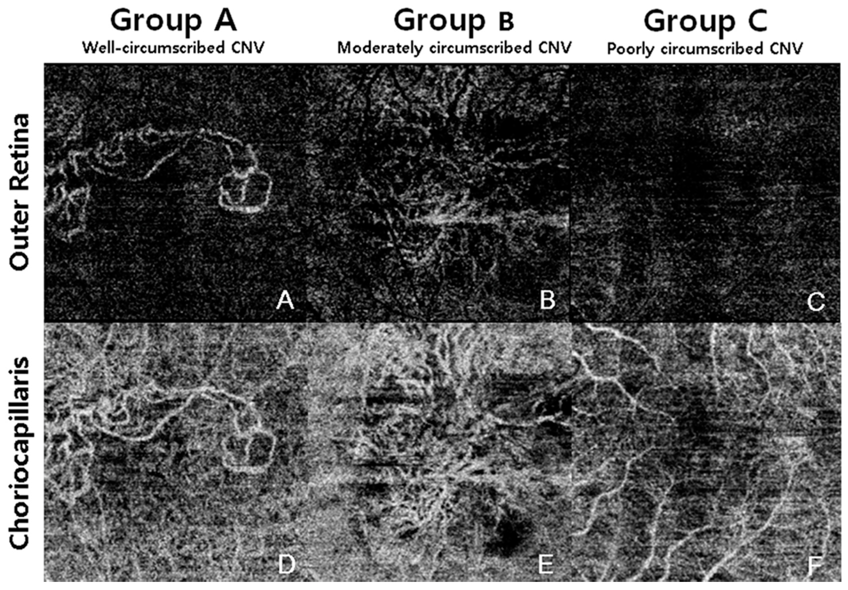
| Group A; well-circumscribed | 18 (34.6) |
| Group B; moderately circumscribed | 24 (46.2) |
| Group C; poorly circumscribed | 10 (19.2) |
| Detection sensitivity, % | 80.70 |
| CNV detection score, points | 2.15 ± 0.72 |
| Group A (Well Circumscribed) | Group B (Moderately Circumscribed) | Group C (Poorly Circumscribed) | p-Value | |
|---|---|---|---|---|
| No. of eyes | 18 | 24 | 10 | - |
| Mean age, years | 74.5 ± 2.0 | 72.8 ± 9.4 | 71.9 ± 9.3 | 0.687 * |
| Sex (female, %) | 10 (55.6) | 9 (37.5) | 5 (50) | 0.491 † |
| BCVA, logMAR | 0.63 ± 0.40 | 0.45 ± 0.30 | 0.51 ± 0.49 | 0.279 ‡ |
| IOP, mmHg | 14.2 ± 3.3 | 15.0 ± 3.1 | 15.8 ± 4.0 | 0.221 ‡ |
| No. of treatment naive eyes | 2 | 5 | 0 | 0.326 § |
| No. of intravitreal injection | 4.2 ± 3.4 | 6.2 ± 5.2 | 6.2 ± 4.7 | 0.838 ‡ |
| Disease duration, months | 14.5 ± 14.1 | 17.9 ± 15.6 | 18.9 ± 14.6 | 0.524 ‡ |
| PED max height, μm | 167.2 ± 167.3 | 163.6 ± 144.9 | 174.7 ± 115.5 | 0.515 ‡ |
| PED max width, μm | 2493.4 ± 1364.6 | 2425.7 ± 1425.2 | 1926.0 ± 1317.7 | 0.421 ‡ |
| Choroidal thickness, μm | 210.0 ± 81.6 | 213.9 ± 103.6 | 235.4 ± 118.7 | 0.422 ‡ |
| Central fovea thickness, μm | 203.0 ± 65.7 | 297.5 ± 179.1 | 272.7 ± 121.7 | 0.534 ‡ |
| CNV area, mm2 | 2.2 ± 1.3 | 1.9 ± 2.5 | - | 0.816 ∥ |
| SRF existence, no. of eyes | 7 | 13 | 4 | 0.561 † |
| SRF height in eyes with SRF, μm | 135.7 ± 76.9 | 100.8 ± 62.5 | 106.0 ± 56.4 | 0.492 ‡ |
| Type 1 CNV | Type 2 CNV | Type 3 CNV | p-Value | |
|---|---|---|---|---|
| No. of eyes | 34 | 9 | 9 | - |
| Sex (female, %) | 13 (38.2) | 2 (22.2) | 9 (100) | 0.01 * |
| Mean age, years | 73.6 ± 9.2 | 71.56 ± 8.1 | 73.4 ± 8.6 | 0.622 † |
| No. of treatment naive eyes | 2 | 3 | 2 | 0.052 * |
| No. of intravitreal injection | 6.3 ± 4.5 | 5.3 ± 5.7 | 2.7 ± 2.1 | 0.121 † |
| IOP, mmHg | 14.09 ± 2.84 | 15.25 ± 3.96 | 14.77 ± 2.24 | 0.232 † |
| BCVA, logMAR | 0.52 ± 0.43 | 0.53 ± 0.29 | 0.53 ± 0.24 | 0.643 ‡ |
| Disease duration, months | 19.4 ± 14.1 | 13.8 ± 16.3 | 10.7 ± 14.1 | 0.064 ‡ |
| PED max height, μm | 168.7 ± 151.9 | 147.4 ± 78.2 | 180.1 ± 171.5 | 0.822 ‡ |
| PED max width, μm | 2466.7 ± 1,398.9 | 2543.8 ± 1,001.3 | 1733 ± 1,470.8 | 0.310 ‡ |
| Choroidal thickness, μm | 232.5 ± 101.1 | 234.2 ± 90.5 | 139.6 ± 51.1 | 0.016 § |
| Center fovea thickness, μm | 252.6 ± 119.3 | 198.5 ± 75.7 | 349.2 ± 214.8 | 0.422 ‡ |
| CNV area, mm2 | 1.95 ± 1.28 | 3.52 ± 3.41 | 0.51 ± 0.83 | 0.006 ∥ |
| SRF height in eyes with SRF, μm | 100.0 ± 51.9 | 104.1 ± 48.9 | 165.0 ± 96.2 | - |
| SRF existence, no. of eyes | 14 | 6 | 4 | 0.466 * |
| Total (n = 52) | Type 1 CNV (n = 34) | Type 2 CNV (n = 9) | Type 3 CNV (n = 9) | p-Value | |
|---|---|---|---|---|---|
| Group A (well-circumscribed) | 18 | 10 | 5 | 3 | |
| Group B (moderately circumscribed) | 24 | 15 | 4 | 5 | |
| Group C (poorly circumscribed) | 10 | 9 | 0 | 1 | |
| Detection sensitivity, % | 80.7 (42/52) | 73.5 (25/34) | 100 (9/9) | 88.9 (8/9) | 0.159 * |
| CNV detection score, point | 2.15 ± 0.72 | 2.03 ± 0.76 | 2.56 ± 0.53 | 2.22 ± 0.63 | 0.158 † |
| Group A | Group B | Group C | p-Value | |
|---|---|---|---|---|
| No. of eyes | 10 | 15 | 9 | |
| Mean age | 77.3 ± 7.60 | 71.6 ± 10.2 | 72.9 ± 9.3 | 0.329 † |
| Sex (female, %) | 6 (60) | 3 (30) | 4 (44.5) | 0.158 † |
| BCVA, logMAR | 0.66 ± 0.51 | 0.40 ± 0.29 | 0.53 ± 0.52 | 0.364 † |
| IOP, mmHg | 13.2 ± 2.8 | 15.1 ± 2.7 | 15.8 ± 4.3 | 0.195 * |
| No. of treatment naive eyes | 0 | 2 | 0 | 0.492 † |
| No. of intravitreal injection | 5.0 ± 2.7 | 7.1 ± 5.1 | 6.3 ± 4.9 | 0.541 ‡ |
| Disease duration, months | 15.6 ± 10.4 | 21.6 ± 15.6 | 19.8 ± 15.2 | 0.597 * |
| PED max height, μm | 1193.3 ± 214.6 | 144.9 ± 122.9 | 181.1 ± 120.6 | 0.72 ‡ |
| PED max width, μm | 2729.2 ± 1473.2 | 2577.2 ± 1386.1 | 1990.9 ± 1380.6 | 0.489 ‡ |
| Choroidal thickness, μm | 205.1 ± 73.9 | 239.5 ± 111.0 | 251.2 ± 114.2 | 0.588 ‡ |
| Center fovea thickness, μm | 214.6 ± 67.3 | 264.8 ± 140.1 | 274.7 ± 128.9 | 0.491 ‡ |
| CNV area, mm2 | 2.31 ± 1.27 | 1.70 ± 1.27 | - | 0.249 |
| SRF existence, number of eyes | 3 | 7 | 4 | 0.747 † |
| SRF height in eyes with SRF, μm | 139.3 ± 57.7 | 79.7 ± 43.0 | 106.0 ± 56.5 | 0.532 ‡ |
© 2019 by the authors. Licensee MDPI, Basel, Switzerland. This article is an open access article distributed under the terms and conditions of the Creative Commons Attribution (CC BY) license (http://creativecommons.org/licenses/by/4.0/).
Share and Cite
Yeo, J.H.; Chung, H.; Kim, J.T. Swept-Source Optical Coherence Tomography Angiography According to the Type of Choroidal Neovascularization. J. Clin. Med. 2019, 8, 1272. https://doi.org/10.3390/jcm8091272
Yeo JH, Chung H, Kim JT. Swept-Source Optical Coherence Tomography Angiography According to the Type of Choroidal Neovascularization. Journal of Clinical Medicine. 2019; 8(9):1272. https://doi.org/10.3390/jcm8091272
Chicago/Turabian StyleYeo, Joon Hyung, Hum Chung, and Jee Taek Kim. 2019. "Swept-Source Optical Coherence Tomography Angiography According to the Type of Choroidal Neovascularization" Journal of Clinical Medicine 8, no. 9: 1272. https://doi.org/10.3390/jcm8091272
APA StyleYeo, J. H., Chung, H., & Kim, J. T. (2019). Swept-Source Optical Coherence Tomography Angiography According to the Type of Choroidal Neovascularization. Journal of Clinical Medicine, 8(9), 1272. https://doi.org/10.3390/jcm8091272


