Determining the Significance of Coronary Plaque Lesions: Physiological Stenosis Severity and Plaque Characteristics


Abstract
:1. Introduction
2. The Significance of Coronary Artery Atherosclerotic Disease
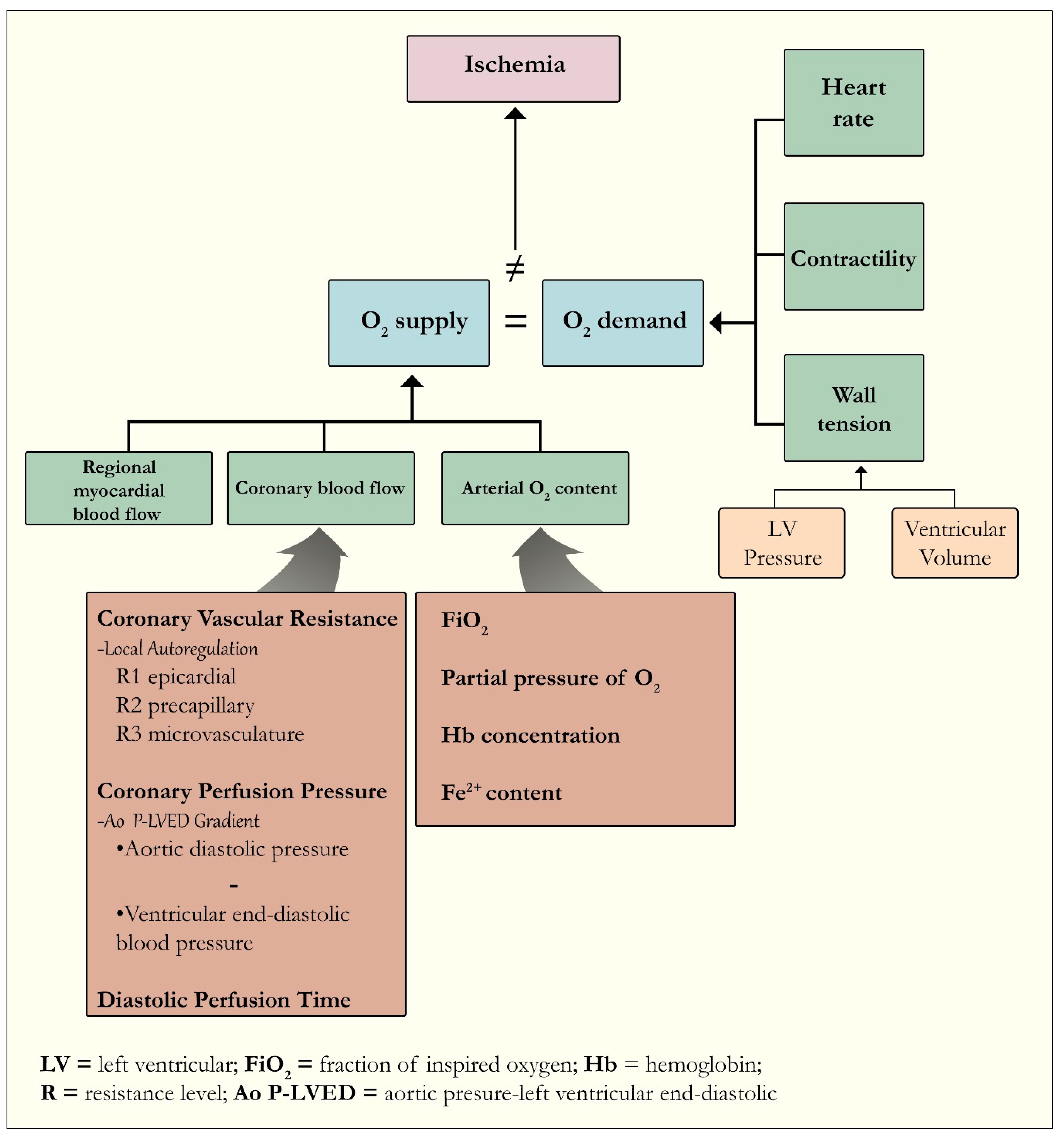
2.1. The Coronary Supply and Myocardial Demand Relationship
2.2. Coronary Artery Disease
“Culprit” Plaque versus “Vulnerable” Plaque
2.3. High-Risk Plaque Characteristics
2.4. Morphological and Functional Assessment Tools
Intravascular Ultrasound (IVUS) and Optical Coherence Tomography (OCT)
3. Principles of the Invasive Physiologic Assessment Tools
3.1. Fractional Flow Reserve
3.2. Instantaneous Wave-Free Ratio (iFR) and Other Physiologic Tools
4. The Era of Physiological Stenosis Severity Assessment
4.1. Physiologic-guided Culprit Vessel versus Complete Revascularization: Significance and Outcomes
4.2. Functional Assessment of Coronary Lesions in Special Populations: Patients with Severe Aortic Stenosis
4.3. Coronary Physiology Assessment in the Catheterization Laboratory
5. The Implications of the Addition of Plaque Characteristics to Traditional FFR Assessment
5.1. Noninvasive Plaque Morphology Evaluation: Focus on Coronary CT Angiography
5.1.1. The Evolution of CCTA
5.1.2. The Link between Plaque Characteristics on CCTA and Hemodynamic Significance by FFR
5.1.3. Unanswered Questions for Patients with Fractional Flow Reserve > 0.80
5.2. Intravascular Imaging Techniques: IVUS and OCT
5.2.1. IVUS and FFR
5.2.2. OCT and FFR
5.3. The Future of Coronary Revascularization
6. Conclusions
Author Contributions
Funding
Conflicts of Interest
References
- Bech, G.J.; De Bruyne, B.; Pijls, N.H.; de Muinck, E.D.; Hoorntje, J.C.; Escaned, J.; Stella, P.R.; Boersma, E.; Bartunek, J.; Koolen, J.J.; et al. Fractional flow reserve to determine the appropriateness of angioplasty in moderate coronary stenosis: A randomized trial. Circulation 2001, 103, 2928–2934. [Google Scholar] [CrossRef] [PubMed] [Green Version]
- Neumann, F.J.; Sousa-Uva, M.; Ahlsson, A.; Alfonso, F.; Banning, A.P.; Benedetto, U.; Byrne, R.A.; Collet, J.P.; Falk, V.; Head, S.J.; et al. 2018 ESC/EACTS Guidelines on myocardial revascularization. Eur. Heart J. 2019, 40, 87–165. [Google Scholar] [CrossRef]
- Zimmermann, F.M.; Ferrara, A.; Johnson, N.P.; van Nunen, L.X.; Escaned, J.; Albertsson, P.; Erbel, R.; Legrand, V.; Gwon, H.C.; Remkes, W.S.; et al. Deferral vs. performance of percutaneous coronary intervention of functionally non-significant coronary stenosis: 15-year follow-up of the DEFER trial. Eur. Heart J. 2015, 36, 3182–3188. [Google Scholar] [CrossRef] [Green Version]
- van Nunen, L.X.; Zimmermann, F.M.; Tonino, P.A.L.; Barbato, E.; Baumbach, A.; Engstrøm, T.; Klauss, V.; MacCarthy, P.A.; Manoharan, G.; Oldroyd, K.G.; et al. Fractional flow reserve versus angiography for guidance of PCI in patients with multivessel coronary artery disease (FAME): 5-year follow-up of a randomised controlled trial. Lancet 2015, 386, 1853–1860. [Google Scholar] [CrossRef]
- Xaplanteris, P.; Fournier, S.; Pijls, N.H.J.; Fearon, W.F.; Barbato, E.; Tonino, P.A.L.; Engstrom, T.; Kaab, S.; Dambrink, J.H.; Rioufol, G.; et al. Five-Year outcomes with PCI guided by fractional flow reserve. N. Engl. J. Med. 2018, 379, 250–259. [Google Scholar] [CrossRef] [PubMed]
- Lee, J.M.; Jung, J.H.; Hwang, D.; Park, J.; Fan, Y.; Na, S.H.; Doh, J.H.; Nam, C.W.; Shin, E.S.; Koo, B.K. Coronary flow reserve and microcirculatory resistance in patients with intermediate coronary stenosis. J. Am. Coll. Cardiol. 2016, 67, 1158–1169. [Google Scholar] [CrossRef]
- Xaplanteris, P.; de Hemptinne, Q. The Relationship Between Plaque Morphology and its Hemodynamic Significance. 2018. Available online: https://www.acc.org/latest-in-cardiology/articles/2018/10/30/09/08/the-relationship-between-plaque-morphology-and-its-hemodynamic-significance (accessed on 31 December 2019).
- Calvert, P.A.; Obaid, D.R.; O’Sullivan, M.; Shapiro, L.M.; McNab, D.; Densem, C.G.; Schofield, P.M.; Braganza, D.; Clarke, S.C.; Ray, K.K.; et al. Association between IVUS findings and adverse outcomes in patients with coronary artery disease: The VIVA (VH-IVUS in vulnerable atherosclerosis) study. JACC Cardiovasc. Imaging 2011, 4, 894–901. [Google Scholar] [CrossRef] [Green Version]
- Puchner, S.B.; Liu, T.; Mayrhofer, T.; Truong, Q.A.; Lee, H.; Fleg, J.L.; Nagurney, J.T.; Udelson, J.E.; Hoffmann, U.; Ferencik, M. High-risk plaque detected on coronary CT angiography predicts acute coronary syndromes independent of significant stenosis in acute chest pain: Results from the ROMICAT-II trial. J. Am. Coll. Cardiol. 2014, 64, 684–692. [Google Scholar] [CrossRef] [Green Version]
- Lee, J.M.; Choi, G.; Koo, B.K.; Hwang, D.; Park, J.; Zhang, J.; Kim, K.J.; Tong, Y.; Kim, H.J.; Grady, L.; et al. Identification of high-risk plaques destined to cause acute coronary syndrome using coronary computed tomographic angiography and computational fluid dynamics. JACC Cardiovasc. Imaging 2019, 12, 1032–1043. [Google Scholar] [CrossRef]
- Lanza, G.A.; Crea, F. Primary coronary microvascular dysfunction: Clinical presentation, pathophysiology, and management. Circulation 2010, 121, 2317–2325. [Google Scholar] [CrossRef] [Green Version]
- Taqueti, V.R.; Di Carli, M.F. Coronary microvascular disease pathogenic mechanisms and therapeutic options: JACC state-of-the-art review. J. Am. Coll. Cardiol. 2018, 72, 2625–2641. [Google Scholar] [CrossRef] [PubMed]
- Libby, P.; Theroux, P. Pathophysiology of coronary artery disease. Circulation 2005, 111, 3481–3488. [Google Scholar] [CrossRef] [PubMed] [Green Version]
- Khuddus, M.A.; Pepine, C.J.; Handberg, E.M.; Bairey Merz, C.N.; Sopko, G.; Bavry, A.A.; Denardo, S.J.; McGorray, S.P.; Smith, K.M.; Sharaf, B.L.; et al. An intravascular ultrasound analysis in women experiencing chest pain in the absence of obstructive coronary artery disease: A substudy from the National Heart, Lung and Blood Institute-Sponsored Women’s Ischemia Syndrome Evaluation (WISE). J. Interv. Cardiol. 2010, 23, 511–519. [Google Scholar] [CrossRef] [PubMed]
- Murthy, V.L.; Naya, M.; Taqueti, V.R.; Foster, C.R.; Gaber, M.; Hainer, J.; Dorbala, S.; Blankstein, R.; Rimoldi, O.; Camici, P.G.; et al. Effects of sex on coronary microvascular dysfunction and cardiac outcomes. Circulation 2014, 129, 2518–2527. [Google Scholar] [CrossRef] [Green Version]
- Taqueti, V.R.; Hachamovitch, R.; Murthy, V.L.; Naya, M.; Foster, C.R.; Hainer, J.; Dorbala, S.; Blankstein, R.; Di Carli, M.F. Global coronary flow reserve is associated with adverse cardiovascular events independently of luminal angiographic severity and modifies the effect of early revascularization. Circulation 2015, 131, 19–27. [Google Scholar] [CrossRef] [Green Version]
- Deussen, A.; Ohanyan, V.; Jannasch, A.; Yin, L.; Chilian, W. Mechanisms of metabolic coronary flow regulation. J. Mol. Cell. Cardiol. 2012, 52, 794–801. [Google Scholar] [CrossRef]
- Feigl, E.O. Coronary physiology. Physiol. Rev. 1983, 63, 1–205. [Google Scholar] [CrossRef]
- Duncker, D.J.; Merkus, D. Acute adaptations of the coronary circulation to exercise. Cell Biochem. Biophys. 2005, 43, 17–36. [Google Scholar] [CrossRef]
- Camici, P.G.; Crea, F. Coronary microvascular dysfunction. N. Engl. J. Med. 2007, 356, 830–840. [Google Scholar] [CrossRef] [Green Version]
- Deussen, A.; Loncar, R. Metabolic aspects of myocardial ischemia. Z. Kardiol. 1998, 87 (Suppl. 2), 37–40. [Google Scholar] [CrossRef]
- Crossman, D.C. The pathophysiology of myocardial ischaemia. Heart 2004, 90, 576–580. [Google Scholar] [CrossRef] [PubMed] [Green Version]
- Heusch, G. Myocardial ischemia: Lack of coronary blood flow or myocardial oxygen supply/demand imbalance? Circ. Res. 2016, 119, 194–196. [Google Scholar] [CrossRef] [PubMed] [Green Version]
- Sandoval, Y.; Jaffe, A.S. Type 2 myocardial infarction: JACC review topic of the week. J. Am. Coll. Cardiol. 2019, 73, 1846–1860. [Google Scholar] [CrossRef] [PubMed]
- Libby, P. Inflammation in atherosclerosis. Nature 2002, 420, 868–874. [Google Scholar] [CrossRef] [PubMed]
- Naghavi, M.; Libby, P.; Falk, E.; Casscells, S.W.; Litovsky, S.; Rumberger, J.; Badimon, J.J.; Stefanadis, C.; Moreno, P.; Pasterkamp, G.; et al. From vulnerable plaque to vulnerable patient: A call for new definitions and risk assessment strategies: Part I. Circulation 2003, 108, 1664–1672. [Google Scholar] [CrossRef] [PubMed]
- Saybolt, M.D.; Lilly, S.M.; Patel, D.; Hamamdzic, D.; Llano, R.; Fenning, R.S.; Madden, S.; Wilensky, R.L. The vulnerable artery: Early and rapid deposition of lipid in coronary arteries is associated with subsequent development of thin-cap fibroatheromas. EuroIntervention 2016, 11, e1612–e1618. [Google Scholar] [CrossRef] [PubMed]
- Janoudi, A.; Shamoun, F.E.; Kalavakunta, J.K.; Abela, G.S. Cholesterol crystal induced arterial inflammation and destabilization of atherosclerotic plaque. Eur. Heart J. 2016, 37, 1959–1967. [Google Scholar] [CrossRef] [Green Version]
- Valente, A.J.; Irimpen, A.M.; Siebenlist, U.; Chandrasekar, B. OxLDL induces endothelial dysfunction and death via TRAF3IP2: Inhibition by HDL3 and AMPK activators. Free Radic. Biol. Med. 2014, 70, 117–128. [Google Scholar] [CrossRef] [Green Version]
- Vandenabeele, P.; Galluzzi, L.; Vanden Berghe, T.; Kroemer, G. Molecular mechanisms of necroptosis: An ordered cellular explosion. Nat. Rev. Mol. Cell Biol. 2010, 11, 700–714. [Google Scholar] [CrossRef]
- Karunakaran, D.; Geoffrion, M.; Wei, L.; Gan, W.; Richards, L.; Shangari, P.; DeKemp, E.M.; Beanlands, R.A.; Perisic, L.; Maegdefessel, L.; et al. Targeting macrophage necroptosis for therapeutic and diagnostic interventions in atherosclerosis. Sci. Adv. 2016, 2, e1600224. [Google Scholar] [CrossRef] [Green Version]
- Stefanadis, C.; Antoniou, C.K.; Tsiachris, D.; Pietri, P. Coronary atherosclerotic vulnerable plaque: Current perspectives. J. Am. Heart Assoc. 2017, 6, e005543. [Google Scholar] [CrossRef] [Green Version]
- Glagov, S.; Weisenberg, E.; Zarins, C.K.; Stankunavicius, R.; Kolettis, G.J. Compensatory enlargement of human atherosclerotic coronary arteries. N. Engl. J. Med. 1987, 316, 1371–1375. [Google Scholar] [CrossRef] [PubMed]
- Clarkson, T.B. Remodeling of coronary arteries in human and nonhuman primates. JAMA J. Am. Med. Assoc. 1994, 271, 289–294. [Google Scholar] [CrossRef]
- Schoenhagen, P.; Ziada, K.M.; Kapadia, S.R.; Crowe, T.D.; Nissen, S.E.; Tuzcu, E.M. Extent and direction of arterial remodeling in stable versus unstable coronary syndromes: An intravascular ultrasound study. Circulation 2000, 101, 598–603. [Google Scholar] [CrossRef] [PubMed] [Green Version]
- Virmani, R.; Kolodgie, F.D.; Burke, A.P.; Farb, A.; Schwartz, S.M. Lessons from sudden coronary death: A comprehensive morphological classification scheme for atherosclerotic lesions. Arterioscler. Thromb. Vasc. Biol. 2000, 20, 1262–1275. [Google Scholar] [CrossRef] [PubMed] [Green Version]
- Farb, A.; Burke, A.P.; Tang, A.L.; Liang, T.Y.; Mannan, P.; Smialek, J.; Virmani, R. Coronary plaque erosion without rupture into a lipid core. A frequent cause of coronary thrombosis in sudden coronary death. Circulation 1996, 93, 1354–1363. [Google Scholar] [CrossRef] [PubMed]
- Ambrose, J.A.; Tannenbaum, M.A.; Alexopoulos, D.; Hjemdahl-Monsen, C.E.; Leavy, J.; Weiss, M.; Borrico, S.; Gorlin, R.; Fuster, V. Angiographic progression of coronary artery disease and the development of myocardial infarction. J. Am. Coll. Cardiol. 1988, 12, 56–62. [Google Scholar] [CrossRef] [Green Version]
- Virmani, R.; Burke, A.P.; Kolodgie, F.D.; Farb, A. Pathology of the thin-cap fibroatheroma: A type of vulnerable plaque. J. Interv. Cardiol. 2003, 16, 267–272. [Google Scholar] [CrossRef]
- Kolodgie, F.D.; Burke, A.P.; Farb, A.; Gold, H.K.; Yuan, J.; Narula, J.; Finn, A.V.; Virmani, R. The thin-cap fibroatheroma: A type of vulnerable plaque: The major precursor lesion to acute coronary syndromes. Curr. Opin. Cardiol. 2001, 16, 285–292. [Google Scholar] [CrossRef]
- Waxman, S.; Mittleman, M.A.; Zarich, S.W.; Fitzpatrick, P.J.; Lewis, S.M.; Leeman, D.E.; Shubrooks, S.J.; Snyder, J.T.; Muller, J.E.; Nesto, R.W. Angioscopic assessment of coronary lesions underlying thrombus. Am. J. Cardiol. 1997, 79, 1106–1109. [Google Scholar] [CrossRef]
- Yock, P.G.; Linker, D.T.; Angelsen, B.A. Two-Dimensional intravascular ultrasound: Technical development and initial clinical experience. J. Am. Soc. Echocardiogr. 1989, 2, 296–304. [Google Scholar] [CrossRef]
- Mintz, G.S.; Nissen, S.E.; Anderson, W.D.; Bailey, S.R.; Erbel, R.; Fitzgerald, P.J.; Pinto, F.J.; Rosenfield, K.; Siegel, R.J.; Tuzcu, E.M.; et al. American College of Cardiology clinical expert consensus document on standards for the acquisition, measurement and reporting of intravascular ultrasound studies (IVUS): A report of the American College of Cardiology task force on clinical expert consensus documents. J. Am. Coll. Cardiol. 2001, 37, 1478–1492. [Google Scholar] [PubMed] [Green Version]
- Schoenhagen, P.; Nissen, S.E.; Tuzcu, E.M. Coronary arterial remodeling: From bench to bedside. Curr. Atheroscler. Rep. 2003, 5, 150–154. [Google Scholar] [CrossRef] [PubMed]
- Ge, J.; Chirillo, F.; Schwedtmann, J.; Gorge, G.; Haude, M.; Baumgart, D.; Shah, V.; von Birgelen, C.; Sack, S.; Boudoulas, H.; et al. Screening of ruptured plaques in patients with coronary artery disease by intravascular ultrasound. Heart 1999, 81, 621–627. [Google Scholar] [CrossRef] [PubMed] [Green Version]
- Kim, S.H.; Hong, M.K.; Park, D.W.; Lee, S.W.; Kim, Y.H.; Lee, C.W.; Kim, J.J.; Park, S.W.; Park, S.J. Impact of plaque characteristics analyzed by intravascular ultrasound on long-term clinical outcomes. Am. J. Cardiol. 2009, 103, 1221–1226. [Google Scholar] [CrossRef] [PubMed]
- Sano, K.; Kawasaki, M.; Ishihara, Y.; Okubo, M.; Tsuchiya, K.; Nishigaki, K.; Zhou, X.; Minatoguchi, S.; Fujita, H.; Fujiwara, H. Assessment of vulnerable plaques causing acute coronary syndrome using integrated backscatter intravascular ultrasound. J. Am. Coll. Cardiol. 2006, 47, 734–741. [Google Scholar] [CrossRef]
- Goertz, D.E.; Frijlink, M.E.; Tempel, D.; van Damme, L.C.; Krams, R.; Schaar, J.A.; Ten Cate, F.J.; Serruys, P.W.; de Jong, N.; van der Steen, A.F. Contrast harmonic intravascular ultrasound: A feasibility study for vasa vasorum imaging. Invest. Radiol. 2006, 41, 631–638. [Google Scholar] [CrossRef]
- Low, A.F.; Tearney, G.J.; Bouma, B.E.; Jang, I.K. Technology insight: Optical coherence tomography—Current status and future development. Nat. Clin. Pract. Cardiovasc. Med. 2006, 3, 154–162. [Google Scholar] [CrossRef]
- Yabushita, H.; Bouma, B.E.; Houser, S.L.; Aretz, H.T.; Jang, I.K.; Schlendorf, K.H.; Kauffman, C.R.; Shishkov, M.; Kang, D.H.; Halpern, E.F.; et al. Characterization of human atherosclerosis by optical coherence tomography. Circulation 2002, 106, 1640–1645. [Google Scholar] [CrossRef]
- Sinclair, H.; Bourantas, C.; Bagnall, A.; Mintz, G.S.; Kunadian, V. OCT for the identification of vulnerable plaque in acute coronary syndrome. JACC Cardiovasc. Imaging 2015, 8, 198–209. [Google Scholar] [CrossRef] [Green Version]
- Pijls, N.H.; Van Gelder, B.; Van der Voort, P.; Peels, K.; Bracke, F.A.; Bonnier, H.J.; el Gamal, M.I. Fractional flow reserve: A useful index to evaluate the influence of an epicardial coronary stenosis on myocardial blood flow. Circulation 1995, 92, 3183–3193. [Google Scholar] [CrossRef] [PubMed]
- Pijls, N.H.; van Son, J.A.; Kirkeeide, R.L.; De Bruyne, B.; Gould, K.L. Experimental basis of determining maximum coronary, myocardial, and collateral blood flow by pressure measurements for assessing functional stenosis severity before and after percutaneous transluminal coronary angioplasty. Circulation 1993, 87, 1354–1367. [Google Scholar] [CrossRef] [PubMed] [Green Version]
- De Bruyne, B.; Pijls, N.H.; Bartunek, J.; Kulecki, K.; Bech, J.W.; De Winter, H.; Van Crombrugge, P.; Heyndrickx, G.R.; Wijns, W. Fractional flow reserve in patients with prior myocardial infarction. Circulation 2001, 104, 157–162. [Google Scholar] [CrossRef] [PubMed] [Green Version]
- Bartunek, J.; Van Schuerbeeck, E.; de Bruyne, B. Comparison of exercise electrocardiography and dobutamine echocardiography with invasively assessed myocardial fractional flow reserve in evaluation of severity of coronary arterial narrowing. Am. J. Cardiol. 1997, 79, 478–481. [Google Scholar] [CrossRef]
- Pijls, N.H.; De Bruyne, B.; Peels, K.; Van Der Voort, P.H.; Bonnier, H.J.; Bartunek, J.; Koolen, J.J. Measurement of fractional flow reserve to assess the functional severity of coronary-artery stenoses. N. Engl. J. Med. 1996, 334, 1703–1708. [Google Scholar] [CrossRef] [PubMed]
- Lotfi, A.; Jeremias, A.; Fearon, W.F.; Feldman, M.D.; Mehran, R.; Messenger, J.C.; Grines, C.L.; Dean, L.S.; Kern, M.J.; Klein, L.W.; et al. Expert consensus statement on the use of fractional flow reserve, intravascular ultrasound, and optical coherence tomography: A consensus statement of the Society of Cardiovascular Angiography and Interventions. Catheter. Cardiovasc. Interv. 2014, 83, 509–518. [Google Scholar] [CrossRef]
- Levine, G.N.; Bates, E.R.; Blankenship, J.C.; Bailey, S.R.; Bittl, J.A.; Cercek, B.; Chambers, C.E.; Ellis, S.G.; Guyton, R.A.; Hollenberg, S.M.; et al. 2011 ACCF/AHA/SCAI guideline for percutaneous coronary intervention: A report of the American College of Cardiology Foundation/American Heart Association task force on practice guidelines and the Society for Cardiovascular Angiography and Interventions. Circulation 2011, 124, e574–e651. [Google Scholar]
- Agarwal, S.K.; Kasula, S.; Edupuganti, M.M.; Raina, S.; Shailesh, F.; Almomani, A.; Payne, J.J.; Pothineni, N.V.; Uretsky, B.F.; Hakeem, A. Clinical decision-making for the hemodynamic “Gray zone” (FFR 0.75-0.80) and long-term outcomes. J. Invasive Cardiol. 2017, 29, 371–376. [Google Scholar] [CrossRef]
- Adjedj, J.; De Bruyne, B.; Flore, V.; Di Gioia, G.; Ferrara, A.; Pellicano, M.; Toth, G.G.; Bartunek, J.; Vanderheyden, M.; Heyndrickx, G.R.; et al. Significance of intermediate values of fractional flow reserve in patients with coronary artery disease. Circulation 2016, 133, 502–508. [Google Scholar] [CrossRef] [Green Version]
- Ahn, J.M.; Park, D.W.; Shin, E.S.; Koo, B.K.; Nam, C.W.; Doh, J.H.; Kim, J.H.; Chae, I.H.; Yoon, J.H.; Her, S.H.; et al. Fractional flow reserve and cardiac events in coronary artery disease: data from a prospective IRIS-FFR registry (Interventional Cardiology Research Incooperation Society Fractional Flow Reserve). Circulation 2017, 135, 2241–2251. [Google Scholar] [CrossRef]
- Jeremias, A.; Maehara, A.; Genereux, P.; Asrress, K.N.; Berry, C.; De Bruyne, B.; Davies, J.E.; Escaned, J.; Fearon, W.F.; Gould, K.L.; et al. Multicenter core laboratory comparison of the instantaneous wave-free ratio and resting Pd/Pa with fractional flow reserve: The RESOLVE study. J. Am. Coll. Cardiol. 2014, 63, 1253–1261. [Google Scholar] [CrossRef] [PubMed] [Green Version]
- Sen, S.; Escaned, J.; Malik, I.S.; Mikhail, G.W.; Foale, R.A.; Mila, R.; Tarkin, J.; Petraco, R.; Broyd, C.; Jabbour, R.; et al. Development and validation of a new adenosine-independent index of stenosis severity from coronary wave-intensity analysis: Results of the ADVISE (A Denosine Vasodilator Independent Stenosis Evaluation) study. J. Am. Coll. Cardiol. 2012, 59, 1392–1402. [Google Scholar] [CrossRef] [PubMed] [Green Version]
- Samady, H.; Gogas, B.D. Does flow during rest and relaxation suffice? J. Am. Coll. Cardiol. 2013, 61, 1436–1439. [Google Scholar] [CrossRef] [PubMed] [Green Version]
- Johnson, N.P.; Kirkeeide, R.L.; Asrress, K.N.; Fearon, W.F.; Lockie, T.; Marques, K.M.; Pyxaras, S.A.; Rolandi, M.C.; van ‘t Veer, M.; De Bruyne, B.; et al. Does the instantaneous wave-free ratio approximate the fractional flow reserve? J. Am. Coll. Cardiol. 2013, 61, 1428–1435. [Google Scholar] [CrossRef] [Green Version]
- Berry, C.; van ‘t Veer, M.; Witt, N.; Kala, P.; Bocek, O.; Pyxaras, S.A.; McClure, J.D.; Fearon, W.F.; Barbato, E.; Tonino, P.A.; et al. VERIFY (VERification of instantaneous wave-free ratio and fractional flow reserve for the assessment of coronary artery stenosis severity in everyday practice): A multicenter study in consecutive patients. J. Am. Coll. Cardiol. 2013, 61, 1421–1427. [Google Scholar] [CrossRef] [Green Version]
- De Rosa, S.; Polimeni, A.; De Velli, G.; Conte, M.; Sorrentino, S.; Spaccarotella, C.; Mongiardo, A.; Sabatino, J.; Contarini, M.; Indolfi, C. Reliability of instantaneous wave-free ratio (iFR) for the evaluation of left main coronary artery lesions. J. Clin. Med. 2019, 8, 1143. [Google Scholar] [CrossRef] [Green Version]
- De Rosa, S.; Polimeni, A.; Petraco, R.; Davies, J.E.; Indolfi, C. Diagnostic performance of the instantaneous wave-free ratio: Comparison with fractional flow reserve. Circ. Cardiovasc. Interv. 2018, 11, e004613. [Google Scholar] [CrossRef]
- Indolfi, C.; Mongiardo, A.; Spaccarotella, C.; Torella, D.; Caiazzo, G.; Polimeni, A.; Sorrentino, S.; Micieli, M.; Sabatino, J.; Curcio, A.; et al. The instantaneous wave-free ratio (iFR) for evaluation of non-culprit lesions in patients with acute coronary syndrome and multivessel disease. Int. J. Cardiol. 2015, 178, 46–54. [Google Scholar] [CrossRef]
- Gould, K.L.; Lipscomb, K.; Hamilton, G.W. Physiologic basis for assessing critical coronary stenosis. Instantaneous flow response and regional distribution during coronary hyperemia as measures of coronary flow reserve. Am. J. Cardiol. 1974, 33, 87–94. [Google Scholar] [CrossRef]
- Fearon, W.F.; Balsam, L.B.; Farouque, H.M.; Caffarelli, A.D.; Robbins, R.C.; Fitzgerald, P.J.; Yock, P.G.; Yeung, A.C. Novel index for invasively assessing the coronary microcirculation. Circulation 2003, 107, 3129–3132. [Google Scholar] [CrossRef]
- Melikian, N.; Vercauteren, S.; Fearon, W.F.; Cuisset, T.; MacCarthy, P.A.; Davidavicius, G.; Aarnoudse, W.; Bartunek, J.; Vanderheyden, M.; Wyffels, E.; et al. Quantitative assessment of coronary microvascular function in patients with and without epicardial atherosclerosis. EuroIntervention 2010, 5, 939–945. [Google Scholar] [CrossRef] [PubMed]
- Luo, C.; Long, M.; Hu, X.; Huang, Z.; Hu, C.; Gao, X.; Du, Z. Thermodilution-Derived coronary microvascular resistance and flow reserve in patients with cardiac syndrome X. Circ. Cardiovasc. Interv. 2014, 7, 43–48. [Google Scholar] [CrossRef] [PubMed] [Green Version]
- Hannan, E.L.; Samadashvili, Z.; Walford, G.; Holmes, D.R., Jr.; Jacobs, A.K.; Stamato, N.J.; Venditti, F.J.; Sharma, S.; King, S.B., III. Culprit vessel percutaneous coronary intervention versus multivessel and staged percutaneous coronary intervention for ST-segment elevation myocardial infarction patients with multivessel disease. JACC Cardiovasc. Interv. 2010, 3, 22–31. [Google Scholar] [CrossRef] [PubMed] [Green Version]
- Kornowski, R.; Mehran, R.; Dangas, G.; Nikolsky, E.; Assali, A.; Claessen, B.E.; Gersh, B.J.; Wong, S.C.; Witzenbichler, B.; Guagliumi, G.; et al. Prognostic impact of staged versus “one-time” multivessel percutaneous intervention in acute myocardial infarction: Analysis from the HORIZONS-AMI (harmonizing outcomes with revascularization and stents in acute myocardial infarction) trial. J. Am. Coll. Cardiol. 2011, 58, 704–711. [Google Scholar] [CrossRef] [PubMed] [Green Version]
- Hideo-Kajita, A.; Garcia-Garcia, H.M.; Kuku, K.O.; Beyene, S.S.; Azizi, V.; Meirovich, Y.F.; Melaku, G.D.; Dheendsa, A.; Brathwaite, E.J.; Desale, S.; et al. Clinical outcomes of complete revascularization using either angiography-guided or fractional flow reserve-guided drug-eluting stent implantation in non-culprit vessels in ST elevation myocardial infarction patients: Insights from a study based on a systematic review and meta-analysis. Int. J. Cardiovasc. Imaging 2018, 34, 1349–1364. [Google Scholar]
- Pijls, N.H.; Fearon, W.F.; Tonino, P.A.; Siebert, U.; Ikeno, F.; Bornschein, B.; van’t Veer, M.; Klauss, V.; Manoharan, G.; Engstrom, T.; et al. Fractional flow reserve versus angiography for guiding percutaneous coronary intervention in patients with multivessel coronary artery disease: 2-Year follow-up of the FAME (Fractional Flow Reserve Versus Angiography for Multivessel Evaluation) study. J. Am. Coll. Cardiol. 2010, 56, 177–184. [Google Scholar] [CrossRef] [Green Version]
- Layland, J.; Oldroyd, K.G.; Curzen, N.; Sood, A.; Balachandran, K.; Das, R.; Junejo, S.; Ahmed, N.; Lee, M.M.; Shaukat, A.; et al. Fractional flow reserve vs. angiography in guiding management to optimize outcomes in non-ST-segment elevation myocardial infarction: The British Heart Foundation FAMOUS-NSTEMI randomized trial. Eur. Heart J. 2015, 36, 100–111. [Google Scholar]
- Levine, G.N.; Bates, E.R.; Blankenship, J.C.; Bailey, S.R.; Bittl, J.A.; Cercek, B.; Chambers, C.E.; Ellis, S.G.; Guyton, R.A.; Hollenberg, S.M.; et al. 2015 ACC/AHA/SCAI focused update on primary percutaneous coronary intervention for patients with st-elevation myocardial infarction: An update of the 2011 ACCF/AHA/SCAI guideline for percutaneous coronary intervention and the 2013 ACCF/AHA guideline for the management of st-elevation myocardial infarction: A Report of the American College of Cardiology/American Heart Association Task Force on Clinical Practice guidelines and the Society for Cardiovascular Angiography and Interventions. Circulation 2016, 133, 1135–1147. [Google Scholar]
- Hoole, S.P.; Brown, A.J.; Jaworski, C.; McCormick, L.M.; Clarke, S.C.; West, N.E. Interpretation of fractional flow reserve in ST-elevation myocardial infarction culprit lesions. Coron. Artery Dis. 2015, 26, 495–502. [Google Scholar] [CrossRef]
- Cuculi, F.; Dall’Armellina, E.; Manlhiot, C.; De Caterina, A.R.; Colyer, S.; Ferreira, V.; Morovat, A.; Prendergast, B.D.; Forfar, J.C.; Alp, N.J.; et al. Early change in invasive measures of microvascular function can predict myocardial recovery following PCI for ST-elevation myocardial infarction. Eur. Heart J. 2014, 35, 1971–1980. [Google Scholar] [CrossRef] [Green Version]
- Dziewierz, A.; Siudak, Z.; Rakowski, T.; Zasada, W.; Dubiel, J.S.; Dudek, D. Impact of multivessel coronary artery disease and noninfarct-related artery revascularization on outcome of patients with ST-elevation myocardial infarction transferred for primary percutaneous coronary intervention (from the EUROTRANSFER Registry). Am. J. Cardiol. 2010, 106, 342–347. [Google Scholar] [CrossRef] [PubMed]
- Goldstein, J.A.; Demetriou, D.; Grines, C.L.; Pica, M.; Shoukfeh, M.; O’Neill, W.W. Multiple complex coronary plaques in patients with acute myocardial infarction. N. Engl. J. Med. 2000, 343, 915–922. [Google Scholar] [CrossRef] [PubMed]
- de Waard, G.A.; Hollander, M.R.; Teunissen, P.F.; Jansen, M.F.; Eerenberg, E.S.; Beek, A.M.; Marques, K.M.; van de Ven, P.M.; Garrelds, I.M.; Danser, A.H.; et al. Changes in coronary blood flow after acute myocardial infarction: Insights from a patient study and an experimental porcine model. JACC Cardiovasc. Interv. 2016, 9, 602–613. [Google Scholar] [CrossRef]
- Lee, J.M.; Kim, H.K.; Lim, K.S.; Park, J.K.; Choi, K.H.; Park, J.; Hwang, D.; Rhee, T.M.; Yang, J.H.; Shin, E.S.; et al. Influence of local myocardial damage on index of microcirculatory resistance and fractional flow reserve in target and nontarget vascular territories in a porcine microvascular injury model. JACC Cardiovasc. Interv. 2018, 11, 717–724. [Google Scholar] [CrossRef] [PubMed]
- Smits, P.C.; Abdel-Wahab, M.; Neumann, F.J.; Boxma-de Klerk, B.M.; Lunde, K.; Schotborgh, C.E.; Piroth, Z.; Horak, D.; Wlodarczak, A.; Ong, P.J.; et al. Fractional flow reserve-guided multivessel angioplasty in myocardial infarction. N. Engl. J. Med. 2017, 376, 1234–1244. [Google Scholar] [CrossRef]
- Wald, D.S.; Morris, J.K.; Wald, N.J.; Chase, A.J.; Edwards, R.J.; Hughes, L.O.; Berry, C.; Oldroyd, K.G.; Investigators, P. Randomized trial of preventive angioplasty in myocardial infarction. N. Engl. J. Med. 2013, 369, 1115–1123. [Google Scholar] [CrossRef] [PubMed] [Green Version]
- Toma, M.; Buller, C.E.; Westerhout, C.M.; Fu, Y.; O’Neill, W.W.; Holmes, D.R., Jr.; Hamm, C.W.; Granger, C.B.; Armstrong, P.W.; APEX-AMI Investigators. Non-Culprit coronary artery percutaneous coronary intervention during acute ST-segment elevation myocardial infarction: Insights from the APEX-AMI trial. Eur. Heart J. 2010, 31, 1701–1707. [Google Scholar]
- Muller, O.; Mangiacapra, F.; Ntalianis, A.; Verhamme, K.M.; Trana, C.; Hamilos, M.; Bartunek, J.; Vanderheyden, M.; Wyffels, E.; Heyndrickx, G.R.; et al. Long-Term follow-up after fractional flow reserve-guided treatment strategy in patients with an isolated proximal left anterior descending coronary artery stenosis. JACC Cardiovasc. Interv. 2011, 4, 1175–1182. [Google Scholar] [CrossRef] [Green Version]
- Li, J.; Elrashidi, M.Y.; Flammer, A.J.; Lennon, R.J.; Bell, M.R.; Holmes, D.R.; Bresnahan, J.F.; Rihal, C.S.; Lerman, L.O.; Lerman, A. Long-Term outcomes of fractional flow reserve-guided vs. angiography-guided percutaneous coronary intervention in contemporary practice. Eur. Heart J. 2013, 34, 1375–1383. [Google Scholar]
- Goel, S.S.; Ige, M.; Tuzcu, E.M.; Ellis, S.G.; Stewart, W.J.; Svensson, L.G.; Lytle, B.W.; Kapadia, S.R. Severe aortic stenosis and coronary artery disease—Implications for management in the transcatheter aortic valve replacement era: A comprehensive review. J. Am. Coll. Cardiol. 2013, 62, 1–10. [Google Scholar] [CrossRef] [Green Version]
- Wiegerinck, E.M.; van de Hoef, T.P.; Rolandi, M.C.; Yong, Z.; van Kesteren, F.; Koch, K.T.; Vis, M.M.; de Mol, B.A.; Piek, J.J.; Baan, J., Jr. Impact of aortic valve stenosis on coronary hemodynamics and the instantaneous effect of transcatheter aortic valve implantation. Circ. Cardiovasc. Interv. 2015, 8, e002443. [Google Scholar] [CrossRef] [PubMed] [Green Version]
- Ahmad, Y.; Vendrik, J.; Eftekhari, A.; Howard, J.P.; Cook, C.; Rajkumar, C.; Malik, I.; Mikhail, G.; Ruparelia, N.; Hadjiloizou, N.; et al. Determining the predominant lesion in patients with severe aortic stenosis and coronary stenoses: A multicenter study using intracoronary pressure and flow. Circ. Cardiovasc. Interv. 2019, 12, e008263. [Google Scholar] [CrossRef] [PubMed]
- Ahmad, Y.; Gotberg, M.; Cook, C.; Howard, J.P.; Malik, I.; Mikhail, G.; Frame, A.; Petraco, R.; Rajkumar, C.; Demir, O.; et al. Coronary hemodynamics in patients with severe aortic stenosis and coronary artery disease undergoing transcatheter aortic valve replacement: Implications for clinical indices of coronary stenosis severity. JACC Cardiovasc. Interv. 2018, 11, 2019–2031. [Google Scholar] [CrossRef] [PubMed]
- Pesarini, G.; Scarsini, R.; Zivelonghi, C.; Piccoli, A.; Gambaro, A.; Gottin, L.; Rossi, A.; Ferrero, V.; Vassanelli, C.; Ribichini, F. Functional assessment of coronary artery disease in patients undergoing transcatheter aortic valve implantation: Influence of pressure overload on the evaluation of lesions severity. Circ. Cardiovasc. Interv. 2016, 9, e004088. [Google Scholar] [CrossRef] [Green Version]
- Clarke, J.D.; Kennedy, R.; Duarte Lau, F.; Lancaster, G.I.; Zarich, S.W. Invasive evaluation of the microvasculature in acute myocardial infarction: Coronary flow reserve versus the index of microcirculatory resistance. J. Clin. Med. 2019, 9, 86. [Google Scholar] [CrossRef] [Green Version]
- Min, J.K.; Wann, S. Indications for coronary and cardiac computed tomographic angiography. Cardiol. Rev. 2007, 15, 87–96. [Google Scholar] [CrossRef]
- Min, J.K.; Shaw, L.J.; Berman, D.S. The present state of coronary computed tomography angiography a process in evolution. J. Am. Coll. Cardiol. 2010, 55, 957–965. [Google Scholar] [CrossRef] [Green Version]
- Budoff, M.J.; Dowe, D.; Jollis, J.G.; Gitter, M.; Sutherland, J.; Halamert, E.; Scherer, M.; Bellinger, R.; Martin, A.; Benton, R.; et al. Diagnostic performance of 64-multidetector row coronary computed tomographic angiography for evaluation of coronary artery stenosis in individuals without known coronary artery disease: Results from the prospective multicenter ACCURACY (Assessment by Coronary Computed Tomographic Angiography of Individuals Undergoing Invasive Coronary Angiography) trial. J. Am. Coll. Cardiol. 2008, 52, 1724–1732. [Google Scholar]
- Miller, J.M.; Rochitte, C.E.; Dewey, M.; Arbab-Zadeh, A.; Niinuma, H.; Gottlieb, I.; Paul, N.; Clouse, M.E.; Shapiro, E.P.; Hoe, J.; et al. Diagnostic performance of coronary angiography by 64-row CT. N. Engl. J. Med. 2008, 359, 2324–2336. [Google Scholar] [CrossRef] [Green Version]
- Meijboom, W.B.; Meijs, M.F.; Schuijf, J.D.; Cramer, M.J.; Mollet, N.R.; van Mieghem, C.A.; Nieman, K.; van Werkhoven, J.M.; Pundziute, G.; Weustink, A.C.; et al. Diagnostic accuracy of 64-slice computed tomography coronary angiography: A prospective, multicenter, multivendor study. J. Am. Coll. Cardiol. 2008, 52, 2135–2144. [Google Scholar] [CrossRef] [Green Version]
- Nakazato, R.; Shalev, A.; Doh, J.H.; Koo, B.K.; Gransar, H.; Gomez, M.J.; Leipsic, J.; Park, H.B.; Berman, D.S.; Min, J.K. Aggregate plaque volume by coronary computed tomography angiography is superior and incremental to luminal narrowing for diagnosis of ischemic lesions of intermediate stenosis severity. J. Am. Coll. Cardiol. 2013, 62, 460–467. [Google Scholar] [CrossRef] [PubMed] [Green Version]
- Motoyama, S.; Sarai, M.; Harigaya, H.; Anno, H.; Inoue, K.; Hara, T.; Naruse, H.; Ishii, J.; Hishida, H.; Wong, N.D.; et al. Computed tomographic angiography characteristics of atherosclerotic plaques subsequently resulting in acute coronary syndrome. J. Am. Coll. Cardiol. 2009, 54, 49–57. [Google Scholar] [CrossRef] [PubMed] [Green Version]
- Versteylen, M.O.; Kietselaer, B.L.; Dagnelie, P.C.; Joosen, I.A.; Dedic, A.; Raaijmakers, R.H.; Wildberger, J.E.; Nieman, K.; Crijns, H.J.; Niessen, W.J.; et al. Additive value of semiautomated quantification of coronary artery disease using cardiac computed tomographic angiography to predict future acute coronary syndrome. J. Am. Coll. Cardiol. 2013, 61, 2296–2305. [Google Scholar] [CrossRef] [PubMed]
- Lee, J.M.; Choi, K.H.; Koo, B.K.; Park, J.; Kim, J.; Hwang, D.; Rhee, T.M.; Kim, H.Y.; Jung, H.W.; Kim, K.J.; et al. Prognostic implications of plaque characteristics and stenosis severity in patients with coronary artery disease. J. Am. Coll. Cardiol. 2019, 73, 2413–2424. [Google Scholar] [CrossRef] [PubMed]
- Park, H.B.; Heo, R.; Hartaigh, B.Ó.; Cho, I.; Gransar, H.; Nakazato, R.; Leipsic, J.; Mancini, G.B.J.; Koo, B.K.; Otake, H.; et al. Atherosclerotic plaque characteristics by CT angiography identify coronary lesions that cause ischemia: A direct comparison to fractional flow reserve. JACC Cardiovasc. Imaging 2015, 8, 1–10. [Google Scholar] [CrossRef] [PubMed]
- Liu, Y.; Chow, B.J.; Dwivedi, G. Computed tomography quantification of coronary plaque volume may provide further perspective on intermediate severity stenoses. Cardiovasc. Diagn. Ther. 2015, 5, 71–73. [Google Scholar]
- Nakazato, R.; Park, H.B.; Berman, D.S.; Gransar, H.; Koo, B.K.; Erglis, A.; Lin, F.Y.; Dunning, A.M.; Budoff, M.J.; Malpeso, J.; et al. Noninvasive fractional flow reserve derived from computed tomography angiography for coronary lesions of intermediate stenosis severity: Results from the DeFACTO study. Circ. Cardiovasc. Imaging 2013, 6, 881–889. [Google Scholar] [CrossRef] [Green Version]
- Norgaard, B.L.; Leipsic, J.; Gaur, S.; Seneviratne, S.; Ko, B.S.; Ito, H.; Jensen, J.M.; Mauri, L.; De Bruyne, B.; Bezerra, H.; et al. Diagnostic performance of noninvasive fractional flow reserve derived from coronary computed tomography angiography in suspected coronary artery disease: The NXT trial (Analysis of coronary blood flow using CT angiography: Next steps). J. Am. Coll. Cardiol. 2014, 63, 1145–1155. [Google Scholar] [CrossRef] [Green Version]
- Nakazato, R.; Park, H.B.; Gransar, H.; Leipsic, J.A.; Budoff, M.J.; Mancini, G.B.; Erglis, A.; Berman, D.S.; Min, J.K. Additive diagnostic value of atherosclerotic plaque characteristics to non-invasive FFR for identification of lesions causing ischaemia: Results from a prospective international multicentre trial. EuroIntervention 2016, 12, 473–481. [Google Scholar] [CrossRef] [Green Version]
- Rizvi, A.; Hartaigh, B.O.; Danad, I.; Han, D.; Lee, J.H.; Gransar, H.; Szymonifka, J.; Lin, F.Y.; Min, J.K. Diffuse coronary artery disease among other atherosclerotic plaque characteristics by coronary computed tomography angiography for predicting coronary vessel-specific ischemia by fractional flow reserve. Atherosclerosis 2017, 258, 145–151. [Google Scholar] [CrossRef] [Green Version]
- Gaur, S.; Ovrehus, K.A.; Dey, D.; Leipsic, J.; Botker, H.E.; Jensen, J.M.; Narula, J.; Ahmadi, A.; Achenbach, S.; Ko, B.S.; et al. Coronary plaque quantification and fractional flow reserve by coronary computed tomography angiography identify ischaemia-causing lesions. Eur. Heart J. 2016, 37, 1220–1227. [Google Scholar] [CrossRef] [PubMed]
- Baskaran, L.; Hartaigh, B.Ó.; Schulman-Marcus, J.; Gransar, H.; Lin, F.; Min, J.K. Dense calcium and lesion-specific ischemia: A comparison of CCTA with fractional flow reserve. Atherosclerosis 2017, 260, 163–168. [Google Scholar] [CrossRef] [PubMed] [Green Version]
- Driessen, R.S.; Stuijfzand, W.J.; Raijmakers, P.G.; Danad, I.; Min, J.K.; Leipsic, J.A.; Ahmadi, A.; Narula, J.; van de Ven, P.M.; Huisman, M.C.; et al. Effect of plaque burden and morphology on myocardial blood flow and fractional flow reserve. J. Am. Coll. Cardiol. 2018, 71, 499–509. [Google Scholar] [CrossRef] [PubMed]
- Koh, J.S.; Koo, B.K.; Kim, J.H.; Yang, H.M.; Park, K.W.; Kang, H.J.; Kim, H.S.; Oh, B.H.; Park, Y.B. Relationship between fractional flow reserve and angiographic and intravascular ultrasound parameters in ostial lesions: Major epicardial vessel versus side branch ostial lesions. JACC Cardiovasc. Interv. 2012, 5, 409–415. [Google Scholar] [CrossRef] [PubMed] [Green Version]
- Waksman, R.; Legutko, J.; Singh, J.; Orlando, Q.; Marso, S.; Schloss, T.; Tugaoen, J.; DeVries, J.; Palmer, N.; Haude, M.; et al. FIRST: Fractional flow reserve and intravascular ultrasound relationship study. J. Am. Coll. Cardiol. 2013, 61, 917–923. [Google Scholar] [CrossRef] [Green Version]
- Jin, X.J.; Tahk, S.J.; Yang, H.M.; Lim, H.S.; Yoon, M.H.; Choi, S.Y.; Choi, B.J.; Hwang, G.S.; Seo, K.W.; Shin, J.S.; et al. The relationship between intravascular ultrasound-derived percent total atheroma volume and fractional flow reserve in the intermediate stenosis of proximal or middle left anterior descending coronary artery. Int. J. Cardiol. 2015, 185, 56–61. [Google Scholar] [CrossRef] [PubMed]
- Brown, A.J.; Giblett, J.P.; Bennett, M.R.; West, N.E.J.; Hoole, S.P. Anatomical plaque and vessel characteristics are associated with hemodynamic indices including fractional flow reserve and coronary flow reserve: A prospective exploratory intravascular ultrasound analysis. Int. J. Cardiol. 2017, 248, 92–96. [Google Scholar] [CrossRef]
- Gonzalo, N.; Escaned, J.; Alfonso, F.; Nolte, C.; Rodriguez, V.; Jimenez-Quevedo, P.; Banuelos, C.; Fernandez-Ortiz, A.; Garcia, E.; Hernandez-Antolin, R.; et al. Morphometric assessment of coronary stenosis relevance with optical coherence tomography: A comparison with fractional flow reserve and intravascular ultrasound. J. Am. Coll. Cardiol. 2012, 59, 1080–1089. [Google Scholar] [CrossRef] [Green Version]
- Burzotta, F.; Nerla, R.; Hill, J.; Paraggio, L.; Leone, A.M.; Byrne, J.; Porto, I.; Niccoli, G.; Aurigemma, C.; Trani, C.; et al. Correlation between frequency-domain optical coherence tomography and fractional flow reserve in angiographically-intermediate coronary lesions. Int. J. Cardiol. 2018, 253, 55–60. [Google Scholar] [CrossRef]
- Usui, E.; Yonetsu, T.; Kanaji, Y.; Hoshino, M.; Yamaguchi, M.; Hada, M.; Hamaya, R.; Kanno, Y.; Murai, T.; Lee, T.; et al. Efficacy of optical coherence tomography-derived morphometric assessment in predicting the physiological significance of coronary stenosis: Head-to-Head comparison with intravascular ultrasound. EuroIntervention 2018, 13, e2210–e2218. [Google Scholar] [CrossRef]
- Leone, A.M.; Burzotta, F.; Aurigemma, C.; De Maria, G.L.; Zambrano, A.; Zimbardo, G.; Arioti, M.; Cerracchio, E.; Vergallo, R.; Trani, C.; et al. Prospective randomized comparison of fractional flow reserve versus optical coherence tomography to guide revascularization of intermediate coronary stenoses: One-Month results. J. Am. Heart Assoc. 2019, 8, e012772. [Google Scholar] [CrossRef] [PubMed]
- Tanaka, S.; Noda, T.; Segawa, T.; Iwama, M.; Minagawa, T.; Watanabe, S.; Minatoguchi, S. Relation between functional stenosis and tissue characterization of intermediate coronary plaques in patients with stable coronary heart disease. J. Cardiol. 2010, 55, 296–302. [Google Scholar] [CrossRef] [PubMed] [Green Version]
- Kang, J.; Koo, B.K.; Hu, X.; Lee, J.M.; Hahn, J.Y.; Yang, H.M.; Shin, E.S.; Nam, C.W.; Doh, J.H.; Lee, B.K.; et al. Comparison of fractional flow reserve and intravascular ultrasound-guided intervention strategy for clinical outcomes in patients with intermediate stenosis (FLAVOUR): Rationale and design of a randomized clinical trial. Am. Heart J. 2018, 199, 7–12. [Google Scholar] [CrossRef] [PubMed]
- Prati, F.; Regar, E.; Mintz, G.S.; Arbustini, E.; Di Mario, C.; Jang, I.K.; Akasaka, T.; Costa, M.; Guagliumi, G.; Grube, E.; et al. Expert review document on methodology, terminology, and clinical applications of optical coherence tomography: Physical principles, methodology of image acquisition, and clinical application for assessment of coronary arteries and atherosclerosis. Eur. Heart J. 2010, 31, 401–415. [Google Scholar] [CrossRef]
- Tearney, G.J.; Regar, E.; Akasaka, T.; Adriaenssens, T.; Barlis, P.; Bezerra, H.G.; Bouma, B.; Bruining, N.; Cho, J.M.; Chowdhary, S.; et al. Consensus standards for acquisition, measurement, and reporting of intravascular optical coherence tomography studies: A report from the International Working Group for Intravascular Optical Coherence Tomography Standardization and Validation. J. Am. Coll. Cardiol. 2012, 59, 1058–1072. [Google Scholar] [CrossRef] [Green Version]
- Shiono, Y.; Kitabata, H.; Kubo, T.; Masuno, T.; Ohta, S.; Ozaki, Y.; Sougawa, H.; Orii, M.; Shimamura, K.; Ishibashi, K.; et al. Optical coherence tomography-derived anatomical criteria for functionally significant coronary stenosis assessed by fractional flow reserve. Circ. J. 2012, 76, 2218–2225. [Google Scholar] [CrossRef] [Green Version]
- Zafar, H.; Ullah, I.; Dinneen, K.; Matiullah, S.; Hanley, A.; Leahy, M.J.; Sharif, F. Evaluation of hemodynamically severe coronary stenosis as determined by fractional flow reserve with frequency domain optical coherence tomography measured anatomical parameters. J. Cardiol. 2014, 64, 19–24. [Google Scholar] [CrossRef] [Green Version]
- Pawlowski, T.; Prati, F.; Kulawik, T.; Ficarra, E.; Bil, J.; Gil, R. Optical coherence tomography criteria for defining functional severity of intermediate lesions: A comparative study with FFR. Int. J. Cardiovasc. Imaging 2013, 29, 1685–1691. [Google Scholar] [CrossRef]
- Reith, S.; Battermann, S.; Hellmich, M.; Marx, N.; Burgmaier, M. Correlation between optical coherence tomography-derived intraluminal parameters and fractional flow reserve measurements in intermediate grade coronary lesions: A comparison between diabetic and non-diabetic patients. Clin. Res. Cardiol. 2015, 104, 59–70. [Google Scholar] [CrossRef]
- Køber, L.; Engstrøm, T. A more complete picture of revascularization in STEMI. N. Engl. J. Med. 2019, 381, 1472–1474. [Google Scholar] [CrossRef]
- Mehta, S.R.; Wood, D.A.; Storey, R.F.; Mehran, R.; Bainey, K.R.; Nguyen, H.; Meeks, B.; Di Pasquale, G.; Lopez-Sendon, J.; Faxon, D.P.; et al. Complete revascularization with multivessel PCI for myocardial infarction. N. Engl. J. Med. 2019, 381, 1411–1421. [Google Scholar] [CrossRef] [PubMed] [Green Version]


| Rupture Propensity Characteristics |
|---|
| Thin cap fibroatheroma morphology |
| Plaque fissuring |
| Active inflammation (monocyte/macrophage and sometimes T-cell infiltration) |
| Presence of superficial calcified nodule |
| Intraplaque hemorrhage |
| Endothelial dysfunction |
| Positive (outward) remodeling |
| Erosion Propensity Characteristics |
| Endothelial dysfunction |
| Denudation of endothelial cell layer with underlying thrombogenic milieu ± thrombus formation |
| Signs of effects of extrinsic factors |
| ≥90% vessel lumen stenosis |
| ST-Segment Elevation Myocardial Infarction (STEMI)-Only Trials | ||||||
|---|---|---|---|---|---|---|
| Study | Year | Study Design | (n) | Population | Revascularization Strategy | Outcome |
| PRAMI | 2013 | Single-blinded Randomized | 465 | Patients with acute STEMI and multivessel disease (MVD) | After successful infarct-related artery (IRA) primary percutaneous coronary intervention (P-PCI): randomization to preventive PCI (angiographic assessment of N-IRA) vs. IRA-only | Preventive PCI reduces risk of major adverse cardiac events (MACE) |
| CvLPRIT | 2015 | Single-blinded Randomized | 296 | Patients with acute STEMI and MVD | After successful IRA P-PCI: randomization to complete revascularization (including angiographic assessment of all N-IRAs) or IRA-only | Complete revascularization lowered MACE at 12-months |
| DANAMI-3—PRIMULTI | 2015 | Open-label Randomization | 627 | Patients with acute STEMI and >50% stenosis in ≥1 N-IRA | After successful IRA P-PCI: randomization to FFR-guided complete revascularization vs. IRA-only | Complete revascularization reduced risk of adverse events (mainly through ↓ repeat revascularization) |
| COMPARE-ACUTE | 2017 | Randomized Prospective | 885 | Patients with acute STEMI and >50% stenosis in ≥1 N-IRA | After successful IRA P-PCI: randomization to FFR-guided complete revascularization vs. IRA-only (with FFR assessment of non-culprit lesions) | Complete revascularization reduced risk of MACCE [86] |
| COMPLETE | 2019 | Single-blinded Randomized | 4041 | Patients with acute STEMI and ≥70% stenosis or FFR < 0.80 in ≥1 N-IRA | After successful IRA P-PCI: randomization to staged FFR-guided complete revascularization vs. IRA-only | Complete revascularization was associated with reduced risk of death from cardiovascular causes |
| NSTE-ACS and Stable IHD Studies | ||||||
| DEFER | 2001 | Prospective Randomized | 325 | Patients undergoing elective PTCA for >50% stenosis of native coronary artery | Once FFR > 0.75, patients randomized to deferral vs. performance of PTCA. If FFR < 0.75, then PTCA was performed | No benefit to intervening on non-significant lesions |
| FAME | 2009 | Prospective Randomized | 1005 | Patients undergoing PCI with ≥50% stenosis in ≥2 vessels. (Including non-acute ACS) | Angiographic-guided revascularization vs. FFR-guided PCI (measurement of all indicated stenoses) | Routine FFR-guidance reduces risk of death, non-fatal MI and repeat revascularization |
| Muller et al. [89] | 2011 | Single-center Observational Study | 730 | Patients with SIHD and proximal LAD stenosis 30–70% + other vessel disease <30% | Revascularization vs. OMT based on FFR <0.80. | Medical therapy in patients with non-significant lesions is associated with favorable long-term survival. |
| Mayo Registry [90] | 2013 | Single-center Retrospective Registry Study | 7358 | Patients with NSTE-ACS and SIHD | Performance of FFR-guided PCI in ≥1 vessel vs. Angiography alone. | FFR-guided decision making is associated with freedom from MACE |
| FAME2 | 2012 | Randomized Placebo Controlled Blinded | 888 | Patients with stable angina with ≥1 vessel with ≥50% stenosis | Randomized to revascularization vs. OMT. Significant lesions = FFR ≤ 0.80 | FFR-guided PCI in combination with OMT is superior to OMT alone in reducing risk of urgent revascularization for ACS symptoms. |
| RIPCORD | 2014 | Prospective Cohort Study | 200 | Patients undergoing elective diagnostic angiogram with ≥1 vessel with ≥30% stenosis. | FFR assessment of all epicardial vessels or major branches of ≥2.25 mm diameter. Significant lesions = < 0.80 (lowest of 2 measurements) | FFR is better at identifying stenoses and influences medical management |
| FAMOUS-NSTEMI | 2014 | Single-blinded, Prospective Randomized Controlled Parallel group | 350 | Patients with recent NSTEMI, ≥1 CAD risk factor, planned PCI within 72h. Diagnostic angiogram with ≥1 vessel with ≥30% stenosis | FFR measurement in all lesions ≥30% stenosis. Revascularization by PCI or CABG vs. medical therapy. Significant lesions = FFR < 0.80 | No significant difference in outcomes or quality of life |
| PRIME-FFR | 2017 | Nationwide Prospective Study (POST-IT + R3F registries) | 1983 | Patients having undergone routine use of FFR at the time of diagnostic angiography | Operator discretion of FFR evaluation following angiographic assessment | No difference in MACE in patients with ACS at 1 year. FFR-based deferral was safe |
| IRIS-FFR [61] | 2017 | Prospective registry | 5846 | Patients who underwent FFR measurement of ≥1 coronary lesion. | Revascularization was generally recommended for FFR < 0.75 and deferred with FFR > 0.80. FFR values 0.75–0.80, operator dependent | Revascularization of significant lesions (FFR ≤ 0.75) associated with ↓MACE. OMT for FFR > 0.75 is reasonable. |
| Study | Year | (n) | Number of Lesions | Plaque Characteristics | Outcome |
|---|---|---|---|---|---|
| CTA | |||||
| Nakazato et al. [102] | 2013 | 58 | 58 | Diameter stenosis, MLD, MLA, %APV | %APV enhances identification of ischemic lesions of intermediate stenosis severity compared to diameter stenosis, MLD, and MLA. |
| Park et al. [106] | 2015 | 252 | 407 | %APV, PR, LAP, SC. | PR is an independent predictor of ischemia for all lesions, while %APV and LAP are only useful in lesions with >50% stenosis. |
| Gaur et al. [112] | 2016 | 254 | 484 | PV, NCP, CP, LD-NCP, PR | LD-NCP is an independent predictor of ischemia. FFR values were inversely related to PV, irrespective of severity of stenosis. |
| Nakazato et al. [110] | 2016 | 252 | 407 | PR, LAP, SC | Strong correlation between PR and ischemic lesions. SC or LAP had no significant correlation. PR superior to CT stenosis plus FFRCT for detection of ischemic lesions. |
| Rizvi et al. [111] | 2017 | 252 | 407 | PD, MLD, PR, LAP | PD may enhance the accuracy of CCTA in detecting ischemic lesions. |
| Baskaran et al. [113] | 2017 | 249 | 399 | DCV, LPV, NCV, AS | DCV is not an independent predictor of ischemia although it serves as a marker for aggregate LPV, which, in turn, can predict ischemia. |
| Driessen et al. [114] | 2018 | 208 | 415 | Plaque length, PV, PB, NCP, CP, partial calcified plaque, LAP, PR, SC | NCP, LAP, PR, and SC were independently associated with ischemia. |
| IVUS | |||||
| Koh et al. [115] | 2012 | 77 | 93 | MLA, PB, %AS, RI, NR. | Significant correlation between ischemic lesions and MLA, PB, and percent area stenosis. |
| Waksman et al. [116] | 2013 | 350 | 367 | Total lumen area, proximal lumen area, distal lumen area, %AS | Significant correlation between MLA and FFR. Plaque morphology characteristics have no correlation with FFR. |
| Jin et al. [117] | 2015 | 130 | 130 | MLA, PB, lesion length, %TAV. | Independent predictors of ischemia included MLA and %TAV. |
| Brown et al. [118] | 2017 | 89 | 92 | PV, PB, MLA, % fibrous, % fibrofatty, % necrotic core, % dense calcium. | Independent predictors of ischemia included MLA and PB. No significant association between plaque composition and FFR. |
| OCT | |||||
| Gonzalo et al. [119] | 2012 | 56 | 61 | MLA, MLD, lesion length, reference lumen area, eccentricity lumen index, % AS. | OCT has moderate diagnostic ability in identifying severe coronary stenoses. It has superior ability compared to IVUS in vessels <3 mm. |
| Burzotta et al. [120] | 2018 | 40 | 40 | MLA, mRLA, %AS, major plaque ulceration, intracoronary thrombi. | Independent predictors of FFR include MLA, %AS, plaque ulceration, and intracoronary thrombus presence. |
| Usui et al. [121] | 2018 | 186 | 203 | MLA, %AS. | OCT-MLA is superior to IVUS-MLA in predicting FFR <0.75. However, intravascular imaging is not a substitute for FFR. |
| Leone et al. [122] | 2019 | 350 | 446 | MLA, pRLA, dRLA, and mRLA. | Early data at 1 month show that patients in the FFR branch underwent less revascularization compared to patients in the OCT branch. Thus, OCT is associated with increased total costs and contrast-induced AKI. No difference in MACE or angina episodes. |
© 2020 by the authors. Licensee MDPI, Basel, Switzerland. This article is an open access article distributed under the terms and conditions of the Creative Commons Attribution (CC BY) license (http://creativecommons.org/licenses/by/4.0/).
Share and Cite
Clarke, J.-R.D.; Duarte Lau, F.; Zarich, S.W. Determining the Significance of Coronary Plaque Lesions: Physiological Stenosis Severity and Plaque Characteristics. J. Clin. Med. 2020, 9, 665. https://doi.org/10.3390/jcm9030665
Clarke J-RD, Duarte Lau F, Zarich SW. Determining the Significance of Coronary Plaque Lesions: Physiological Stenosis Severity and Plaque Characteristics. Journal of Clinical Medicine. 2020; 9(3):665. https://doi.org/10.3390/jcm9030665
Chicago/Turabian StyleClarke, John-Ross D., Freddy Duarte Lau, and Stuart W. Zarich. 2020. "Determining the Significance of Coronary Plaque Lesions: Physiological Stenosis Severity and Plaque Characteristics" Journal of Clinical Medicine 9, no. 3: 665. https://doi.org/10.3390/jcm9030665
APA StyleClarke, J.-R. D., Duarte Lau, F., & Zarich, S. W. (2020). Determining the Significance of Coronary Plaque Lesions: Physiological Stenosis Severity and Plaque Characteristics. Journal of Clinical Medicine, 9(3), 665. https://doi.org/10.3390/jcm9030665