Additional Value of Machine-Learning Computed Tomographic Angiography-Based Fractional Flow Reserve Compared to Standard Computed Tomographic Angiography
Abstract
:1. Introduction
2. Materials and Methods
2.1. Patient Population
2.2. Risk-Stratification
2.3. cCTA Acquisition and Analysis
2.4. Analysis of Computed Tomography Angiography based Fractional Flow Reserve
2.5. Coronary Angiography
2.6. Statistical Analysis
3. Results
3.1. Demographics and Study Population
3.2. Risk Stratification
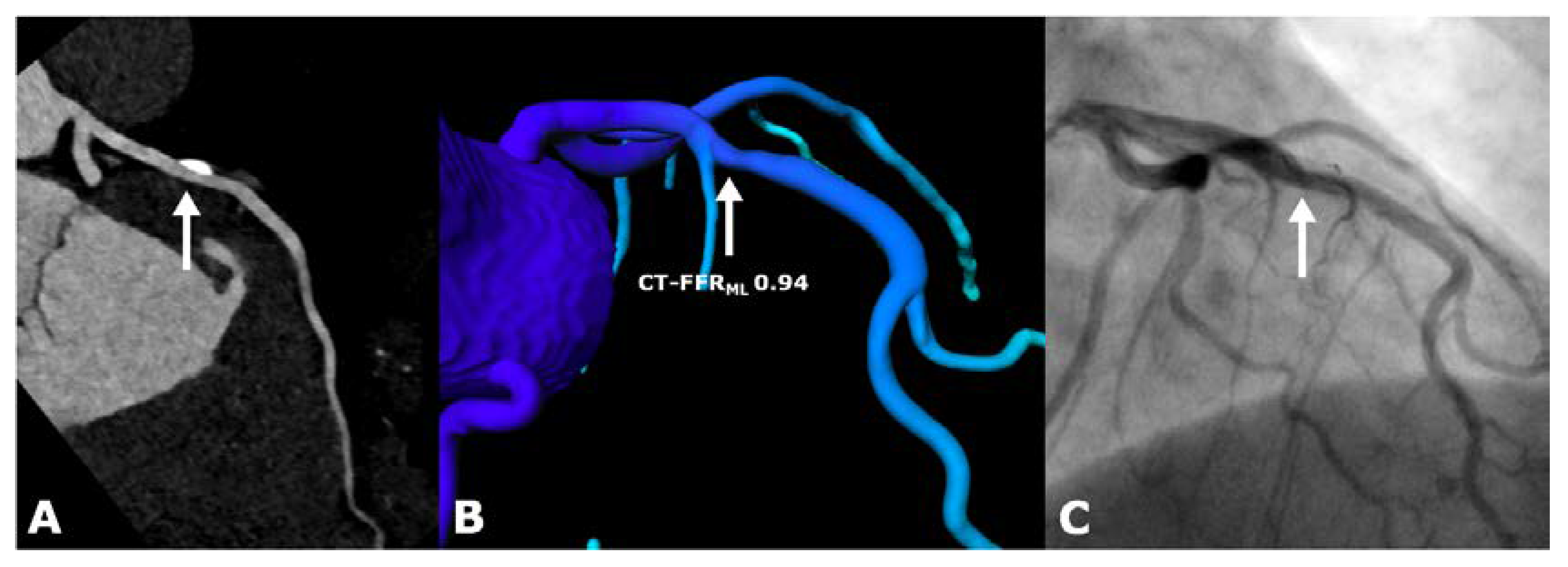
3.3. cCTA and CT-FFR
3.4. CT-FFR Analysis and Reclassification
3.5. Invasive Coronary Intervention
3.6. Radiation Exposure
4. Discussion
5. Limitations
6. Conclusions
Author Contributions
Funding
Acknowledgments
Conflicts of Interest
Abbreviations
| CAD | coronary artery disease |
| CCS | chronic coronary syndrome |
| cCTA | coronary computed tomography angiography |
| CT-FFRCFD | CT-derived fractional flow reserve based on computational fluid dynamics |
| CT-FFRML | CT-derived fractional flow reserve based on machine learning |
| FFR | fractional flow reserve |
| ICA | invasive coronary angiography |
| iwFR | instantaneous wave-free ratio |
| OMT | optima medical therapy |
References
- Schoepf, U.J.; Zwerner, P.L.; Savino, G.; Herzog, C.; Kerl, J.M.; Costello, P. Coronary CT angiography. Radiology 2007, 244, 48–63. [Google Scholar] [CrossRef] [PubMed]
- Douglas, P.S.; Hoffmann, U.; Patel, M.R.; Mark, D.B.; Al-Khalidi, H.R.; Cavanaugh, B.; Cole, J.; Dolor, R.J.; Fordyce, C.B.; Huang, M.; et al. Outcomes of anatomical versus functional testing for coronary artery disease. N. Engl. J. Med. 2015, 372, 1291–1300. [Google Scholar] [CrossRef] [PubMed] [Green Version]
- Newby, D.E.; Adamson, P.D.; Berry, C.; Boon, N.A.; Dweck, M.R.; Flather, M.; Forbes, J.; Hunter, A.; Lewis, S.; MacLean, S.; et al. Coronary CT Angiography and 5-Year Risk of Myocardial Infarction. N. Engl. J. Med. 2018, 379, 924–933. [Google Scholar] [PubMed]
- Knuuti, J.; Wijns, W.; Saraste, A.; Capodanno, D.; Barbato, E.; Funck-Brentano, C.; Prescott, E.; Storey, R.F.; Deaton, C.; Cuisset, T.; et al. 2019 ESC Guidelines for the diagnosis and management of chronic coronary syndromes. Eur. Heart J. 2019, 41, 407. [Google Scholar] [CrossRef] [PubMed]
- Karlsberg, R.P.; Budoff, M.J.; Thomson, L.E.; Friedman, J.D.; Berman, D.S. Reduction in downstream test utilization following introduction of coronary computed tomography in a cardiology practice. Int. J. Cardiovasc. Imaging 2010, 26, 359–366. [Google Scholar] [CrossRef] [PubMed] [Green Version]
- Montalescot, G.; Sechtem, U.; Achenbach, S.; Andreotti, F.; Arden, C.; Budaj, A.; Bugiardini, R.; Crea, F.; Cuisset, T.; Di Mario, C.; et al. 2013 ESC guidelines on the management of stable coronary artery disease: The Task Force on the management of stable coronary artery disease of the European Society of Cardiology. Eur. Heart J. 2013, 34, 2949–3003. [Google Scholar] [PubMed]
- Norgaard, B.L.; Leipsic, J.; Gaur, S.; Seneviratne, S.; Ko, B.S.; Ito, H.; Jensen, J.M.; Mauri, L.; De Bruyne, B.; Bezerra, H.; et al. Diagnostic performance of noninvasive fractional flow reserve derived from coronary computed tomography angiography in suspected coronary artery disease: The NXT trial (Analysis of Coronary Blood Flow Using CT Angiography: Next Steps). J. Am. Coll. Cardiol. 2014, 63, 1145–1155. [Google Scholar] [CrossRef] [Green Version]
- Neumann, F.J.; Sousa-Uva, M.; Ahlsson, A.; Alfonso, F.; Banning, A.P.; Benedetto, U.; Byrne, R.A.; Collet, J.P.; Falk, V.; Head, S.J.; et al. 2018 ESC/EACTS Guidelines on myocardial revascularization. Eur. Heart J. 2018, 40, 87–165. [Google Scholar] [CrossRef]
- De Bruyne, B.; Pijls, N.H.; Kalesan, B.; Barbato, E.; Tonino, P.A.; Piroth, Z.; Jagic, N.; Mobius-Winkler, S.; Rioufol, G.; Witt, N.; et al. Fractional flow reserve-guided PCI versus medical therapy in stable coronary disease. N. Engl. J. Med. 2012, 367, 991–1001. [Google Scholar] [CrossRef] [Green Version]
- Xaplanteris, P.; Fournier, S.; Pijls, N.H.J.; Fearon, W.F.; Barbato, E.; Tonino, P.A.L.; Engstrom, T.; Kaab, S.; Dambrink, J.H.; Rioufol, G.; et al. Five-Year Outcomes with PCI Guided by Fractional Flow Reserve. N. Engl. J. Med. 2018, 379, 250–259. [Google Scholar] [CrossRef]
- Pijls, N.H.; van Schaardenburgh, P.; Manoharan, G.; Boersma, E.; Bech, J.W.; van’t Veer, M.; Bar, F.; Hoorntje, J.; Koolen, J.; Wijns, W.; et al. Percutaneous coronary intervention of functionally nonsignificant stenosis: 5-Year follow-up of the DEFER Study. J. Am. Coll. Cardiol. 2007, 49, 2105–2111. [Google Scholar] [CrossRef] [PubMed] [Green Version]
- Tonino, P.A.; De Bruyne, B.; Pijls, N.H.; Siebert, U.; Ikeno, F.; van’t Veer, M.; Klauss, V.; Manoharan, G.; Engstrom, T.; Oldroyd, K.G.; et al. Fractional flow reserve versus angiography for guiding percutaneous coronary intervention. N. Engl. J. Med. 2009, 360, 213–224. [Google Scholar] [CrossRef] [PubMed] [Green Version]
- Gotberg, M.; Christiansen, E.H.; Gudmundsdottir, I.J.; Sandhall, L.; Danielewicz, M.; Jakobsen, L.; Olsson, S.E.; Ohagen, P.; Olsson, H.; Omerovic, E.; et al. Instantaneous Wave-free Ratio versus Fractional Flow Reserve to Guide PCI. N. Engl. J. Med. 2017, 376, 1813–1823. [Google Scholar] [CrossRef] [PubMed] [Green Version]
- Davies, J.E.; Sen, S.; Dehbi, H.M.; Al-Lamee, R.; Petraco, R.; Nijjer, S.S.; Bhindi, R.; Lehman, S.J.; Walters, D.; Sapontis, J.; et al. Use of the Instantaneous Wave-free Ratio or Fractional Flow Reserve in PCI. N. Engl. J. Med. 2017, 376, 1824–1834. [Google Scholar] [CrossRef] [PubMed] [Green Version]
- Hochman, J.S. International Study of Comparative Health Effectiveness with Medical and Invasive Approaches-ISCHEMIA. In Proceedings of the American Heart Association Annual Scientific Sessions (AHA 2019), Philadelphia, PA, USA, 16–18 November 2019. [Google Scholar]
- Spertus, J.A. International Study of Comparative Health Effectiveness with Medical and Invasive Approaches-ISCHEMIA. In Proceedings of the American Heart Association Annual Scientific Sessions (AHA 2019), Philadelphia, PA, USA, 16–18 November 2019. [Google Scholar]
- Baumann, S.; Renker, M.; Hetjens, S.; Fuller, S.R.; Becher, T.; Lossnitzer, D.; Lehmann, R.; Akin, I.; Borggrefe, M.; Lang, S.; et al. Comparison of Coronary Computed Tomography Angiography-Derived vs Invasive Fractional Flow Reserve Assessment: Meta-Analysis with Subgroup Evaluation of Intermediate Stenosis. Acad. Radiol. 2016, 23, 1402–1411. [Google Scholar] [CrossRef] [PubMed]
- Douglas, P.S.; Pontone, G.; Hlatky, M.A.; Patel, M.R.; Norgaard, B.L.; Byrne, R.A.; Curzen, N.; Purcell, I.; Gutberlet, M.; Rioufol, G.; et al. Clinical outcomes of fractional flow reserve by computed tomographic angiography-guided diagnostic strategies vs. usual care in patients with suspected coronary artery disease: The prospective longitudinal trial of FFR(CT): Outcome and resource impacts study. Eur. Heart J. 2015, 36, 3359–3367. [Google Scholar]
- Grunau, G.L.; Min, J.K.; Leipsic, J. Modeling of fractional flow reserve based on coronary CT angiography. Curr. Cardiol. Rep. 2013, 15, 336. [Google Scholar] [CrossRef]
- Tesche, C.; De Cecco, C.N.; Baumann, S.; Renker, M.; McLaurin, T.W.; Duguay, T.M.; Bayer, R.R., 2nd; Steinberg, D.H.; Grant, K.L.; Canstein, C.; et al. Coronary CT Angiography-derived Fractional Flow Reserve: Machine Learning Algorithm versus Computational Fluid Dynamics Modeling. Radiology 2018, 288, 64–72. [Google Scholar] [CrossRef] [Green Version]
- Arad, Y.; Spadaro, L.A.; Goodman, K.; Newstein, D.; Guerci, A.D. Prediction of coronary events with electron beam computed tomography. J. Am. Coll. Cardiol. 2000, 36, 1253–1260. [Google Scholar] [CrossRef] [Green Version]
- van Rosendael, A.R.; Shaw, L.J.; Xie, J.X.; Dimitriu-Leen, A.C.; Smit, J.M.; Scholte, A.J.; van Werkhoven, J.M.; Callister, T.Q.; DeLago, A.; Berman, D.S.; et al. Superior Risk Stratification with Coronary Computed Tomography Angiography Using a Comprehensive Atherosclerotic Risk Score. JACC Cardiovasc. Imaging 2019, 12, 1987–1997. [Google Scholar] [CrossRef] [Green Version]
- Bittencourt, M.S.; Hulten, E.; Polonsky, T.S.; Hoffman, U.; Nasir, K.; Abbara, S.; Di Carli, M.; Blankstein, R. European Society of Cardiology-Recommended Coronary Artery Disease Consortium Pretest Probability Scores More Accurately Predict Obstructive Coronary Disease and Cardiovascular Events Than the Diamond and Forrester Score: The Partners Registry. Circulation 2016, 134, 201–211. [Google Scholar] [CrossRef] [PubMed] [Green Version]
- QxMD Medical Incorporated. Pre-Test Probability of CAD (CAD Consortium). Available online: https://qxmd.com/calculate/calculator_287/pre-test-probability-of-cad-cad-consortium (accessed on 15 February 2019).
- Agatston, A.S.; Janowitz, W.R.; Hildner, F.J.; Zusmer, N.R.; Viamonte, M., Jr.; Detrano, R. Quantification of coronary artery calcium using ultrafast computed tomography. J. Am. Coll. Cardiol. 1990, 15, 827–832. [Google Scholar] [CrossRef] [Green Version]
- Raff, G.L.; Abidov, A.; Achenbach, S.; Berman, D.S.; Boxt, L.M.; Budoff, M.J.; Cheng, V.; DeFrance, T.; Hellinger, J.C.; Karlsberg, R.P.; et al. SCCT guidelines for the interpretation and reporting of coronary computed tomographic angiography. J. Cardiovasc. Comput. Tomogr. 2009, 3, 122–136. [Google Scholar] [CrossRef] [PubMed]
- Levine, G.N.; Bates, E.R.; Blankenship, J.C.; Bailey, S.R.; Bittl, J.A.; Cercek, B.; Chambers, C.E.; Ellis, S.G.; Guyton, R.A.; Hollenberg, S.M.; et al. 2011 ACCF/AHA/SCAI Guideline for Percutaneous Coronary Intervention: A report of the American College of Cardiology Foundation/American Heart Association Task Force on Practice Guidelines and the Society for Cardiovascular Angiography and Interventions. Circulation 2011, 124, e574–e651. [Google Scholar]
- Baumann, S.; Chandra, L.; Skarga, E.; Renker, M.; Borggrefe, M.; Akin, I.; Lossnitzer, D. Instantaneous wave-free ratio (iFR((R))) to determine hemodynamically significant coronary stenosis: A comprehensive review. World J. Cardiol. 2018, 10, 267–277. [Google Scholar] [CrossRef]
- Patel, M.R.; Dai, D.; Hernandez, A.F.; Douglas, P.S.; Messenger, J.; Garratt, K.N.; Maddox, T.M.; Peterson, E.D.; Roe, M.T. Prevalence and predictors of nonobstructive coronary artery disease identified with coronary angiography in contemporary clinical practice. Am. Heart J. 2014, 167, 846–852.e2. [Google Scholar] [CrossRef]
- Nagel, E.; Greenwood, J.P.; McCann, G.P.; Bettencourt, N.; Shah, A.M.; Hussain, S.T.; Perera, D.; Plein, S.; Bucciarelli-Ducci, C.; Paul, M.; et al. Magnetic Resonance Perfusion or Fractional Flow Reserve in Coronary Disease. N. Engl. J. Med. 2019, 380, 2418–2428. [Google Scholar] [CrossRef]
- Fairbairn, T.A.; Nieman, K.; Akasaka, T.; Norgaard, B.L.; Berman, D.S.; Raff, G.; Hurwitz-Koweek, L.M.; Pontone, G.; Kawasaki, T.; Sand, N.P.; et al. Real-world clinical utility and impact on clinical decision-making of coronary computed tomography angiography-derived fractional flow reserve: Lessons from the ADVANCE Registry. Eur. Heart J. 2018, 39, 3701–3711. [Google Scholar] [CrossRef] [Green Version]
- Baumann, S.; Renker, M.; Akin, I.; Borggrefe, M.; Schoepf, U.J. FFR-Derived From Coronary CT Angiography Using Workstation-Based Approaches. JACC Cardiovasc. Imaging 2017, 10, 497–498. [Google Scholar] [CrossRef]
- Schwartz, F.R.; Koweek, L.M.; Norgaard, B.L. Current Evidence in Cardiothoracic Imaging: Computed Tomography-derived Fractional Flow Reserve in Stable Chest Pain. J. Thorac. Imaging 2019, 34, 12–17. [Google Scholar] [CrossRef]
- Benton, S.M., Jr.; Tesche, C.; De Cecco, C.N.; Duguay, T.M.; Schoepf, U.J.; Bayer, R.R., 2nd. Noninvasive Derivation of Fractional Flow Reserve From Coronary Computed Tomographic Angiography: A Review. J. Thorac. Imaging 2018, 33, 88–96. [Google Scholar] [CrossRef] [PubMed]
- Renker, M.; Schoepf, U.J.; Wang, R.; Meinel, F.G.; Rier, J.D.; Bayer, R.R., 2nd; Mollmann, H.; Hamm, C.W.; Steinberg, D.H.; Baumann, S. Comparison of diagnostic value of a novel noninvasive coronary computed tomography angiography method versus standard coronary angiography for assessing fractional flow reserve. Am. J. Cardiol. 2014, 114, 1303–1308. [Google Scholar] [CrossRef] [PubMed]
- Mastrodicasa, D.; Albrecht, M.H.; Schoepf, U.J.; Varga-Szemes, A.; Jacobs, B.E.; Gassenmaier, S.; De Santis, D.; Eid, M.H.; van Assen, M.; Tesche, C.; et al. Artificial intelligence machine learning-based coronary CT fractional flow reserve (CT-FFRML): Impact of iterative and filtered back projection reconstruction techniques. J. Cardiovasc. Comput. Tomogr. 2018, 13, 331–335. [Google Scholar] [CrossRef] [PubMed]
- Hlatky, M.A.; De Bruyne, B.; Pontone, G.; Patel, M.R.; Norgaard, B.L.; Byrne, R.A.; Curzen, N.; Purcell, I.; Gutberlet, M.; Rioufol, G.; et al. Quality-of-Life and Economic Outcomes of Assessing Fractional Flow Reserve with Computed Tomography Angiography: PLATFORM. J. Am. Coll. Cardiol. 2015, 66, 2315–2323. [Google Scholar] [CrossRef] [Green Version]
- Villines, T.C. Can CT-derived FFR better inform clinical decision-making and improve outcomes in stable ischaemic heart disease? Eur. Heart J. 2018, 39, 3712–3714. [Google Scholar] [CrossRef]
- Cook, C.M.; Petraco, R.; Shun-Shin, M.J.; Ahmad, Y.; Nijjer, S.; Al-Lamee, R.; Kikuta, Y.; Shiono, Y.; Mayet, J.; Francis, D.P.; et al. Diagnostic Accuracy of Computed Tomography-Derived Fractional Flow Reserve: A Systematic Review. JAMA Cardiol. 2017, 2, 803–810. [Google Scholar] [CrossRef]
- Ding, W.Y.; Nair, S.; Appleby, C. Diagnostic accuracy of instantaneous wave free-ratio in clinical practice. J. Interv. Cardiol. 2017, 30, 564–569. [Google Scholar] [CrossRef]
- Bamberg, F.; Boiselle, P.M.; Choe, Y.H.; Funabashi, N.; Schoepf, U.J. Expert opinion: Should coronary CT angiography be used as a screening test? J. Thorac. Imaging 2012, 27, 339. [Google Scholar] [CrossRef]
- Loewe, C.; Stadler, A. Computed tomography assessment of hemodynamic significance of coronary artery disease: CT perfusion, contrast gradients by coronary CTA, and fractional flow reserve review. J. Thorac. Imaging 2014, 29, 163–172. [Google Scholar] [CrossRef]


| Demographics | Mean Value ± Standard Deviation or Frequency (%) |
|---|---|
| Age, mean ± SD (years) | 63 ± 11 |
| Male, no (%) | 65 (74) |
| BMI, mean ± SD [kg/m2] | 29 ± 5 |
| Cardiovascular risk factors | |
| Hypertension *, no. (%) | 69 (78) |
| Hyperlipidemia **, no. (%) | 39 (44) |
| Diabetes mellitus, no. (%) | 13 (15) |
| Smoker, no. (%) | 34 (39) |
| Family history of CAD, no. (%) | 19 (22) |
| Angina type | |
| Typical angina, no. (%) | 19 (22) |
| Atypical angina, no. (%) | 12 (14) |
| Non-cardiac chest pain, no. (%) | 53 (60) |
| Pretest probability ***, mean ± SD (%) | 25 ± 21 |
| Baseline medication | |
| aspirin, no. (%) | 24 (27) |
| P2Y12 inhibitor, no. (%) | 3 (3) |
| statin, no. (%) | 31 (35) |
| beta-blocker, no. (%) | 32 (36) |
| calcium channel blocker (CCB), no. (%) | 15 (17) |
| angiotensin-converting-enzyme inhibitor or AT1 receptor blocker (ARB), no. (%) | 46 (52) |
| nitrates, no. (%) | 1 (1) |
| Baseline blood values | |
| Cholesterol, mean ± SD [mg/dl] | 195 ± 59 |
| High-density lipoprotein, mean ± SD [mg/dl] | 51 ± 20 |
| Low-density lipoprotein, mean ± SD [mg/dl] | 117 ± 45 |
| Triglycerides, mean ± SD [mg/dl] | 213 ± 213 |
| Hemoglobin A1c, mean ± SD [%] | 6 ± 1 |
| Creatine kinase, mean ± SD [U/l] | 188 ± 254 |
| Creatine kinase muscle-brain type, mean ± SD [U/l] | 45 ± 86 |
| High sensitive Troponin-I > 0.045, no. (%) | 4 (5) |
| Total | Not Revascularized, (Median) | Revascularized, (Median) | OR | 95% CI | AUC | p-Value | |
|---|---|---|---|---|---|---|---|
| Pre-test probability (%) | 88 | 14.00 | 20.50 | 1.010 | 0.990–1.032 | 0.578 | 0.3212 |
| Agatston score | 79 | 207.20 | 429.20 | 1.055 | 1.000–1.001 | 0.631 | 0.1448 |
| Comprehensive CTA score | 88 | 7.16 | 10.79 | 1.110 | 1.020–1.208 | 0.633 | 0.0158 |
| Total | Not revascularized, (Mean ± SD) | Revascularized, (Mean ± SD) | OR | 95% CI | AUC | p–Value | |
| CT-FFRML | 88 | 0.89 ± 0.08 | 0.58 ± 0.15 | 0.138 | 0.062–0.309 | 0.958 | <0.0001 |
| Coronary Computed Tomography Angiography—Lesion Location | |
|---|---|
| Left main truncus *, no. (%) | 0 (0) |
| Left anterior descending *, no. (%) | 60 (68) |
| Left circumflex artery *, no. (%) | 11 (13) |
| Right coronary artery *, no. (%) | 17 (19) |
| Stenosis grade in cCTA [26] | |
| Moderate: 50–69% diameter stenosis, no. (%) | 35 (40) |
| Severe: 70–99% diameter stenosis, no. (%) | 53 (60) |
| Occluded, no (%) | 0 (0) |
| Risk stratification and Image Quality | |
| Agatston score *, mean ± SD | 553 ± 651 |
| Agatston score *, Range | 6–3264 |
| Agatston score *, no. of patients >400 (%) | 33 (38) |
| Comprehensive CTA score **, mean ± SD | 9.72 ± 5.47 |
| Image quality ***, mean ± SD | 4.2 ± 0.8 |
| Radiation Exposure | |
| Mean ± SD [mGy*cm] | 617 ± 406 |
| Median [mGy*cm] | 553 |
| Contrast agent ± SD [mL] | 80.8 ± 15.4 |
| CT-FFRML | Total no. (%) | ||
|---|---|---|---|
| ≤0.80 | >0.80 | ||
| obstructive CAD | 37 (42.0%) | 3 (3.4%) | 40 (45.5%) |
| no obstructive CAD | 3 (3.4%) | 45 (51.1%) | 48 (54.5%) |
| total no. (%) | 40 (45.5%) | 48 (54.5%) | 88 (100%) |
| Invasive Coronary Angiography | |
|---|---|
| Contrast agent ± SD [mL] | 158 ± 106 |
| X-ray time ± SD [min] | 7.4 ± 6.2 |
| Dose area product ± SD [cGy*cm2] | 11,900 ± 21,080 |
| One-vessel disease, no. (%) | 35 (40) |
| Two-vessel disease, no. (%) | 11 (13) |
| Three-vessel disease, no. (%) | 4 (5) |
© 2020 by the authors. Licensee MDPI, Basel, Switzerland. This article is an open access article distributed under the terms and conditions of the Creative Commons Attribution (CC BY) license (http://creativecommons.org/licenses/by/4.0/).
Share and Cite
Lossnitzer, D.; Chandra, L.; Rutsch, M.; Becher, T.; Overhoff, D.; Janssen, S.; Weiss, C.; Borggrefe, M.; Akin, I.; Pfleger, S.; et al. Additional Value of Machine-Learning Computed Tomographic Angiography-Based Fractional Flow Reserve Compared to Standard Computed Tomographic Angiography. J. Clin. Med. 2020, 9, 676. https://doi.org/10.3390/jcm9030676
Lossnitzer D, Chandra L, Rutsch M, Becher T, Overhoff D, Janssen S, Weiss C, Borggrefe M, Akin I, Pfleger S, et al. Additional Value of Machine-Learning Computed Tomographic Angiography-Based Fractional Flow Reserve Compared to Standard Computed Tomographic Angiography. Journal of Clinical Medicine. 2020; 9(3):676. https://doi.org/10.3390/jcm9030676
Chicago/Turabian StyleLossnitzer, Dirk, Leonard Chandra, Marlon Rutsch, Tobias Becher, Daniel Overhoff, Sonja Janssen, Christel Weiss, Martin Borggrefe, Ibrahim Akin, Stefan Pfleger, and et al. 2020. "Additional Value of Machine-Learning Computed Tomographic Angiography-Based Fractional Flow Reserve Compared to Standard Computed Tomographic Angiography" Journal of Clinical Medicine 9, no. 3: 676. https://doi.org/10.3390/jcm9030676
APA StyleLossnitzer, D., Chandra, L., Rutsch, M., Becher, T., Overhoff, D., Janssen, S., Weiss, C., Borggrefe, M., Akin, I., Pfleger, S., & Baumann, S. (2020). Additional Value of Machine-Learning Computed Tomographic Angiography-Based Fractional Flow Reserve Compared to Standard Computed Tomographic Angiography. Journal of Clinical Medicine, 9(3), 676. https://doi.org/10.3390/jcm9030676


