Article

Anti-Inflammatory and T-Cell Immunomodulatory Effects of Banana Peel Extracts and Selected Bioactive Components in LPS-Challenged In Vitro and In Vivo Models

1
Graduate Programs of Nutrition Science, School of Life Science, National Taiwan Normal University, Taipei 106209, Taiwan
2
School of Medicine for International Students, I-Shou University, Kaohsiung 82445, Taiwan
3
Department of Obstetrics & Gynecology, E-Da Hospital/E-Da Dachang Hospital, Kaohsiung 82445, Taiwan
4
Department of Health Management, College of Medicine, I-Shou University, Kaohsiung 82445, Taiwan
5
Department of Medicinal Botanicals and Foods on Health Applications, Da-Yeh University, Changhua 51591, Taiwan
6
Department of Nutrition, I-Shou University, Kaohsiung 82445, Taiwan
7
Department of Chemical Engineering, I-Shou University, Kaohsiung 82445, Taiwan
*
Authors to whom correspondence should be addressed.
Agriculture 2023, 13(2), 451; https://doi.org/10.3390/agriculture13020451
Submission received: 20 December 2022 / Revised: 9 February 2023 / Accepted: 10 February 2023 / Published: 14 February 2023
(This article belongs to the Section Agricultural Product Quality and Safety)

Abstract

:
Banana peel (BP) has potent antioxidative properties; however, the anti-inflammatory potential of BP and its related bioactive components remain unclear. This study used solvent extraction and gas chromatography–mass spectrometry (GC–MS) to isolate and identify the active fractions and compounds in BP. BP was extracted with 95% ethanol (BP-95E) and partitioned with an ethyl acetate (EA) and water mixture to obtain the BP-95E-EA and BP-95E-H2O fractions. The BP-95E-EA fractions were further partitioned with n-hexane (Hex) and methanol (MeOH) mixtures to obtain BP-95E-EA-Hex and BP-95E-EA-MeOH subfractions, and the BP-95E-H2O fractions were partitioned with n-butanol (BuOH) to obtain BP-95E-H2O-BuOH subfractions and the H2O residual. The results show that the BP-95E-H2O-BuOH subfractions exhibited the most potent inhibition of interleukin-6 (IL-6) and tumor necrosis factor-α (TNF-α) secretion while down-regulating inducible nitric oxide synthase (iNOS) and cyclooxygenase-2 (COX-2) protein expression in lipopolysaccharide (LPS)-stimulated RAW264.7 macrophages. In this active subfraction, five non-polyphenol compounds were identified, namely, 5-hydroxyethyl furfural (5-HMF), guaiol, oleic acid, linoleic acid, and oleamide. 5-HMF, guaiol, and oleamide were the most effective at suppressing IL-6 and TNF-α secretion. The in vivo immunomodulatory action of BP was evaluated in an LPS-induced endotoxemia model of BALB/c mice. Oral administration of BP-95E-H2O-BuOH extracts (42 and 166 mg/kg b.w.) for two weeks lowered the serum levels of IL-6 and TNF-α and normalized the activated T-cell population, as evidenced by an increase in CD3CD69 and decrease in IFN-γ/IL-4 (Th1/Th2) in mice with systemic inflammation. Our findings reveal that BP exhibits anti-inflammatory and T-cell immunomodulatory effects that may contribute to delaying endotoxemia-associated disorders.

1. Introduction

According to the Food and Agriculture Organization (FAO) estimation, bananas are the most widely consumed and traded agricultural crop in the world, accounting for 13.6% of global fruit production. The average global banana production increased from 69 million tons in 2000–2002 to 119.8 million tons in 2020, with a value of approximately USD 31 billion [1]. Banana peel (BP) is a waste byproduct that accounts for around 35–40% of the total fruit weight; as a result, over 41.9 million tons of BP are discarded each year and may cause environmental problems. Therefore, increasing the recycling and reuse of BP would appear to be an environmentally friendly approach to waste management and increase its added value [2,3].
Mohapatra et al. reported that BP has the potential to be a good feed material for livestock and poultry since it provides 3% carbohydrates, 6% protein, 3% fat, 3.8–11% crude fat, 43.2–49% dietary fiber, and 1% vitamin C [4]. BP is also used as a biopolymer in water purification [5] and is an organic fertilizer, recently producing ethanol, methane, and cellulose [2]. In aquaculture, the dietary administration of BP extracts for 120 days enhanced prawns’ resistance to pathogens by stimulating immune responses and phagocytic activities. In traditional and folk medicine, BP is used to treat trauma, diarrhea in infancy [6], burn wounds, depression [7], and inflammatory-related illnesses. However, the scientific evidence underlying its therapeutic activities remains unclear.
It has been reported that BP is rich in dietary fiber and pectin [8], contains over 40 polyphenol antioxidants, and exhibits potent antioxidant and antimicrobial properties. The polyphenols in BP can be categorized into four subgroups: hydroxycinnamic acids, flavonols, flavan-3-ols, and catecholamines. Among these compounds, ferulic acid and rutin are the two most abundant constituents [9]. The administration of gallocatechin-rich BP extract (106.6 µg/mL) exhibits wound-healing potential as evidenced by a decrease in the epithelization period and an increase in the hydroxyproline content over the experimental period [7]. This is consistent with the beneficial wound-healing data of Atzingen et al. (2013), who employed a 4% BP gel from unripe Musa sapientum L. to treat skin lesions caused by surgical excision in rats [10]. Of note, variety, maturity, cultivation, and drying condition all have an impact on the bioactive compositions of BP [9,11]. For example, the contents of chlorophyll decreased by 90% as BP turned from green to yellow, whereas carotenoids and flavonoids increased by 50% and 27%, respectively. Compared to heat-dry methods, microwave irradiation of BP at 960 watts for six minutes preserved the main bioactive components and antioxidant activity [11]. The contents of phenolics and proanthocyanidins and antioxidant capacity increased with the ripening process [12]. The gallocatechin contents and antioxidant activity of BP against lipid peroxidation are greater than those of banana pulp [13]. Fresh unripe BP has the most significant antioxidant capacity, while fresh ripe BP can only suppress nitric oxide (NO) generation [14].
BP extracts may also possess anti-microbial and anti-inflammatory properties. Water and ethanol extracts of BP have shown antimicrobial activities against Gram-positive and Gram-negative bacteria [15,16,17]. In an emergency, the inside of BP possesses antibacterial characteristics and can be placed directly around wounds or relieve swelling following a mosquito bite [7]. Wang et al. also reported that polyphenol-rich BP extracts (200 mg/kg body weight) could ameliorate hepatic injuries and inhibit the production of proinflammatory cytokines and oxidative stress in CCl4-treated mice [18]. These findings suggest that BP could be a functional food for human nutrition and health maintenance.
Inflammatory cytokines and the activation of macrophages and T lymphocytes have a role in the development of cancer [19], insulin resistance [20], and cardiovascular disease [21]. Several studies have reported that acute phase endotoxemia, tissue damage, organ failure, shock, and even COVID-19 mortality may result from the overproduction of pro-inflammatory mediators and cell adhesion molecules [22,23]. Observations have indicated that CD4+ T cells, regulatory T cells (Tregs), and natural killer T cells (NKTs) can modulate the activation and inactivation of macrophages in response to extrinsic stimuli such as LPS, leading to an inflammatory response. In response, macrophages may release a cascade of pro-inflammatory cytokines, including IL-1, IL-6, IL-12, TNF-α, and IFN-γ [24]. Accordingly, the present study investigated the anti-inflammatory and T-cell immunomodulatory effects of BP in LPS-treated RAW264.7 macrophages and an LPS-challenged mouse model. The solvent extraction and GC–MS technique were also employed to isolate and identify the active fractions and non-polyphenol compounds in BP.

2. Materials and Methods

2.1. Reagents

The chemical reagents lipopolysaccharide (LPS, E. coli serotype O55:B5), ammonium pyrrolidine dithiocarbamate (PDTC), concanavalin A (Con A), bovine serum albumin, 5-hydroxyethyl furfural (product No. 53407, ≥98.0%), guaiol (product No. 29242, ≥99.0%), oleic acid, linoleic acid (product No. O1008, ≥ 99.0%), oleamide (product No. 08393, ≥98.5%), and 3(4,5-dimethylthiazol-2-yl)-2,5-diphenyltetrazolium bromide (MTT) were purchased from Sigma Chemicals Co. (St Louis, MO, USA). Anti-iNOS, anti-COX-2, and anti-GAPDH antibodies were purchased from Cell Signaling Technology (Danvers, MA, USA). IL-6 and TNF-α enzyme-linked immunosorbent assay (ELISA) kits were purchased from eBioscience (Minneapolis, MN, USA). All other chemicals were of reagent or analytical grade.

2.2. Plant Materials and Preparation of Banana Peel Extracts

The raw materials of bananas (Musa sp., cavendish (AAA) subgroup) were purchased from fruit and vegetable markets in Kaohsiung city and authenticated by the Taiwan Banana Research Institute (Pingtung City, Taiwan). Briefly, the bananas were washed, and their pulps were removed. The remaining fresh peels were defined as wet banana peels (wBPs) in this study. To prepare dry banana peels (dBPs), the wet peels were placed in a circulating oven at 60 °C for three days. To prepare BP extracts, 10 kg of wBPs and dBPs was cut into small pieces and extracted five times with 95% ethanol (95E) at room temperature. After gauze filtration, the filtrates were collected and concentrated with a vacuum evaporator to obtain the wBP-95E and dBP-95E extracts, respectively. Both of the BP-95E extracts were then partitioned with ethyl acetate (EA) and water mixtures (1:1) in a separatory funnel; the EA and water layers were collected and concentrated to obtain the BP-95E-EA and BP-95E-H2O fractions, respectively. Finally, the BP-95E-EA fractions were further partitioned with n-hexane (Hex) and methanol (MeOH) mixtures (1:1) to obtain the BP-95E-EA-Hex and BP-95E-EA-MeOH subfractions, whereas the BP-95E-H2O fractions were partitioned with n-butanol (BuOH) to obtain the BP-95E-H2O-BuOH subfractions and the H2O residual. The partitioning scheme of the wBPs and dBPs for liquid–liquid extraction is shown in Figure 1.
GC–MS analysis was performed using an Agilent 7890B-GC and 5977A-MSD system (Agilent Technologies, Santa Clara, CA, USA) with an electron impact mode (70 eV) injector and an Agilent data system. The GC column was an HP-5ms Ultra Inert capillary column (30 m × 0.25 mm, film thickness 0.25 μm, Agilent 19091S-433UI, USA). The injector and detector temperatures were set at 250 °C. The oven temperature was 70 °C, which was held for 10 min, and programmed to increase to 180 °C (hold for 5 min) at a rate of 2.5 °C/min. Then, it was raised to 250 °C at a rate of 2 °C/min, and kept at 250 °C for 6 min. The carrier gas was helium at a flow rate of 0.8 mL/min. Diluted samples of 2.0 μL were injected manually in the splitless mode. The components were identified by comparison of their mass spectra with the NIST MS 14.0 database (Gaithersburg, MD, USA).

2.3. Cell Culture and Treatment

The RAW264.7 cells (Bioresource Collection and Research Center, Hsinchu, Taiwan; BCRC60001) were cultured in Dulbecco’s modified Eagle’s medium (DMEM) supplemented with 10% heat-inactivated fetal bovine serum in a humidified incubator at 37 °C with 5% CO2. While conducting the experiments, the cells were seeded on either a 96-well plate (cell density of 5 × 10³ cells/well) or 6 cm dishes (cell density of 7 × 10⁵ cells/well) for the indicated assays. The cells were pretreated with BP extracts or their identified compounds for 1 h and then stimulated with 100 ng/mL LPS. The cell viability was measured with the MTT assay [11]. Briefly, 55 μL of DMEM containing 1 mg/mL of MTT was loaded onto the plate and incubated for 3 h. Then, 100 mL of isopropanol containing 0.04 N HCl was added to each well to dissolve the colorful crystals. The cell viability was determined by measuring the absorbance at 570 nm in an ELISA plate reader (Model 550, BioRad Laboratories, Hercules, CA, USA). Additionally, the culture media were collected to analyze the release of the pro-inflammatory cytokines after 48 h of stimulation. In the Western blotting assay, the cell lysates were collected to detect the target protein (iNOS and COX-2) expressions after 12 h of stimulation.

2.4. Western Blotting

The cells were lysed with lysis buffer at 4 °C, and the supernatants were collected and analyzed immediately. The protein concentrations were detected with a BCA Protein Assay Kit (Pierce, Rockford, IL, USA). A total of 25 μg from the total cell lysates was subjected to SDS-polyacrylamide gel electrophoresis, and the proteins were transferred onto a polyvinylidene difluoride membrane (Millipore, Temecula, CA, USA). The membrane was incubated with primary anti-iNOS (1:5000) and anti-COX-2 antibodies (1:7500) at 4 °C. The membrane was then incubated with goat anti-rabbit IgG, or goat anti-mouse IgG conjugated to horse radish peroxidase was used as a secondary antibody. Enhanced chemiluminescence plus Western blotting detection reagents (Amersham Bioscience, Uppsala, Sweden) were used to detect the protein signals, and Quantity One software (Bio-Rad) was used to measure the band intensities. Polyclonal mouse anti-human GAPDH antibodies served as the internal control.

2.5. LPS-Induced Systemic Inflammation in Mice

Six-week-old female BALB/c mice (n = 88) were purchased from the National Animal Center (Taipei, Taiwan). All mice were maintained in an air-conditioned room at 23 ± 2 °C on a regulated 12 h light/dark cycle and fed a standard chow diet (Lab Rodent Chow 5001, Ralston Purina Inc., St. Louis, MO, USA) for adaptation. At the age of 9 weeks, the mice were randomized and divided into four groups (22 mice in each group): the control group, the L-BP group (BP-95E-H2O-BuOH extracts with 42 mg/kg b.w.), the H-BP group (BP-95E-H2O-BuOH extracts with 166 mg/kg b.w.), and the PDTC group (which served as an NF-κB inhibitor for the anti-inflammatory control). All mice were fed a chow diet and daily tube-fed with either the indicated sample or distilled water at a volume of 100 μL. After two weeks, the mice were injected intraperitoneally (i.p.) with 15 μg LPS/kg body weight (b.w.) to induce systemic inflammation as described previously [25]. The PDTC group mice were i.p. injected with 50 mg PDTC/kg b.w., a dose with anti-inflammatory effects, 1 h before the LPS challenge. The body temperatures and survival times of ten mice in each group were recorded to monitor their lifespan. Six hours after the LPS challenge, the remaining 12 mice were euthanized, and the spleen cells and peritoneal exudate cells (PECs) were collected for the analysis of cytokines and T-cell populations. All animal studies were conducted according to the Animal Protection Act and the regulations of the Animal Care and Use Committee of the Council of Agriculture, Executive Yuan, and approved by the Institutional Animal Care and Use Committee (IACUC) of I-Shou University (IACUC-ISU-102-16).

2.6. Murine Primary Spleen and PEC Cell Culture

After the mice were sacrificed, the spleen cells and PECs were collected under sterile conditions. The culture medium used in the experiments was RPMI-1640 supplemented with 10% FBS. The spleen cells, at a concentration of 5 × 10⁶ cells/mL/well, were treated with or without Con A (5 μg/mL) for 24 h. The PECs were seeded at 1.5 × 10⁶ cells/mL/well and treated with or without LPS (10 μg/mL) for 48 h. After stimulation, the cell supernatants were collected for the measurement of cytokine secretion.

2.7. Nitric Oxide Determination

The level of nitrite to represent the nitric oxide (NO) production was analyzed using Griess reagent, which was freshly prepared by mixing reagent A (1% sulfanilamide in 2.5% phosphoric acid) and reagent B (0.1% N-1-naphthylethylene diamide dihydrochloride in water) at a ratio of 1:1. Briefly, the cell supernatant at the volume of 80 μL was added to the well, and then the comparable volume of Griess reagent was added. After a 10 min reaction, the 540 nm absorbance of each well was measured by an ELISA plate reader. The NO concentrations were calculated using a NaNO2 standard curve.

2.8. Pro-Inflammatory and Anti-Inflammatory Cytokines Assay

The secretion of cytokines, including IL-4, IL-6, IL-10, TNF-α, and IFN-γ, was measured with commercial ELISA kits (eBioscience, Minneapolis, MN, USA). Briefly, primary anti-cytokine antibodies were coated onto 96-well plates overnight. The wells in the plates were washed with washing buffer and blocked with blocking solution for 1 h. The diluted cell supernatants or sera were added to the wells for 2 h of incubation. Next, the wells were washed, and biotin-conjugated anti-cytokine antibodies were added for 1 h. The wells were then washed, and horseradish-peroxidase-conjugated streptavidin was added for 30 min. Following the washes, the coloring reagent tetramethylbenzidine (TMB, Clinical Science Products, Mansfield, MA, USA) was added to wells. The absorbance was measured by an ELISA plate reader at 620 nm. The data were calculated according to the standard curves of the cytokines.

2.9. Flow Cytometry

Phenotypic analysis of the spleen cells from the mice was performed by flow cytometry. Cells at a density of 2 × 10⁶ were suspended in 1 mL of PBS (with 0.1% sodium azide) and incubated at 4 °C for 30 min with fluorescein isothiocyanate- or phycoerythrin-conjugated monoclonal antibodies (BD Pharmingen, San Diego, CA, USA). The cells were washed and resuspended in 1.0 mL of PBS (with 0.1% sodium azide) and subjected to fluorescence-activated cell sorting (FACScan analysis). A total of 10,000 cells were counted, and the frequencies of cell surface markers (CD3, CD4, CD25, and CD69) were determined using appropriate software (FACScan, Becton Dickinson, Mountain View, CA, USA).

2.10. Statistical Analysis

Each experiment was performed at least three times. The data are expressed as the means ± SD or means ± SEM. The significant difference compared to the control group was statistically analyzed using Student’s t-test using the SAS software program (SAS/STATA version 8.2; SAS Institute, Cary, NC, USA). The statistical comparison between different survival curves was analyzed using Cox’s proportional hazards regression test (STATA version 9.0; Stata Corp., College Station, TX, USA). The relationship was analyzed using the simple correlation of the SAS program. Statistical significance is expressed as p < 0.05.

3. Results and Discussions

3.1. Anti-Inflammatory Effect of BP Extracts in LPS-Stimulated Macrophages

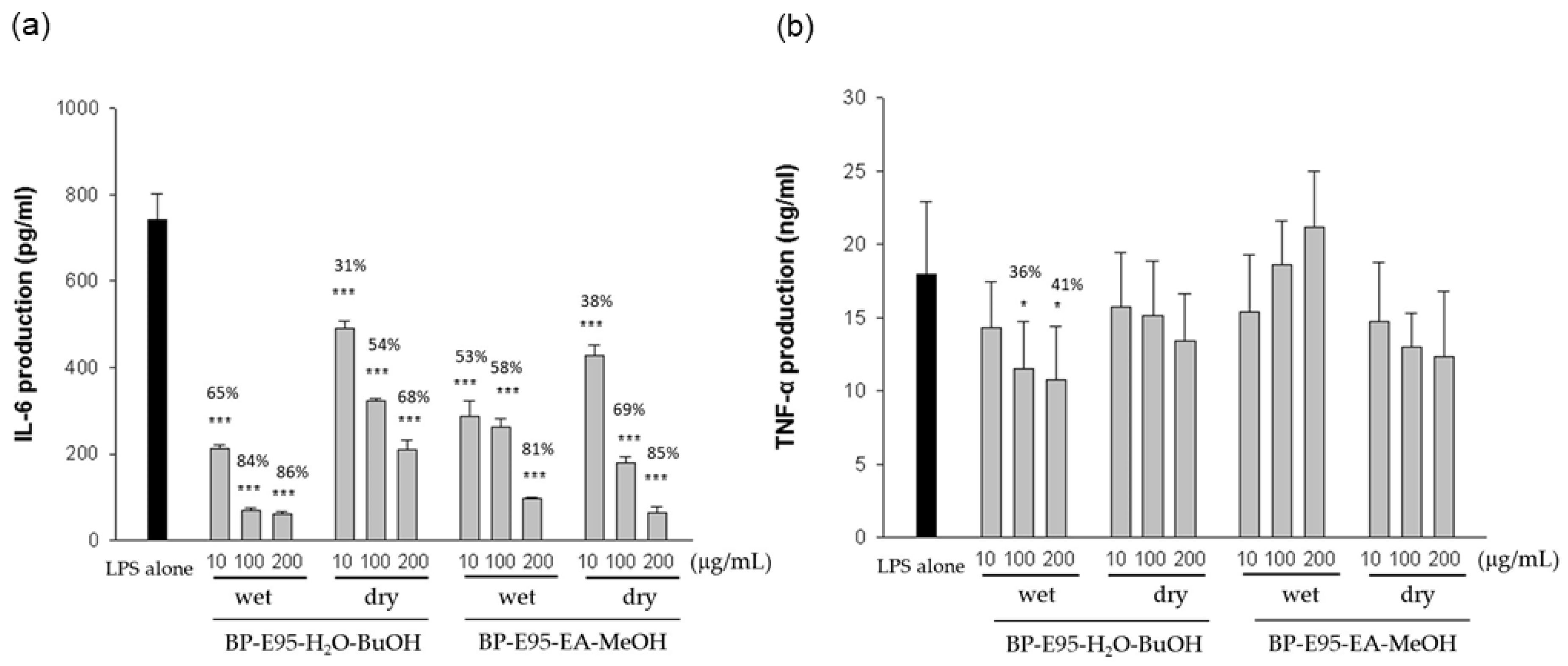
Given that drying conditions and extracted solvents may affect the chemical and bioactive properties of dried BP [11,26], the BP was divided into fresh wet banana peels (wBPs) and dry banana peels (dBPs) with different heat-drying procedures in this study. We examined the anti-inflammatory effects of wet and dry BP extracts obtained from the liquid–liquid solvent extraction in the LPS-stimulated RAW264.7 macrophages. The results in Figure 2a show that the BP extracts from the BuOH (wBP-95E-H2O-BuOH and dBP-95E-H2O-BuOH) and methanol (wBP-95E-EA-MeOH and dBP-95E-EA-MeOH) subfractions display significant suppression of IL-6 production in the LPS-stimulated RAW264.7 cells. In addition, only the BP-95E-H2O-BuOH subfractions inhibited TNF-α production (Figure 2b). We also detected expressions of inflammation-related proteins and found that the BP-95E-H2O-BuOH subfractions exhibited significant inhibitory effects on the expressions of iNOS and COX-2 (Figure 3). However, wBP-95E-EA-MeOH at a concentration of 200 µg/mL reduced iNOS expression but increased COX-2 expression, showing diverse effects on the inflammatory response at a specific dose. However, the extracts, other than BP-95E-H2O-BuOH and wBP-95E-EA-MeOH, had slight or moderate inhibitory effects on these cytokines and protein expressions. Accordingly, the active wBP-95E-H2O-BuOH subfractions were chosen to verify their anti-inflammatory activity in vivo.

3.2. Identification of Bioactive Compounds in BP-95E-H2O-BuOH Subfractions

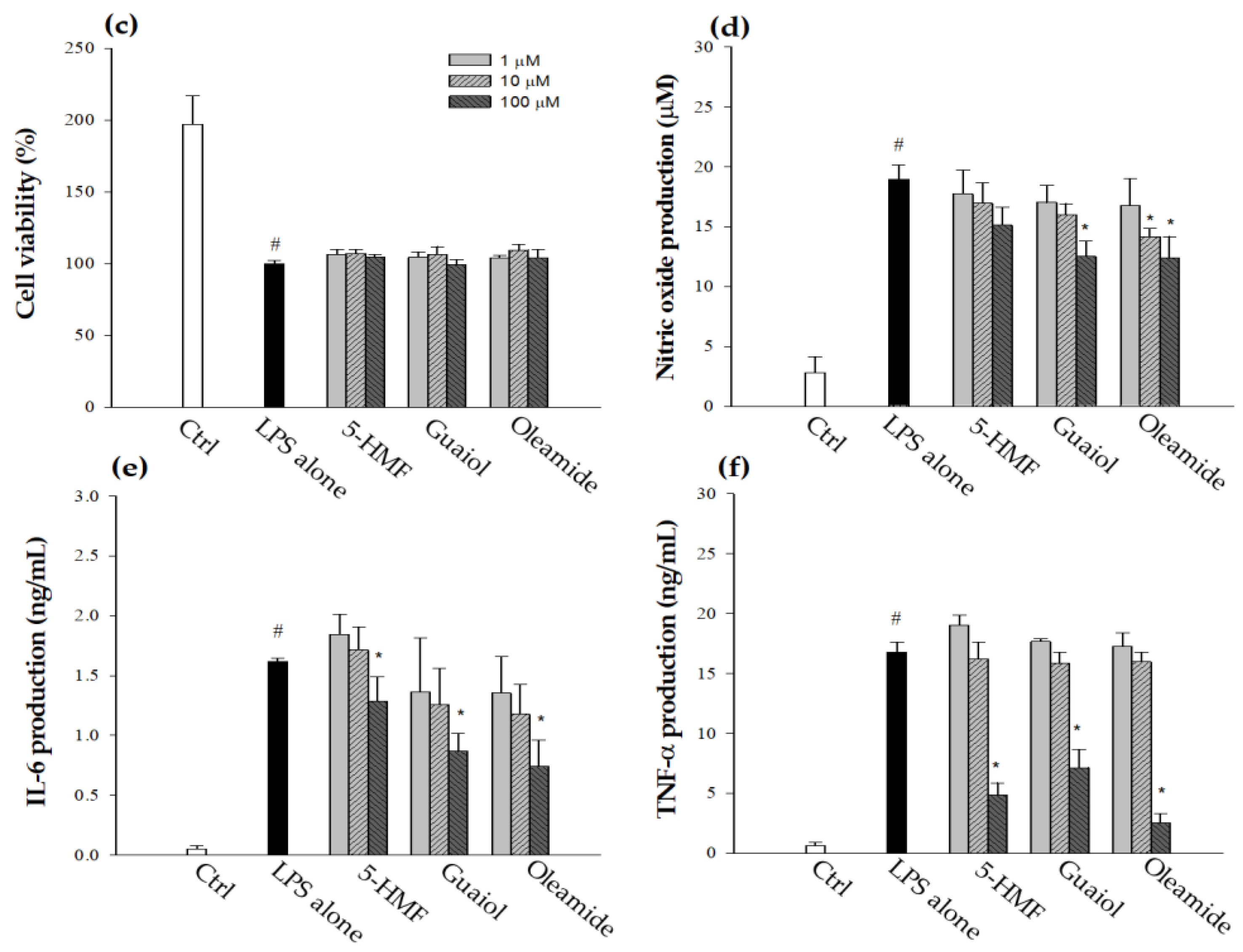
Five compounds of the BP-95E-H2O-BuOH subfractions were identified from the GC–MS analysis (Figure 4a,b), and their anti-inflammatory potencies were analyzed. The experimental data show that 5- hydroxymethyl furfural (5-HMF, 1), guaiol (2), and oleamide (5) at a concentration of 100 µM significantly inhibited NO production (Figure 4d) and IL-6 (Figure 4e) and TNF-α (Figure 4f) secretion. A recent study indicated that 5-HMF (100 µM) isolated from Armillaria mellea inhibited NO, IL-6, and TNF-α production in LPS-stimulated microglial cells [27]. Another study showed the protective effects of oleamides on neurofunction through the suppression of iNOS and COX-2 proteins [28]. Our study is the first to report that these three compounds in BP extracts inhibited the production of pro-inflammatory cytokines and may be the crucial constituents that contribute to the anti-inflammatory properties of the BP-95E-H2O-BuOH subfractions.
A review study summarized that there are many biologically active compounds in banana peel, including flavonoids, tannins, phenolic acids, alkaloids, saponins, carotenoids, sterols, triterpenes, and catecholamines [2]. Another study also reported that banana peel contains high amounts of oleic acid and linoleic acid [29]. However, no studies have reported that 5-hydroxymethylfurfural (5-HMF), guaiol, and oleamide exist in banana peel. This study identified HMF, guaiol, and oleamide for the first time in BP using GC. The current study determined that BP-E95-H2O-BuOH subfraction and its non-polyphenol components 5-HMF, guaiol, and oleamide exhibited significant anti-inflammatory properties. 5-HMF is a highly valuable chemical derived from biomass because it is relatively chemically reactive, and many derivatives can be produced by its oxidation, hydrogenation, and condensation [30]. It is worth investigating more renewable biomass wastes, such as food and agricultural wastes. Recently, a chemical transforming method successfully converted components of banana peel to 5-HMF at a high yield rate [31], implying that 5-HMF or its precursors reasonably exist in banana peel. Here, the anti-inflammatory potencies of these compounds were explored. Since oleic acid and linoleic acid at high doses (100–1000 μM) yielded slight inhibitory effects on the secretions of pro-inflammatory mediators in the RAW264.7 cells in our tests and in a previous study [32], we only showed the anti-inflammatory effects of three compounds in the RAW cell model. Guaiol has been shown to exist in the essential oil extracted from the leaves of Calycorectes sellowianus O. Berg and to exhibit anti-inflammatory effects through the reduction of neutrophil chematoxins [33]. In another study, Moon et al. [34] found that oleamide inhibits inflammatory responses in LPS-stimulated RAW264.7 macrophages and alleviates paw edema in a rat model of inflammation induced by carrageenan. The results of this study indicate that, in addition to the polyphenolic compounds found in BP, other non-polyphenolic components may also possess anti-inflammatory potential, which warrants further investigation.

3.3. In Vivo Anti-Ianflammatory Effects of BP-95E-H2O-BuOH Subfractions

After a two-week supplement of BP-95E-H2O-BuOH subfractions, the mice were challenged with LPS, and the serum and cellular cytokine profiles were assayed. As shown in Table 1, the serum levels of TNF-α and IL-6 in the H-BP group were lower than those of the control group. The PDTC group, a positive control, showed an inhibitory effect on the production of these two cytokines, which is in accordance with our previous studies [25,35]. Next, for cellular events, we harvested the PECs from mice and cultured PECs in the absence and presence of LPS to determine the production of pro-inflammatory mediators. As shown in Table 1, the levels of NO and TNF-α were not affected, but IL-6 was significantly reduced by the BP-95E-H2O-BuOH treatment. In parallel, the PDTC treatment significantly reduced the production of IL-6 and TNF-α. Therefore, the BP-95E-H2O-BuOH subfractions of BP could attenuate the inflammatory responses in LPS-challenged mice.

3.4. Effects of BP-95E-H2O-BuOH extracts on the Regulation of Lymphocytes

In general, murine spleen cells under a non-stimulated status have lower cytokine production; however, in mice suffering from LPS challenge, spleen cells will produce high levels of inflammation-related cytokines [8]. In the present study, spleen cells produced detectable levels of IFN-γ, IL-4, IL-6, and IL-10 without stimulus (Table 2). While these cells were stimulated with Con A, a mitogen leading T cells to polyclonal proliferation, the levels of the above cytokines increased remarkably. We found that the mice administered BP-95E-H2O-BuOH extracts in the test groups with 166 mg/k b.w. (H-BP group) could significantly inhibit the release of IFN-γ and IL-6 and slightly increase the release of IL-4 and IL-10. Since the percentage of macrophages in spleen cells is about 12–16% [36], the cytokine network of macrophages, dendritic cells, and T cells plays a crucial role in regulating pro-inflammation and anti-inflammation. IFN-γ is one of the T helper type 1 (Th1)-derived cytokines and can communicate with macrophages for expanding inflammation [37]. IL-4 and IL-10, Th2-derived cytokines, can suppress excess inflammation. Thus, the Th1/Th2 ratio can be used for the concept of pro-/anti-inflammation [38]. Several studies have reported that levels of LPS determine Th1 or Th2 responses to the antigen. Animals sanitized with a high concentration of LPS can lead to a Th1 response; however, treatment with a low dose of LPS failed to produce IFN-γ and resulted in Th2 airway inflammation after LPS challenge [39,40].
This study used a high dose of LPS at 15 mg/kg BW to induce acute inflammation, making the cytokine profile tend toward Th1 polarity. Varma et al. reported that IFN-γ-producing cells mainly belong to NK cells (around 60%), and around 25% are NKT cells in the spleens of LPS-challenged mice [41]. It is known that CD69 glycoprotein expresses on the membranes of activated T and NK cells. Thus, cells with CD3 and CD69 membrane proteins are often regarded as activated T cells and NKT cells. According to our data, the CD3CD69 cell population in the BP-95E-H2O-BuOH group was significantly lower than that of the control (Table 2), which suggests that a BP-95E-H2O-BuOH supplement can regulate the LPS-induced activations of NKT and T cells and lead to reduced IFN-γ production. These data show that BP-95E-H2O-BuOH extracts can modulate T-cell activation and the production of inflammation-related cytokines in LPS-challenged mice.

3.5. Effects of BP Extracts on the Survival Rate and Time in LPS-Challenged Mice

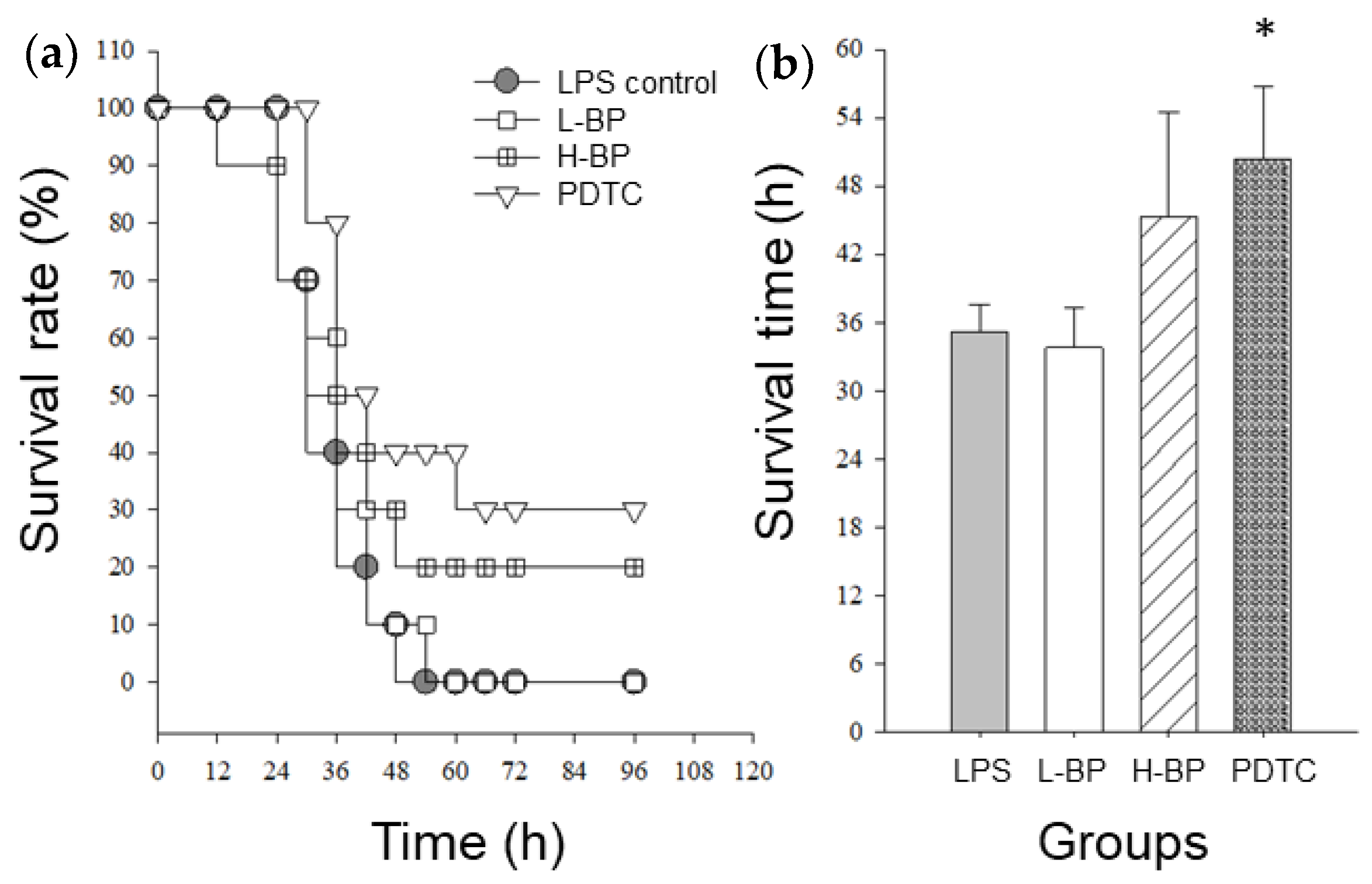
The levels of inflammation-related cytokines were negatively correlated with the lifespans of LPS-challenged mice [25]. In this study, the survival rate of the H-BP group (40%) was higher than that of the control group (20%) at 42 h after the LPS challenge (Figure 3). At the end of this experiment, none of the control or L-BP mice survived, but the survival rates of the H-BP and PDTC groups were 20 and 30%, respectively. Based on the average survival time of the mice, the H-BP group had a longer lifespan than the control group (45.3 ± 9.02 h vs. 35.2 ± 2.37 h, p = 0.20). Although no significant effect between the BP-95E-H2O-BuOH (L-BP and H-BP groups) and the control was observed in this experiment, our data suggest that the regulation of the inflammation-related cytokine profile of a mouse can partly reflect its lifespan in an LPS-challenged model. Some plant extracts, such as Dioscoreae Rhizoma [42], Prunella vulgaris [43], and Salvia miltiorrhiza bunge [44], have been suggested to alleviate inflammatory illnesses, including gouty arthritis, endotoxemia, allergic asthma, and anaphylaxi, by inhibiting TNF-α production. Possible ways to limit signal transductions have been postulated, including protein kinase C, tyrosine kinase, and NF-κB activation. PDTC, an NF-κB inhibitor, has been shown to protect mice from lethal endotoxic shock when injected 0.5 or 1 h before LPS induction [45]. As the positive control group in this study, PDTC effectively decreased serum IL-6, TNF-α, and IFN-γ following LPS exposure, resulting in the lowest mortality among the three groups (Figure 5).

4. Conclusions

The BP-95E-H2O-BuOH subfractions of BP had the most effective anti-inflammatory and T-cell immunomodulatory effects in LPS-challenged in vitro and in vivo models. The anti-inflammation effects of BP extracts are likely due to decreasing the population of CD3CD69 cells (activated T cells) and up-regulating the anti-inflammatory cytokines (IL-4 and IL-10). Notably, 5-HMF, guaiol, and oleamide showed significant anti-inflammatory activity on LPS-stimulated cells, the bioactive components reported in the BP extracts. In summary, our data indicate that BP can inhibit inflammatory mediators and modulate T-cell activation, implying that BP may be a medicinal material capable of preventing inflammation-related disorders.

Author Contributions

All authors contributed to this study. Y.-H.H.: Methodology, writing—original draft preparation. C.K.: Methodology, software, validation. C.-C.C.: Methodology, resources. F.-K.C.: Methodology, resources. T.-Y.S.: Methodology, resources. J.-Y.H.: Conceptualization, resources, supervision. C.-H.W.: Conceptualization, writing—review and editing, Supervision. All authors have read and agreed to the published version of the manuscript.

Funding

This research was supported by the grants MOST102-2632-B-214-001-MY3, MOST 109-2314-B-214-004, and MOST 111-2221-E-003-033.

Institutional Review Board Statement

The animal study protocol was approved by the IACUC of I-Shou University (IACUC-ISU-102-16).

Informed Consent Statement

Not applicable.

Data Availability Statement

Not applicable.

Conflicts of Interest

The authors declare no conflict of interest.

References

  1. Mezzavilla, M.; Notarangelo, M.; Concas, M.P.; Catamo, E.; Gasparini, P.; Grillotti, M.G.; Robino, A. Investigation of the link between PROP taste perception and vegetables consumption using FAOSTAT data. Int. J. Food Sci. Nutr. 2019, 70, 484–490. [Google Scholar] [CrossRef] [PubMed]
  2. Hikal, W.M.; Ahl, H.A.H.S.-A.; Bratovcic, A.; Tkachenko, K.G.; Sharifi-Rad, J.; Kačániová, M.; Elhourri, M.; Atanassova, M. Banana Peels: A Waste Treasure for Human Being. Evidence-Based Complement. Altern. Med. 2022, 2022, 7616452. [Google Scholar] [CrossRef] [PubMed]
  3. Acevedo, S.A.; Carrillo, J.D.; Flórez-López, E.; Grande-Tovar, C.D. Recovery of Banana Waste-Loss from Production and Processing: A Contribution to a Circular Economy. Molecules 2021, 26, 5282. [Google Scholar] [CrossRef] [PubMed]
  4. Mohapatra, D.; Mishra, S.; Sutar, N. Banana and its by-product utilisation: An overview. J. Sci. Ind. Res. 2010, 69, 323–329. [Google Scholar]
  5. Annadurai, G.; Juang, R.-S.; Lee, D. Adsorption of heavy metals from water using banana and orange peels. Water Sci. Technol. 2003, 47, 185–190. [Google Scholar] [CrossRef]
  6. Fries, J.H.; Waldron, R.J. Dehydrated banana in the dietetic management of diarrheas of infancy. J. Pediatr. 1950, 37, 367–372. [Google Scholar] [CrossRef]
  7. Pereira, A.; Maraschin, M. Banana (Musa spp.) from peel to pulp: Ethnopharmacology, source of bioactive compounds and its relevance for human health. J. Ethnopharmacol. 2015, 160, 149–163. [Google Scholar] [CrossRef]
  8. Emaga, T.H.; Robert, C.; Ronkart, S.N.; Wathelet, B.; Paquot, M. Dietary fibre components and pectin chemical features of peels during ripening in banana and plantain varieties. Bioresour. Technol. 2008, 99, 4346–4354. [Google Scholar] [CrossRef]
  9. Vu, H.T.; Scarlett, C.J.; Vuong, Q.V. Phenolic compounds within banana peel and their potential uses: A review. J. Funct. Foods 2018, 40, 238–248. [Google Scholar] [CrossRef]
  10. Atzingen, D.A.N.C.V.; Gragnani, A.; Veiga, D.F.; Abla, L.E.F.; Cardoso, L.L.F.; Ricardo, T.; dos Anjos Mendonça, A.R.; Ferreira, L.M. Unripe Musa sapientum peel in the healing of surgical wounds in rats. Acta Cir. Bras. 2013, 28, 33–38. [Google Scholar] [CrossRef]
  11. Vu, H.T.; Scarlett, C.J.; Vuong, Q.V. Effects of drying conditions on physicochemical and antioxidant properties of banana (Musa cavendish) peels. Dry. Technol. 2017, 35, 1141–1151. [Google Scholar] [CrossRef]
  12. Vu, H.T.; Scarlett, C.J.; Vuong, Q.V. Changes of phytochemicals and antioxidant capacity of banana peel during the ripening process; with and without ethylene treatment. Sci. Hortic. 2019, 253, 255–262. [Google Scholar] [CrossRef]
  13. Someya, S.; Yoshiki, Y.; Okubo, K. Antioxidant compounds from bananas (Musa Cavendish). Food Chem. 2002, 79, 351–354. [Google Scholar] [CrossRef]
  14. MPharm, S.R. Anti-inflammatory and antioxidant activities of extracts from Musa sapientum peel. J. Med. Assoc. Thai. 2012, 95, S142–S146. [Google Scholar]
  15. Chabuck, Z.A.G.; Al-Charrakh, A.H.; Hindi, N.K.K.; Hindi, S.K.K. Antimicrobial effect of aqueous banana peel extract, Iraq. Res. Gate. Pharm. Sci. 2013, 1, 73–75. [Google Scholar]
  16. Kapadia, S.P.; Pudakalkatti, P.S.; Shivanaikar, S. Detection of antimicrobial activity of banana peel (Musa paradisiaca L.) on Porphyromonas gingivalis and Aggregatibacter actinomycetemcomitans: An in vitro study. Contemp. Clin. Dent. 2015, 6, 496–499. [Google Scholar] [CrossRef] [PubMed]
  17. Singh, C.R.; Kathiresan, K.; Boopathy, N.S.; Anandhan, S.; Govindan, T. Evaluation of microbial potential of different coloured banana peels. Int. J. Preclin. Pharm. Res. 2013, 4, 62–64. [Google Scholar]
  18. Wang, R.; Feng, X.; Zhu, K.; Zhao, X.; Suo, H. Preventive activity of banana peel polyphenols on CCl4-induced experimental hepatic injury in Kunming mice. Exp. Ther. Med. 2016, 11, 1947–1954. [Google Scholar] [CrossRef]
  19. Propper, D.J.; Balkwill, F.R. Harnessing cytokines and chemokines for cancer therapy. Nat. Rev. Clin. Oncol. 2022, 19, 237–253. [Google Scholar] [CrossRef]
  20. Al-Mansoori, L.; Al-Jaber, H.; Prince, M.S.; Elrayess, M.A. Role of Inflammatory Cytokines, Growth Factors and Adipokines in Adipogenesis and Insulin Resistance. Inflammation 2022, 45, 31–44. [Google Scholar] [CrossRef]
  21. Soysal, P.; Arik, F.; Smith, L.; Jackson, S.E.; Isik, A.T. Inflammation, Frailty and Cardiovascular Disease. Adv. Exp. Med. Biol. 2020, 1216, 55–64. [Google Scholar] [CrossRef]
  22. Mohammad, S.; Thiemermann, C. Role of Metabolic Endotoxemia in Systemic Inflammation and Potential Interventions. Front. Immunol. 2020, 11, 594150. [Google Scholar] [CrossRef]
  23. Yu, Z.; Li, X.; Zhao, J.; Sun, S. Identification of hospitalized mortality of patients with COVID-19 by machine learning models based on blood inflammatory cytokines. Front. Public Health 2022, 10, 1001340. [Google Scholar] [CrossRef]
  24. Okeke, E.B.; Okwor, I.; Uzonna, J.E. Regulatory T Cells Restrain CD4+ T Cells from Causing Unregulated Immune Activation and Hypersensitivity to Lipopolysaccharide Challenge. J. Immunol. 2014, 193, 655–662. [Google Scholar] [CrossRef]
  25. Chao, W.W.; Hong, Y.H.; Chen, M.L.; Lin, B.F. Inhibitory effects of Angelica sinensis ethyl acetate extract and major compounds on NF-κB trans-activation activity and LPS-induced inflammation. J. Ethnopharmacol. 2010, 129, 244–249. [Google Scholar] [CrossRef]
  26. Vu, H.T.; Scarlett, C.J.; Vuong, Q.V. Maximising recovery of phenolic compounds and antioxidant properties from banana peel using microwave assisted extraction and water. J. Food Sci. Technol. 2019, 56, 1360–1370. [Google Scholar] [CrossRef]
  27. Geng, Y.; Zhu, S.; Cheng, P.; Lu, Z.-M.; Xu, H.-Y.; Shi, J.-S.; Xu, Z.-H. Bioassay-guided fractionation of ethyl acetate extract from Armillaria mellea attenuates inflammatory response in lipopolysaccharide (LPS) stimulated BV-2 microglia. Phytomedicine 2017, 26, 55–61. [Google Scholar] [CrossRef] [PubMed]
  28. Oh, Y.T.; Lee, J.Y.; Lee, J.; Lee, J.H.; Kim, J.E.; Ha, J.; Kang, I. Oleamide suppresses lipopolysaccharide-induced expression of iNOS and COX-2 through inhibition of NF-κB activation in BV2 murine microglial cells. Neurosci. Lett. 2010, 474, 148–153. [Google Scholar] [CrossRef]
  29. Zaini, H.M.; Roslan, J.; Saallah, S.; Munsu, E.; Sulaiman, N.S.; Pindi, W. Banana peels as a bioactive ingredient and its potential application in the food industry. J. Funct. Foods 2022, 92, 105054. [Google Scholar] [CrossRef]
  30. Iriondo, A.; Agirre, I.; Viar, N.; Requies, J. Value-Added Bio-Chemicals Commodities from Catalytic Conversion of Biomass Derived Furan-Compounds. Catalysts 2020, 10, 895. [Google Scholar] [CrossRef]
  31. Leamon, A.A.A.; Venegas, M.P.; Orsat, V.; Auclair, K.; Dumont, M.-J. Semisynthetic transformation of banana peel to enhance the conversion of sugars to 5-hydroxymethylfurfural. Bioresour. Technol. 2022, 362, 127782. [Google Scholar] [CrossRef] [PubMed]
  32. Okada, Y.; Tsuzuki, Y.; Sato, H.; Narimatsu, K.; Hokari, R.; Kurihara, C.; Watanabe, C.; Tomita, K.; Komoto, S.; Kawaguchi, A.; et al. Trans fatty acids exacerbate dextran sodium sulphate-induced colitis by promoting the up-regulation of macrophage-derived proinflammatory cytokines involved in T helper 17 cell polarization. Clin. Exp. Immunol. 2013, 174, 459–471. [Google Scholar] [CrossRef] [PubMed]
  33. Apel, M.A.; Lima, M.E.; Sobral, M.; Young MC, M.; Cordeiro, I.; Schapoval, E.E.; Henriques, A.T.; Moreno, P.R.H. Anti-inflammatory activity of essential oil from leaves of Myrciaria tenella and Calycorectes sellowianus. Pharm. Biol. 2010, 48, 433–438. [Google Scholar] [CrossRef] [PubMed]
  34. Moon, S.-M.; Lee, S.A.; Hong, J.H.; Kim, J.-S.; Kim, D.K.; Kim, C.S. Oleamide suppresses inflammatory responses in LPS-induced RAW264.7 murine macrophages and alleviates paw edema in a carrageenan-induced inflammatory rat model. Int. Immunopharmacol. 2018, 56, 179–185. [Google Scholar] [CrossRef] [PubMed]
  35. Hong, Y.H.; Weng, L.W.; Chang, C.C.; Hsu, H.F.; Wang, C.P.; Wang, S.W.; Houng, J.Y. Anti-inflammatory effects of Siegesbeckia orientalis ethanol extract in in vitro and in vivo models. Biomed. Res. Int. 2014, 2014, 329712. [Google Scholar] [CrossRef]
  36. Gomez, C.R.; Karavitis, J.; Palmer, J.L.; Faunce, D.E.; Ramirez, L.; Nomellini, V.; Kovacs, E.J. Interleukin-6 Contributes to Age-Related Alteration of Cytokine Production by Macrophages. Mediat. Inflamm. 2010, 2010, 475139. [Google Scholar] [CrossRef]
  37. Burke, J.D.; Young, H.A. IFN-gamma: A cytokine at the right time, is in the right place. Semin. Immunol. 2019, 43, 101280. [Google Scholar] [CrossRef]
  38. Liu, C.-J.; Lin, J.-Y. Anti-inflammatory effects of phenolic extracts from strawberry and mulberry fruits on cytokine secretion profiles using mouse primary splenocytes and peritoneal macrophages. Int. Immunopharmacol. 2013, 16, 165–170. [Google Scholar] [CrossRef]
  39. Na, H.; Cho, M.; Chung, Y. Regulation of Th2 Cell Immunity by Dendritic Cells. Immune Netw. 2016, 16, 1–12. [Google Scholar] [CrossRef]
  40. Eisenbarth, S.C.; Piggott, D.A.; Huleatt, J.W.; Visintin, I.; Herrick, C.A.; Bottomly, K. Lipopolysaccharide-enhanced, toll-like receptor 4-dependent T helper cell type 2 responses to inhaled antigen. J. Exp. Med. 2002, 196, 1645–1651. [Google Scholar] [CrossRef]
  41. Varma, T.K.; Lin, C.Y.; Toliver-Kinsky, T.E.; Sherwood, E.R. Endotoxin-Induced Gamma Interferon Production: Contributing Cell Types and Key Regulatory Factors. Clin. Vaccine Immunol. 2002, 9, 530–543. [Google Scholar] [CrossRef] [PubMed]
  42. Yu, D.; Wang, Y.; Yu, C.; Song, M.; Zhou, Q.; Liu, S. High-throughput serum metabolomics analysis of gouty arthritis rat treated by total saponins of Rhizoma Dioscoreae Makino by UPLC–Q/TOF–MS. Biomed. Chromatogr. 2020, 34, e4867. [Google Scholar] [CrossRef] [PubMed]
  43. Wang, S.-J.; Wang, X.-H.; Dai, Y.-Y.; Ma, M.-H.; Rahman, K.; Nian, H.; Zhang, H. Prunella vulgaris: A Comprehensive Review of Chemical Constituents, Pharmacological Effects and Clinical Applications. Curr. Pharm. Des. 2019, 25, 359–369. [Google Scholar] [CrossRef] [PubMed]
  44. Luo, J.; Zhang, L.; Zhang, X.; Long, Y.; Zou, F.; Yan, C.; Zou, W. Protective effects and active ingredients of Salvia miltiorrhiza Bunge extracts on airway responsiveness, inflammation and remodeling in mice with ovalbumin-induced allergic asthma. Phytomedicine 2019, 52, 168–177. [Google Scholar] [CrossRef] [PubMed]
  45. Liu, S.F.; Ye, X.; Malik, A.B. Inhibition of NF-κB activation by pyrrolidine dithiocarbamate prevents in vivo expression of proinflammatory genes. Circulation 1999, 100, 1330–1337. [Google Scholar] [CrossRef]
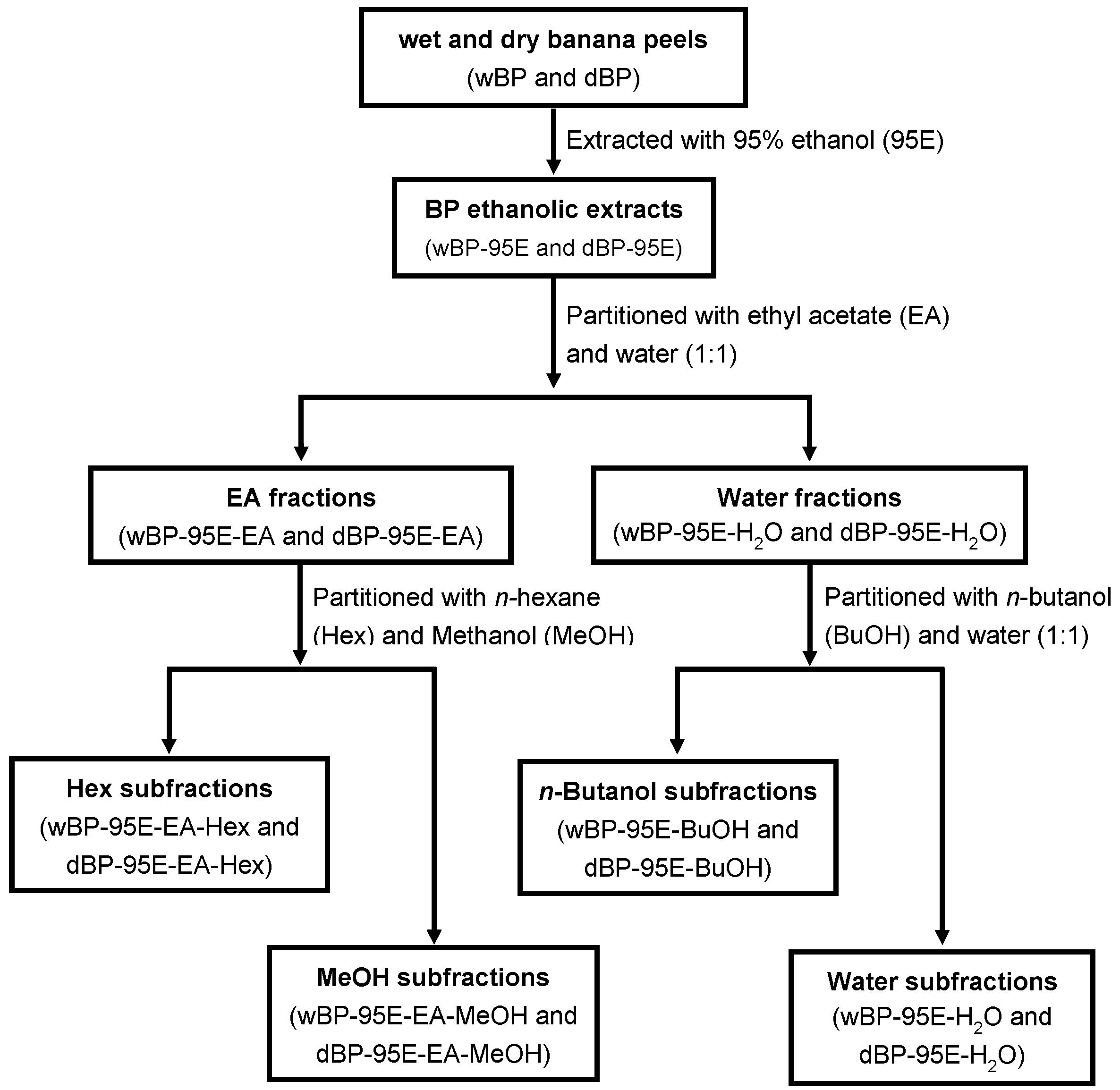
Figure 1. Extraction, separation, and fractionation of anti-inflammatory components from banana peel. BP-95E-H2O-BuOH subfractions exhibited the most potent anti-inflammatory and T-cell immunomodulatory properties.2.3. Gas Chromatography–Mass (GC–MS) Spectrometry Analysis.
Agriculture 13 00451 g001
Figure 2. Effects of butanol and methanol subfractions of BP on the release of IL-6 (a) and TNF-α (b) in LPS-stimulated RAW264.7 cells. Cells were preincubated with or without the indicated BP subfractions for 1 h and then stimulated with LPS (100 ng/mL) for 48 h. Cytokine secretions in culture media were measured by commercial ELISA kits. The levels of IL-6 and TNF-α in untreated control (without LPS) were undetectable. A significant difference from the LPS alone is indicated as * p < 0.05 or *** p < 0.001 according to Student’s t-test. The values in the column represent the inhibition percentage (%) of cytokine secretions by BP extracts. The fresh peel was defined as wet banana peel. The wet banana peel placed in a circulating oven at 60 °C for three days was defined as dry peel. BP, banana peel; E95, 95% ethanol extract; BuOH, butanol subfraction; MeOH, methanol subfraction.
Agriculture 13 00451 g002
Figure 3. Effects of butanol and methanol subfractions of BP on iNOS or COX-2 protein expressions in LPS-stimulated RAW264.7 cells. Cells were pretreated with the indicated BP extracts for 1 h and then stimulated with LPS (100 ng/mL) for 12 h. Whole-cell lysates were subjected to Western blotting analysis using the anti-COX-2 and anti-iNOS antibodies or an anti-GAPDH antibody as a loading control (a). The extents of protein expression are expressed as multiples of LPS alone (b,c) and quantified by densitometric analysis using Quantity One software (Bio-Rad). Bar values are means ± SD of three independent experiments in these assays. A significant difference from the LPS control is indicated as * p < 0.05, ** p < 0.01, or *** p < 0.001 according to Student’s t-test.
Agriculture 13 00451 g003
Figure 4. (a) GC–MS spectrometry profile of BP-95E-H2O-BuOH subfractions of BP. (b) Five non-polyphenol components in BP-95E-H2O-BuOH subfractions were identified by comparison of their mass spectra with the NIST MS 14.0 database (Gaithersburg) as follows: (1) 5-hydroxymethyl furfural (5-HMF, retention time (RT) = 23.9 min), (2) guaiol (RT = 42.0 min), (3) oleic acid (RT = 67.1 min), (4) linoleic acid (RT = 76.7 min), and (5) oleamide (RT = 77.1 min). (c) RAW264.7 cells were treated with 5-HMF, guaiol, and oleamideat for 48 h, and the cell viability was measured with MTT assay. The levels of (d) NO, (e) IL-6, and (f) TNF-α were determined in culture media of LPS-challenged RAW264.7 cells with or without 5-HMF, guaiol, or oleamideat, as described in Section 2. The bars with the symbols # and * represent significant differences (p < 0.05) from the blank and the control, respectively, according to Student’s t-test.
Agriculture 13 00451 g004aAgriculture 13 00451 g004b
Figure 5. The effects of BP-95E-H2O-BuOH extract administration on the survival rate of mice challenged with LPS. The BALB/c mice were tube-fed with BP-95E-H2O-BuOH extracts at doses of 44 (L-BP group) and 166 mg/kg b.w. (H-BP group) for two weeks before the LPS challenge, whereas the mice in the PDTC group were i.p. injected with 50 mg PDTC/kg b.w. 1 h before the LPS challenge. The survival of each group of mice was recorded and is expressed as the survival rate (a) and survival time (b). A significant difference from the control is indicated as * p < 0.05 according to Student’s t-test.
Agriculture 13 00451 g005
Table 1. The effects of BP-95E-H2O-BuOH extract administration on the release of pro-inflammatory mediators in serum and PECs from LPS-challenged mice.
GroupsControl aL-BPH-BPPDTC
Serum b
IL-6755 ± 88661 ± 78556 ± 75 *562 ± 43 *
TNF-α0.99 ± 0.110.66 ± 0.10 *0.69 ± 0.13 *0.14 ± 0.04 **
PECs (spontaneous, without LPS) c
NO10.30 ± 1.7411.50 ± 2.359.83 ± 2.1910.20 ± 1.83
IL-68.10 ± 0.525.18 ± 0.69 **5.14 ± 0.69 **4.55 ± 0.73 **
TNF-α0.18 ± 0.020.15 ± 0.020.18 ± 0.020.10 ± 0.02 *
PECs (LPS stimulation)
NO18.50 ± 3.8717.40 ± 3.8516.30 ± 3.6116.90 ± 3.44
IL-623.50 ± 3.4511.50 ± 2.61 *11.30 ± 1.99 **12.80 ± 3.69 *
TNF-α0.69 ± 0.110.56 ± 0.140.77 ± 0.140.22 ± 0.07 **
a Mice were sacrificed six hours after LPS injection. The BALB/c mice were tube-fed BP-95E-H2O-BuOH extracts at doses of 44 (L-BP group) and 166 mg/kg b.w. (H-BP group) for two weeks before the LPS challenge, whereas the mice in the PDTC group were i.p. injected with 50 mg PDTC/kg b.w. 1 h before the LPS challenge. Values are expressed as means ± SEM (n = 12 mice/group) and were statistically analyzed using Student’s t-test. A significant difference from the control in the same row is indicated at * p < 0.05 and ** p < 0.01. b The mice sera were collected for the measurement of IL-6 and TNF-α. The cytokines levels are represented as ng/mL. c PECs (peritoneal exudate cells) were stimulated with and without 10 μg/mL LPS for macrophage activation. The cytokines and NO levels are represented as ng/mL and μM, respectively. The levels of basic cytokine released in normal cell culture conditions without LPS stimulation were spontaneous.
Table 2. The modulatory effects of BP-95E-H2O-BuOH extracts on T-cell-related cytokine secretion in spleen cells from LPS-challenged mice.
GroupsControl aL-BPH-BPPDTC
Spontaneous (without Con A) b
IFN-γ0.28 ± 0.050.11 ± 0.01 **0.09 ± 0.02 **0.12 ± 0.02 *
IL-46.52 ± 1.066.38 ± 2.075.19 ± 1.3810.4 ± 2.49
IL-60.09 ± 0.010.08 ± 0.010.08 ± 0.020.15 ± 0.04
IL-100.21 ± 0.040.23 ± 0.060.21 ± 0.090.32 ± 0.18
Con A stimulation c
IFN-γ5.35 ± 1.063.33 ± 1.102.38 ± 0.47 *2.94 ± 0.95 *
IL-424.8 ± 9.2534.1 ± 11.164.2 ± 16.790.10 ± 32.90
IL-60.63 ± 0.100.70 ± 0.170.25 ± 0.06 *0.83 ± 0.18 *
IL-100.31 ± 0.130.61 ± 0.200.81 ± 0.241.98 ± 0.37 *
T-cell populations d
CD3CD6923.8 ± 4.5619.3 ± 5.2415.9 ± 4.17 *18.3 ± 3.41 *
CD3CD4CD2565.3 ± 4.8967.4 ± 5.8965.6 ± 1.6263.4 ± 2.90
a Mice were sacrificed six h after LPS injection. The BALB/c mice were tube-fed BP-95E-H2O-BuOH extracts at doses of 44 (L-BP group) and 166 mg/kg b.w. (H-BP group) for two weeks before the LPS challenge, whereas the mice in the PDTC group were i.p. injected with 50 mg PDTC/kg b.w. 1 h before the LPS challenge. Murine spleen cells were obtained for primary cell culture, and 24 h following culture, the supernatants were collected for cytokine assay. Values are expressed as the means ± SEM (n = 12 mice/group) and were statistically analyzed by using Student’s t-test. A significant difference from the control in the same row is indicated as * p < 0.05 and ** p < 0.01. b The levels of basic cytokine release in normal cell culture conditions without Con A stimulation were spontaneous. IL-4 levels are expressed as pg/mL, while others are ng/mL. c Cells were treated with Con A (5 μg/mL) for T-cell activation. d CD3CD69 cell population indicates the percentage of activated T cells in splenocytic T cells; CD3CD4CD25 cell population refers to the percentage of regulatory T cells in splenocytic T helper cells.
Disclaimer/Publisher’s Note: The statements, opinions and data contained in all publications are solely those of the individual author(s) and contributor(s) and not of MDPI and/or the editor(s). MDPI and/or the editor(s) disclaim responsibility for any injury to people or property resulting from any ideas, methods, instructions or products referred to in the content.