Article

Exploration of Piezo Channels in Bread Wheat (Triticum aestivum L.)

1
Department of Botany, Panjab University, Chandigarh 160014, India
2
Department of Biotechnology, Panjab University, Chandigarh 160014, India
*
Author to whom correspondence should be addressed.
Agriculture 2023, 13(4), 783; https://doi.org/10.3390/agriculture13040783
Submission received: 11 February 2023 / Revised: 22 March 2023 / Accepted: 27 March 2023 / Published: 29 March 2023
(This article belongs to the Section Crop Genetics, Genomics and Breeding)

Abstract

:
Piezo channels belong to an important class of cell membrane-bound, Ca2+-permeable, mechanosensitive channels consisting of a pore and multiple transmembrane helices. In plants, the functional aspects of Piezo channels have been less studied than other mechanosensitive channels. However, a few studies that have been carried out indicate the involvement of Piezo channels in stress response and developmental processes. In our analysis, we identified a total of three Piezo genes in the Triticum aestivum genome. The phylogenetic analysis revealed the monocot and dicot-specific clustering of Piezo proteins. The gene and protein structure analysis indicated their conserved architecture. The promoter region of each of the three Piezo genes contained light-, growth-and development-, hormone-, and stress-responsive cis-regulatory elements. Moreover, the differential expression of Piezo genes in tissue developmental stages and under abiotic and biotic stress conditions indicated their probable role in plant growth and development and various stresses. The quantitative real-time polymerase chain reaction (qRT-PCR) analysis suggested that TaPiezo1-D might be involved in Ca2+ homeostasis. In addition, protein–protein interaction indicated their precise role in glucose, hormone and stress responses. The miRNA interaction analysis further suggested their participation in signaling cascades and biological processes. The present study will extend our understanding about Piezo channels in Ca2+ mediated signaling in plants under various stresses and provide a path for the functional validation of TaPiezo genes in future research.

1. Introduction

Plants sense various mechanical stimuli, including touch, sound, gravity, osmotic pressure etc., throughout their life cycle. These mechanical stimuli cause plasma membrane deformation, leading to the activation of various channels, which allow the ion permeation and downstream signaling cascade [1]. These channels are known as stretch-activated channels or mechanosensitive (MS) channels. Five types of MS channels have been identified: mechanosensitive channel of small conductance-like (MscS-like or MSL); mid1-complementing activity (MCA); two-pore potassium channel (TPK); hyperosmolality-induced [Ca2+] increase (OSCA) and Piezo channels in plants [2]. In contrast to the MSL, MCA, TPK and OSCA, the Piezo channels are relatively less explored.
Piezo channels are primarily known for their contribution in various aspects of animal mechanosensation [3,4,5]. Initially, Piezo1 and Piezo2 were discovered in mouse (Mus musculus) as Ca2+-permeable ion channels [6]. Moreover, Piezo proteins were reported as regulator of membrane potential and Ca2+ signaling [5,6,7,8]. The structural studies of Piezo1 by high-resolution cryo-electron microscopy revealed its three-bladed propeller shape, consisting of an arched transmembrane region and ion-conducting central pore [9,10,11]. The homologous proteins of Piezo have been reported in fungi, animals and plants [6]. In the majority of plants, one to three Piezo genes have been reported [12]. Most of the species of algae, bryophytes and pteridophytes comprised only one Piezo gene. However, Ostreococcus lucimarinus, Physcomitrella patens and Sphagnum fallax, each harbor two Piezo genes [12,13]. Monocot species, for instance, Brachypodium distachyon, Brachypodium stacei, Oryza sativa Japonica, Sorghum bicolor, Setaria italica, Setaria viridis, Zea mays etc., consisted of only one Piezo gene [12,13]. In the case of dicot species, one Piezo gene was found in each of Arabidopsis thaliana, Nicotiana attenuate, Solanum species etc., two genes in each of Medicago truncatula, Vigna radiata, Vitis vinifera, Eucalyptus grandis, Ricinus communis, Brassica rapa etc., and three genes in Glycine max and Gossypium raimondii [12,13].
In A. thaliana, the mutant AtPiezo showed abnormal root growth on agar medium, which indicated their involvement in normal root growth. Additionally, AtPiezo channel facilitates the influx of Ca2+ ions in the root cap upon mechanical stimuli, and therefore, it acts as a mechanosensory protein [12,14]. Furthermore, AtPiezo mutants show enhanced susceptibility to viral infection, which revealed the role of AtPiezo in biotic stress responses [15]. In P. patens, the vacuolar morphology is reported to controlled by PpPiezo1 and PpPiezo2 by facilitating the Ca2+ efflux from the vacuole in response to hypo-osmotic stress [16]. In O. sativa, the Piezo gene exhibited significant expression in roots, leaves, pistils, lemmas and ovaries [13]. Moreover, the occurrence of gibberellin-, abscisic acid (ABA)- and jasmonic acid-related cis-regulatory elements in the Piezo gene of O. sativa suggested its participation in stress-related signal transduction [13].
To date, no Piezo channel has been identified in major food crop, i.e., T. aestivum. In the current study, we identified three Piezo genes in the genome of T. aestivum. Furthermore, using in silico approaches, various analyses such as that of chromosomal localization, phylogenetic, gene structure, cis-regulatory elements, and protein structure have been carried out. To elucidate the functioning of Piezo genes, their expression patterns were analyzed in three developmental stages of five tissues and heat, drought, salt and fungal stresses using the available high-throughput RNA sequence data. To validate the functioning of Piezo protein in Ca2+ homeostasis, the quantitative real-time polymerase chain reaction (qRT-PCR) of one Piezo gene homeolog, i.e., TaPiezo1-D, was performed. Protein–protein and miRNA interaction analyses were also carried out. Collectively, this study will create a path for the functional validation of TaPiezo proteins in the future.

2. Materials and Methods

2.1. Identification of TaPiezos and Their Chromosomal Distribution

Firstly, the bidirectional BLAST hit approach was used to find the Piezo proteins of T. aestivum. The protein model sequences were retrieved from the IWGSC (IWGSC RefSeq assembly v2.0, accessed on 13 June 2022) and Ensembl Plant databases. The known protein sequences of Piezo channel of A. thaliana and O. sativa were used as queries in the BLASTp search as opposed to the above-retrieved protein sequences of T. aestivum. The occurrence of a functionally conserved domain in all TaPiezo proteins was confirmed using the Conserved Domain Database search and SMART server [17,18].
The sub-genomic and chromosomal information of TaPiezo genes was retrieved from the Ensembl Plant database. Furthermore, the chromosomal maps were prepared through MapInspect (mapinspect.software.in-former.com/, accessed on 14 January 2023). The homeologous clustering of identified Piezo was based on ≥90% sequence similarity [19,20,21]. The Piezo of T. aestivum were named according to the recommended guidelines of gene symbolization (http://wheat.pw.usda.gov/ggpages/wgc/98/Intro.htm, accessed on 17 July 2022).

2.2. Phylogenetic Analysis

The full-length protein sequences of Piezo channels from eight plants, namely, A. thaliana, B. rapa, B. distachyon, G. max, N. attenuate, O. sativa, S. bicolor, and Z. mays, along with T. aestivum, were used for the phylogenetic analysis. Firstly, the Muscle algorithm program was used for the alignment of protein sequences. Then, the phylogenetic tree was created using the protein sequence alignment on MEGA X software [22]. The neighbor-joining method was employed with 1000 bootstrap replicates.

2.3. Gene Structure Analysis

To construct the gene structure, the coding sequence (CDS) and genomic sequence of each TaPiezo gene were aligned using the GSDS 2.0 server [23]. The gene structure was demonstrated in the form of exon–intron patterning and intron phase distribution.

2.4. Cis-Regulatory Element Analysis

The cis-regulatory elements were predicted among the 1.5 kb upstream promoter of each TaPiezo gene using the PlantCARE database [24].

2.5. Physicochemical, Motif Analyses and Multiple Sequence Alignment

The physicochemical characterizations of Piezo proteins including length, isoelectric point (pI), molecular weight (MW) and grand average of hydropathicity (GRAVY) value were carried out using the online Expasy tool [25]. The number of transmembrane helices, the signal peptide and the subcellular localization of each Piezo protein were examined through the TMHMM-2.0 (https://services.healthtech.dtu.dk/service.php?TMHMM-2.0, accessed on 19 July 2022), SignalP 5.0. (https://services.healthtech.dtu.dk/service.php?SignalP-5.0, accessed on 19 July 2022) and Cello server [26], respectively. Moreover, the 15 conserved motifs were predicted through MEME (Multiple Expectation Maximization for Motif Elicitation) suite version 5.1.1 server at default parameters, which were further scanned through the scan motif server [27] (https://myhits.sib.swiss/cgi-bin/motif_scan, accessed on 21 July 2022). Furthermore, to analyze the conserved residues, the multiple sequence alignment of Piezo proteins was carried out using the ClustalW in Bioedit [28]. The 3-dimensional (3D) structure of TaPiezo1-A was predicted using the Pyre2 (http://www.sbg.bio.ic.ac.uk/phyre2/html/page.cgi?id=index, accessed on 13 January 2023) and illustrated by the Pymol (https://pymol.org/2/, accessed on 15 January 2023).

2.6. Expression Profiling of Piezo Genes

To decipher the putative functioning of Piezo genes, the expression profiling of these genes was performed under several tissue developmental stages and stress treatments. An openly accessible URGI database (https://urgi.versailles.inra.fr/files/RNASeqWheat, accessed on 26 July 2022) was used to retrieve high-throughput RNA sequence data.
To investigate the expression profiling of TaPiezo in tissue developmental stages, the high-throughput RNA sequence data, derived from the root, leaf, stem, spike, and grain tissues generated in two biological replicates, were used [29,30]. The developmental stages were: z10, z13, z39 for roots; z30, z32, z65 for stem; z10, z23, z71 for leaf; z32, z39 z65 for spike; and z71, z75, z85 for grain. The expression values were calculated in the form of fragments per kilobase of transcripts per million mapped (FPKM) reads through Trinity packages. Furthermore, the Expression ATLAS was used for the confirmation of expression data [31,32].
The effect of abiotic stresses on TaPiezo was studied by performing the expression profiling with the usage of high-throughput RNA sequence data produced in duplicates after 1 and 6 h of heat stress (HS) (40 °C), drought stress (DS) (20% Poly Ethylene Glycol (PEG) and combined heat and drought stress (HD) from leaf tissue [33]. During salt stress, 6, 12, 24 and 48 h of salt-treated (150 mM NaCl) root tissue was used for the production of RNA sequence data [34].
Moreover, the effect of. Blumeria graminis (Bgt) and Puccinia striiformis (Pst) were studied by performing expression profiling. The high-throughput RNA sequence data was extracted after 24, 48 and 72 h of fungal infestation in triplicate [35]. The expression readings were computed into FPKM reads through the Trinity package [31]. Furthermore, the heat maps were made through the Hierarchical Clustering Explorer (HCE) 3.5 software and hierarchical clustering was performed with the Euclidean distance method [36].

2.7. qRT-PCR Analysis

Firstly, sodium hypochlorite (1.2%) along with 10% ethanol were used for surface sterilization of T. aestivum (cv. Chinese spring) seeds. After washing with double autoclaved water, the seeds were kept at 4 °C for stratification. The next day, seeds were allowed to germinate at room temperature. Thereafter, the germinated seedlings were placed in a plant growth chamber at 16 h of light and 8 h of the dark period, 60% relative humidity and 22 °C temperature. After 7 days, the CaCl2 (20 mM) treatment was given to the seedlings with Murashige and Skoog media. Subsequently, the samples were collected in liquid nitrogen after 6, 12, 24 and 48 h of stress treatment. The total RNA from root tissues was isolated using Spectrum TM Plant Total RNA kit (Sigma, Saint Louis, MI, USA). For the production of contamination free RNA, the isolated samples were treated using the TURBO DNA-free™ Kit (Invitrogen, Carlsbad, CA, USA). The quality of isolated RNA samples were examined via agarose gel electrophoresis, and quantity via Nanodrop spectrophotometer. Subsequently, the cDNAs were prepared with the Superscript III First-Strand Synthesis Super-mix (Invitrogen, Carlsbad, CA, USA) from one microgram of RNA. The qRT-PCR experiment was carried out at CFX96 Real-Time PCR (BioRad, Hercules, CA, USA) using SYBR Green, cDNA and gene-specific primers, following recommended procedures [37]. Moreover, for the internal control, an ADP-ribosylation factor of T. aestivum (TaARF1) was utilized during the experiment (Table 1). The delta-delta CT (2−ΔΔCT) method was employed to compute the expression values, and the experiment was practiced in three biological replicates (n = 3), which were displayed as mean standard deviation (SD) [38,39].

2.8. Protein–Protein and miRNA-Interaction Analysis

The protein–protein interactions of TaPiezo proteins were predicted through the STRING server (http://stringdb.org, accessed on 27 July 2022) accessed on 27 July 2022) [40]. The psRNATarget database (http://plantgrn.Noble.org/psRNATarget/, accessed on 28 July 2022) was explored to find the interacting miRNAs of TaPiezo genes [41]. Furthermore, the interaction networks have been prepared using the Cytoscape tool (https://cytoscape.org/download.html, accessed on 14 November 2022).

3. Results

3.1. Identification and Chromosomal Localization of TaPiezo

A total of three TaPiezo genes (TraesCS3A02G192500.1, TraesCS3B02G222000.1 and TraesCS3D02G196400.1) were identified in the T. aestivum genome through a BLASTp search of Piezo sequences of A. thaliana (AT2G48060) and O. sativa (Os01g0388500). These three genes shared ≥90% sequence similarity with and were derived from A, B and D sub-genome and were therefore considered homoeologous genes. The genes TraesCS3A02G192500.1, TraesCS3B02G222000.1 and TraesCS3D02G196400.1 were named as TaPiezo1-A, TaPiezo1-B and TaPiezo1-D, respectively. The chromosomal positions of TaPiezo1-A, TaPiezo1-B and TaPiezo1-D were on the short arm of chromosome 3A, 3B and 3D, respectively (Figure 1; Table S1). All the TaPiezo genes were found on the forward strand of their respective chromosomes.

3.2. Phylogenetic Analysis

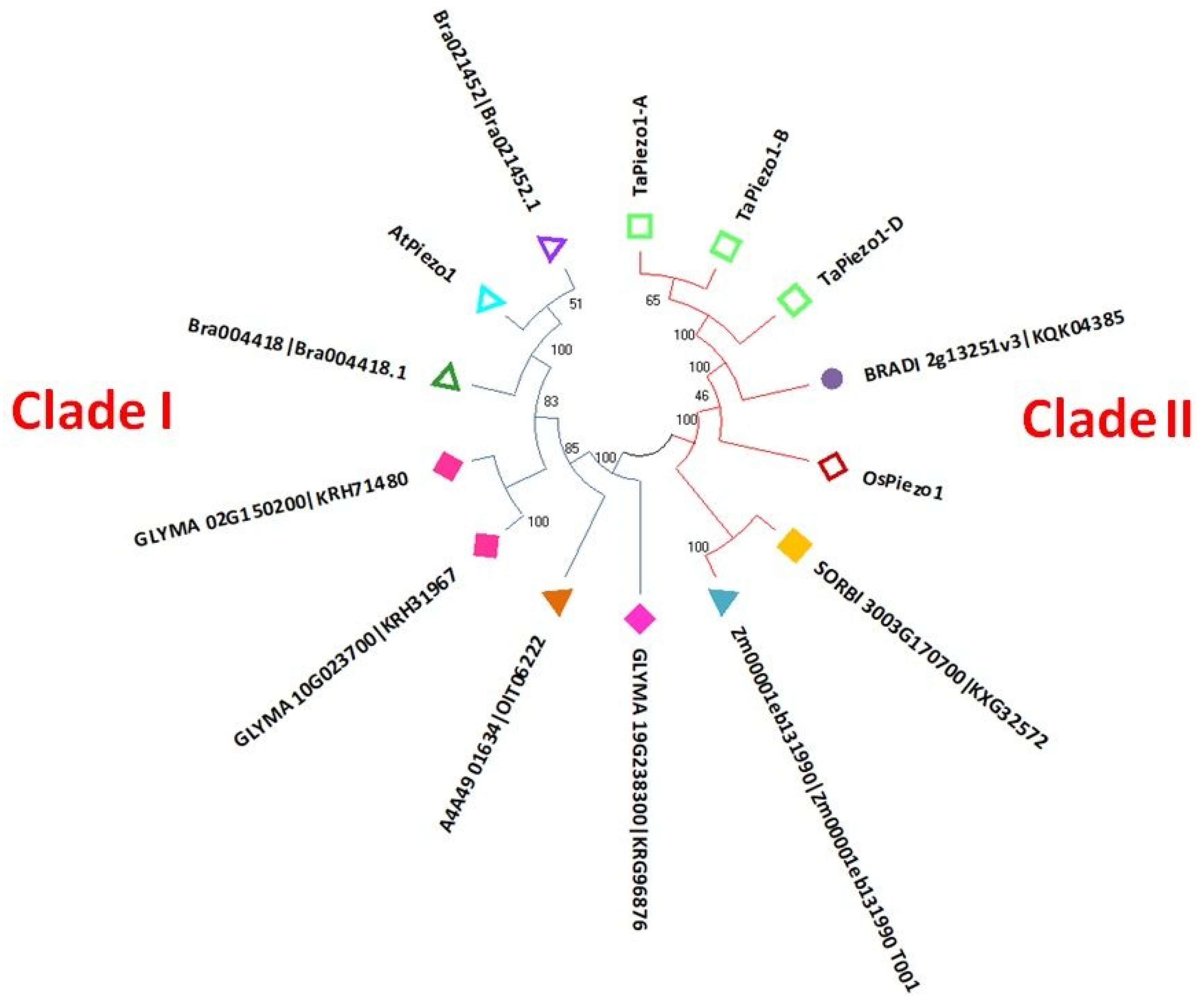
To study the evolutionary relationships, the phylogenetic tree was constructed with full-length Piezo protein sequences of A. thaliana, G. max, B. rapa, B. distachyon, N. attenuate, O. sativa, T. aestivum, S. bicolor and Z. mays (Figure 2). The phylogenetic tree showed the clustering of Piezos into two groups: I and II. Piezo proteins of dicot species were clustered in group I, while Piezo proteins of monocots were clustered in group II. Moreover, the Piezo proteins of T. aestivum were closely clustered in the phylogenetic tree (Figure 2).

3.3. Gene Structure Analysis

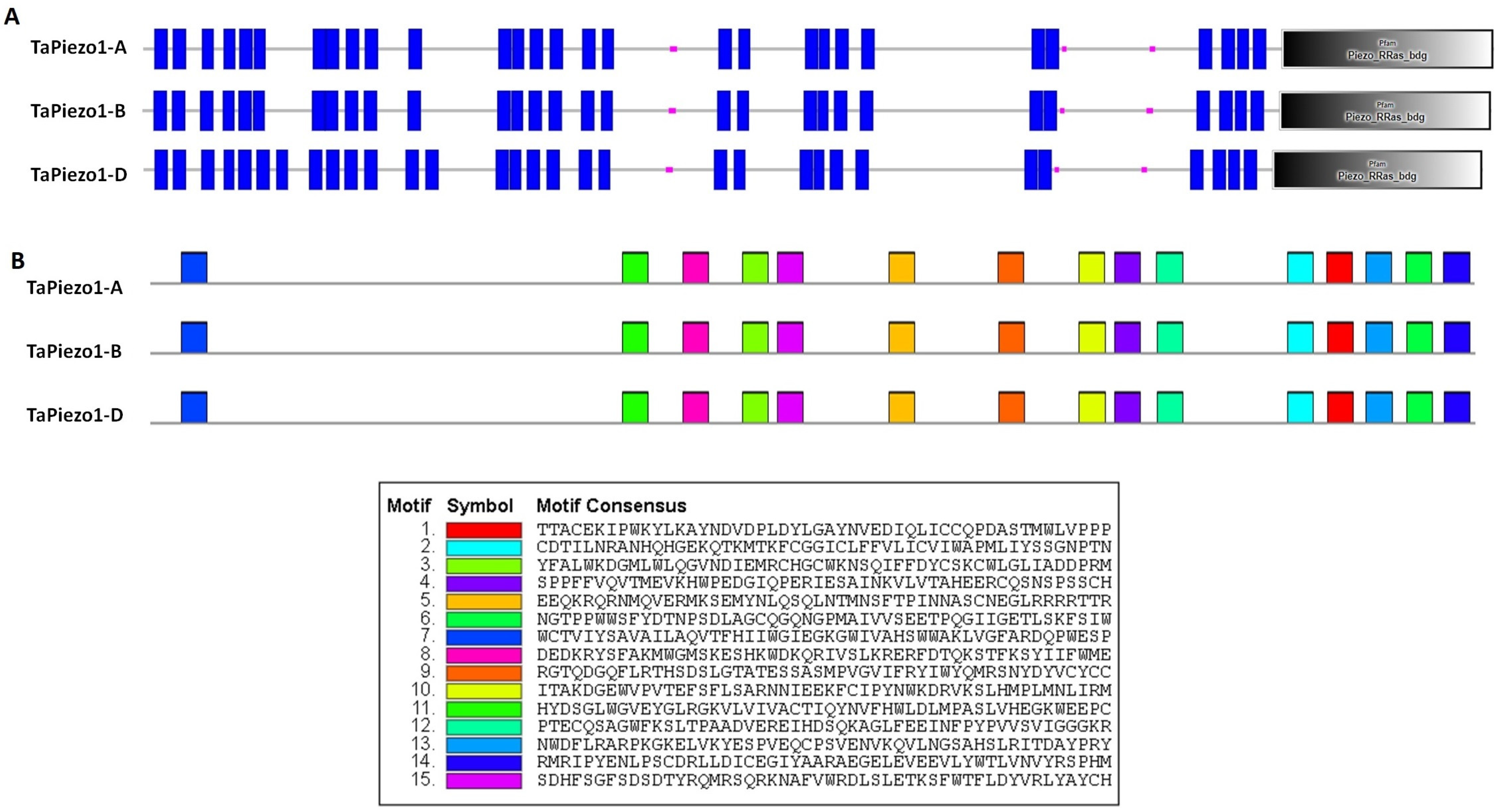
The gene structure analysis was carried out to investigate the length of CDS, the number of introns and exons, their organization, and intron phase distribution. The predicted length of CDS for each TaPiezo1-A and TaPiezo1-D was 7494 base pair (bp), while TaPiezo1-B was 7492 bp. Each of the TaPiezo genes showed the presence of 21 exons and 20 introns. The patterns of exons and introns were found to be similar among all Piezo genes of T. aestivum (Figure 3; Table S1). The majority of the introns (42%) of TaPiezo genes were found in intron phase 1, followed by phase 0 (35%) and phase 2 (23%) (Figure 3).

3.4. Cis-Regulatory Element Analysis

The analysis of cis-regulatory elements is a significant approach for studying the involvement of Piezo channels in a diverse range of biological processes. In the current analysis, we have identified a wide range of cis-regulatory elements that were light, growth and development, stress and hormone responsive (Table 2). The predicted light-responsive cis-regulatory elements were G-Box, Sp1, G-box, TCCC-motif, AE-box, ACE, TCT-motif, I-box and Box 4. The cis-regulatory elements which showed their connection with various plant growth and developmental processes were O2-site (zein metabolism regulation), RY-element (seed-specific regulation), CAT-box (meristem expression), GCN4_motif (endosperm development), CCGTCC-box (meristem specific activation), etc. (Table 2). Several identified stress-related cis-regulatory elements were LTR (low-temperature responsive), GC-motif (anoxic specific inducibility), ARE (anaerobic induction), W-box (responsive to pathogenic attack), MBS (involved in drought inducibility), TC-rich repeats (defense and stress-responsive), DRE (dehydration responsive element), MYB, MYC (drought-responsive elements), WRE3 (wound responsive), etc. In addition to these, the most common hormone-responsive cis-regulatory elements found in the Piezo genes were ABRE, ABRE4, ABRE3a, AT~ABRE (Abscisic acid-responsive), TGA-element (Auxin responsive), P-box (gibberellin responsive), CGTCA-motif and TGACG-motif (methyl jasmonate responsive), and TCA-element (salicylic acid-responsive) (see Table 2).

3.5. Protein Characterization

The length of each TaPiezo1-A and TaPiezo1-D protein was 2497 amino acid (aa) residues, while the length of TaPiezo1-B protein was 2496 aa residues. The calculated MW of TaPiezo1-A and TaPiezo1-D was 286.7 kilodaltons (kDa) (Table S1), while the MW of TaPiezo1-B was 286.4 kDa. The isoelectric points calculated for TaPiezo1-A, TaPiezo1-B and TaPiezo1-D were 8.17, 8.24 and 8.21, respectively (Table S1). The GRAVY values for TaPiezo1-A, TaPiezo1-B and TaPiezo1-D were 0.229, 0.230 and 0.226, respectively. The sub-cellular localization analysis predicted the plasma membrane localization of TaPiezo channels. In addition, no signal peptide was found in the TaPiezo proteins (Table S1).
Domains are considered the structural and functional units of proteins, and therefore, it is important to study the domain composition of a protein. In the current study, domain analysis at Pfam and SMART server suggested the occurrence of a very conserved domain, known as the Piezo R-Ras-binding domain (PF12166) in all the identified Piezo proteins of T. aestivum (Figure 4A; Table S2). This conserved domain comprised 392 aa residues in each TaPiezo protein. Additionally, the TaPiezo1-A and TaPiezo1-B consisted of 31 transmembrane helices, while TaPiezo1-D consisted of 33 transmembrane helices (Figure 4A).
A total of 15 motifs have been predicted in Piezo proteins of T. aestivum (Figure 4B). All the motifs were predominately found in each TaPiezo protein and revealed their conserved nature. Motifs 2, 6 and 14 were identified as part of the conserved Piezo R-Ras-binding domain, and motifs 7 and 9 were identified as part of transmembrane helices. Furthermore, casein kinase II phosphorylation and protein kinase C phosphorylation sites were also found in the majority of the motifs of TaPiezo proteins. However, cAMP- and cGMP-dependent protein kinase phosphorylation, ASN-glycosylation, tyrosine kinase phosphorylation and N-myristoylation sites were detected in motifs 8, 9 and 13, and, 5 and 13, and, 3, 9, 11 and 14, respectively.
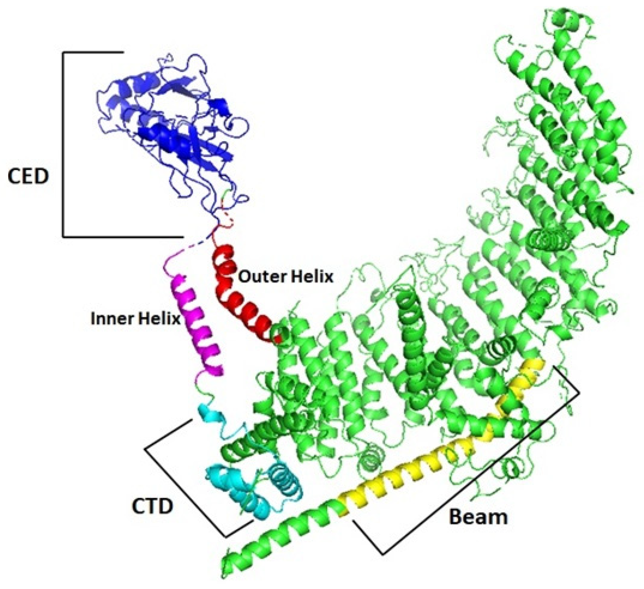
Furthermore, the Piezo protein sequences of A. thaliana, T. aestivum, O. sativa and M. musculus were aligned using the ClustalW to highlight the conversed residues (Figure S1). All the Piezo proteins consisted of a beam domain, outer helix (OH), central cap domain (CED), inner helix (IN) and C-terminal domain (CTD) (Figure S1). Moreover, it was analyzed that OH, CED, IN and CTD were evolutionarily more conserved as compared to the beam domain in all the species. All the domains were also highlighted in the 3D structure of the TaPiezo1-A protein (Figure 5).

3.6. Expression Profiling of Piezo Genes in Tissue Developmental Stages

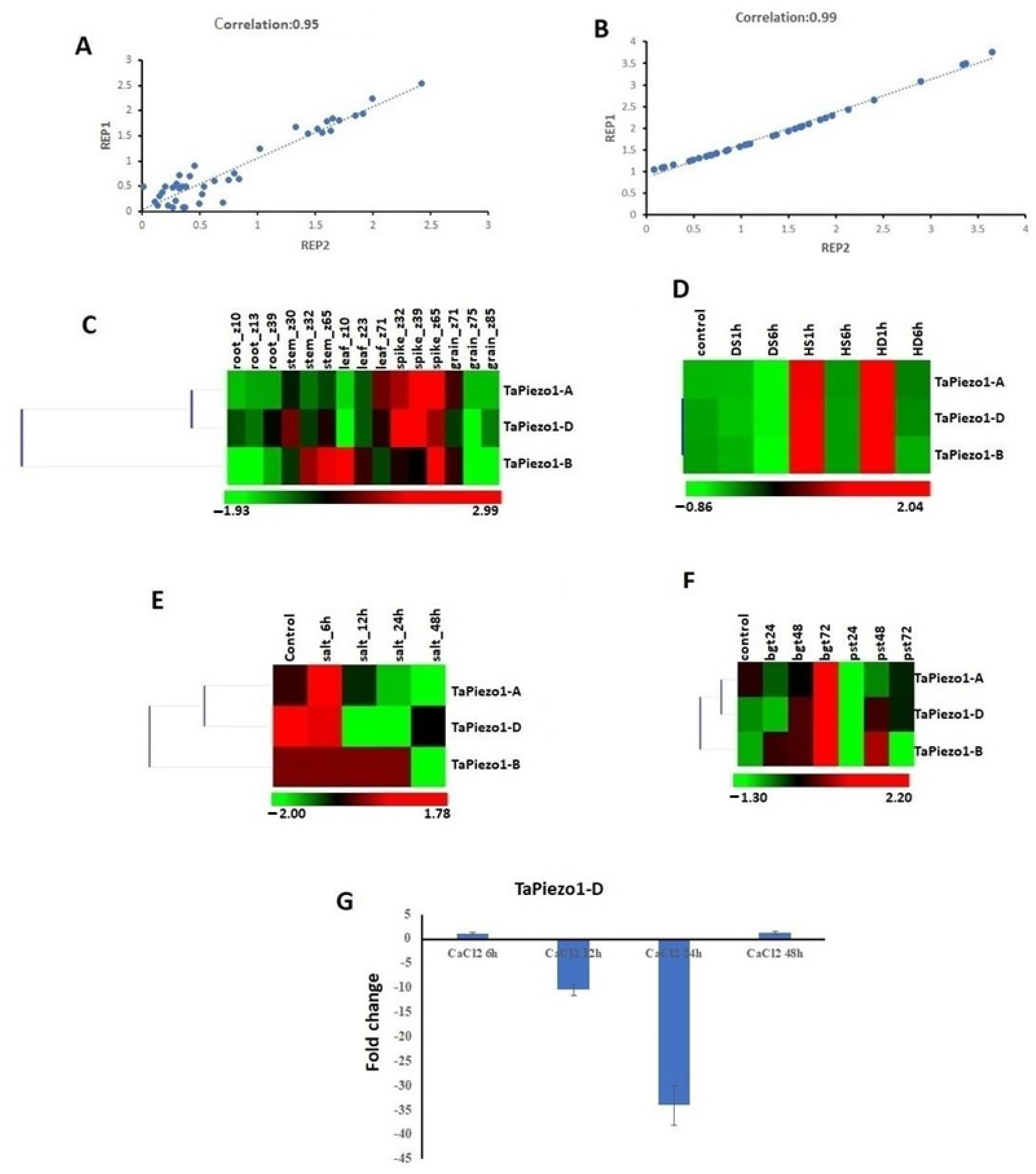
To study the probable functioning of TaPiezo genes in plant growth and development, their expression profiling was carried out in three developmental stages of the root, stem, leaf, spike and grain tissues using the high-throughput RNA sequence data [29,30]. Furthermore, the reliability of expression was confirmed by correlation analysis. The Pearson correlation value was 0.95 for various tissue developmental stages and 0.99 for stress conditions (Figure 6A,B). TaPiezo1-A exhibited low expression values during the various tissue development stages, except spike tissues (Figure 6C). On the other hand, TaPiezo1-D showed comparatively elevated expression during multiple tissue development stages including stem, leaf, root, spike and grain tissues, while TaPiezo1-B showed expression in stem, leaf and spike tissues. All the TaPiezo genes showed significant upregulation in the spike developmental stages. Overall analysis indicated that TaPiezo genes expressed in several tissue developmental stages, however, lacked tissue specificity.

3.7. Expression Profiling of Piezo Genes in Abiotic and Biotic Stresses

To analyze the effect of abiotic stresses on TaPiezo genes, their expression values are retrieved from leaf tissues under HS, DS and HD, and from root tissues under salt stress using the respective high-throughput RNA sequence data as described in Materials and Methods [33,34]. All the genes showed significant upregulation at one hour of HS and HD. TaPiezo1-A showed a 5.9- and 9.2-fold higher expression at one hour of HS and HD stress as compared to control, respectively (Figure 6D). However, TaPiezo1-B exhibited a 4.6- and 5.9-fold upregulated expression at one hour of HS and HD, respectively. The gene TaPiezo1-D was 3.5- and 5.2-fold upregulated at the one hour of HS and HD stress, respectively. Moreover, TaPiezo1-A and TaPiezo1-D also showed upregulation to some extent at six hours of HS and HD treatments (Figure 6D).
During salt stress, the gene TaPiezo1-A showed upregulation (two-fold) at six hours of stress, and downregulation at 12, 24 and 48 h (Figure 6E). The gene TaPiezo1-D was found to be downregulated at 6, 12, 24 and 48 h, while TaPiezo1-B at 48 h of salt stress (Figure 6E).
To study the effect of biotic stresses on TaPiezos, their expression values were retrieved from leaf tissues under the stress of two fungal pathogens, i.e., Bgt and Pst using the high-throughput RNA sequence data [35]. The gene TaPiezo1-A was upregulated (two-fold) at 72 h of Bgt infestation (Figure 6F). The gene TaPiezo1-B exhibited significant upregulation, i.e., 3-, 3.2- and 6.5-fold at 24, 48 and 72 h of Bgt infestation and 4-fold at 48 h of Pst infestation, respectively. The gene TaPiezo1-D showed 1.7- and 3.2-fold upregulation after 48 and 72 h of Bgt infestation, and 1.6- and 1.3-fold after 48 and 72 h of Pst infestation, respectively (Figure 6F).

3.8. qRT-PCR Analysis

The qRT-PCR analysis was carried out to study the effect of CaCl2 on the Piezo genes of T. aestivum in the root tissues and its role in Ca2+ homeostasis and signaling. One of the homeologous gene, TaPiezo1-D, was chosen for the qRT-PCR analysis because all the identified genes showed very high similarity. TaPiezo1-D gene expression was almost unaffected at six hours, while it was significantly downregulated at 12 and 24 h of CaCl2 treatment (Figure 6G). At 48 h, its expression was restored.

3.9. Protein–Protein Interaction

To explore the coordinated functions of TaPiezo proteins in developmental processes, plant metabolism, and stress responses, the protein–protein interaction analysis was performed using the STRING server. The results indicated the interaction of TaPiezo1-A, TaPiezo1-B and TaPiezo1-D with seven proteins, including RNA-dependent RNA polymerase 1 (RDR1), Dolichyl-diphosphooligosacchar—e--protein glycosyltransferase subunit STT3A and STT3B, putative E3 ubiquitin-protein ligase XBAT31, Gamma-soluble NSF attachment protein (GSNAP), Protein pleiotropic regulatory locus 1 (PLR1), and Ribulose-1,5-bisphosphate carboxylase/oxygenase large subunit (RBCL) (Figure 7A).

3.10. miRNAs Interaction Analysis

A total of 13 known miRNAs of T. aestivum showed interaction with TaPiezo genes. Among them, 11 miRNAs (tae-miR2676a, ta-miR125a, ta-miR2004a, tae-miR1132a, tae-miR535a, ta-miR007b, ta-miR056a, ta-miR101a, ta-miR104a, ta-miR145a and ta-miR2081) showed interaction with their targeted transcripts through cleavage mechanism (Figure 7B; Table S3). However, two miRNAs (ta-miR007a and tae-miR168a) showed translation inhibition as their mode of action. The majority of miRNAs interacted with all the Piezo genes of T. aestivum. However, the ta-miR007a interacted with TaPiezo1-A and TaPiezo1-D, and ta-miR056a interacted with only TaPiezo1-B gene (Figure 7B).

4. Discussion

In our study, the identification and characterization of TaPiezo channels have been performed to explore their structural features and roles in T. aestivum. After an extensive BLAST search, three TaPiezo genes were identified in the allohexaploid genome of T. aestivum, which were comparatively greater as compared to diploid plants such as O. sativa, A. thaliana and Z. mays, etc., due to its higher ploidy level [12,13]. In addition, previously, the relationship between a higher number of Piezo genes and ploidy composition was observed in tetraploid plant species such as G. max and G. hirsutum [12,13].
During evolutionary analysis, the clustering of Piezo proteins of monocots and dicots in separate groups of phylogenetic tree pointed towards the divergence among them. As reported in previous studies, the monocots possess only single Piezo and dicots possess variable number of Piezo genes [12,13].
The length of CDS, the number of exons and introns of the Piezo genes of T. aestivum were found comparable with the Piezo of A. thaliana [12]. The presence of a similar pattern of exons and introns among the Piezos of T. aestivum and intron distribution revealed their conserved architecture..
Furthermore, the existence of numerous cis-regulatory elements in the promoter of TaPiezo pointed towards their diverse functionality. They were predicted to be involved in stress (abiotic and biotic), hormone, light-related signaling, and plant growth and developmental processes. Previous research also indicated the presence of light, methyl jasmonate, ABA, and meristem specific cis-regulatory elements in the promoter of Piezo of O. sativa [13]. Several studies reported the elevation of Ca2+ ions in response to stress response and developmental processes [42,43,44,45]. Therefore, we have speculated that TaPiezo genes could facilitate the influx of Ca2+ ions, which further triggers the downstream signaling cascade.
The length of TaPiezo proteins and the number of transmembrane helices were comparable with Piezo proteins studied in various animals and plants [12,13,46]. The predicted subcellular localization of TaPiezo was plasma membrane, similarly to previous studies [13,46,47]. The presence of transmembrane helices further confirmed the plasma membranous localization of the TaPiezo proteins. In the current analysis, we have found the conserved beam, OH, CAP, IH and CTD, and similar findings were reported in earlier studies [10,12,13,48]. The OH, CAP, IH and CTD were the core-forming structure of Piezo proteins and were found to be highly conserved in animals as well as plants [10]. In contrast to them, the beam domain facilitates the transmission of mechanical signals to the CTD of Piezo proteins [10]. The less the beam domain was conserved suggested that there might be diversity between the signal transmission in Piezo proteins of different species.
The significant expression of all the TaPiezo genes in the spike indicated their probable contribution in reproductive development. TaPiezo1-A might be specifically involved in spike development due to its higher expression in this tissue only. However, the higher expression of TaPiezo1-B and TaPiezo1-D in numerous tissues suggested their involvement in both vegetative and reproductive tissue development. An earlier study demonstrated the GUS activity of AtPiezo gene in the root, leaf petioles, flower petals, silique, stalks, guard tissues, vascular tissues and pollen tissues [12,15]. Moreover, the AtPiezo was reported to be expressed in the columella and cap cells of the root tip, where it showed mechanical activity through Ca2+ elevation at the time of root growth [14]. The Piezo of O. sativa was reported to be significantly expressed in vegetative (root and leaf) and reproductive organs (pistil, ovary and lemmas) [13].
The modulated expression of TaPiezo genes at different hours of HS, DS, HD, salt, Bgt and Pst stresses suggested their putative functioning in defense, which was also supported by the presence of various stress-related cis-regulatory elements. An earlier report also demonstrated a precise role of AtPiezo in the suppression of systemic infection of the Cucumber and Turnip mosaic virus [15]. The altered expression of TaPiezo1-D gene under CaCl2 stress suggested that it might be associated with Ca2+ homeostasis and signaling.
The interaction of TaPiezos with other proteins and miRNAs suggested their involvement in several biological processes as well as stress-responsive signaling pathways. For instance, the involvement of RDR1 in plant defense and resistance against viruses and fungus suggested that the TaPiezo proteins might be an important player in biotic stress responses [49,50,51,52]. The interaction of TaPiezo proteins with the STT3a subunit suggested their roles in adaptive responses to salt/osmotic stress [53,54]. Another identified interacting protein, PRL1, was reported as the global regulator of sugar, stress and hormone responses [55,56,57], thus revealing the diverse functionality of TaPiezo proteins. Moreover, the interaction of TaPiezo proteins with RBCL indicated their linkage with carbon dioxide fixation during photosynthesis. The involvement of interacting miRNAs in blast disease resistance and controlling the plant height, panicle architecture and grain shape [58,59] pointed toward the precise role of TaPiezo in plant immunity as well as growth. The induced expression of miRNA168a under ABA and other abiotic stresses suggested the probable involvement of TaPiezos in plant stress responses [60]. The miRNA168a was reported to be responsive to a fungal pathogen Phytophthora infestans in Solanum lycopersicum [61,62]. Furthermore, the involvement of miRNA1132 in fruit development revealed the probable functioning of TaPiezo in reproductive development [63].
In conclusion, an inclusive analysis of the Piezo gene family in T. aestivum indicated their plausible participation from development to defense. The presence of cis-regulatory elements of TaPiezo genes revealed their probable functioning in a wide range of physiological processes. The expression profiling of TaPiezo genes indicated their role in plant developmental processes and stress responses. Their interaction with other proteins and miRNAs associated with defense responses further suggested their participation in stress response. Hence, the present analysis will facilitate a way to further explore the functions and their validation in future studies.

Supplementary Materials

The following supporting information can be downloaded at: https://www.mdpi.com/article/10.3390/agriculture13040783/s1, Table S1. Characterization of Piezo genes and proteins in T. aestivum; Table S2. Domain analysis of Piezo proteins of T. aestivum; Table S3. The predicted interacting miRNAs of TaPiezo proteins. Figure S1. Multiple sequence alignment of Piezo protein of A. thaliana, M. musculus, O. sativa and T. aestivum.

Author Contributions

S.K.U. conceived the idea and designed the experiments. A.K., M. and A.S. accomplished the whole experiments. Data analysis was performed by S.K.U., K.S. and A.K., A.K. and S.K.U. wrote the manuscript. All authors have read and agreed to the published version of the manuscript.

Funding

This research has been accomplished without external funding.

Institutional Review Board Statement

Not applicable.

Informed Consent Statement

Not applicable.

Data Availability Statement

Not applicable.

Acknowledgments

We are thankful to the Panjab University, Chandigarh, India, for providing us the research facilities, and Science and Engineering Board (SERB), Government of India (CRG/2021/000040), CSIR and UGC for the Core Research Grant and fellowships, respectively.

Conflicts of Interest

The authors declare no conflict of interest.

References

  1. Hamilton, E.S.; Schlegel, A.M.; Haswell, E.S. United in diversity: Mechanosensitive ion channels in plants. Annu. Rev. Plant Biol. 2015, 66, 113–137. [Google Scholar] [CrossRef] [PubMed] [Green Version]
  2. Hamant, O.; Haswell, E.S. Life behind the wall: Sensing mechanical cues in plants. BMC Biol. 2017, 15, 59. [Google Scholar] [CrossRef] [PubMed] [Green Version]
  3. Kim, S.E.; Coste, B.; Chadha, A.; Cook, B.; Patapoutian, A. The role of Drosophila Piezo in mechanical nociception. Nature 2012, 483, 209–212. [Google Scholar] [CrossRef] [PubMed] [Green Version]
  4. Ranade, S.S.; Woo, S.H.; Dubin, A.E.; Moshourab, R.A.; Wetzel, C.; Petrus, M.; Mathur, J.; Bégay, V.; Coste, B.; Mainquist, J.; et al. Piezo2 is the major transducer of mechanical forces for touch sensation in mice. Nature 2014, 516, 121–125. [Google Scholar] [CrossRef] [PubMed] [Green Version]
  5. Murthy, S.E.; Dubin, A.E.; Patapoutian, A. Piezos thrive under pressure: Mechanically activated ion channels in health and disease. Nat. Rev. Mol. Cell Biol. 2017, 18, 771–783. [Google Scholar] [CrossRef] [PubMed]
  6. Coste, B.; Mathur, J.; Schmidt, M.; Earley, T.J.; Ranade, S.; Petrus, M.J.; Dubin, A.E.; Patapoutian, A. Piezo1 and Piezo2 are essential components of distinct mechanically activated cation channels. Science 2010, 330, 55–60. [Google Scholar] [CrossRef] [PubMed] [Green Version]
  7. Rode, B.; Shi, J.; Endesh, N.; Drinkhill, M.J.; Webster, P.J.; Lotteau, S.J.; Bailey, M.A.; Yuldasheva, N.Y.; Ludlow, M.J.; Cubbon, R.M.; et al. Piezo1 channels sense whole body physical activity to reset cardiovascular homeostasis and enhance performance. Nat. Commun. 2017, 8, 350. [Google Scholar] [CrossRef]
  8. Wu, J.; Lewis, A.H.; Grandl, J. Touch, tension, and transduction–the function and regulation of Piezo ion channels. Trends Biochem. Sci. 2017, 42, 57–71. [Google Scholar] [CrossRef] [Green Version]
  9. Ge, J.; Li, W.; Zhao, Q.; Li, N.; Chen, M.; Zhi, P.; Li, R.; Gao, N.; Xiao, B.; Yang, M. Architecture of the mammalian mechanosensitive Piezo1 channel. Nature 2015, 527, 64–69. [Google Scholar] [CrossRef]
  10. Saotome, K.; Murthy, S.E.; Kefauver, J.M.; Whitwam, T.; Patapoutian, A.; Ward, A.B. Structure of the mechanically activated ion channel Piezo1. Nature 2018, 554, 481–486. [Google Scholar] [CrossRef]
  11. Zhao, Q.; Zhou, H.; Chi, S.; Wang, Y.; Wang, J.; Geng, J.; Wu, K.; Liu, W.; Zhang, T.; Dong, M.Q.; et al. Structure and mechanogating mechanism of the Piezo1 channel. Nature 2018, 554, 487–492. [Google Scholar] [CrossRef] [PubMed]
  12. Fang, X.; Liu, B.; Shao, Q.; Huang, X.; Li, J.; Luan, S.; He, K. AtPiezo plays an important role in root cap mechanotransduction. Int. J. Mol. Sci. 2021, 22, 467. [Google Scholar] [CrossRef] [PubMed]
  13. Heng, H.; Guoqiang, H.; Jin, S.; Fengli, Z.; Dabing, Z. Bioinformatics analysis for Piezo in rice. Reprod. Breed. 2021, 1, 108–113. [Google Scholar] [CrossRef]
  14. Mousavi, S.A.; Dubin, A.E.; Zeng, W.Z.; Coombs, A.M.; Do, K.; Ghadiri, D.A.; Keenan, W.T.; Ge, C.; Zhao, Y.; Patapoutian, A. PIEZO ion channel is required for root mechanotransduction in Arabidopsis thaliana. Proc. Natl. Acad. Sci. USA 2021, 118, e2102188118. [Google Scholar] [CrossRef]
  15. Zhang, Z.; Tong, X.; Liu, S.Y.; Chai, L.X.; Zhu, F.F.; Zhang, X.P.; Zou, J.Z.; Wang, X.B. Genetic analysis of a Piezo-like protein suppressing systemic movement of plant viruses in Arabidopsis thaliana. Sci. Rep. 2019, 9, 3187. [Google Scholar] [CrossRef]
  16. Radin, I.; Richardson, R.A.; Weiner, E.R.; Bascom, C.S.; Bezanilla, M.; Haswell, E.S. Regulation of vacuole morphology by PIEZO channels in spreading earth moss. BioRxiv 2020, 2020-08. [Google Scholar] [CrossRef]
  17. Marchler-Bauer, A.; Bo, Y.; Han, L.; He, J.; Lanczycki, C.J.; Lu, S.; Chitsaz, F.; Derbyshire, M.K.; Geer, R.C.; Gonzales, N.R.; et al. CDD/SPARCLE: Functional classification of proteins via subfamily domain architectures. Nucleic Acids Res. 2017, 45, D200–D203. [Google Scholar] [CrossRef] [Green Version]
  18. Letunic, I.; Doerks, T.; Bork, P. SMART: Recent updates, new developments and status in 2015. Nucleic Acids Res. 2015, 43, D257–D260. [Google Scholar] [CrossRef]
  19. Kaur, A.; Taneja, M.; Tyagi, S.; Sharma, A.; Singh, K.; Upadhyay, S.K. Genome-wide characterization and expression analysis suggested diverse functions of the mechanosensitive channel of small conductance-like (MSL) genes in cereal crops. Sci. Rep. 2020, 10, 16583. [Google Scholar] [CrossRef]
  20. Madhu; Kaur, A.; Tyagi, S.; Singh, K.; Upadhyay, S.K. Exploration of glutathione reductase for abiotic stress response in bread wheat (Triticum aestivum L.). Plant Cell Rep. 2022, 41, 639–654. [Google Scholar] [CrossRef]
  21. Kaur, A.; Sharma, A.; Dixit, S.; Singh, K.; Upadhyay, S.K. OSCA Genes in Bread Wheat: Molecular Characterization, Expression Profiling, and Interaction Analyses Indicated Their Diverse Roles during Development and Stress Response. Int. J. Mol. Sci. 2022, 23, 14867. [Google Scholar] [CrossRef]
  22. Kumar, S.; Stecher, G.; Li, M.; Knyaz, C.; Tamura, K. MEGA X: Molecular evolutionary genetics analysis across computing platforms. Mol. Biol. Evol. 2018, 35, 1547. [Google Scholar] [CrossRef] [PubMed]
  23. Hu, B.; Jin, J.; Guo, A.Y.; Zhang, H.; Luo, J.; Gao, G. GSDS 2.0: An upgraded gene feature visualization server. Bioinformatics 2015, 31, 1296–1297. [Google Scholar] [CrossRef] [PubMed] [Green Version]
  24. Lescot, M.; Déhais, P.; Thijs, G.; Marchal, K.; Moreau, Y.; Van de Peer, Y.; Rouzé, P.; Rombauts, S. PlantCARE, a database of plant cis-acting regulatory elements and a portal to tools for in silico analysis of promoter sequences. Nucleic Acids Res. 2002, 30, 325–327. [Google Scholar] [CrossRef]
  25. Gasteiger, E.; Hoogland, C.; Gattiker, A.; Wilkins, M.R.; Appel, R.D.; Bairoch, A. Protein identification and analysis tools on the ExPASy server. In The proteomics Protocols Handbook; Humana Press: Totowa, NJ, USA, 2005; pp. 571–607. [Google Scholar]
  26. Yu, C.S.; Chen, Y.C.; Lu, C.H.; Hwang, J.K. Prediction of protein subcellular localization. Proteins Struct. Funct. Bioinform. 2006, 64, 643–651. [Google Scholar] [CrossRef] [PubMed]
  27. Bailey, T.L.; Boden, M.; Buske, F.A.; Frith, M.; Grant, C.E.; Clementi, L.; Ren, J.; Li, W.W.; Noble, W.S. MEME SUITE: Tools for motif discovery and searching. Nucleic Acids Res. 2009, 37, 202–208. [Google Scholar] [CrossRef] [PubMed]
  28. Hall, T.; Biosciences, I.; Carlsbad, C.J.G.B.B. BioEdit: An important software for molecular biology. GERF Bull. Biosci. 2011, 2, 60–61. [Google Scholar]
  29. Choulet, F.; Alberti, A.; Theil, S.; Glover, N.; Barbe, V.; Daron, J.; Pingault, L.; Sourdille, P.; Couloux, A.; Paux, E.; et al. Structural and functional partitioning of bread wheat chromosome 3B. Science 2014, 345, 1249721. [Google Scholar] [CrossRef]
  30. Pingault, L.; Choulet, F.; Alberti, A.; Glover, N.; Wincker, P.; Feuillet, C.; Paux, E. Deep transcriptome sequencing provides new insights into the structural and functional organization of the wheat genome. Genome Biol. 2015, 16, 29. [Google Scholar] [CrossRef] [Green Version]
  31. Haas, B.J.; Papanicolaou, A.; Yassour, M.; Grabherr, M.; Blood, P.D.; Bowden, J.; Couger, M.B.; Eccles, D.; Li, B.; Lieber, M.; et al. De novo transcript sequence reconstruction from RNA-seq using the Trinity platform for reference generation and analysis. Nat. Protoc. 2013, 8, 1494–1512. [Google Scholar] [CrossRef]
  32. Papatheodorou, I.; Fonseca, N.A.; Keays, M.; Tang, Y.A.; Barrera, E.; Bazant, W.; Burke, M.; Füllgrabe, A.; Fuentes, A.M.P.; George, N.; et al. Expression Atlas: Gene and protein expression across multiple studies and organisms. Nucleic Acids Res. 2018, 46, D246–D251. [Google Scholar] [CrossRef] [PubMed]
  33. Liu, Z.; Xin, M.; Qin, J.; Peng, H.; Ni, Z.; Yao, Y.; Sun, Q. Temporal transcriptome profiling reveals expression partitioning of homeologous genes contributing to heat and drought acclimation in wheat (Triticum aestivum L.). BMC Plant Boil. 2015, 15, 1–20. [Google Scholar] [CrossRef] [PubMed] [Green Version]
  34. Zhang, Y.; Liu, Z.; Khan, A.A.; Lin, Q.; Han, Y.; Mu, P.; Liu, Y.; Zhang, H.; Li, L.; Meng, X.; et al. Expression partitioning of homeologs and tandem duplications contribute to salt tolerance in wheat (Triticum aestivum L.). Sci. Rep. 2016, 6, 21476. [Google Scholar] [CrossRef] [PubMed] [Green Version]
  35. Zhang, H.; Yang, Y.; Wang, C.; Liu, M.; Li, H.; Fu, Y.; Wang, Y.; Nie, Y.; Liu, X.; Ji, W. Large-scale transcriptome comparison reveals distinct gene activations in wheat responding to stripe rust and powdery mildew. BMC Genom. 2014, 15, 898. [Google Scholar] [CrossRef] [PubMed] [Green Version]
  36. Seo, J.; Gordish-Dressman, H.; Hoffman, E.P. An interactive power analysis tool for microarray hypothesis testing and generation. Bioinformatics 2006, 22, 808–814. [Google Scholar] [CrossRef] [Green Version]
  37. Sharma, A.; Tyagi, S.; Alok, A.; Singh, K.; Upadhyay, S.K. Thaumatin-like protein kinases: Molecular characterization and transcriptional profiling in five cereal crops. Plant Sci. 2020, 290, 110317. [Google Scholar] [CrossRef]
  38. Livak, K.J.; Schmittgen, T.D. Analysis of relative gene expression data using real-time quantitative PCR and the 2−ΔΔCT method. Methods 2001, 25, 402–408. [Google Scholar] [CrossRef]
  39. Sharma, A.; Sharma, H.; Rajput, R.; Pandey, A.; Upadhyay, S.K. Molecular Characterization Revealed the Role of Thaumatin-Like Proteins of Bread Wheat in Stress Response. Front. Plant Sci. 2022, 12, 1664-462X. [Google Scholar] [CrossRef]
  40. Franceschini, A.; Szklarczyk, D.; Frankild, S.; Kuhn, M.; Simonovic, M.; Roth, A.; Lin, J.; Minguez, P.; Bork, P.; Von Mering, C.; et al. STRING v9. 1: Protein-protein interaction networks, with increased coverage and integration. Nucleic Acids Res. 2012, 41, D808–D815. [Google Scholar] [CrossRef] [Green Version]
  41. Dai, X.; Zhuang, Z.; Zhao, P.X. psRNATarget: A plant small RNA target analysis server (2017 release). Nucleic Acids Res. 2018, 46, W49–W54. [Google Scholar] [CrossRef] [Green Version]
  42. Knight, H. Calcium signaling during abiotic stress in plants. In International Review of Cytology; Academic Press: Cambridge, MA, USA, 1999; Volume 195, pp. 269–324. [Google Scholar]
  43. Aldon, D.; Mbengue, M.; Mazars, C.; Galaud, J.P. Calcium signalling in plant biotic interactions. Int. J. Mol. Sci. 2018, 19, 665. [Google Scholar] [CrossRef] [PubMed] [Green Version]
  44. Frachisse, J.M.; Thomine, S.; Allain, J.M. Calcium and plasma membrane force-gated ion channels behind development. Curr. Opin. Plant Biol. 2020, 53, 57–64. [Google Scholar] [CrossRef] [PubMed]
  45. Pei, Z.M.; Murata, Y.; Benning, G.; Thomine, S.; Klüsener, B.; Allen, G.J.; Grill, E.; Schroeder, J.I. Calcium channels activated by hydrogen peroxide mediate abscisic acid ssignalling in guard cells. Nature 2000, 406, 731–734. [Google Scholar] [CrossRef]
  46. Coste, B.; Xiao, B.; Santos, J.S.; Syeda, R.; Grandl, J.; Spencer, K.S.; Kim, S.E.; Schmidt, M.; Mathur, J.; Dubin, A.E.; et al. Piezo proteins are pore-forming subunits of mechanically activated channels. Nature 2012, 483, 176–181. [Google Scholar] [CrossRef] [Green Version]
  47. Guo, Y.R.; MacKinnon, R. Structure-based membrane dome mechanism for Piezo mechanosensitivity. Elife 2017, 6, e33660. [Google Scholar] [CrossRef]
  48. Woo, S.H.; Lukacs, V.; de Nooij, J.C.; Zaytseva, D.; Criddle, C.R.; Francisco, A.; Jessell, T.M.; Wilkinson, K.A.; Patapoutian, A. Piezo2 is the principal mechanotransduction channel for proprioception. Nat. Neurosci. 2015, 18, 1756–1762. [Google Scholar] [CrossRef] [Green Version]
  49. Rakhshandehroo, F.; Takeshita, M.; Squires, J.; Palukaitis, P. The influence of RNA-dependent RNA polymerase 1 on potato virus Y infection and on other antiviral response genes. Mol. Plant Microbe Interact. 2009, 22, 1312–1318. [Google Scholar] [CrossRef] [PubMed] [Green Version]
  50. Liao, Y.W.; Sun, Z.H.; Zhou, Y.H.; Shi, K.; Li, X.; Zhang, G.Q.; Xia, X.J.; Chen, Z.X.; Yu, J.Q. The role of hydrogen peroxide and nitric oxide in the induction of plant-encoded RNA-dependent RNA polymerase 1 in the basal defense against Tobacco mosaic virus. PLoS ONE 2013, 8, e76090. [Google Scholar] [CrossRef]
  51. Liao, Y.W.; Liu, Y.R.; Liang, J.Y.; Wang, W.P.; Zhou, J.; Xia, X.J.; Zhou, Y.H.; Yu, J.Q.; Shi, K. The relationship between the plant-encoded RNA-dependent RNA polymerase 1 and alternative oxidase in tomato basal defense against Tobacco mosaic virus. Planta 2015, 241, 641–650. [Google Scholar] [CrossRef]
  52. Cao, J.Y.; Xu, Y.P.; Li, W.; Li, S.S.; Rahman, H.; Cai, X.Z. Genome-wide identification of Dicer-like, Argonaute, and RNA-dependent RNA polymerase gene families in Brassica species and functional analyses of their Arabidopsis homologs in resistance to Sclerotinia sclerotiorum. Front. Plant Sci. 2016, 7, 1614. [Google Scholar] [CrossRef] [Green Version]
  53. Koiwa, H.; Li, F.; McCully, M.G.; Mendoza, I.; Koizumi, N.; Manabe, Y.; Nakagawa, Y.; Zhu, J.; Rus, A.; Pardo, J.M.; et al. The STT3a subunit isoform of the Arabidopsis oligosaccharyltransferase controls adaptive responses to salt/osmotic stress. Plant Cell. 2003, 15, 2273–2284. [Google Scholar] [CrossRef] [PubMed] [Green Version]
  54. Kang, J.S.; Frank, J.; Kang, C.H.; Kajiura, H.; Vikram, M.; Ueda, A.; Kim, S.; Bahk, J.D.; Triplett, B.; Fujiyama, K.; et al. Salt tolerance of Arabidopsis thaliana requires maturation of N-glycosylated proteins in the Golgi apparatus. Proc. Natl. Acad. Sci. USA 2008, 105, 5933–5938. [Google Scholar] [CrossRef] [PubMed] [Green Version]
  55. Németh, K.; Salchert, K.; Putnoky, P.; Bhalerao, R.; Koncz-Kálmán, Z.; Stankovic-Stangeland, B.; Bakó, L.; Mathur, J.; Ökrész, L.; Stabel, S.; et al. Pleiotropic control of glucose and hormone responses by PRL1, a nuclear WD protein, in Arabidopsis. Genes Dev. 1998, 12, 3059–3073. [Google Scholar] [CrossRef] [PubMed] [Green Version]
  56. Baruah, A.; Šimková, K.; Hincha, D.K.; Apel, K.; Laloi, C. Modulation of 1O2-mediated retrograde signaling by the PLEIOTROPIC RESPONSE LOCUS 1 (PRL1) protein, a central integrator of stress and energy signaling. Plant J. 2009, 60, 22–32. [Google Scholar] [CrossRef] [PubMed]
  57. Flores-Pérez, Ú.; Pérez-Gil, J.; Closa, M.; Wright, L.P.; Botella-Pavía, P.; Phillips, M.A.; Ferrer, A.; Gershenzon, J.; Rodríguez-Concepción, M. Pleiotropic regulatory locus 1 (PRL1) integrates the regulation of sugar responses with isoprenoid metabolism in Arabidopsis. Mol. Plant. 2010, 3, 101–112. [Google Scholar] [CrossRef]
  58. Sun, M.; Shen, Y.; Li, H.; Yang, J.; Cai, X.; Zheng, G.; Zhu, Y.; Jia, B.; Sun, X. The multiple roles of OsmiR535 in modulating plant height, panicle branching and grain shape. Plant Sci. 2019, 283, 60–69. [Google Scholar] [CrossRef]
  59. Zhang, L.L.; Huang, Y.Y.; Zheng, Y.P.; Liu, X.X.; Zhou, S.X.; Yang, X.M.; Liu, S.L.; Li, Y.; Li, J.L.; Zhao, S.L.; et al. Osa-miR535 targets SQUAMOSA promoter binding protein-like 4 to regulate blast disease resistance in rice. Plant J. 2022, 110, 166–178. [Google Scholar] [CrossRef]
  60. Li, W.; Cui, X.; Meng, Z.; Huang, X.; Xie, Q.; Wu, H.; Jin, H.; Zhang, D.; Liang, W. Transcriptional regulation of Arabidopsis MIR168a and argonaute1 homeostasis in abscisic acid and abiotic stress responses. Plant Physiol. 2012, 158, 1279–1292. [Google Scholar] [CrossRef] [Green Version]
  61. Cui, J.; Jiang, N.; Hou, X.; Wu, S.; Zhang, Q.; Meng, J.; Luan, Y. Genome-wide identification of lncRNAs and analysis of ceRNA networks during tomato resistance to Phytophthora infestans. Phytopathology 2020, 110, 456–464. [Google Scholar] [CrossRef]
  62. Hou, X.; Cui, J.; Liu, W.; Jiang, N.; Zhou, X.; Qi, H.; Meng, J.; Luan, Y. LncRNA39026 enhances tomato resistance to Phytophthora infestans by decoying miR168a and inducing PR gene expression. Phytopathology 2020, 110, 873–880. [Google Scholar] [CrossRef]
  63. Wu, J.; Wang, D.; Liu, Y.; Wang, L.; Qiao, X.; Zhang, S. Identification of miRNAs involved in pear fruit development and quality. BMC Genom. 2014, 15, 953. [Google Scholar] [CrossRef] [PubMed] [Green Version]
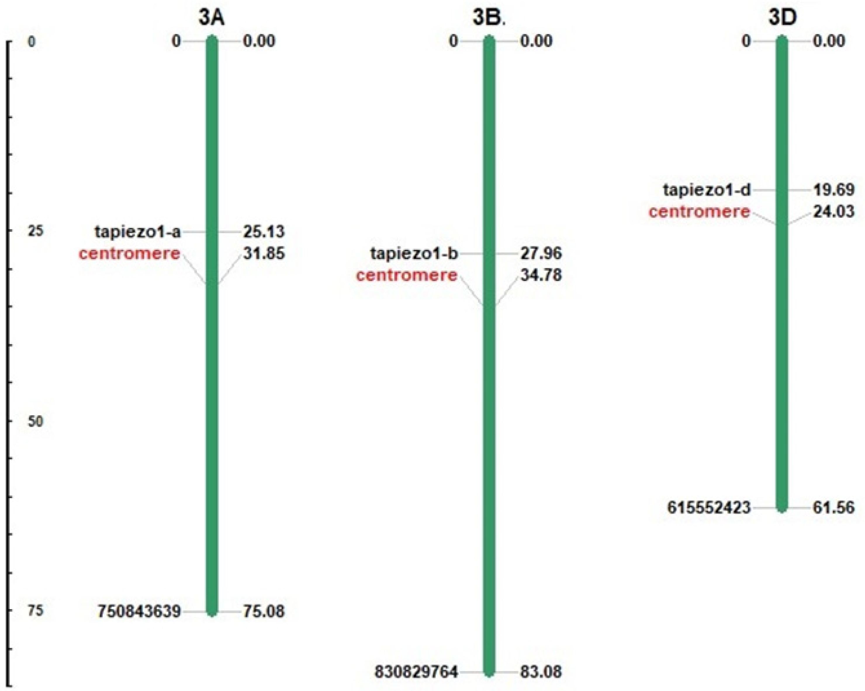
Figure 1. The chromosomal position of TaPiezo genes. TaPiezo genes are found on chromosomes 3A, 3B and 3D of T. aestivum.
Agriculture 13 00783 g001
Figure 2. Phylogenetic analysis of Piezo proteins of A. thaliana (AtPiezo1), O. sativa (OsPiezo1), T. aestivum (TaPiezo1-A, TaPiezo1-B and TaPiezo1-D), B. rapa (Bra004418|Bra004418.1, Bra021452|Bra021452.1), B. distachyon (BRADI_2g13251v3|KQK04385), G. max (GLYMA_19G238300|KRG96876, GLYMA_10G023700|KRH31967, GLYMA_02G150200|KRH71480), N. attenuate (A4A49_01634|OIT06222), S. bicolor (SORBI_3003G170700|KXG32572), and Z. mays (Zm00001eb131990|Zm00001eb131990_T001). The phylogenetic tree represents the clustering of Piezo proteins into two clades, and each clade is colored differently.
Agriculture 13 00783 g002
Figure 3. Gene structure analysis of TaPiezo genes. The figure represents the exon–intron organization and intron phase distribution in each TaPiezo gene, generated by the GSDS 2.0.
Agriculture 13 00783 g003
Figure 4. Protein structure analysis of TaPiezo proteins. (A) represents the schematic diagram of TaPiezo proteins obtained from the SMART server. (B) represents the arrangement of 15 conserved motifs of each TaPiezo protein, represented as colored boxes.
Agriculture 13 00783 g004
Figure 5. The three-dimensional structure of TaPiezo1-A protein. The figure displays the predicted 3D structure of TaPiezo1-A protein. The beam domain, outer helix (OH), central cap domain (CED), inner helix (IN) and C-terminal domain (CTD) are highlighted in the figure with different colors.
Agriculture 13 00783 g005
Figure 6. Correlation, expression and qRT-PCR analysis. Figure shows correlation analyses between replicates of expression data in (A) tissue developmental stages and (B) stress conditions. Heat map (C) shows the clustering and expression profiling TaPiezo genes in tissue developmental stages. The Heat maps (DF) show the expression profile of TaPiezo genes under (D) drought stress, heat stress, and heat–drought stress, (E) salt stress and (F) during the treatment of Blumeria graminis (Bgt) and Puccinia striiformis (Pst), respectively. The low and high expressions are represented by green and red colors, respectively, in the color bar. Graph (G) shows the qRT-PCR results of TaPiezo1-D under CaCl2 stress at 6, 12, 24, and 48 h of treatments. The bar graphs indicate the fold change in TaPiezo1-D gene expression under treatments as compared to the control condition.
Agriculture 13 00783 g006
Figure 7. Protein–protein and miRNA interaction analysis of Piezo of T. aestivum. The network (A) shows the protein–protein interactions, where TaPiezo proteins are represented with red and predicted interacting proteins with blue. The interaction network (B) of predicted miRNA with TaPiezo genes. The interaction networks were built using the Cytoscape tool.
Agriculture 13 00783 g007
Table 1. List of qRT-PCR primers.
Gene Name5′-3′
TaARF_Forward primerTGATAGGGAACGTGTTGTTGAGGC
TaARF_Reverse primerAGCCAGTCAAGACCCTCGTACAAC
TaPiezo1-D_Forward primerAGGAGAGGATTTCACAATTGGAGGCTG
TaPiezo1-D_Reverse primerCTTCAACCAAAGAAAGGACAGCAGCAG
Table 2. Various types of Cis-regulatory elements of TaPiezo genes.
Gene NameLight ResponseGrowth and DevelopmentStress ResponseHormone Response
TaPiezo1-AG-box, TCT-motif, I-box, TCCC-motif, G-Box, Box 4CCGTCC-box, GCN4_motif,
CCGTCC motif,
AAGAA-motif
ARE, as-1, LTR, STRE, WRE3, A-box, W box, MYB, GC-motif, Myb-binding site, MYB-like sequence, TC-rich repeatsTCA-element, ABRE, CGTCA-motif,
TGA-element,
TGACG-motif
TaPiezo1-BAE-box, G-box,
G-Box,
O2-site, RY-element, CCGTCC-box,
CCGTCC motif
MYC, Myb-binding site, STRE, MYB recognition site, MYB, WRE3, LTR, ARE, Myb, as-1, A-boxTGACG-motif, ABRE, CGTCA-motif
TaPiezo1-DSp1, G-box,
TCT-motif, ACE
CCGTCC motif, O2-site, CCGTCC-box, CAT-boxW box, LTR, MYB-like sequence, WRE3, GC-motif, TC-rich repeats, MYC, A-box, box S, Myb, MBS, STRE, as-1, Myb-binding site, DRE core, MYB recognition site, MYBCGTCA-motif, AT~ABRE, ABRE4, TGACG-motif, TATC-box, P-box, ABRE3a, ABRE
Disclaimer/Publisher’s Note: The statements, opinions and data contained in all publications are solely those of the individual author(s) and contributor(s) and not of MDPI and/or the editor(s). MDPI and/or the editor(s) disclaim responsibility for any injury to people or property resulting from any ideas, methods, instructions or products referred to in the content.