Article

Study on Revealing Peanut-Related Disease Prevention Gene Clusters via Whole Transcriptome Sequencing

1
Forestry College, Shandong Agricultural University, Tai’an 271018, China
2
College of Chemistry and Material Science, Shandong Agricultural University, Tai’an 271018, China
3
Taishan Forest Ecosystem Research Station/Key Laboratory of National Forestry and Grassland Administration for Silviculture of the Lower Yellow River, Tai’an 271018, China
*
Author to whom correspondence should be addressed.
These authors contributed equally to this work.
Agriculture 2023, 13(8), 1608; https://doi.org/10.3390/agriculture13081608
Submission received: 17 June 2023 / Revised: 1 August 2023 / Accepted: 3 August 2023 / Published: 15 August 2023
(This article belongs to the Section Crop Genetics, Genomics and Breeding)

Abstract

:
Root rot caused by Fusarium oxysporum is a serious root disease that affects the growth and yield of peanut crops worldwide. Identifying resistance genes is of great significance for both conventional and molecular breeding. At present, there are a few identified disease resistance genes, and the defense response mechanism against this pathogen remains unclear. Using transcriptome analysis techniques, 21 differentially expressed genes (DEGs) were identified in plants treated with Bacillus thuringiensis TG5 and carbendazim. Among these DEGs, the TIFY10A protein gene, pyruvate decarboxylase, and UDP glycosyltransferase 74B1 genes play an important role in early resistance to root rot in peanut plants, leading to regulatory factors that trigger immunity. Our study provides important information on the response mechanism of peanut plants to F. oxysporum-induced root rot and the protective benefits of colonization by the microbial pesticide, B. thuringiensis.

1. Introduction

Peanut (Arachis hypogaea L.) is one of the most important cash crops in the world.
During its entire growth cycle, it is vulnerable to various pathogenic microorganisms; therefore, cultivating and selecting resistant varieties are some of the most economical and effective ways to prevent and control diseases. Disease resistance genes are one of the important means by which peanut plants are protected against pathogenic microorganisms. Current research shows that the early and timely activation of plant-defense-related genes in peanut are key to an effective response to pathogenic attack. Transcriptomic techniques are increasingly used to screen plant transcriptomes for resistance-related genes. Previous studies have shown that the positive regulation of the phosphorylation of calmodulin-binding protein 60-LIKE G, mediated by calcium-dependent protein kinase, can improve the resistance of peanut plants to soil-borne fungal pathogens and increase the expression of immune response-related genes [1,2]. The overexpression of peanut pathogenesis-related protein genes, in response to pathogen stress or abiotic stimuli, plays an important role in the defense system of peanut plants [3,4]. Furthermore, oxidative lipids, lipoxygenase (LOX), and related oxidative metabolites in peanut plants are involved in the specific defense mechanism of peanut plants against fungal infection [5,6]. More research is needed to better understand the response of peanut plants to pathogens and the disease resistance genes involved.
Many transcription factors in plants, especially the JASMONATE ZIM-domain, PEAPOD, zinc finger inflorescence, and TIFY, are crucial in regulating plant stress responses [7,8,9]. Several pathways, including the pyruvic acid decarboxylase-mediated plant metabolic pathway [10], the cytochrome P450 monooxygenase-regulated triterpene modification pathway [11], and the cinnamic acid 4-hydroxylase (SdC4H, CYP73A111)-regulated biosynthetic pathway of secondary metabolites [12], also play important roles in providing a host defense response in plants. Various environmental challenges, including exposure to high levels of salt, drought, chemical pesticides, pathogenic microorganisms, and intense light, can cause an increase in the number of reactive oxygen species (ROS), high levels of which are harmful to plants. The accumulation of antioxidant substances in peanut plants is shown to have positive effects on the defense mechanism of peanut plants against glyphosate stress [13,14,15,16]. Further research is required to better understand the interaction between transcription factors [17], metabolic pathways, and various environmental factors in the stress response of peanut plants against pathogenic microorganisms.
Root rot in peanut plants caused by F. oxysporum is a serious root disease that affects the growth and yield of this crop worldwide. Existing research shows that F. oxysporum produces fusaric acid, which reduces host resistance, and cell wall-degrading enzymes are also produced that degrade the host cell wall and compromise tissue protection, releasing polysaccharides that promote the growth of pathogenic fungi [18]. Some Bacillus have a strong antagonistic effect on peanut powdery mildew [19]. Bacillus pumilus is reported to have an inhibitory effect on peanut blight in South China [20] and Pseudomonas fluorescens is a good biocontrol inhibitor in plants [21]. Furthermore, a commercialized preparation of B. thuringiensis (Bt) inhibits the damage to leaves caused by Spodoptera frugiperda and is an efficient substitute for chemical pesticides [22,23]. However, the mechanism of B. thuringiensis-induced peanut plant resistance to root rot caused by F. oxysporum infection requires further research.
The mechanism of microbial pesticides and traditional chemical pesticides that combat F. oxysporum in peanut plants is poorly understood, and thus far, no studies have explored the changes in the expressions of various functional gene clusters following F. oxysporum infection in peanut plants, as well as the changes in effector levels that mediate resistance responses. In this paper, based on the Illumina high-throughput sequencing transcriptome method, we examined the mechanism of microbial pesticides that forces peanut plants to cope with F. oxysporum stress by identifying new genes and metabolic pathways. This study provided a putative candidate gene cluster for differential expression in peanut plant cells as well as the metabolic pathways associated with resistance to F. oxysporum infection and resistance gene expression. It can further verify the potential candidate gene cluster and further develop marker detection methods to reduce the contamination level of F. oxysporum in peanut production.

2. Materials and Methods

2.1. Experimental Setup

We used 18 flowerpots (22 cm in diameter) filled with 2500 g of industrial sand, and six seeds were planted in each pot. The pots were watered thoroughly and seedlings emerged, usually after approximately 14 d. Three tests groups, CK (control), B. thuringiensis (TG5; NCBI SUB12195433, access number CP110119.1), and carbendazim (Car) treatments, were set up, and each treatment was repeated thrice. Nine pots of peanut seedlings were inoculated with F. oxysporum (25 mL of fermentation liquid) via irrigation of the roots. After 7 d of normal culture, 62.5 µL of 5 × 10⁶ CFU of B. thuringiensis was added to three pots of the TG5 treatment group, 62.5 µL of carbendazim (1:800 dilution) was added to the Car treatment group, and 62.5 µL of purified water was added to the CK treatment group. The root transcriptome, chlorophyll, catalase (CAT), and superoxide dismutase (SOD) levels in peanut plant cells were measured at 0, 24, and 72 h, respectively. This treatment was performed with each sample in triplicate.

2.2. Determination of Chlorophyll Content

We collected leaf tips from the same position for each treatment group. Chlorophyll was extracted from 0.2 g of material and the absorbance measured at 665 and 649 nm using 95% ethanol as a blank control.

2.3. Determination of Soil Enzyme Activity

The activity of SOD in peanut plant leaf tissue was measured using the nitroblue tetrazole (NBT) method (the amount of enzyme required to inhibit a 50% photochemical reduction of NBT was taken as the enzyme activity unit, U). The activity of peroxidase was measured using the guaiacol method (A470 enzyme changed 0.01 times per minute when measured as the activity unit of peroxidase conversion, U).

2.4. Transcriptome Sequencing of Peanut Root Tissue

Peanut root tissue DNA was extracted using a reagent kit CTAB + Adlai RN40 (manufacturer: Adlai, model: RN40). The Huada Gene DNBSEQ platform was used for sequencing. The sequencing data were filtered with SOAPnuke (v1.5.2) [24] by: removing reads containing sequencing adapter; removing reads whose low-quality base ratio (base quality less than or equal to 5) is more than 20%; and removing reads whose unknown base (‘N’ base) ratio is more than 5%. Afterward clean reads were obtained and stored in FASTQ format. The clean reads were mapped to the reference genome using HISAT2 (v2.0.4) [25]. Bowtie2 (v2.2.5) [26] was applied to align the clean reads to the reference coding gene set, and then, the expression level of the gene was calculated using StringTie (v2.1.2) [27]. Clean reads were compared with the reference genome based on Hisat [28] to obtain positional information on the reference genome or gene, as well as the unique sequence feature information of the sequencing sample. Based on the Hisat2 alignment results between the reads of each sample and the reference genome sequence, GATK4 software [28] was used to identify single-base mismatches between the sequencing sample and the reference genome as well as potential SNP sites. Furthermore, it was possible to analyze whether these SNP loci affected the expression level of genes or the types of protein products.

2.5. Transcriptome Data Analysis Method

To detect differentially expressed genes (DEGs), fold change ≥ 2 and FDR < 0.05 were used as the screening criteria. Hierarchical clustering analysis was performed on the screened DEGs and cluster analysis on genes with the same or similar expression patterns. Then, we further classified and conducted an enrichment analysis on the three major functional classes of gene ontology (GO), molecular function (MF), cellular component (CC), and biological process (BP).
In a pathway significance enrichment analysis, the KEGG Pathway database was used as a reference, and hypergeometric tests were conducted to identify pathways that were substantially enriched in DEGs compared to the entire genome background. A DEG interaction network was constructed based on the results of differential expression analysis and the interaction relationships included in the database. For species included in the database, the interactions between the target gene set were directly extracted from the database to construct the interaction network. For species not included in the database, BLAST (Version0.5.1) software was used to sequence and compare the target gene with the proteins in the database, search for homologous proteins, and construct an interaction network based on the interactions between homologous proteins. The transcription factor analysis was based on AnimalTFDB and PlantTFDB, which are classifications and annotations whole-genome transcription factors and cofactors containing 97 animal and 165 plant genomes, respectively. Transcription factors were divided into 73 families and cofactors were divided into 83 families and 6 major categories based on DNA binding domains. The corresponding species was found using the corresponding transcription factors.

2.6. Statistical Analysis

Data were analyzed using Excel 2003 and IBM SPSS 23. Duncan’s multiple range test (5% probability level) was used for one-way analysis of variance (ANOVA) to compare the average value of different treatment methods (p < 0.05). Single-factor ANOVA was used to test the homogeneity of variance for real-time data. If necessary, the description (D) test was used to compare the average values between different treatments (p < 0.05). Data represent mean ± SEM.

3. Results

3.1. Picture of Peanut Plant Disease Resistance

After inoculation with F. oxysporum and cultivation for 7 d, compared with the control, Car treatment initially thickened and blackened the peanut plants leaves; however, the leaves soon recovered. After TG5 treatment, the leaves became thicker and the leaf veins grew stronger, resulting in a significant increase in the number of root nodules, root area, leaf area, and net length of the peanut roots. Notably, the control effect of Car on root rot was not significant in the medium to long term (Figure 1); however, the damage to peanut roots was significant, such that it was difficult for the plant to recover in a short growth cycle.

3.2. Analysis of Chlorophyll and Protective Enzymes in Peanut Leaves

Chlorophyll is the main pigment that characterizes the photosynthetic capacity of plants. Compared with the control, a change in chlorophyll content in peanut leaves was observed after TG5 treatment (Figure 2). The chlorophyll content in peanut leaves after TG5 treatment was superior to the control, although the change in the number of individual leaves was not very significant. Given the number of leaves, there was a significant difference in chlorophyll content between the entire plant and the entire plot, resulting in the significant accumulation of substances caused by photosynthesis. The accumulation of ROS caused membrane lipid peroxidation and the loss of membrane differential permeability in plants, leading to defense reactions such as allergic necrosis and phytoalexin production. SOD and CAT eliminated ROS and superoxide anion in plants, thus providing plant disease resistance. Compared with the control, after 72 h of TG5 treatment, the contents of SOD and CAT in peanut plants increased by 23.2 and 14.0%, respectively, significantly increasing the stress resistance of the peanut plants (p < 0.05). Compared with Car treatment, the effect of TG5 on SOD and peroxidase in peanut plants was similar and less harmful to the environment.

3.3. Sample Clustering and Principal Component Analysis

A total of 27 samples were tested in this project, resulting in 287.29 Gb of data with a Q30 base percentage of 90.19% or higher. Raw data obtained via sequencing were filtered, and these filtered clean reads were matched to the reference sequences. Sequence alignment was performed between the clean reads of each sample and the designated reference genome, with an alignment efficiency ranging from 74.39 to 97.93%. After comparing the sequencing reads to the reference genome and reconstructing the transcript, a total of 5955 new genes were detected, of which 5599 were functionally annotated.

3.4. Gene Expression Level Analysis

Fragments per kilobase of transcript per million fragments mapped (FPKM), as an indicator measure of transcript or gene expression level, can be directly used to compare differences in gene expression between different samples. Transcriptome data were used to detect gene expressions with high-sensitivity sequencing, as well as FPKM values of protein coding gene expression levels across six orders of magnitude. As shown in the quadrant diagram, we intuitively observed the dispersion of the expression levels of individual sample genes compared to the overall gene expression levels of different samples (Figure 3).

3.5. Differential Gene Expression Cluster Analysis

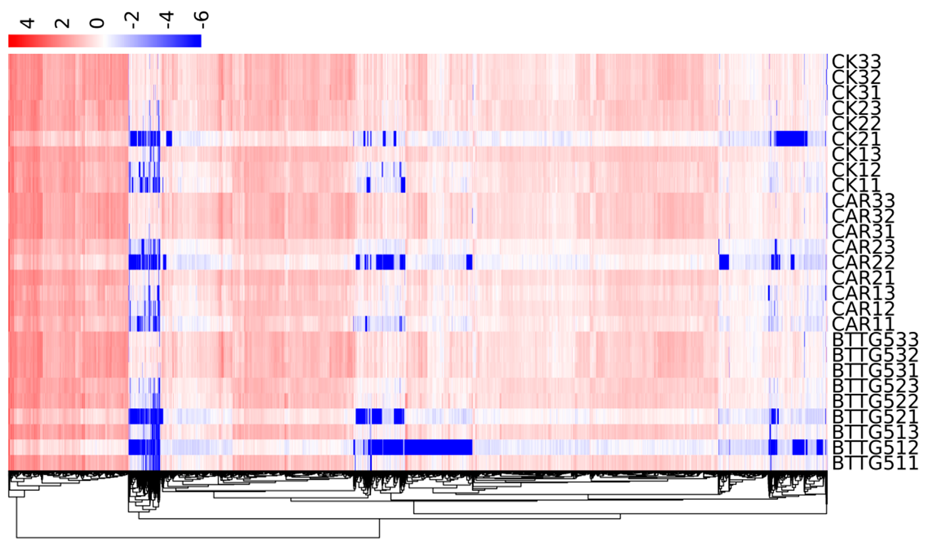
Figure 4 shows the gene expressions in different groups over different time sequences. There were significant gene differences in different time sequences and between different groups. A hierarchical clustering analysis was performed on the screened DEGs to cluster genes with the same or similar expression patterns.

3.6. Gene Ontology (GO) Analysis between TG5 Strains and Carbazim Treatment Groups

GO comprehensively demonstrates the attributes of genes and gene products in organisms. GO was divided into three functional categories: MF, CC, and BP. GO classification statistics of DEGs are shown in Figure S1.

3.7. Gene Regulation Analysis in Biochemical Process

Our data show that, after 72 h, TG5 treatment increased the expression levels of NADPH cytochrome P450 reductase 2, L-ascorbate peroxidase, calmodulin, and NADPH oxidase genes by 184, 205, 180, and 303% (p < 0.05), respectively, compared with the control values (Figure 5A). The expression levels of calmodulin and NADPH oxidase genes increased by 147 and 154%, respectively, after treatment with Car treatment (p < 0.05).
Compared with the control, our data show that, after 72h, the gene expression levels of transglucanase/hydrolase protein 22, protein TIFY10A, and inorganic phosphate transporter 1–3 in xyloglucan treated with TG5 increased by 237, 189, and 174%, respectively (p < 0.05), whereas Car treatment increased the gene expression levels of transglucanase/hydrolase protein 22 and protein TIFY10A in xyloglucan by 220 and 139%, respectively (p < 0.05; Figure 5B).

3.8. Pathogenesis-Related Regulatory Genes Analysis

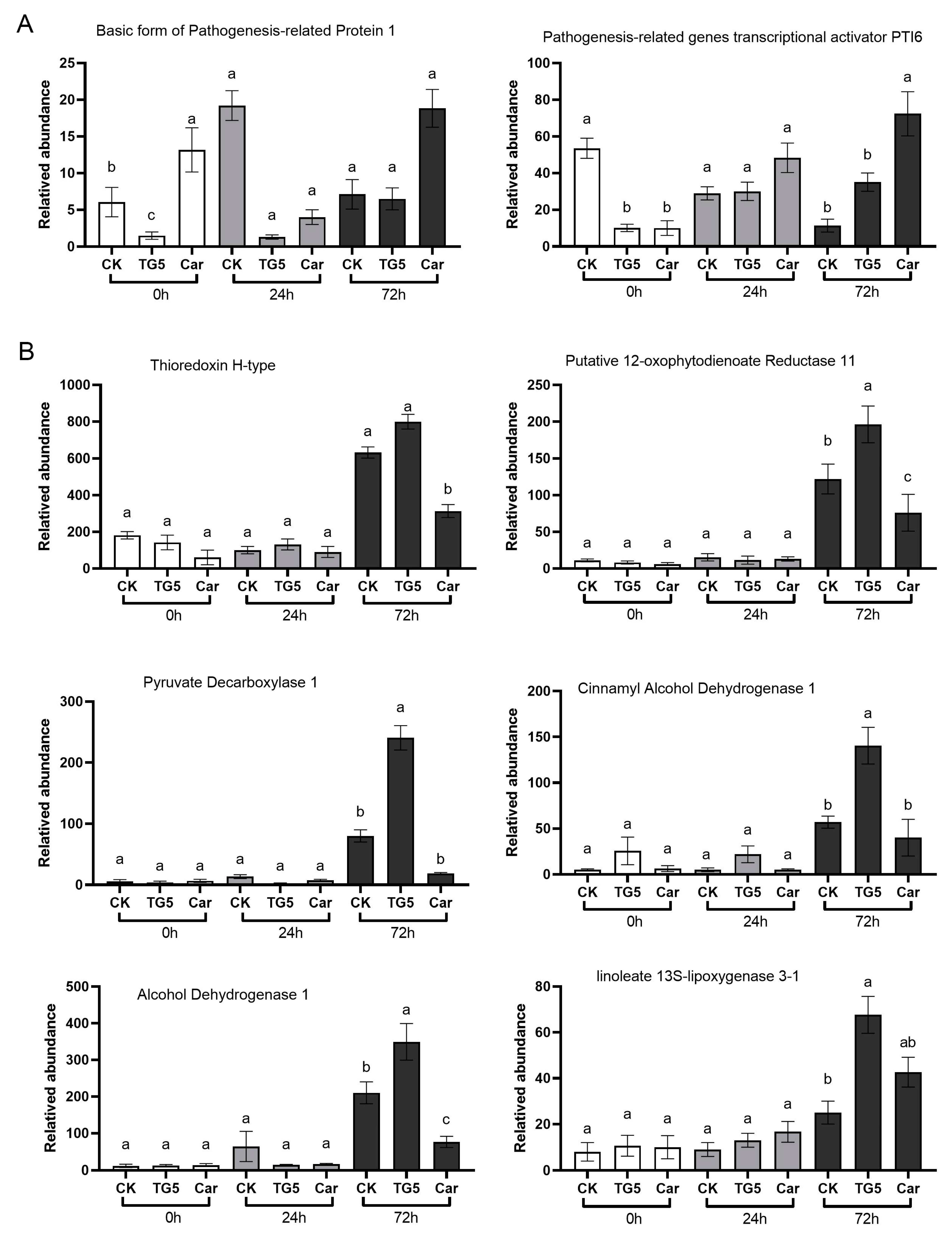
Pathogenesis-related proteins are produced when plants respond to stress, pathogen attacks, or abiotic stimulation, and play an important role in plant defense systems [3]. Our data show that, after 72 h, TG5 treatment decreased the expression of pathogenesis- related protein 1 gene by 12.0% (p < 0.05) and increased the expression of PTI6 pathogenesis-related gene by 316% (p < 0.05) compared with the control. Meanwhile, Car treatment increased the gene expression levels of pathogenesis-related protein 1 and pathogenesis-related gene transcriptional activator PTI6 by 268 and 668%, respectively (p < 0.05; Figure 6A).

3.9. TG5-Specific Expression Gene Analysis

Compared with the control, our data show that after 72 h, TG5 treatment increased the gene expression levels of thioredoxin H-type, 12-oxygenated plant diene reductase 11, pyruvate decarboxylase 1, pyruvate decarboxylase 2, cinnamyl alcohol dehydrogenase 10, and alcohol dehydrogenase 1 by 125, 158, 256, 243, 302, and 165%, respectively (p < 0.05), whereas Car treatment reduced the expression levels of these genes by 50.9, 40, 84, 75, 29, and 62%, respectively (p < 0.05; Figure 6B). Furthermore, compared with the control, TG5 treatment increased the expression levels of UDP glycosyltransferase 74B1, aldehyde dehydrogenase family member H1, and phospholipid hydrogen peroxide glutathione peroxidase genes by 236, 196, and 166%, respectively (p < 0.05), whereas carbendazim treatment reduced the expression levels of these genes by 59, 25, and 22%, respectively (p < 0.05; Figure 6C).

3.10. Difference Analysis in KEGG Metabolic Pathway between TG5 and Car Treatment Groups

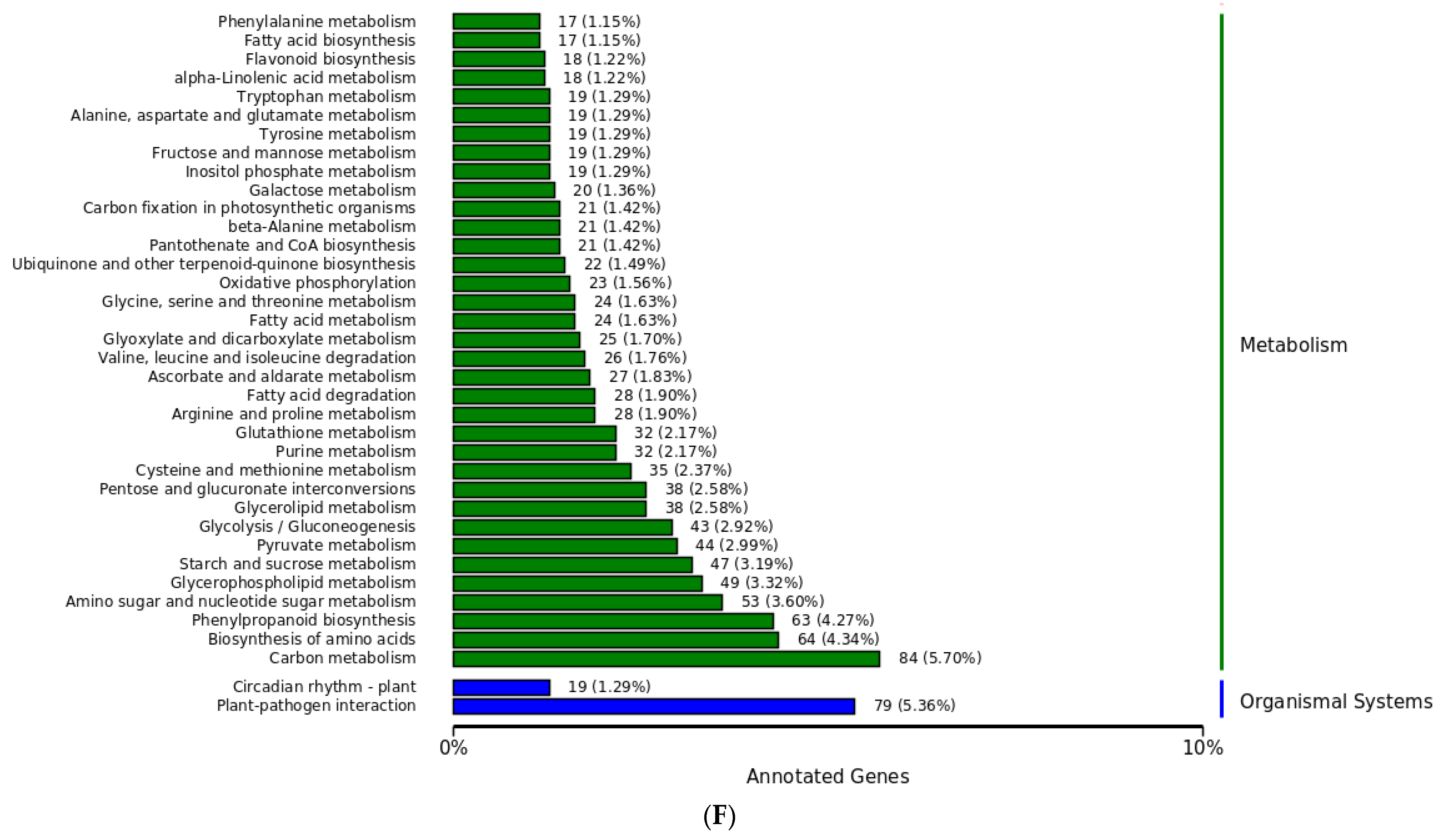
Our data show the gene pathways of influence during host–pathogen interactions (Figure 7). A pathway check was performed to examine DEGs during host–pathogen interactions. These pathways represent three types of metabolic paths, MF, CC, and BP, as well as other metabolic pathways, including linoleic acid 13S LOX, NADPH cytochrome P450 reductase 2, and 12-oxo plant dienoic acid reductase 11. In this case, the metabolic pathway was Arabidopsis K000591 (linoleic acid metabolism). The metabolic pathway of abscisic acid receptor PYL4 and NADPH oxidase was Arabidopsis K004016 (MAPK signaling pathway plant), and for xylanase endoglucanase/hydrolase protein 22 and protein TIFY10A, it was the Arabidopsis K004075 (plant hormone signal transduction) pathway. The pathway for disease progression-related protein 1, disease-related gene transcriptional activator PTI6, and calmodulin metabolism was Arabidopsis K004626 (plant–pathogen interaction). The metabolic pathway of L-ascorbate peroxidase and abscisic acid 8’-hydroxylase was Arabidopsis K000906 (carotenoid biosynthesis); for phospholipid hydrogen peroxide and glutathione peroxidase, it was Arabidopsis K000480 (glutathione metabolism); and for UDP glycosyltransferase 74B1, it was Arabidopsis K000966 (glucoside biosynthesis). The H1 metabolic channel of alcohol dehydrogenase 1 and aldehyde dehydrogenase family 3 was Arabidopsis K000620 (pyruvate metabolism).

4. Discussion

After 72 h of treatment with TG5 bacterial agent in peanut plants, the gene expression levels of TIFY10A, UDP-glycosyl transferase 74B1, disease-related gene transcriptional activator PTI6, NADPH oxidase, phospholipid hydrogen peroxide, and glutathione peroxidase genes increased by 189, 236, 316, 303, and 166%, respectively, compared with the control values (p < 0.05). However, Car treatment increased the gene expression levels of TIFY10A, NADPH oxidase gene, pathogenesis-related protein 1, and pathogenesis-related transcriptional activator PTI6 by 139, 154, 268, and 668%, respectively, compared with the control values (p < 0.05).
The pathogenic mechanism of peanut root rot caused by F. oxysporum has not yet been clearly defined, and the use of high-quality reference genomes has created a huge opportunity to improve our current understanding of this mechanism. Current research has found that there are 145 metabolic pathways related to peanut pathogenicity. Among these, linoleic acid metabolism, plants with the MAPK signaling pathway, plant hormone signaling transduction, plant–pathogen interactions, carotenoid biosynthesis, glutathione metabolism, glucoside biosynthesis, and acetone acid metabolism are the main metabolic pathways.
Plant stress-resistant gene clusters mainly include members of the LOX gene cluster, transcription factor gene cluster, protective enzyme gene cluster [5], secondary metabolism, and the associated regulation gene cluster. LOX produces fatty acid metabolites in plants, including hydroxy fatty acids, epoxy hydroxy fatty acids, and trihydroxy fatty acids that participate in pathogen-specific defense mechanisms against fungal infection. An increase in LOX activity in infected rice plants regulates the oxidation of linoleic acid to 9-hydroperoxy-10, 12, 15-octadecatrienoic acid, and its metabolites, which exhibit antibacterial activity against various pathogens [6]. An increase in lipid peroxidase levels (represented by malondialdehyde content) and CAT content in peanut plant leaves under glyphosate stress induces changes in plant antioxidant status, serving as an effective defense mechanism against glyphosate toxicity [4]. The continuous and cumulative induction of phenylalanine ammonia lyase, peroxidase, polyphenol oxidase, superoxide dismutase, CAT, LOX, and other defense enzymes, as well as pathogenesis-related proteins, enhanced the resistance of peanut plants to leaf miner and ear rot [29]. This study confirms the above results. After the peanut plants were treated with Car and TG5, NADPH oxidase expression increased, which induced and enhanced the stress response of the plants to F. oxysporum. WRKY transcription factors are found to participate in growth and development but are also involved in plant defense and stress responses via regulating a variety of downstream stress response genes [30,31]. In contrast to the above studies, transcription factor protein TIFY10A and pathogenesis protein activator PTI6 jointly mediate the effects of carbendazim and the biological pesticide TG5 agent, inducing the resistance of peanut plants to F. oxysporum stress.
As an important glycosylation catalyst, UDP flavonoid 3-O-glucosyltransferase responds to plant stress by activating plant hormones and increasing related metabolites. UDP glycosyltransferases genes play an important role in plant secondary metabolism [32,33]. They can serve as structural genes in the final step of anthocyanin biosynthesis, significantly increasing plant anthocyanin content by inhibiting the feedback of jasmonate signaling [34], as well as participating in plant detoxification processes via interactions with transcription factors [35,36]. This study found that UDP glycosyltransferase and its related gene cluster played an important role in resisting the stress caused by F. oxysporum infection in peanut plants.
The traditional chemical fungicide carbendazim is known to have an effective antibacterial effect on F. oxysporum [8,37]. Therefore, using plant growth-promoting rhizobacteria and other microbial technology to partially or completely replace carbendazim pesticide is an essential method for the prevention and control of F. oxysporum-induced root rot in peanut plants.

5. Conclusions

We believe that the overexpression of transcription factor TIFY protein, pathogenesis transcription activator PTI6, pathogenesis-related protein 1, and NADPH-oxidase-related genes in peanut plants is central to the mechanism of Car-induced resistance in peanut plants in response to root rot caused by F. oxysporum. Simultaneously, the colonization of peanut plants with the TG5 strain can improve antibacterial properties through the overexpression of phospholipid hydroperoxides, glutathione peroxidase, and NADPH oxidase, which work to eliminate free radicals and antioxidant substances. The overexpression of protein TIFY10A and pathogenesis-related transcriptional activator PTI6 genes in peanut plants helps to improve their bacterial resistance. The UDP glycosyltransferase 74B1 gene and its related gene cluster is the main control gene cluster elicited by B. thuringiensis TG5 in peanut plants, increasing their resistance to F. oxysporum-induced root rot.

Supplementary Materials

The following supporting information can be downloaded at: https://www.mdpi.com/article/10.3390/agriculture13081608/s1, Figure S1: Differential expression gene GO annotation classification statistical chart.

Author Contributions

Writing—original draft, H.D. and N.L.; Writing—review & editing, C.L. All authors have read and agreed to the published version of the manuscript.

Funding

This research received no external funding.

Conflicts of Interest

The authors declare no conflict of interest.

References

  1. Yuan, P.; Tanaka, K.; Poovaiah, B.W. Calcium/calmodulin-mediated defense signaling: What Is looming on the horizon for AtSR1/CAMTA3-mediated signaling in plant immunity. Front. Plant Sci. 2022, 12, 795353. [Google Scholar] [CrossRef] [PubMed]
  2. Li, W.; Sun, J.; Zhang, X.; Ahmad, N.; Hou, L.; Zhao, C.; Pan, J.; Tian, R.; Wang, X.; Zhao, S. The Mechanisms Underlying Salt Resistance Mediated by Exogenous Application of 24-Epibrassinolide in Peanut. Int. J. Mol. Sci. 2022, 23, 6376. [Google Scholar] [CrossRef] [PubMed]
  3. Jain, S.; Kumar, D.; Jain, M.; Chaudhary, P.; Deswal, R.; Sarin, N.B. Ectopic overexpression of a salt stress-induced pathogenesis-related class 10 protein (PR10) gene from peanut (Arachis hypogaea L.) affords broad spectrum abiotic stress tolerance in transgenic tobacco. Plant Cell Tissue Organ Cult. (PCTOC) 2012, 109, 19–31. [Google Scholar] [CrossRef]
  4. Senthilraja, G.; Anand, T.; Kennedy, J.; Raguchander, T.; Samiyappan, R. Plant growth promoting rhizobacteria (PGPR) and entomopathogenic fungus bioformulation enhance the expression of defense enzymes and pathogenesis-related proteins in groundnut plants against leafminer insect and collar rot pathogen. Physiol. Mol. Plant Pathol. 2013, 82, 10–19. [Google Scholar] [CrossRef]
  5. Kato, T.; Maeda, Y.; Hirukawa, T.; Namai, T.; Yoshioka, N. Lipoxygenase Activity Increment in Infected Tomato Leaves and Oxidation Product of Linolenic Acid by Its In Vitro Enzyme Reaction. Biosci. Biotechnol. Biochem. 1992, 56, 373–375. [Google Scholar] [CrossRef]
  6. An, J.-U.; Lee, I.-G.; Ko, Y.-J.; Oh, D.-K. Microbial Synthesis of Linoleate 9S-Lipoxygenase Derived Plant C18 Oxylipins from C18 Polyunsaturated Fatty Acids. J. Agric. Food Chem. 2019, 67, 3209–3219. [Google Scholar] [CrossRef] [PubMed]
  7. Liu, X.; Zhao, C.; Yang, L.; Zhang, Y.; Wang, Y.; Fang, Z.; Lv, H. Genome-Wide Identification, Expression Profile of the TIFY Gene Family in Brassica oleracea var. capitata, and Their Divergent Response to Various Pathogen Infections and Phytohormone Treatments. Genes 2020, 11, 127. [Google Scholar] [CrossRef] [Green Version]
  8. Sen, S. Genome-wide TIFY family in Arachis hypogaeain the perspective of legume JAZs. Crop Sci. Biotechnol. 2022, 25, 465–488. [Google Scholar] [CrossRef]
  9. Zheng, L.L.; Wan, Q.; Wang, H.G.; Guo, C.L.; Niu, X.L.; Zhang, X.F.; Zhang, R.; Chen, Y.H.; Luo, K. Genome-wide identification and expression of TIFY family in cassava (Manihot esculenta Crantz). Front. Plant Sci. 2022, 13, 1017840. [Google Scholar] [CrossRef]
  10. Wang, X.; Liu, Y.; Han, Z.; Chen, Y.; Huai, D.; Kang, Y.; Wang, Z.; Yan, L.; Jiang, H.; Lei, Y.; et al. Integrated Transcriptomics and Metabolomics Analysis Reveal Key Metabolism Pathways Contributing to Cold Tolerance in Peanut. Front. Plant Sci. 2021, 12, 752474. [Google Scholar] [CrossRef]
  11. Pramesti, I.; Shuhei, Y.; Pisanee, S. Comparative analysis of NADPH-Cytochrome P450 reductases from legumes for heterologous production of triterpenoids in transgenic saccharomyces cerevisiae. Front. Plant Sci. 2021, 12, 762546. [Google Scholar]
  12. Yoshimi, Y.; Ayaka, M. Functional characterization of nadph-cytochrome p450 reductase and cinnamic acid 4-hydroxylase encoding genes from Scoparia dulcis L. Bot. Stud. 2020, 61, 284. [Google Scholar]
  13. Muñoz, V.; Ibáñez, F.; Figueredo, M.S.; Fabra, A. An oxidative burst and its attenuation by bacterial peroxidase activity is required for optimal establishment of the Arachis hypogaea-Bradyrhizobium sp. symbiosis. J. Appl. Microbiol. 2016, 121, 244–253. [Google Scholar] [CrossRef] [Green Version]
  14. Radwan, D.E.M.; Fayez, K.A. Photosynthesis, antioxidant status and gas-exchange are altered by glyphosate application in peanut leaves. Photosynthetica 2016, 54, 307–316. [Google Scholar] [CrossRef]
  15. Monika, P.; Dhara, F.; Kumar, A.P. Silicon-induced mitigation of drought stress in peanut genotypes (Arachis hypogaea L.) through ion homeostasis, modulations of antioxidative defense system, and metabolic regulations. Plant Physiol. Biochem. PPB 2021, 166, 290–313. [Google Scholar]
  16. Raza, A.; Sharif, Y.; Chen, K.; Wang, L.; Fu, H.; Zhuang, Y.; Chitikineni, A.; Chen, H.; Zhang, C.; Varshney, R.K.; et al. Genome-Wide Characterization of Ascorbate Peroxidase Gene Family in Peanut (Arachis hypogea L.) Revealed Their Crucial Role in Growth and Multiple Stress Tolerance. Front. Plant Sci. 2022, 13, 962182. [Google Scholar] [CrossRef] [PubMed]
  17. Guo, F.; Hou, L.; Ma, C.; Li, G.; Lin, R.; Zhao, Y.; Wang, X. Comparative transcriptome analysis of the peanut semi-dwarf mutant 1 reveals regulatory mechanism involved in plant height. Gene 2021, 791, 145722. [Google Scholar] [CrossRef]
  18. Zhang, R.; Jiang, Z.; Li, M.H.; Xi, P.G. Research progress on the pathogenic mechanism and related pathogenic genes of Fusarium oxysporum. Chin. J. Agric. 2011, 27, 254–258. [Google Scholar]
  19. Chen, P.; He, W.; Shen, Y.; Zhu, L.Y.; Yao, X.Z.; Sun, R.B.; Dai, C.C.; Sun, B.; Chen, Y. Interspecific neighbor stimulates peanut growth through modulating root endophytic microbial community construction. Front. Plant Sci. 2022, 13, 830666. [Google Scholar] [CrossRef]
  20. Basavaraju, S.; Devi, M.C. Mechanism of biological control of root rot of groundnut caused by Macrophomina phaseolina using Pseudomonas fluorescens. Indian Phytopathol. 2012, 65, 360–365. [Google Scholar]
  21. Kumar, C.J.; Priyanka, S.; Arpit, S. Microbial enzyme, 1-aminocyclopropane-1-carboxylic acid (ACC) deaminase: An elixir for plant under stress. Physiol. Mol. Plant Pathol. 2021, 115, 101664. [Google Scholar]
  22. Torres-Cab, W.J.; Ruiz-Sanchez, E.; Reyes-Ramírez, A.; Lugo-García, G.A.; Tucuch-Haas, J.I.; Pierre, J.F. Field evaluation of microbial insecticides against fall armyworm, Spodoptera frugiperda (smith) and corn earworm, helicoverpa zea (boddie) (lepidoptera: Noctuidae), in maize. Arch. Phytopathol. Plant Prot. 2022, 55, 1713–1723. [Google Scholar] [CrossRef]
  23. Li, R.; Li, Y.; Kristiansen, K.; Wang, J. SOAP: Short oligonucleotide alignment program. Bioinformatics 2008, 24, 713–714. [Google Scholar] [CrossRef] [PubMed] [Green Version]
  24. Kim, D.; Langmead, B.; Salzberg, S.L. HISAT: A fast spliced aligner with low memory requirements. Nat. Methods 2015, 12, 357–360. [Google Scholar] [CrossRef] [Green Version]
  25. Langmead, B.; Salzberg, S.L. Fast gapped-read alignment with Bowtie 2. Nat. Methods 2012, 9, 357–359. [Google Scholar] [CrossRef] [Green Version]
  26. Pertea, M.; Pertea, G.M.; Antonescu, C.M.; Chang, T.-C.; Mendell, J.T.; Salzberg, S.L. StringTie enables improved reconstruction of a transcriptome from RNA-seq reads. Nat. Biotechnol. 2015, 33, 290–295. [Google Scholar] [CrossRef] [Green Version]
  27. Xu, J.X.; Li, Z.Y.; Lv, X.; Yan, H.; Zhou, G.-Y.; Cao, L.-X.; Yang, Q.; He, Y.-H. Isolation and characterization of Bacillus subtilis strain 1-L-29, an endophytic bacteria from Camellia oleifera with antimicrobial activity and efficient plant-root colonization. PLoS ONE 2020, 15, e0232096. [Google Scholar] [CrossRef]
  28. McKenna, A.; Hanna, M.; Banks, E.; Sivachenko, A.; Cibulskis, K.; Kernytsky, A.; Garimella, K.; Altshuler, D.; Gabriel, S.; Daly, M.; et al. The Genome Analysis Toolkit: A MapReduce framework for analyzing next-generation DNA sequencing data. Genome Res. 2010, 20, 1297–1303. [Google Scholar] [CrossRef] [Green Version]
  29. Liu, Y.; Liu, S.; Luo, X.; Wu, X.; Ren, J.; Huang, X.; Feng, S.; Lin, X.; Ren, M.; Dong, P. Antifungal activity and mechanism of thymol against Fusarium oxysporum, a pathogen of potato dry rot, and its potential application. Postharvest Biol. Technol. 2022, 192, 112025. [Google Scholar] [CrossRef]
  30. Zhu, H.; Jiang, Y.; Guo, Y.; Huang, J.B.; Zhou, M.H.; Tang, Y.Y.; Sui, J.M.; Wang, J.S.; Qiao, L.X. A novel salt inducible WRKY transcription factor gene, AhWRKY75, confers salt tolerance in transgenic peanut. Plant Physiol. Biochem. 2021, 160, 175–183. [Google Scholar] [CrossRef]
  31. Kurnool, K.; Gunupuru, L.R.; Merum, P.; Ambekar, N.K.; Vennapusa, A.R.; Uppala, L.; Boya, V.; Anthony Johnson, A.M.; Chinta, S. A Novel WRKY Transcription Factor, MuWRKY3 (Macrotyloma uniflorum Lam. Verdc.) Enhances Drought Stress Tolerance in Transgenic Groundnut (Arachis hypogaea L.) Plants. Front. Plant Sci. 2018, 9, 346. [Google Scholar]
  32. Xie, D.W.; Yang, X.; He, R.H.; Huo, H.; Ye, Z.C.; Ren, X.H.; Yuan, H.M.; Dai, Z.G.; Sun, J.; Su, J.G. Comprehensive analysis of the UDP glycosyltransferase gene family in flax [Linum usitatissimum L.] and functional verification of the role of LuUGT175 in the regulation of lignin biosynthesis. Ind. Crops Prod. 2022, 188, 115720. [Google Scholar] [CrossRef]
  33. Chávez, A.; Castillo, N.; López-Tubau, J.M.; Atanasov, K.E.; Fernandez-Crespo, E.; Camanes, G.; Altabella, T.; Ferrer, A. Tomato STEROL GLYCOSYL TRANSFERASE 1 silencing unveils a major role of steryl glycosides in plant and fruit development. Environ. Exp. Bot. 2023, 206, 105181. [Google Scholar] [CrossRef]
  34. Wang, X.; Wang, J.; Cui, H.; Yang, W.; Yu, B.; Zhang, C.; Wen, J.; Kang, J.; Wang, Z.; Yang, Q. The UDP-glycosyltransferase MtUGT84A1 regulates anthocyanin accumulation and plant growth via JA signaling in Medicago truncatula. Environ. Exp. Bot. 2022, 201, 104972. [Google Scholar] [CrossRef]
  35. Guo, F.; Zhu, X.; Zhao, C.; Zhao, S.; Pan, J.; Zhao, Y.; Wang, X.; Hou, L. Transcriptome Analysis and Gene Expression Profiling of the Peanut Small Seed Mutant Identified Genes Involved in Seed Size Control. Int. J. Mol. Sci. 2022, 23, 9726. [Google Scholar] [CrossRef] [PubMed]
  36. Gaobo, Y.; Qiusen, C.; Fengqiong, C. Glutathione Promotes Degradation and Metabolism of Residual Fungicides by Inducing UDP-Glycosyltransferase Genes in Tomato. Front. Plant Sci. 2022, 13, 893508. [Google Scholar]
  37. Yol, E.; Upadhyaya, H.D.; Uzun, B. Molecular diagnosis to identify new sources of resistance to sclerotinia blight in groundnut (Arachis hypogaea L.). Euphytica 2015, 203, 367–374. [Google Scholar] [CrossRef]
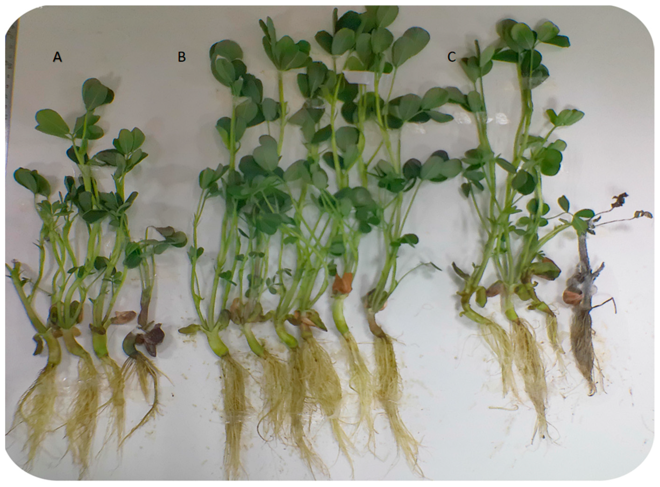
Figure 1. Image of Fusarium oxysporum infection in peanut plants; (A) F. oxysporum-infected peanut plants, (B) F. oxysporum-infected peanut plants after TG5 bacterial fertilizer treatment, and (C) F. oxysporum-infected peanut plants after carbendazim treatment.
Agriculture 13 01608 g001
Figure 2. Changes in chlorophyll and defense enzymes in peanuts. Different letters indicate significant differences between mean values.
Agriculture 13 01608 g002
Figure 3. Sample fragments per kilobase of transcript per million fragments mapped (FPKM); density distribution comparison diagram and box plot.
Agriculture 13 01608 g003
Figure 4. Cluster diagram of differentially expressed gene expression patterns. Blue and red represent low and high expressions, respectively.
Agriculture 13 01608 g004
Figure 5. Analysis of genes promoting stress resistance in peanut plants. Different letters indicate significant differences between mean values. Note, (A) NADPH cytochrome P450 reductase 2, L-ascorbate peroxidase, calmodulin, and NADPH oxidase genes; (B) Xyloglucan Endotransglucosylase, protein TIFY10A, and inorganic phosphate transporter 1–3 genes.
Agriculture 13 01608 g005
Figure 6. Analysis of peanut disease resistance genes. Different letters indicate significant differences between mean values. Note, (A) Genes of basic form of Pathogenesis-related Protein1 and Pathogenesis-related genes transcriptional activatorPTI6; (B) Genes of thioredoxin H-type, 12-oxygenated plant diene reductase 11, pyruvate decarboxylase 1, pyruvate decarboxylase 2, cinnamyl alcohol dehydrogenase 10, and alcohol dehydrogenase 1; (C) Genes of UDP glycosyltransferase 74B1, aldehyde dehydrogenase family member H1, and phospholipid hydrogen peroxide glutathione peroxidase.
Agriculture 13 01608 g006aAgriculture 13 01608 g006b
Figure 7. KEGG classification enrichment results. (A) KEGG classification enrichment: Bt. TG5 treatment (0 h). (B) KEGG classification enrichment: Bt. TG5 treatment (24 h). (C) KEGG classification enrichment: Bt. TG5 treatment (72 h). (D) KEGG classification enrichment: Car treatment (0 h). (E) KEGG classification enrichment: Car treatment (24 h). (F) KEGG Classification enrichment: Car treatment (72 h).
Agriculture 13 01608 g007aAgriculture 13 01608 g007bAgriculture 13 01608 g007cAgriculture 13 01608 g007d
Disclaimer/Publisher’s Note: The statements, opinions and data contained in all publications are solely those of the individual author(s) and contributor(s) and not of MDPI and/or the editor(s). MDPI and/or the editor(s) disclaim responsibility for any injury to people or property resulting from any ideas, methods, instructions or products referred to in the content.