Article

The Two Mycological Sides of Ultraviolet-B Radiation: Harmless for Mushroom Mycelia, Harmful for Mycopathogenic Mould Spores

by
Raquel Hidalgo-Sanz
,
María-Ángeles Del-Castillo-Alonso
,
Susana Sanz
,
Carmen Olarte
,
Javier Martínez-Abaigar
* and
Encarnación Núñez-Olivera
Faculty of Science and Technology, University of La Rioja, Madre de Dios 53, 26006 Logroño, Spain
*
Author to whom correspondence should be addressed.
Agriculture 2024, 14(5), 681; https://doi.org/10.3390/agriculture14050681
Submission received: 20 February 2024 / Revised: 26 March 2024 / Accepted: 24 April 2024 / Published: 26 April 2024
(This article belongs to the Section Crop Protection, Diseases, Pests and Weeds)

Abstract

:
Mycopathogenic moulds are responsible for the greatest crop losses of cultivated mushrooms, thus having a significant negative economic impact on industry. Pesticides are the most common treatment against mycopathogenic moulds, but ultraviolet-B (UV-B, 280–315 nm) radiation could be a more ecological alternative. Thus, we studied the effect of UV-B (at doses from 8 to 192 kJ m−2) on four common mycopathogenic moulds (Cladobotryum mycophilum, Lecanicillium fungicola, Trichoderma aggressivum, and Mycogone perniciosa) under in vitro conditions, using four different culture media. UV-B was tremendously effective in inactivating mould spores even at the lowest dose, with the exception of those of T. aggresivum. Contrarily, UV-B did not present any effect on the development of the host mycelium (Agaricus bisporus), even at the highest dose, when cultivated on Compost Tea medium (CT). This is the most similar medium to the substrate used for commercial mushroom cultivation. UV-B reduced the mould mycelia development in a dose-response manner, but this reduction depended on the species, with the strongly pigmented T. aggressivum as the most tolerant species. Regarding the culture media, all of them (especially CT) absorbed UV-B intensely, contributing to the protection of the mycelia. Overall, UV-B radiation could constitute an ecologically friendly alternative to chemical treatments against mycopathogenic moulds, due to its capacity to inactivate their spores and (in some cases) their mycelia without affecting their hosts.

1. Introduction

Cultivated edible mushrooms are susceptible to diseases caused by bacteria, fungi, and viruses. Among these biotic agents, mycoparasites are responsible for the greatest mushroom crop losses, which have a significant economic impact on industry [1]. The occurrence of mycopathogenic moulds on commercial crops results in reductions in yield and quality, mainly due to basidiome defects, a lower surface area that can be used for cultivation, and the need for early crop termination when the disease becomes epidemic [1,2,3]. Many fungal diseases can affect commercial mushroom crops. Nowadays, the four most serious diseases are cobweb (Cladobotryum mycophilum), dry bubble (Lecanicillium fungicola), wet bubble (Mycogone perniciosa), and green mould (Trichoderma aggressivum) [1].
Mycopathogen development usually implies the appearance of circular colonies on the casing soil or basidiomes. These colonies quickly spread by means of the formation of a more or less evident mycelium. Eventually, colonies of mycelium start to sporulate, producing masses of spores (conidia) either dry (Cladobotryum) or in mucous masses (Trichoderma and Lecanicillium). Spores are easy to release when they are physically disturbed, mainly through watering or picking operations (even air currents from air-conditioning systems are sufficiently strong to mobilise the harmful spores) [3]. Once the conidia are spread throughout the mushroom facilities, they can form secondary colonies on the casing layer or on the basidiomes [2]. In fact, as soon as a primary mycopathogen outbreak is located, it must be treated before sporulation by covering the infected area with a thick damp paper to avoid the release of conidia and the consequent disease dispersion, which magnify infection and increase losses [3,4]. Despite these measures, the use of chemical pesticides may be necessary. In this respect, public policies aimed at reducing the use of chemical pesticides through the use of sustainable agriculture practices [5,6] have led to the intensification of biological control efforts in agriculture. Although there have been attempts to identify biological control agents and environmentally friendly biomolecules that are effective against fungal diseases in mushroom [7,8,9,10], no efficient bio-treatment to control mycopathogen diseases has been described. Thus, this control still relies on the use of chemical fungicides. Moreover, the sensitivity of mycoparasites to approved pesticides is gradually diminishing and signals of resistance have been detected [11,12].
An alternative to chemical pesticides in the fight against mycopathogenic moulds can be found in ultraviolet (UV) radiation. The fungicidal effect of UV-C radiation (100–280 nm wavelength) is well known [13], but its applicability for the control of mycopathogens is strongly limited by its negative effect on cultivated mushrooms in addition to the risks inherent to its manipulation. Conversely, UV-B radiation has a longer and less energetic wavelength (280–315 nm) and, hence, is more easily manageable. Although UV-B only represents a small proportion of the total solar radiation reaching the Earth’s surface (around 0.33% of the visible radiation), it shows a wide range of biological effects [14,15]. The responses of organisms to UV-B have profusely been studied in the case of plants [15,16,17] and mushrooms [18,19]. Under natural ambient levels, or when artificially applied at adequate doses, UV-B acts as a regulatory factor rather than a stressor, and this regulatory role has resulted in many practical applications, including plant and mushroom cultivation [18,19,20,21]. However, UV-B excess can lead to many negative effects on organisms, such as DNA damage and oxidative stress. Specifically, oxidative stress can be caused by an excess of reactive oxygen species (ROS) generated in UV-B-exposed cells [17]. The generation of this excess could represent one of several possible UV-B-induced mechanisms to eliminate harmful organisms for crops, including mycopathogenic moulds. However, as far as we know, the capacity of UV-B to eradicate mycopathogen moulds that specifically cause diseases on mushroom crops (as contemplated in our work) has not been investigated. Nevertheless, the effect of UV-B on different saprobic [14,22] or plant pathogenic [23,24,25,26,27] moulds has been studied, including species potentially pathogenic for mushrooms, such as those belonging to the genus Lecanicillium [28].
Furthermore, it is necessary to point out that the studies carried out in vitro on the effectiveness of diverse treatments, including UV radiation, on the control of pathogenic moulds and their interaction with host cultures have been performed using different culture media [9,23,29,30]. This factor could be responsible for the contradictory results obtained and, thus, the influence of culture media should be experimentally tested.
In this work, we studied the in vitro effect of different doses of UV-B radiation on the mycelia and spore development of the four main mycopathogens of cultivated mushrooms (C. mycophilum, L. fungicola, T. aggressivum, and M. perniciosa), taking into account the influence of the four culture media used. In parallel, we also studied the UV-B effects on their host (Agaricus bisporus). The proposed objective is to make a first approach to the potential of using UV-B in the control of fungal diseases in mushroom crops for its possible application in real culture conditions.

2. Materials and Methods

2.1. Culture Conditions

The four moulds studied were obtained from the collection of the Mushroom Technological Research Center (CTICH) of the Government of La Rioja (Autol, La Rioja, Spain). Their species names, together with their identification codes assigned by the CTICH, are as follows: Cladobotryum mycophilum (Oudem.) W.Gams & Hoozem. (identification code 13900), Lecanicillium fungicola (Preuss) Zare & W.Gams (i.c. L1), Trichoderma aggressivum Samuels & W.Gams (i.c. TA:231120), and Mycogone perniciosa Magnus (i.c. CNC2118). These moulds were previously isolated from diseased commercial crops located in La Rioja (Spain), as in [31].
To evaluate mycelial and spore development, four culture media were employed. Three of them were commercial culture media: Czapek Agar (Cz) (20 g sucrose, 2 g sodium nitrite, 0.5 g magnesium glycerophosphate, 0.35 g potassium sulfate, 0.5 g potassium chloride, 0.01 g ferrous sulphate, and 15 g agar per litre, pH 6.8) (Scharlau, Sentmenat, Spain); Malt Extract Agar (MEA) (12.75 g malt extract, 2.75 g glucose, 2.35 g glycerine, 0.78 g gelatine peptone, and 15 g agar per litre, pH 4.7) (Panreac, Barcelona, Spain); and Potato Dextrose Agar (PDA) (20 g glucose, 200 g potato infusion, and 15 g agar per litre, pH 5.6) (Panreac, Barcelona, Spain). These three media were used due to their different composition, pH, and optical characteristics regarding UV absorption. A fourth culture medium, Compost Tea Agar (CT), was used because of its similarity to the substrate used for commercial mushroom cultivation. For its preparation, 50 g of dry compost was suspended in a litre of distilled water, sterilised for 20 min at 121 °C, and filtered through filter paper. Then, 20 g of agar (Oxoid Ltd., Basingstoke, UK) per litre was added and resterilised, for a final pH of 6.87.

2.2. Irradiation Conditions

Plates with the moulds were irradiated using UV-B radiation lamps (Philips TL 40 W/12, Philips Lighting, Eindhoven, The Netherlands). The lamps were placed at a distance of 55 cm over the plates, providing a UV-B irradiance of 2.2 W m−2. Different UV-B doses (0, 8, 24, 48, 96, and 192 kJ m−2) were applied by varying the exposure periods. This wide range of doses was selected on the basis of our previous experience with fungi and plants [16,18,21]. Control plates (no UV-B exposure) were covered with polycarbonate filters (Macoglass S.L., Valladolid, Spain), which cut off UV radiation. The UV-B doses were provided by covering the plates with methacrylate filters (Macoglass S.L.), which are transparent to UV radiation [18]. This procedure was used to irradiate plates inoculated with both the mycelia and the spore (conidia) suspensions.

2.3. Analysis of UV-B Effects

All measurements were performed in triplicate. To determine the effect of UV-B on the mycelial development of each pathogenic mould, Petri dishes with the four different culture media were inoculated in the centre with a 5 mm diameter agar plug taken from the edge of a 5-day-old PDA pure culture of each mould. All the plates (both irradiated and nonirradiated) were incubated in the dark at 20 °C for 14–16 days.
We also studied the effect of UV-B on the mycelia directly developed from spores (spore development) and not from plugs. For this aim, 0.1 mL of a conidial suspension of each pathogen was sprayed onto the surface of the four different culture media for a final count of around 106 conidia per plate. The pathogen inoculum was prepared on the day of inoculation and consisted of conidia from a 2-week-old culture on PDA, washed with sterile water, and filtered through a polypropylene net filter (25 μm pore size, Millipore®, Merck, Darmstadt, Germany) to remove any remaining mycelium from the culture suspension. The concentration of each conidial suspension was determined using a haemocytometer and was adjusted using sterile water to a concentration of 107 conidia mL−1 [32]. Similarly to the procedure used for mycelial development evaluation, the plates (both irradiated and nonirradiated) were incubated in the dark at 20 °C for 15 days.
The “relative development degree” of the mycelium of each species, developed either from plugs or spores, was measured at different moments along the incubation period. Different parameters were considered, depending on the species, and each parameter was quantified in a scale from 0 to 5. To allow interspecific comparisons, the maximum score established for each mould was made to correspond to a development value of 100%. Thus, for C. mycophylum, we evaluated colony diameter (score of 5 when the main colony covered the entire surface of the plate, i.e., 9 cm diameter), secondary sporulation (score of 5 when it was extended over the entire plate), and coloured secretion production (score of 5 when it was generalized throughout the colony) for a maximum score of 15 points, corresponding to a relative development degree of 100%. For T. aggressivum, we considered colony diameter (following the same criterion to the previous species), secondary colony diameter (score of 5 when the secondary colonies were distributed throughout the surface of the plate), mycelium density (score of 5 when the mycelium was uniform, dense and compact), and colony colour (score of 5 when the mycelium had an intense dark green colour) for a maximum score of 20 points, corresponding to 100%. For L. fungicola and M. perniciosa, only colony diameter (following again the same criterion) was evaluated for a maximum score of 5 points, corresponding to 100%. It is necessary to point out that not all moulds in the different culture media and conditions tested reached 100% development in the period of time considered.
To evaluate the influence of the culture media on the UV-B effects, the UV absorption of the four different media was measured. For this aim, plates of each medium were prepared using three different thicknesses (2, 4, and 6 mm). The plates were placed under the UV-B lamps and the spectral irradiance transmitted through the plates was measured using a spectroradiometer (Macam SR9910, Macam Photometrics Ltd., Livingstone, UK). The percentage of UV absorption of each medium, in relation to the UV emitted by the lamps, was calculated as follows: UV absorption (%) = 100 × ((UV emitted) − (UV transmitted)/UV emitted).
To verify the effect of UV-B on the development of the host mushroom, plates inoculated with the mycelium of both Agaricus bisporus (J.E.Lange) Imbach (as host) and Lecanicillium fungicola (as a representative of pathogenic moulds) were irradiated. Taking into account the needs for A. bisporus growth, the only culture medium used for this experiment was CT, which was the most similar to the commercial culture substrate.

2.4. Statistical Analysis

One-way ANOVA was applied to test the global effect of the UV-B doses on the development of mould mycelia. Two-way ANOVA was applied to test the effect of the type and thickness of the culture media used on their UV absorption. For ANOVAs, it was previously proved that the data met the assumptions of normality (Shapiro–Wilk’s test) and homoscedasticity (Levene’s test). In the case of significant differences, means were then compared by Tukey’s test. For each species and culture medium, Student’s t was applied to compare the effect of UV-B radiation (controls vs. UV-B treatments) on the development of mould mycelia. All the statistical procedures were performed with SPSS 24.0 for Windows (SPSS Inc., Chicago, IL, USA).

3. Results and Discussion

In the present study, we tested the effect of UV-B radiation (at doses from 8 to 192 kJ m−2) on four common mycopathogenic moulds (C. mycophilum, L. fungicola, T. aggressivum, and M. perniciosa) cultivated on four different culture media (Czapek Agar, Cz; Malt Extract Agar, MEA; Potato Dextrose Agar, PDA; and Compost Tea Agar, CT). We tested the effects on both mycelia and spore development, taking also into account the UV-B effects on the host (A. bisporus).

3.1. Effect of UV-B on the Mycelial Development

First, we evaluated the effect of different UV-B doses on the development of the four studied pathogenic moulds using the same culture medium (Cz, Figure 1). This medium was selected on the basis of its low UV-absorption capacity (see Section 3.2), which could anticipate a low protection of the mycelia against UV-B radiation, thus making more evident the negative effects of this radiation on the mycelia development. Overall, the effect was proportional to the applied irradiation dose, which caused a reduced and delayed development of the irradiated mycelia in comparison with the controls.
The negative effects of UV-B varied in intensity depending on the mould analysed, but no species showed any significant effect at the lowest dose used (8 kJ m−2) and only half of the species were affected by the second lowest dose (24 kJ m−2). However, irradiation with the highest doses (96 and 192 kJ m−2) caused a significant reduction in the development of every species. Thus, at the end of the incubation period, the plates irradiated with 192 kJ m−2 only reached 25, 8, and 15% of the maximum development established for C. mycophillum, L. fungicola, and M. perniciosa, respectively. The lowest UV-B effect was detected in T. aggressivum, even using high doses. Indeed, at the end of the incubation period, plates irradiated with 48–192 kJ m−2 reached a development level between 53% and 60% of the maximum established.
The different effect of UV-B on the development of different mould species was expected and had previously been described by several authors. Using in vitro conditions, these differences have frequently been attributed to the different mould pigmentation, which would exert a protective effect against UV-B. This has been pointed out for Aspergillus species [33], a diversity of species with hyaline (Aspergillus terreus and Trichoderma koningii) or darkly pigmented (Cladosporium sphaerospermum and Epicoccum purpurascens) mycelia [22], and other moulds [14,25,28,34,35]. Thus, among the moulds studied in our research, the greater resistance of T. aggressivum to high UV-B doses could be attributed to its pigmentation, which was unique among the four mycopathogens investigated. This is supported by the fact that, in another species of the same genus (T. reesei), deletion of polyketide synthase-encoding genes, which were responsible for the synthesis of green polyketides in Trichoderma conidia, resulted in the loss of green conidial pigmentation and a reduced tolerance to UV radiation [36]. In addition, using a similar molecular approach, it was demonstrated that polyketides were responsible for the UV tolerance of Beauveria bassiana conidia [37]. Overall, UV-absorbing compounds, such as polyketides in Trichoderma spp. [38], confer UV tolerance by reducing the amount of UV radiation that can reach and destroy its molecular targets, such as DNA. Nevertheless, there can be other mechanisms than UV-absorbing compounds, such as mycelium density [34], underlying the tolerance of moulds to UV radiation.
The negative effects of UV-B and the influence of the species on those effects have also been described in moulds pathogenic for different crops. For example, UV-B irradiation reduced the mycelial growth of Penicillium italicum, Aspergillus spp., and Magnaporthe oryzae on, respectively, Satsuma mandarin fruits [24], grape and pistachio media [23], and rice [39]. In some cases, between-strain differences have even been found, as in Lecanicillium sp. on coffee plants [28]. Similar results have been reported in moulds involved in litter decomposition (such as species of Aspergillus, Trichoderma, Cladosporium, Epicoccum, etc.) or inhabiting plant leaves [14,22].

3.2. Influence of Culture Media

In this experiment, the four moulds were inoculated onto the four media used and were irradiated with the highest UV-B dose (192 kJ m−2). Mould development was assessed in control and irradiated samples after 16 days of cultivation (Figure 2).
In all used culture media except CT, there were differences in the development kinetics of the irradiated moulds as compared to their respective controls. These differences were particularly evident for C. mycophilum and M. perniciosa at the third day of incubation and could also be clearly observed at the end of the cultivation period. At this moment, differences between irradiated and control samples were most evident in the Cz medium (in the four species), followed by MEA (in three species) and PDA (only in two species). Interestingly, the effect of UV-B on T. aggressivum was only significant in Cz. As in the previous experiment (Figure 1), the pigmentation of this mould might have exerted a protective effect, minimizing the action of UV-B.
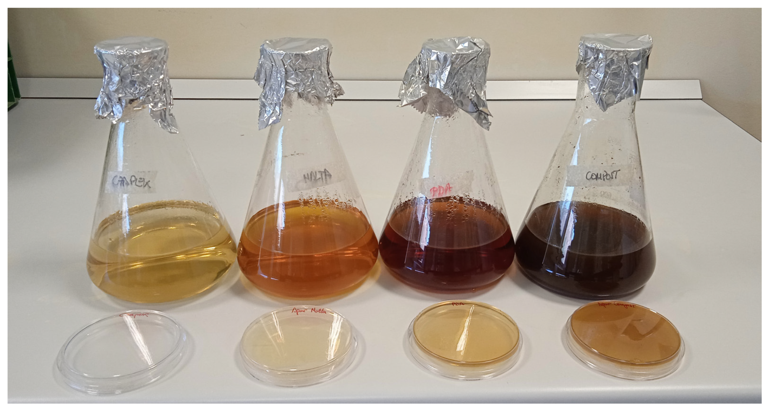
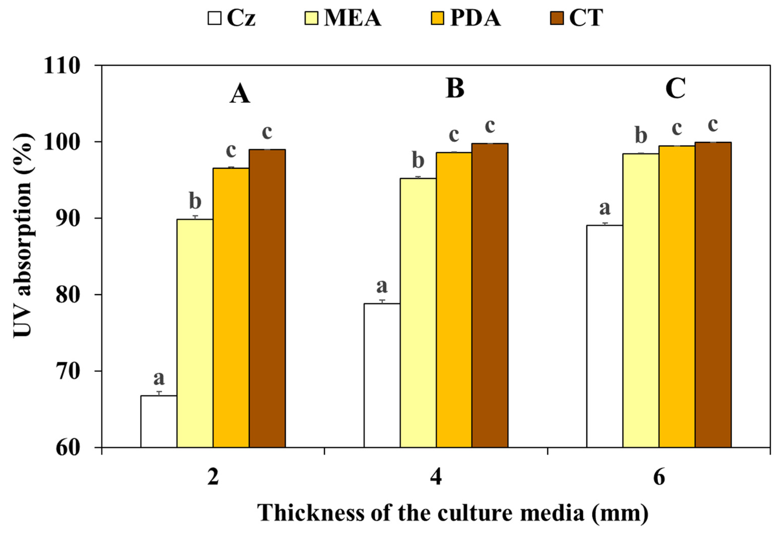
Apart from the influence of the species, the different UV-B effects were probably caused by the UV absorption capacity of the different culture media, which, in turn, was primarily based on their inherent optical characteristics (Figure 3). Nevertheless, we confirmed that their UV absorption also depended on the medium thickness (Figure 4), which should be carefully checked and homogenized in the experiments. Overall, the UV absorption increased with increasing thickness, reaching almost 100% at 6 mm in all media except Cz. This was the medium that significantly absorbed less UV (around 65% at 2 mm and nearly 90% at 6 mm), followed by MEA (whose absorption ranged between 90% and 98%). In contrast, PDA and CT were the most absorbent media, reaching values above 97% even at a thickness of 2 mm.
Consistent with our results, various studies have demonstrated the limited penetration of UV-B in culture media [14], which could be due to their different composition. This would explain the restricted effect of UV-B on mycelial development, especially when using certain media. Therefore, the embedded growth of the mycelia could be a fungal strategy against UV-B radiation [34]. The influence of the culture medium could be an important consideration to explain the differences found in the existing literature regarding UV-B effects. For instance, these effects were different in Aspergillus sp. depending on the media used [23,33] and also in various moulds when comparing Cz and PDA [14].
The limited penetration of UV-B on most media could constrain its applicability in controlling mycopathogenic moulds in vivo. Specifically, CT, which closely resembles the substrate used to cultivate commercial mushrooms, showed the lowest penetration of UV-B and, thus, the greatest protective effect on mycelial development.

3.3. Effect on Spore Development

Unlike the growing mycelia, which would be protected by the substrate, the spores (conidia) released by the mould for propagation would not have the substrate protection until they germinate. This would mean that spores would be more UV-B-sensitive than mycelia, better reflecting the UV-B effects. To test this hypothesis, we exposed mould spores to every UV-B dose and assessed their development in the four media used.
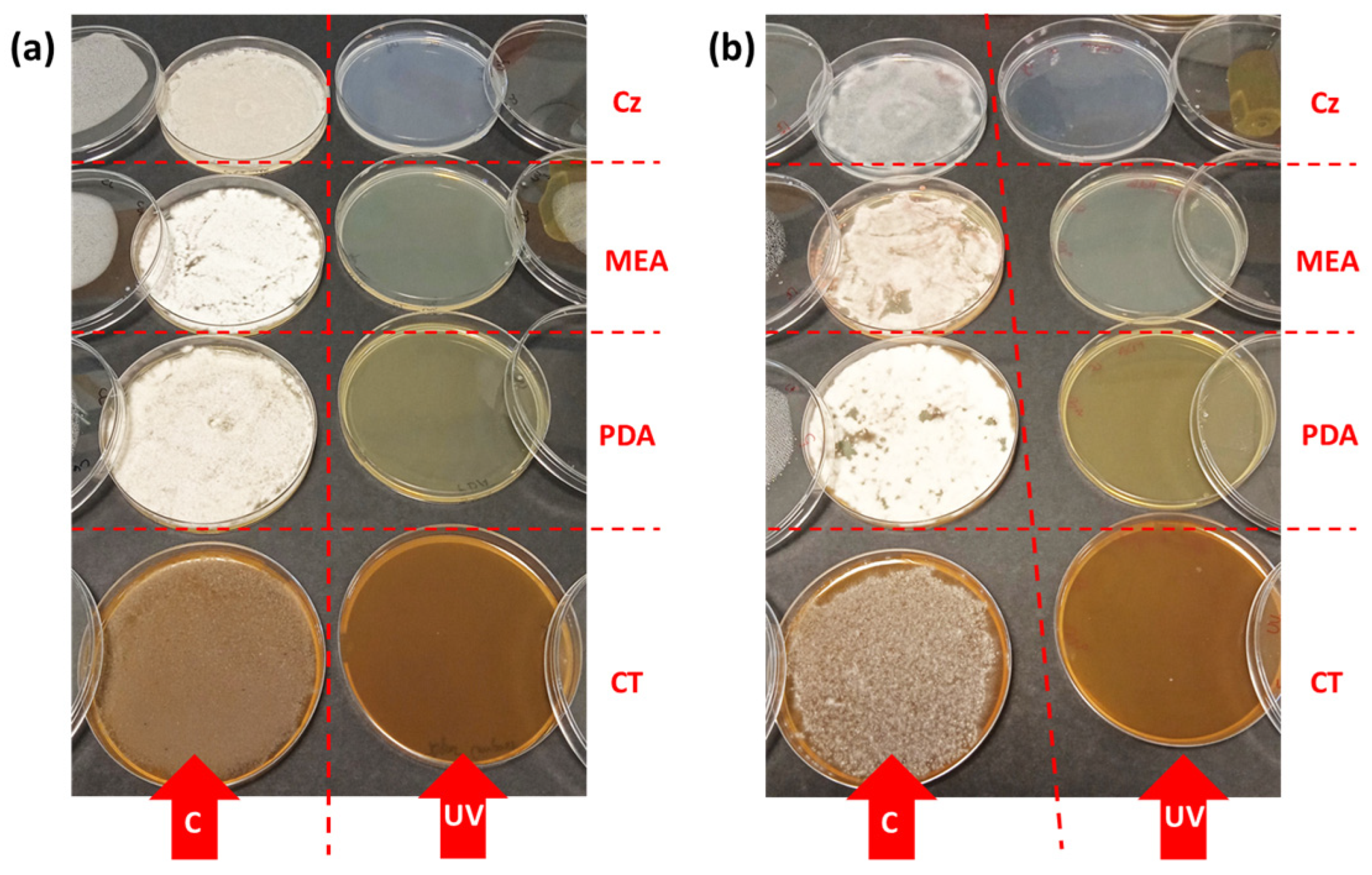
In non-pigmented moulds (C. mycophilum, L. fungicola, and M. perniciosa), there was a rapid spore development in the control plates but total inhibition in the irradiated plates, irrespective of the dose applied or the medium used. Figure 5 shows, as an example, the results obtained in C. mycophilum and L. fungicola.
However, in the pigmented mould T. aggressivum, a greater UV-B spore tolerance and some influence of the culture medium were observed (Table 1). Total inhibition of development occurred at 48 kJ m−2 in the Cz and MEA media and at 96 kJ m−2 in PDA, whereas, in CT, some development was recorded even at the highest dose (192 kJ m−2).
The different UV-B response of the spores can be attributed primarily to their different absorption spectrum (Figure 6), as has been mentioned by other authors [14,24,26,27,28,33]. All of them point out the complete inactivation of spore germination by UV-B in moulds where the spore pigmentation does not exert a protective effect. On the contrary, the pigmented spores produced by certain moulds, such as T. aggressivum, retain their development capacity even after exposure to high UV-B doses. In addition, development capacity increases in those culture media, such as CT, showing a stronger UV absorption and, consequently, a higher protection capacity. Therefore, UV-B would be much more effective in controlling the spread of mycopathogenic moulds through their spores than in reducing the development of their mycelia.

3.4. Effect of UV-B on the Host (Agaricus bisporus) Mycelium

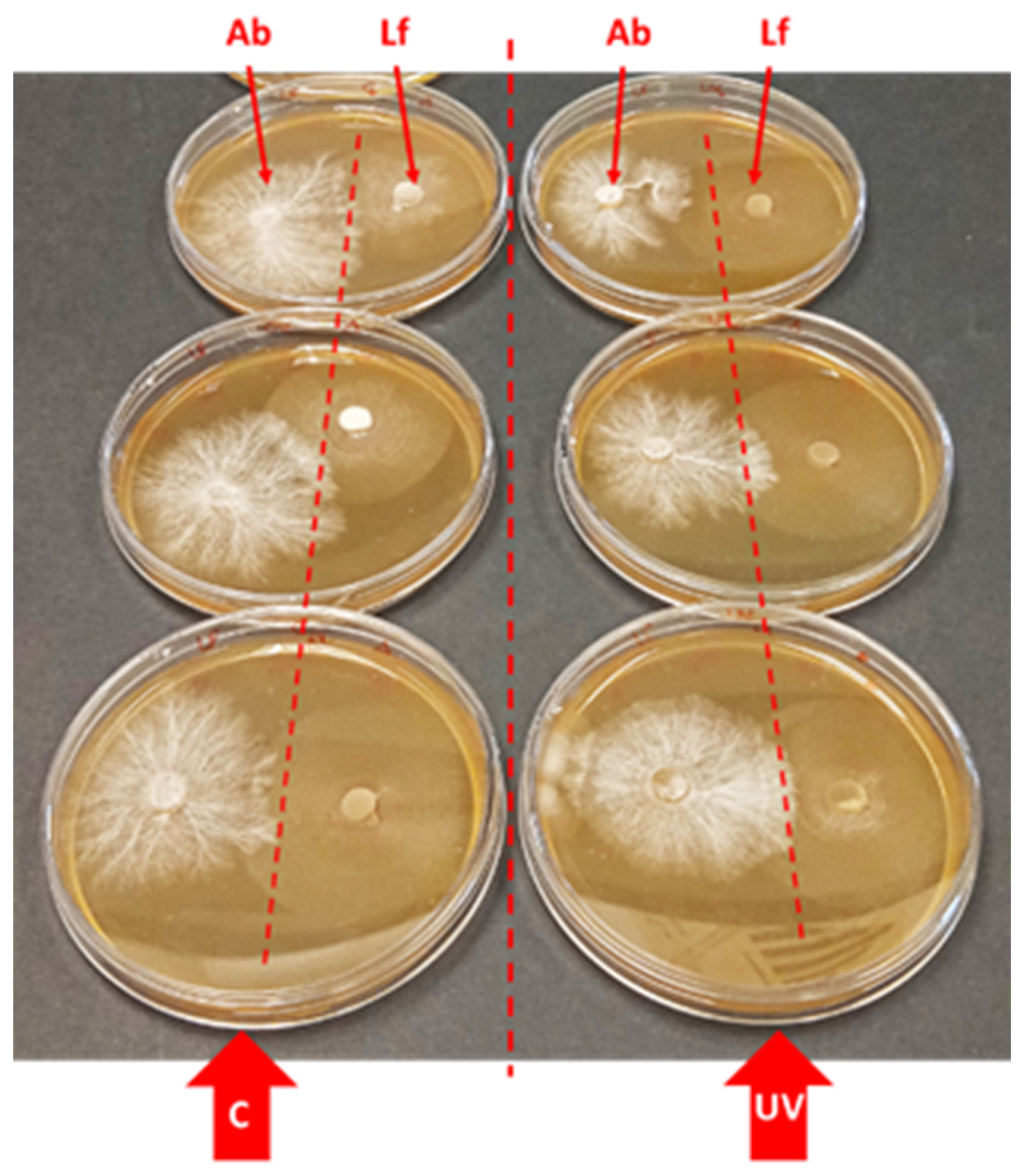
We tested UV-B effects on the development of the mycelium of A. bisporus, a frequent host of the moulds studied (Figure 7). For this aim, we used CT, the most similar medium to that used for commercial mushroom cultivation. None of the UV-B doses used had any significant effect on mycelium development, which was similar in both control and irradiated plates. As a comparative example, the mycelium of L. fungicola was grown in the same plates, confirming that none of the UV-B doses affected its development, as previously found in this species when cultivated on CT (Figure 2).
Much research has been performed on the post-harvest effect of UV-B on cultivated mushrooms, especially for the enhanced production of interesting compounds such as vitamin D [18,40]. However, there are few studies on the pre-harvest UV-B effects on cultivated mushrooms or, in general, on any Basidiomycota species. Specifically, several in vitro studies reported a mycelium growth decrease in UV-B-exposed medicinal or edible species, such as Serpula himantioides [41], Inonotus obliquus [42], and Trametes versicolor and Flammulina velutipes [43]. This reduced growth was accompanied by an increase in photoprotective metabolites with strong antioxidant capacity, such as polyphenolic compounds. These results were obtained employing different culture media, respectively, PDA, Yeast Peptone Dextrose (YPD), and Malt Agar (MA). However, UV-B did not cause any negative impact on the yield of A. bisporus and Pleurotus ostreatus [44,45]. These last results, obtained under crop conditions, support our in vitro results and emphasize the protective role of commercial substrates against the potentially negative effects of UV-B.

4. Conclusions

UV-B proved to be a highly effective tool in inhibiting spore development and thus controlling the spread of mycopathogenic moulds. We demonstrated this on spores grown in plates but it could also be applicable to spores suspended in the air or deposited on the mushrooms, in both cases under commercial cultivation conditions. Spore inactivation was particularly evident in nonpigmented moulds, including the most harmful ones (C. mycophilum and L. fungicola), although pigmented moulds (such as T. aggressivum) exhibited a greater UV-B tolerance. In addition, UV-B did not interfere with the mycelial development of cultivated mushrooms, such as A. bisporus. Nevertheless, the UV-B efficacy in controlling the mycelial development of mycopathogenic moulds was lower than that found for the spores, partly because of its low penetration into the different culture media, which exerted a protective role. Given that the culture medium most similar to the substrate used in the commercial cultivation of edible mushrooms (CT) was the medium that provided the highest protection to the mould mycelium, UV-B would only be relatively useful to control mycopathogenic moulds under commercial conditions. While studies in real crops would be necessary, our in vitro results open the door to the use of UV-B (much less aggressive than UV-C and, therefore, more easily applicable) in controlling the main fungal diseases of cultivated mushrooms, thus emerging as a solid alternative or complement to the use of traditional fungicides. To our knowledge, this is the first study demonstrating simultaneously that UV-B radiation can eliminate spores and (less efficiently) mycelia of mycopathogenic moulds without affecting mushroom mycelia.

Author Contributions

Conceptualization, S.S., C.O., E.N.-O. and J.M.-A.; methodology, S.S., C.O. and E.N.-O.; investigation, R.H.-S., M.-Á.D.-C.-A., S.S. and C.O.; formal analysis, R.H.-S., M.-Á.D.-C.-A. and S.S.; writing—original draft preparation, R.H.-S. and M.-Á.D.-C.-A.; writing—review and editing, S.S., C.O., J.M.-A. and E.N.-O.; visualization, M.-Á.D.-C.-A., R.H.-S. and S.S.; supervision, E.N.-O., J.M.-A. and M.-Á.D.-C.-A.; funding acquisition, E.N.-O. and J.M.-A. All authors have read and agreed to the published version of the manuscript.

Funding

This research was funded by the Government of La Rioja (project “Afianza” 2023/05).

Data Availability Statement

Data supporting reported results are available on request from the corresponding author.

Acknowledgments

The authors thank the collaboration of the Mushroom Technological Research Center (CTICH) of the Goverment of La Rioja (Autol, La Rioja, Spain) for providing the moulds strains. We also thank J. Carrasco for his valuable advice.

Conflicts of Interest

The authors declare no conflicts of interest.

References

  1. Fletcher, J.T.; Gaze, R.H. Mushroom Pest and Disease Control: A Colour Handbook, 1st ed.; Manson Publishing Ltd.: London, UK; Academic Press: San Diego, CA, USA, 2008; 192p. [Google Scholar]
  2. Adie, B.A.T. The Biology and Epidemiology of the Cobweb Disease Pathogen (Cladobotryum spp.) Infecting the Cultivated Mushroom (Agaricus bisporus). Ph.D. Thesis, Imperial College, University of London, London, UK, 2000. [Google Scholar]
  3. Adie, B.; Grogan, H.; Archer, S.; Mills, P. Temporal and spatial dispersal of Cladobotryum conidia in the controlled environment of a mushroom growing room. Appl. Environ. Microbiol. 2006, 72, 7212–7217. [Google Scholar] [CrossRef]
  4. Pyck, N.; Grogan, H. Fungal Diseases of Mushrooms and Their Control; Factsheet 04/15, MushTV Publications; AHDB: Warwickshire, UK, 2015; p. 6. Available online: http://horticulture.ahdb.org.uk/mushtv-publications (accessed on 20 January 2024).
  5. European Commission. Communication from the Commission to the European Parliament, the Council, the European Economic and Social Committee and the Committee of the Regions. “From Farm to Fork” Strategy for a Fair, Healthy and Environmentally-Friendly Food System; European Commission: Brussels, Belgium, 2020. [Google Scholar]
  6. Agencia Estatal Boletín Oficial del Estado. Real Decreto 1050/2022, De 27 De Diciembre, Por El Que Se Modifica El Real Decreto 1311/2012, De 14 De Septiembre, Que Establece El Marco De Actuación Para Conseguir Un Uso Sostenible De Los Productos Fitosanitarios; Agencia Estatal Boletín Oficial del Estado: Madrid, Spain, 2022.
  7. Potočnik, I.; Vukojević, J.; Stajić, M.; Rekanović, E.; Stepanović, M.; Milijašević, S.; Todorović, B. Toxicity of biofungicide Timorex 66 EC to Cladobotryum dendroides and Agaricus bisporus. Crop Prot. 2010, 29, 290–294. [Google Scholar] [CrossRef]
  8. Kosanović, D.; Potočnik, I.; Duduk, B.; Vukojević, J.; Stajić, M.; Rekanović, E.; Milijašević-Marčić, S. Trichoderma species on Agaricus bisporus farms in Serbia and their biocontrol. Ann. Appl. Biol. 2013, 163, 218–230. [Google Scholar] [CrossRef]
  9. Gea, F.J.; Carrasco, J.; Diánez, F.; Santos, M.; Navarro, M.J. Control of dry bubble disease (Lecanicillium fungicola) in button mushroom (Agaricus bisporus) by spent mushroom substrate tea. Eur. J. Plant Pathol. 2014, 138, 711–720. [Google Scholar] [CrossRef]
  10. Geösel, A.; Szabó, A.; Akan, O.; Szarvas, J. Effect of essential oils on mycopathogens of Agaricus bisporus. In Proceedings of the 8th International Conference of Mushroom Biology and Mushroom Products, New Delhi, India, 27 November 2014. [Google Scholar]
  11. Gea, F.J.; Navarro, M.J.; Tello, J.C. Reduced sensitivity of the mushroom pathogen Verticillium fungicola to prochloraz-manganese in vitro. Mycol. Res. 2005, 109, 741–745. [Google Scholar] [CrossRef] [PubMed]
  12. Grogan, H.M. Fungicide control of mushroom cobweb disease caused by Cladobotryum strains with different benzimidazole resistance profiles. Pest Manag. Sci. 2006, 62, 153–161. [Google Scholar] [CrossRef]
  13. Usall, J.; Ippolito, A.; Sisquella, M.; Neri, F. Physical treatments to control postharvest diseases of fresh fruits and vegetables. Postharvest Biol. Technol. 2016, 122, 30–40. [Google Scholar] [CrossRef]
  14. Moody, S.A.; Newsham, K.K.; Ayres, P.G.; Paul, N.D. Variation in the responses of litter and phylloplane fungi to UV-B radiation (290–315 nm). Mycol. Res. 1999, 103, 1469–1477. [Google Scholar] [CrossRef]
  15. Barnes, P.W.; Robson, T.M.; Zepp, R.G.; Bornman, J.F.; Jansen, M.A.K.; Ossola, R.; Wang, Q.W.; Robinson, S.A.; Foereid, B.; Klekociuk, A.R.; et al. Interactive effects of changes in UV radiation and climate on terrestrial ecosystems, biogeochemical cycles, and feedbacks to the climate system. Photochem. Photobiol. Sci. 2023, 22, 1049–1091. [Google Scholar] [CrossRef]
  16. Robson, T.M.; Aphalo, P.J.; Banas, A.K.; Barnes, P.W.; Brelsford, C.C.; Jenkins, G.I.; Kotilainen, T.K.; Labuz, J.; Martínez-Abaigar, J.; Morales, L.O.; et al. A perspective on ecologically relevant plant-UV research and its practical application. Photochem. Photobiol. Sci. 2019, 18, 970–988. [Google Scholar] [CrossRef]
  17. Pescheck, F.; Bilger, W. UV-B Resistance of Plants Grown in Natural Conditions. Annu. Plant Rev. Online 2020, 3, 337–398. [Google Scholar] [CrossRef]
  18. Hidalgo-Sanz, R.; Del-Castillo-Alonso, M.A.; Monforte, L.; Tomás-Las-Heras, R.; Sanz, S.; Olarte, C.; Pérez-Matute, P.; Íñiguez-Martínez, M.; Ene, A.L.; Martínez-Abaigar, J.; et al. Ultraviolet-B radiation, mushrooms, and vitamin D: From technology to bioavailability. LWT 2023, 186, 115210. [Google Scholar] [CrossRef]
  19. Zhong, Y.Y.; Dong, S.T.; Cui, Y.; Dong, X.B.; Xu, H.D.; Li, M. Recent Advances in Postharvest Irradiation Preservation Technology of Edible Fungi: A Review. Foods 2023, 12, 103. [Google Scholar] [CrossRef] [PubMed]
  20. Robson, T.M.; Pieristè, M.; Durand, M.; Kotilainen, T.K.; Aphalo, P.J. The benefits of informed management of sunlight in production greenhouses and polytunnels. Plants People Planet 2022, 4, 314–325. [Google Scholar] [CrossRef]
  21. Del-Castillo-Alonso, M.A.; Monforte, L.; Tomás-Las-Heras, R.; Martínez-Abaigar, J.; Núñez-Olivera, E. To What Extent Are the Effects of UV Radiation on Grapes Conserved in the Resulting Wines? Plants 2021, 10, 1678. [Google Scholar] [CrossRef] [PubMed]
  22. Duguay, K.J.; Klironomos, J.N. Direct and indirect effects of enhanced UV-B radiation on the decomposing and competitive abilities of saprobic fungi. Appl. Soil Ecol. 2000, 14, 157–164. [Google Scholar] [CrossRef]
  23. García Cela, E.; Marín, S.; Sanchís, V.; Crespo-Sempere, A.; Ramos, A.J. Effect of ultraviolet radiation A and B on growth and mycotoxin production by Aspergillus carbonarius and Aspergillus parasiticus in grape and pistachio media. Fungal Biol. 2015, 119, 67–78. [Google Scholar] [CrossRef] [PubMed]
  24. Yamaga, I.; Kuniga, T.; Aoki, S.; Kato, M.; Kobayashi, Y. Effect of ultraviolet-B irradiation on disease development caused by Penicillium italicum in Satsuma mandarin fruits. Hortic. J. 2016, 85, 86–91. [Google Scholar] [CrossRef]
  25. Ulevicius, V.; Peclulyte, D.; Lugauskas, A.; Andriejauskiene, A. Field study on changes in viability of airborne fungal propagules exposed to UV radiation. Environ. Toxicol. 2004, 19, 437–441. [Google Scholar] [CrossRef]
  26. Latorre, B.A.; Rojas, S.; Díaz, G.A.; Chauqui, H. Germicidal effect of UV light on epiphytic fungi isolated from blueberry. Cienc. Investig. Agrar. 2012, 39, 473–480. [Google Scholar] [CrossRef]
  27. Parnell, M.; Burt, P.J.A.; Wilson, K. The influence of exposure to ultraviolet radiation in simulated sunlight on ascospores causing Black Sygatoka disease of banana and plantain. Int. J. Biometeorol. 1998, 42, 22–27. [Google Scholar] [CrossRef]
  28. Galvao, J.A.H.; Bettiol, W. Effects of UV-B radiation on Lecanicillium spp., biological control agents on the coffee leaf rust pathogen. Trop. Plant Pathol. 2014, 38, 392–400. [Google Scholar] [CrossRef]
  29. Calonje, M.; García-Mendoza, C.P.; Pérez-Cabo, A.; Bernardo, D.; Novaes-Ledieu, M. Interaction between the mycoparasite Verticillium fungicola and the vegetative mycelial phase of Agaricus bisporus. Mycol. Res. 2000, 104, 988–992. [Google Scholar] [CrossRef]
  30. Shamsahad, A.; Clift, A.D.; Mansfield, S. Host-parasite interaction between cultivated mushroom, Agaricus bisporus hybrid strain Sylvan A15, and the mycoparasite Verticillium fungicola, a causal agent of dry bubble disease. Aust. Plant Pathol. 2009, 38, 74–78. [Google Scholar] [CrossRef]
  31. Carrasco, J.; Tello, M.L.; de Toro, M.; Tkacz, A.; Poole, P.; Pérez-Clavijo, M.; Preston, G. Casing microbiome dynamics during button mushroom cultivation: Implications for dry and wet bubble diseases. Microbiology 2019, 165, 611–624. [Google Scholar] [CrossRef] [PubMed]
  32. Carrasco, J.; Navarro, M.J.; Santos, M.; Dianez, F.; Gea, F.J. Incidence, identification and pathogenicity of Cladobotryum mycophilum, causal agent of cobweb disease on Agaricus bisporus mushroom crops in Spain. Ann. Appl. Biol. 2016, 168, 214–224. [Google Scholar] [CrossRef]
  33. García Cela, E.; Marín, S.; Reyes, M.; Sanchís, V.; Ramos, A.J. Conidial survival of Aspergillus section Nigri, Flavi and Circumdati under UV-A and UV-B radiation with cycling temperature/light regime. J. Sci. Food Agric. 2015, 96, 2249–2256. [Google Scholar] [CrossRef]
  34. Fourtouni, A.; Manetas, Y.; Christias, C. Effects of UV-B radiation in growth, pigmentation, and spore production in the phytopathogenic fungus Alternaria solani. Can. J. Bot. 1998, 76, 2093–2099. [Google Scholar] [CrossRef]
  35. Ignofo, C.M.; García, C. Influence of conidial color on inactivation of several entomogenous fungi by simulated sunlight. Environ. Entomol. 1992, 21, 913–917. [Google Scholar] [CrossRef]
  36. Atanasova, L.; Knox, B.P.; Kubicek, C.P.; Druzhinina, I.S.; Baker, S.E. The polyketide synthase gene pks4 of Trichoderma reesei provides pigmentation and stress resistance. Eukaryot. Cell 2013, 12, 1499–1508. [Google Scholar] [CrossRef]
  37. Wang, J.; Ma, Y.; Liu, Y.; Tong, S.; Zhu, S.; Jin, D.; Pei, Y.; Fan, Y. A polyketide synthase, BbpksP, contributes to conidial cell wall structure and UV tolerance in Beauveria bassiana. J. Invertebr. Pathol. 2020, 169, 107280. [Google Scholar] [CrossRef] [PubMed]
  38. Lai, C.; Chen, J.; Liu, J.; Tian, D.; Lan, D.; Liu, T.; Wu, B.; Bi, H.; Tang, J. New Polyketides from a Hydrothermal Vent Sediment Fungus Trichoderma sp. JWM29-10-1 and Their Antimicrobial Effects. Mar. Drugs 2022, 20, 720. [Google Scholar] [CrossRef] [PubMed]
  39. Li, X.; Huang, L.; He, Y.; Xie, C.; Zhan, F.; Zu, Y.; Sheng, J.; Li, Y. Effects of enhanced UV-B radiation on the interaction between rice and Magnaporthe oryzae in Yuanyang terrace. Photochem. Photobiol. Sci. 2019, 18, 2965–2976. [Google Scholar] [CrossRef] [PubMed]
  40. Kamweru, P.K.; Tindibale, E.L. Vitamin D and Vitamin D from Ultraviolet-Irradiated Mushrooms. Int. J. Med. Mushrooms 2016, 18, 205–214. [Google Scholar] [CrossRef] [PubMed]
  41. Torres, S.; González-Ramírez, M.; Gavilán, J.; Paz, C.; Palfner, G.; Arnold, N.; Fuentealba, J.; Becerra, J.; Pérez, C.; Cabrera-Pardo, J.R. Exposure to UV-B radiation leads to increased deposition of cell wall-associated xerocomic acid in cultures of Serpulla imantioides. Appl. Environ. Microbiol. 2019, 85, e00870-19. [Google Scholar] [CrossRef]
  42. Zhou, R.; Yang, H.; Lu, T.; Zhao, Y.; Zheng, W. Ultraviolet radiation promotes the production of hispidin polyphenols by medicinal mushroom Inonotus ubiquus. Fungal Biol. 2022, 126, 775–785. [Google Scholar] [CrossRef] [PubMed]
  43. Krsmanovic, N.; Raseta, M.; Miskovic, J.; Bekvalac, K.; Bogavac, M.; Karaman, M.; Isikhuemhen, O.S. Effects of UV Stress in promoting antioxidant activities in fungal species Trametes versicolor (L.) Lloyd and Flammulina velutipes (Curtis) Singer. Antioxidants 2023, 12, 302. [Google Scholar] [CrossRef]
  44. Szabo, A.; Gyepes, A.; Nagy, A.; Abranko, L.; Gyorfi, J. The effect of UVB radiation on the vitamin D2 content of white and cream type button mushrooms (Agaricus bisporus Lange/Imbach) and Oyster Mushrooms (Pleorotus ostreatus (Jacq.) P. Kumm). Acta Aliment. 2012, 41, 187–196. [Google Scholar] [CrossRef]
  45. Kristensen, H.L.; Rosenqvist, E.; Jakobsen, J. Increase of Vitamin D2 by UV-B exposure during the growth phase of white button mushroom (Agaricus bisporus). Food Nutr. Res. 2012, 56, 7114. [Google Scholar] [CrossRef]
Figure 1. Effect of UV-B radiation treatments on the degree of mycelial development (relative units, see Section 2.3) in the four moulds studied, grown on Czapek Agar during the following 14–15 days after irradiation. Solid lines, controls for each dose (in some cases, the control lines for several doses are overlapped); dashed lines, UV-B treatments (yellow, 8 kJ m−2; red, 24 kJ m−2; green, 48 kJ m−2; blue, 96 kJ m−2; and grey, 192 kJ m−2). Means ± standard errors (SE) are shown (n = 3). Different letters show significant differences between doses after 14–15 days of growth (at least p < 0.05).
Agriculture 14 00681 g001
Figure 2. Effect of UV-B radiation treatment (at the highest dose used, 192 kJ m−2) on the degree of mycelial development (relative units, see Section 2.3) in the four moulds studied, grown on the four culture media used (Cz, Czapek Agar; MEA, Malt Extract Agar; PDA, Potato Dextrose Agar; and CT, Compost Tea Agar). Measurements were performed at 3 (blue), 5 (orange), 10 (grey), and 16 (yellow) days of growth after radiation (n = 3). For each species and medium, significant differences between controls (C) and treatment (UV) are shown (***, p < 0.001; *, p < 0.05). Standard errors (SE) are not shown for clarity reasons.
Agriculture 14 00681 g002
Figure 3. Aspect of the four culture media used, both before solidifying (liquid state, flasks) and after solidifying (solid state, plates). From left to right: Czapek Agar (Cz), Malt Extract Agar (MEA), Potato Dextrose Agar (PDA), and Compost Tea Agar (CT).
Agriculture 14 00681 g003
Figure 4. Percentage of UV absorption of the four culture media used (Cz, Czapek Agar; MEA, Malt Extract Agar; PDA, Potato Dextrose Agar; and CT, Compost Tea Agar) as influenced by thickness. Different capital letters show significant differences between thicknesses and different lowercase letters show differences between culture media for each thickness (at least p < 0.05). Means ± standard errors (SE) are shown (n = 3).
Agriculture 14 00681 g004
Figure 5. Development of spores of Cladobotryum mycophilum (a) and Lecanicillium fungicola (b) on the four culture media used (Cz, Czapek Agar; MEA, Malt Extract Agar; PDA, Potato Dextrose Agar; and CT, Compost Tea Agar). Spores were irradiated with UV-B (8 kJ m−2) and then were allowed to develop for 15 days. Nonirradiated control plates (C) and irradiated plates (UV) are shown.
Agriculture 14 00681 g005
Figure 6. UV absorption of spore suspensions of the four moulds used (Ta, Trichoderma aggressivum; Cm, Cladobotryum mycophilum; Lf, Lecanicillium fungicola; and Mp, Mycogone perniciosa). The limit between UV-B and UV-A wavelengths (315 nm) is shown (red dashed line).
Agriculture 14 00681 g006
Figure 7. Dual cultures, per triplicate, of Agaricus bisporus (Ab, left side of each plate) and Lecanicillium fungicola (Lf, right side) grown in Compost Tea Agar after 14 days of incubation at 20 °C. Plates in the left row were not irradiated (control plates, C) and plates in the right row were irradiated with 192 kJ m−2 UV-B (UV).
Agriculture 14 00681 g007
Table 1. Relative development (expressed in percentage) of Trichoderma aggressivum spores cultivated on the four culture media used (Cz, Czapek Agar; MEA, Malt Extract Agar; PDA, Potato Dextrose Agar; and CT, Compost Tea Agar) after 15 days of UV-B irradiation with different doses. The dose “0” is the nonirradiated control.
UV-B Dose
(kJ m−2)
Culture Media
CzMEAPDACT
0100100100100
88080100100
2420208080
48------2060
96---------30
192---------20
Disclaimer/Publisher’s Note: The statements, opinions and data contained in all publications are solely those of the individual author(s) and contributor(s) and not of MDPI and/or the editor(s). MDPI and/or the editor(s) disclaim responsibility for any injury to people or property resulting from any ideas, methods, instructions or products referred to in the content.