Article

The Presence of Arbuscular Mycorrhizal Fungi in the Rhizosphere of Transgenic Rapeseed Overexpressing a Trichoderma Thkel1 Gene Improves Plant Development and Yield

1
Department of Botany and Plant Physiology, Institute for Agribiotechnology Research (CIALE), University of Salamanca, 37185 Salamanca, Spain
2
Department of Plant Production and Forest Resources, University Institute for Research in Sustainable Forest Management (iuFOR), University of Valladolid, 34004 Palencia, Spain
3
Independent Researcher, 37007 Salamanca, Spain
4
Department of Microbiology and Genetics, Institute for Agribiotechnology Research (CIALE), University of Salamanca, 37185 Salamanca, Spain
5
Department of Agricultural and Environmental Sciences, Jaume I University, 12071 Castellón, Spain
*
Authors to whom correspondence should be addressed.
These authors contributed equally to this work.
Agriculture 2024, 14(6), 851; https://doi.org/10.3390/agriculture14060851
Submission received: 10 April 2024 / Revised: 24 May 2024 / Accepted: 25 May 2024 / Published: 29 May 2024
(This article belongs to the Special Issue Beneficial Microbes for Sustainable Crop Production)

Abstract

:
Most of the plants belonging to the family of Brassicaceae are non-hosts for arbuscular mycorrhizal fungi (AMF). These plants are known to produce glucosinolates (GSL), a group of allelopathic compounds, with a role in plant defense. The overexpression of the Thkel1 from Trichoderma harzianum in rapeseed (BnKel) plants, this gene encoding a protein that shares similarities with Brassicaceae plant’s nitrile-specifier and epithiospecifier proteins, modified GSL metabolism, reducing the accumulation of toxic isothiocyanates due to hydrolysis of these secondary metabolites. Here, we have analyzed the effect of AMF application on the GSL profiles and the development and yield of BnKel plants. Our results showed that the reduction of GSL compounds on transgenic plants was not enough to allow the formation of arbuscules and vesicles characteristics of an AMF mycorrhizal association. However, the inoculation of transgenic rapeseed plants expressing Thkel1 with AMF improved seed yield and fatty acid composition of the oilseed, showing a beneficial effect of AMF in these plants. The achievement of this effective beneficial association among mycorrhizas and rapeseed plants opens new opportunities in agribiotechnology for the use of AMF as biofertilizers in Brassicaceae crops with potential application in medical, animal and industrial biotechnology.

1. Introduction

The genus Brassica is probably the most agriculturally relevant group of Brassicaceae, from an agronomic point of view, as it includes more than 30 species and hybrids with economic interest and worldwide distribution [1]. Members of the family Brassicaceae are known to produce glucosinolates (GSL), allelopathic compounds related to plant defense [2,3,4]. These compounds are hydrolyzed by thioglucosidases, also known as myrosinases, to form toxic isothiocyanates, playing a role in plant defense against bacteria, fungi, and insect herbivores, while producing other compounds with different biological activities [3]. The GSL breakdown begins when the spatial separation of GSL and myrosinases is suppressed after tissular damage induced by an attacker. Other groups of proteins, called nitrile-specifier (NSP) and epithiospecifier proteins (ESP) can interact with myrosinases, addressing the reaction to the formation of nitriles or epithionitriles, compounds with less toxicity than the GSL-derived isothiocyanates [3,5].
Beneficial mutualistic symbiosis among plants and fungi are critical features for plant survival and development. In this context, Trichoderma and mycorrhizal fungi are of interest for eco-sustainable agriculture [6,7]. Trichoderma includes soil-borne filamentous fungi that are used as direct biocontrol agents against phytopathogens and in their communication with the plant can induce its growth and prime defense responses to pathogens and environmental stresses [7,8,9]. In addition, several studies have shown that Trichoderma constitutes an important source of genes to generate genetically modified (GM) crops [10]. On the other side, arbuscular mycorrhizal fungi (AMF) are characterized by the formation of arbuscules within the root cells, in these symbiotic associations, the presence of AMF improves water and nutrient uptake for these plants, while the fungus receives photosynthetic carbon in the form of sugars and fatty acids [11,12]. Considering crop species, AMF symbiosis is an outstanding feature from an economic point of view. However, the loss of key genes to establish symbiosis during evolution in some flowering plants, as those of the Brasicaceace family [13], has led them to the inability to form AM symbiotic associations [14]. Recently, it has been suggested that the presence of AMF is not entirely impaired in the non-fungal host Arabidopsis [15], and although it has been reported the development of rudimentary arbuscular mycorrhizal (AM) phenotypes in some Brasicaceae species, it seems clear that it is not a true AMF host [16,17].
In a previous work, we characterized the Thkel1 gene from Trichoderma harzianum, encoding the ThKEL1 protein that contains five repeated Kelch domains and sharing similarity to NSPs and ESPs of plant origin [18]. Kelch domain proteins are key in the regulation of GSL metabolism in plants and several other processes [19]. The overexpression of the Thkel1 gene in Arabidopsis and rapeseed plants led to improved tolerance to abiotic stress conditions and resistance against the leaf pathogens Botrytis cinerea and Phoma lingam, respectively, while facilitating their root colonization by Trichoderma and increasing the production of seeds [18,20]. The aim of the present work was to test whether the reduction of the GSL levels in BnKel plants would allow the formation of arbuscular structures within the roots of these transgenic plants when AMF is applied. Once the establishment of this symbiotic association was verified, the main objective of this work was to test its beneficial effects on seed yielding as well as its potential applications in agrobiotechnology.

2. Materials and Methods

2.1. Plant Material

B. napus cv. Jura and its BnKel1 and BnKel2 transgenic lines expressing the Thkel1 gene from T. harzianum T34, previously described [20], were the plants used in this study. The two independent Thkel1 transgenic lines exhibited similar phenotypes and most of the results shown throughout the manuscript correspond to BnKel2 transgenic line.

2.2. AMF Material

We used, as AM mycorrhizal inoculum, the Miratext-02 formulation (Mirat Fertilizantes, Salamanca, Spain), which contained at least 1 × 10⁶ spore kg−1 of five different AMF species: Glomus microagregatum, Funneliformis mosseae, Claroideoglomus claroideum, Rhizophagus irregularis and R. fasciculatus. Non-inoculated (control) plants received a 5 mL aliquot consisting of a filtrate from the arbuscular mycorrhizal inoculum (<20 μm) to provide a general microbial population free of arbuscular mycorrhizal propagules.

2.3. AMF Inoculation and Plant Growth Conditions

Seeds were germinated in plates on Murashige and Skoog (MS) medium (Duchefa, Haarlem, The Netherlands) complemented with 1% agar and 1% sucrose. Seedlings were grown for seven days under controlled environmental conditions in a growth chamber at 22 °C, 40% relative humidity (RH), and a 16/8 h (day/night) photoperiod at 80–100 μE m−2 s−1. After that, seedlings were transferred into 5 L pots, containing an autoclaved mixture of peat/vermiculite (3:1), and maintained under greenhouse conditions as previously described [20] and watered as needed. AM inoculum was applied to each pot by burying 1 g of Miratext-02 (1000 spore g−1) at 5 cm below the substrate surface just before transplanting the seedlings.
Ten weeks after transplanting (during the formation of floral primordia), roots were separated from the shoots, carefully washed and immediately frozen with liquid nitrogen and pulverized with a mortar. Roots from five plants per condition were pooled and root pools from three biological replicates were considered for statistical analysis. Dry weight was determined in the aerial part of 8-week-old plants. They were kept at 65 °C for 48 h and weighed. Plants of three biological replicates were analyzed for each assayed condition, and five plants were included per replicate.
Siliques were collected at the end of the life cycle (19 weeks) and counted. Three biological replicates were considered, and 15 plants were used per biological replicate and condition.

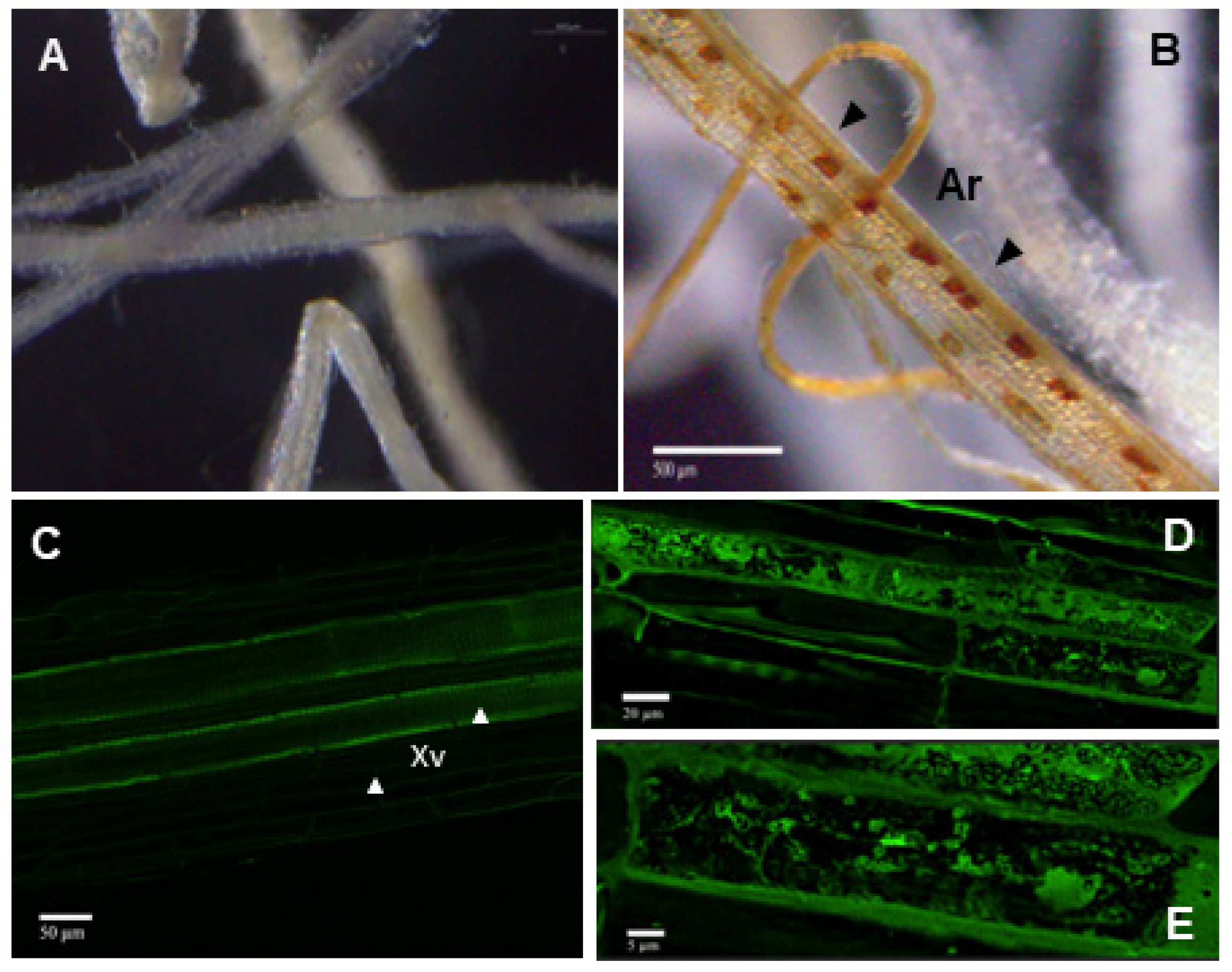
2.4. Visualization of the AMF Structures in Rapeseed Roots

The B. napus roots were immersed in a solution of 10% KOH at 90 °C for 10 min, in order to increase the permeability of their cell walls. Subsequently, three washes were carried out with distilled water to eliminate the KOH and one more with a solution of acetic acid at 2% (v/v). The roots were then transferred to a solution with the Sheaffer Skrip Ink [Cult Pens, Tiverton, UK (5% ink in 2% acetic acid)] for another 10 min. Finally, the excess ink was discarded, and the roots were rinsed with distilled water, where they were kept until their observation [21]. Pictures were taken with a stereoscopic microscope, Leica M205 FA, equipped with a Leica DFC 495 camera, (Leica, Madrid, Spain).
The (WGA)-Alexa Fluor 488 conjugate (Molecular Probes, Eugene, OR, USA) was used for detailed images of the fungal morphology at the roots. Root sections were cleared with KOH as explained before, treated with HCI 0.1N for 30 min, and infiltrated with 2.5 μg ml−1 wheat germ agglutinin in phosphate-buffered saline (PBS 1Xfor 15 min in the dark. Z-stack images were obtained using a laser-scanning confocal fluorescence microscope (Nikon).

2.5. DNA- Detection of AMF at the Roots

The presence of AMF DNA in the roots of rapeseed plants was evaluated by real-time quantitative PCR (qPCR) as previously described [20]. The primers 18S-F and 18S-R were used to amplify the 18S rRNA gene of AMF [18S-F: CTTTCGATGGTAGGATAGAGG; 18S-R: ACAACTTTAATATACGCTATTGGA [22], and the primers BnAct-F and BnAct-R were used to amplify the actin gene of rapeseed, as an internal reference gene, since it is considered a housekeeping gene [BnAct-F: CCCTGGAATTGCTGACCGTA; BnAct-R: TGGAAAGTGCTGAGGGATGC [23]. Amplifications were performed using an ABI PRISM 7000 Sequence Detection System (Applied Biosystems, Foster City, CA, USA) programmed as previously described [24]. Each PCR was performed in triplicate and DNA from three biological replicates (each one including pooled roots of five plants per condition) was used. Cycle threshold values were used to calculate the amount of fungal DNA using standard curves. Values of AMF DNA were referred to the amount of rapeseed DNA in every corresponding sample.

2.6. Phosphorus and Iron Measurement

Phosphorus (P) and iron (Fe) content were determined in the aerial part of 8-week-old plants. All measurements were quantified at the IRNASA analytical service (CSIC, Salamanca, Spain). Five hundred mg of ground samples were used for quantification, and the content of P and Fe was analyzed by means of optical emission spectrometry with source of plasma connected by induction (ICP-OES), using methodology previously described [25]. Plants of three biological replicates were analyzed for each assayed condition, and five plants were included per replicate. Results are expressed in mg g−1 dry weight for P and mg kg−1 dry weight for Fe.

2.7. GintPT and GintAMT2 Gene Expression Analysis by qPCR

Total RNA was isolated from roots using TRIZOL® reagent (Invitrogen Life Technologies, Carlsbad, CA, USA) following the manufacturer’s instructions. Thus, RNA samples were treated with DNase I (Fermentas, Burlington, Canada) and purified using GeneJET RNA Cleanup and Concentration Micro Kit (Thermo Scientific, Waltham, MA, USA). Finally, cDNA was obtained from 1 μg of RNA using the PrimeScriptTM RT reagent mix kit with an oligo (dT) (Takara Inc., Tokyo, Japan).
PCR reactions were performed using the gene-specific primers for AMF transporters GintPT and GintAMT2, as described in Fernandez et al. [15], and the thermocycler as indicated above. Each 10 µL reaction mixture contained 1 μL of cDNA, 5 μL of Kapa Sybr® Fast (Roche, Basel, Switzerland), 3.4 μL of deionized water, and 0.3 μL of each primer pair at a final concentration of 0.1 mM. The PCR program consisted of 40 cycles: denaturation, 95 °C for 3 s; annealing, 58 °C for GinPT and 60 °C for GintAMT2 for 30 s; extension, 72 °C for 1 s. The specificity of the PCR amplification procedure was checked with a heat-dissociation protocol (from 58 to 95 °C) after the final cycle of the PCR. Relative quantification of specific mRNA levels was performed using the comparative method of Livak and Schmittgen [26]. Expression values were normalized using the housekeeping gene 18S previously described. Three different root RNA samples for each treatment were used for analysis (n = 3), with each of them repeated three times. Negative controls without cDNA were used in all the PCR reactions.

2.8. GSL and Oil Profiles

2.8.1. Sample Extraction

Polar and non-polar fractions of oil seeds. Seeds were weighed (c.a. 50 mg, the actual amount was recorded) and crushed using a ball mill and glass beads. The resulting oily paste was thoroughly mixed with 300 μL of pure methanol (LC/MS degree) supplemented with biochanin A at 1 mg L−1 and sonicated for 10 min at room temperature. After centrifugation at 10,000 rpm for 10 min at 4 °C, the supernatant was recovered and combined with 400 μL of ultrapure water and 200 μL of chloroform. The upper water layer was recovered for polar metabolite analysis whereas the lower organic layer was dried down under vacuum and subsequently reconstituted in pure n-butanol (LC/MS degree) for non-polar metabolite analysis described below.
GSL and other semipolar compounds. Freeze-dried root samples (c.a. 10 mg, actual amount was recorded), were extracted in 500 μL of 70% aqueous methanol supplemented with biochanin A at 1 mg L−1 (internal standard, IS) by ultrasonication (10 min) at room temperature. After extraction, samples were centrifuged at 10,000 rpm for 10 min at 4 °C and supernatants were recovered. Prior to LC/ESI-QqTOF-MS analysis, supernatants were filtered through 0.2 μm PTFE syringe filters (Whatman International Inc., Kent, UK). Analyses of polar fractions and GSLs were carried out as described below.

2.8.2. LC/ESI-QqTOF-MS Analyses

Chromatographic separations were performed on a 100 mm × 2.1 mm i.d., 1.6 μm, Luna Omega 1.6u Polar C18 (Phenomenex, Torrance, CA, USA). For non-polar fractions, H2O:cyanuric acid (ACN) (15:85, v/v) supplemented with 0.01% HCOOH and 0.5 mM NH4Ac (A) and n-butanol containing 0.01% HCOOH and 0.5 mM NH4Ac (B) were used as solvents. Analysis of GSLs and polar fractions was achieved using H2O (A) and ACN (B), both supplemented with 0.1% formic acid. In both cases, flow rate was 0.3 mL min−1, column temperature was 40 °C and samples were maintained at 12 °C to slow down degradation. The mass spectrometer was operated in both negative and positive electrospray modes. Argon was used as the collision gas, and nitrogen was used as the nebulizer as well as desolvation gas set at 60 and 800 L h−1, respectively. Exact mass measurements were provided by monitoring the reference compound lockmass leucine enkephalin. After acquisition, mass chromatograms were converted to netCDF and subsequently processed with xcms and CAMERA packages [27]. Identification of compounds was achieved by retention time and mass spectra comparison with pure standards, when available, or tentatively annotated based on mass spectral matching with those available in public databases (Massbank, HMDB and Metlin, La Jolla, CA, USA) or the literature.

2.9. Statistical Analysis

Statistical analysis was carried out with the Statistix 8.0 software. Data were analyzed using one-way ANOVA and the post hoc Tukey’s test at p < 0.05 (data in tables), or by two-way ANOVA followed by Sidak’s multiple comparison test at * p < 0.05; ** p < 0.01; *** p < 0.001; **** p < 0.0001 (for GLS analyses). For analysis of productivity, P and Fe content, and colonization rates, a one-way ANOVA using Tukey’s multiple range test at p < 0.05 was used for pairwise comparisons, indicating the significant differences with different letters.

3. Results

3.1. Presence of AMF in Thkel1-Overexpressing Rapeseed Plants

Wild-type and Thkel1-overexpressing (BnKel) rapeseed plants were inoculated with a pre-commercial AMF inoculum (provided by Mirat Fertilizantes, Salamanca, Spain) containing five different AMF species. In transgenic BnKel plants, some rudimentary structures could be observed, although they could not be identified as true arbuscules or vesicles characteristic of AMF (Figure 1). The presence of AMF at the root samples was further detected by qPCR in the two transgenic lines analyzed (Table 1). No fungal rudimentary structure or DNA was detected in wild-type plants (Figure 1 and Table 1).

3.2. Gene Expression Analysis of Phosphate and Ammonium Transporters of the AMF Rhizophagus Irregularis

We analyzed, in rapeseed roots, the transcript levels of two AMF transporter genes, GintPT (encoding a high-affinity phosphate transporter) and GintAMT2 (encoding a high-affinity ammonium transporter) [15]. The expression of both genes was detected in AMF-BnKel roots, but not in non-inoculated BnKel or wild-type plants inoculated or not with AMF (Figure 2). However, no expression of the two sugar transporter genes, typical of AMF symbiosis, was detected by qPCR (data not shown).

3.3. Changes in P and Fe Content in AMF-BnKel Plants

Compared to the non-inoculated wild-type plants, the levels of the inorganic elements P and Fe decreased in plants when inoculated with AMF. In contrast, the levels of P were higher in the non-inoculated BnKel plants compared to wild-type plants. Both P and Fe contents increased significantly in BnKel plants inoculated with AMF (p < 0.05) (Table 2).

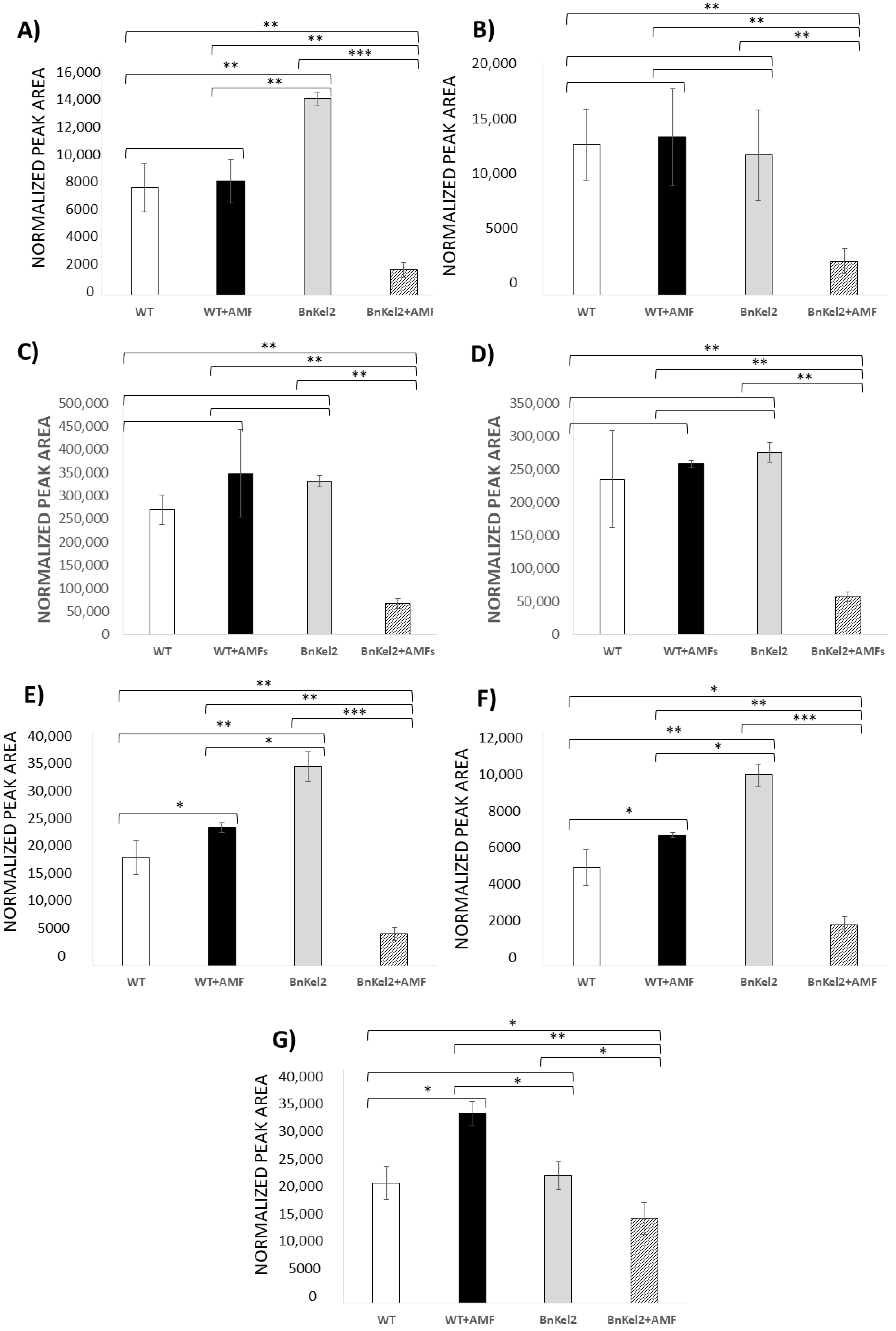
3.4. Reduction of GSL Content in BnKel Roots Inoculated with AMF

Wild-type plants, inoculated or not with AMF, did not show any significant difference in the levels of indole GSL [indole-3 methyl, (I3M), 4-hydroxy indole-3 methyl (4-HO-I3M), 1-methoxy indole-3 methyl (1-MO-I3M) and 4-methoxy indole-3 methyl (4-MO-I3M) GSL], whereas it was observed a significant increase in the content of aliphatic GSL [4-methyl thio butanal (4-MTB) and 5-methyl thio pentanal (5-MTP) GSL] of AMF-inoculated wild-type plants. The levels of both indole and aliphatic GSL significantly decreased in the roots of BnKel2 plants inoculated with AMF (Figure 3A–F). In addition, a significant increase in the levels of a toxic isothiocyanate [4-methoxy indole-3 methyl ITC (4-MO-I3M-ITC)], derived from the hydrolysis of indole GSL, was detected in AMF-inoculated wild-type plants. However, the lowest content of 4-MO-I3M-ITC was observed in AMF-inoculated BnKel2 plants (Figure 3G).

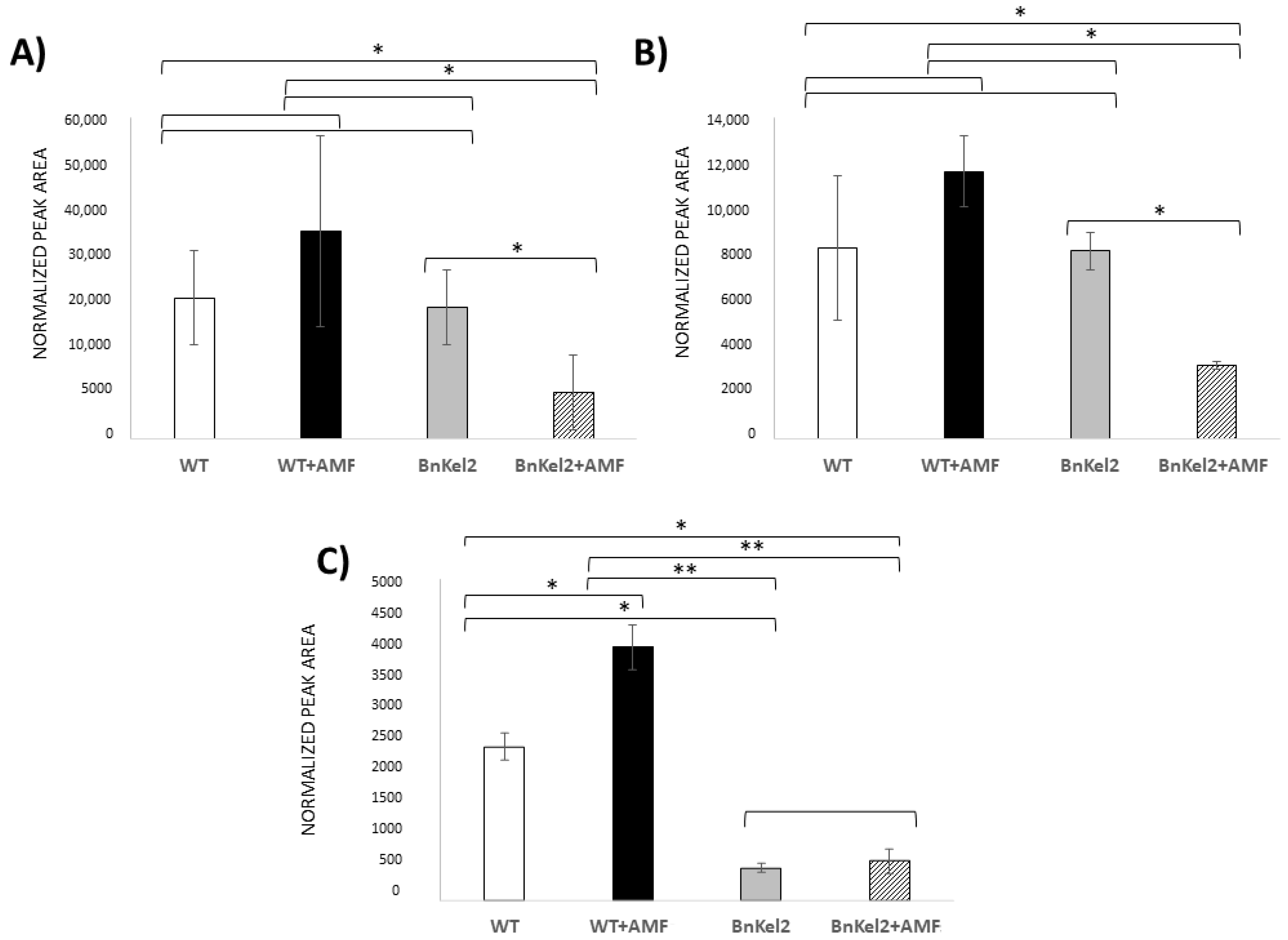
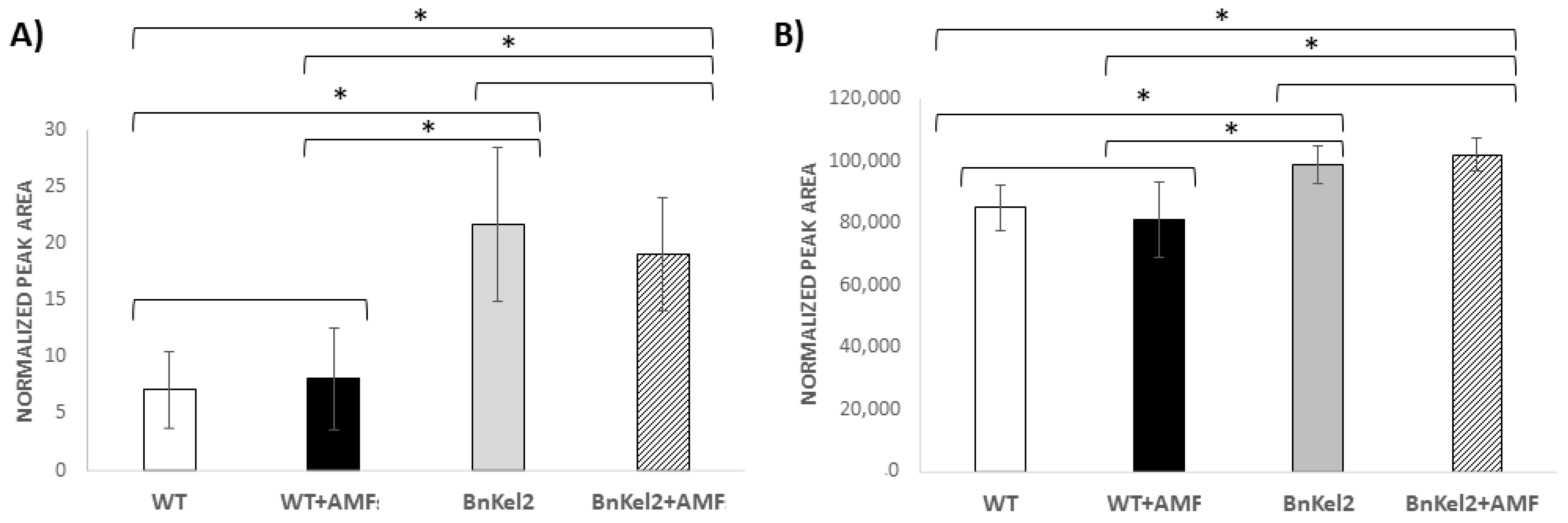
3.5. Reduction of GSL Content in BnKel Seeds and Seed Oil Content

A metabolomic analysis of GSL performed in seeds of wild-type and BnKel2 plants showed a significant increase in the levels of the 4-MO-I3M in seeds of wild-type plants inoculated with AMF. No significant differences were detected in the other GSL determined in wild-type plants. By contrast, a significant decrease in the content of two out of three analyzed GSL was detected in seeds of inoculated BnKel2 plants compared to non-inoculated plants (Figure 4).
Moreover, a significant increase in the levels of octadecatrienoic acid (α-linolenic acid) and glycerophosphocholine was observed in the seeds of BnKel2 plants compared with those of the wild type, regardless of AMF inoculation (Figure 5).

3.6. Rapeseed Seed Yield

A significant increase in seed weight was observed in AMF-inoculated BnKel2 plants compared to those non-inoculated. Regardless of AMF inoculation, seed yield was significantly higher in BnKel2 plants compared with that of the wild type (Table 3). Total seed weight per plant increased by about 1.5 times in the AMF-inoculated BnKel2 plants under optimal growth conditions. Further, shoot dry weight increased when BnKel2 plants were inoculated with AMF compared to wild-type or non-inoculated BnKel2 plants (Supplementary Figure S1). These results indicate that the application of AMF to BnKel plants exerted a beneficial effect on rapeseed yield.

4. Discussion

Many crops, excluding Brassicaceae, can develop mycorrhizal associations that greatly benefit from AMF inoculation [28]. Throughout the evolutionary process, Brassicaceae may have lost their genes related to AMF symbiosis, which seems logical since they were not necessary for their proper development [13]. However, with the present situation of climate change, the need for new technological tools for plant adaptation to adverse environments seems more important than ever. The achievement of an effective beneficial association among mycorrhizas and Brassicaceae plants may be critical in agribiotechnology and opens new opportunities for the use of AMF as biofertilizers in rapeseed crops [6].
Proteins with Kelch domains, including NSP and NSP-like, as ThKEL1, have been associated with the metabolism of GSL and plant defense [2,3,4,19], and have been also related to the mycorrhization process [24,29,30], in a similar way to that observed in legumes nodulation [31]. The Thkel1 gene of T. harzianum T34, which encodes for an NSP-like protein, increases the fungal tolerance to abiotic stress when it is overexpressed in that strain [18]. Furthermore, when Thkel1 was overexpressed in Brassicaceae plants, it played a key role in the T. harzianum root colonization process [24]. These characteristics of the Thkel1 gene have made it a candidate of choice to be transferred to Brassica plants as a biotechnological tool to modify GSL metabolism and perhaps facilitate the interaction among AMF and these plants. To check this out, we challenged Thkel1 overexpressing rapeseed plants with a pre-commercial AMF mixture. No fungal DNA was detected in WT plants (Table 1), but AMF DNA was detected in both BnKel lines, it being indicative that transgenic plants are more sensitive to the presence of AMF symbiotic associations (Figure 1). To support these data, we have obtained similar results regarding the levels of AMF DNA in several independent Arabidopsis thaliana transformants, as well as in transgenic rapeseed plants transformed with Agrobacterium rhizogenes (Supplementary Table S1) [20]. Some studies have shown that different AMFs are able to form colonization points at the roots of some species of Brassicaceae, but without tissue penetration and nutrient exchange [32,33]. More recently, it has been reported that there is some form of interaction between AMF and non-host Arabidopsis plants during the pre-symbiotic stage, whereas the incompatibility appears at later stages, accompanied by an activation of defense responses leading to colonization of the root cortex, but without forming a functional AMF symbiosis [15]. Furthermore, some AMF species can germinate in the presence of roots of wild mustard but without the ability to colonize this Brassicaceae plant [34], which has been found to be due to the formation of allelopathic compounds [35].
Pioneering studies suggested that Brassicaceae plants with low levels of GSL production may establish symbiotic associations with AMF [36]. However, these authors only observed the formation of fungal vesicles in dead cells of the root cortex, concluding that the GSL low content was not the cause of the absence of mycorrhization. Similar results were reported in other ornamental cruciferous plants [37]. Our results show that the application of AMF to BnKel plants increased the P and Fe contents. The higher levels of P content are a known trait in mycorrhizal plants [6,11]. It has been reported that AMF symbiosis can both increase and decrease the Fe uptake, this fact being affected by the fungal species, host plant species and growth conditions [38], where the AMF caused changes in the expression of Fe acquisition and ferric reductase genes [39]. Furthermore, arbuscular formation has been observed in some Brassicaceae species, lacking myrosinase activity and unable to conduct GSL hydrolysis [40]. Another study has also shown that the GSL profile differed among mycorrhizal and non-mycorrhizal Brassicaceae plants. The non-mycorrhizal species accumulated higher levels of the indole GSL 1-MO-I3M and 4-HO-I3M [41], showing a similar profile to that more recently observed in Brassica juncea [42]. In addition, that study pointed out that the type of GSL is more important for the process of colonization by AMF than the quantity of GSL generated [41]. Moreover, it has been reported that the indole GSL pathway metabolites provide resistance to AMF mycorrhization in Arabidopsis [43,44]. In our study, we have observed a reduction of 1-MO-I3M and 4-HO-I3M levels in AMF-BnKel2 plants, as well as a decrease in the isothiocyanate content derived from one of these compounds (4-MO-I3M-ITC) (Figure 3). Thus, the significant reduction of toxic GSL detected in BnKel2 plants could allow them to establish a symbiotic association with AMF. In any case, this decrease is only detected after root–AMF interaction in BnKel plants, although the Thkel1 gene could have a role in this interaction since it has been shown to play an important role during T. harzianum root colonization in Arabidopsis [24].
Recently, the role of GSLs in the control of the crucifer microbiome has been reviewed, highlighting their importance in the absence of effective mycorrhization in this group of plants [44]. Similarly, GSLs are mainly responsible for controlling the fungal diversity of endophytic fungi in crucifers, which is why Brassica crops harbor less endophytic fungal diversity in their roots than other taxonomically distant crops [45].
We have also noted a reduction of the GSL content in seeds (Figure 4). The high concentration of GSL in Brassica seeds reduces the value of the crop and its usefulness for oil extraction [46]. Different approaches to reduce the GSL content in plants have been attempted, albeit without much success, except that performed by mutation of genes encoding GSL transporters [47]. In addition to increased yield and a reduction of GSL content in seeds from BnKel plants, we have also detected changes in the seed oil composition (Figure 4). In this sense, a significant increase in the levels of octadecatrienoic acid, an unsaturated omega-3 fatty acid with important properties for human nutrition, was detected in seeds harvested from BnKel2 plants. This omega-3 fatty acid is considered to have a positive effect on the prevention of cardiovascular disease and obesity and functions as a neuroprotective agent [48]. Another phospholipid metabolism-related compound whose levels were significantly increased in seeds of BnKel2 was glycerophosphocholine (Figure 5), considered important for treating cognitive impairment, as a supplier of the essential dietary amine choline, a precursor of the neurotransmitter acetylcholine, commonly used as a treatment for Alzheimer´s disease and other dementias [49].
Our results show that although true arbuscules were not developed, the application of AMF to BnKel2 plants significantly increases the following parameters: (i) P and Fe contents (Table 2), (ii) total seed weight per plant (Table 3), and (iii) dry weight of the aerial part of the plant (Supplementary Figure S1). These results agree with the increase in yield and product quality observed in mycorrhizal crops [50], and in Brassicaceae plants after the combined application of AMF and T. harzianum [20].
In conclusion, the overexpression of the Thkel1 gene from the fungus T. harzianum in B. napus plants allows transgenic plants to benefit from the presence of arbuscular mycorrhizal fungi at their root rhizosphere, mainly by the modification of the GSL content and their hydrolysis products. The final increase in seed yield and the improvement of seed oil composition are among the most remarkable impacts of these B. napus–AMF interactions, with an impact on the plant’s agronomic value and its potential application in medical, animal and industrial biotechnology.

Supplementary Materials

The following supporting information can be downloaded at: https://www.mdpi.com/article/10.3390/agriculture14060851/s1, Figure S1: Growth of 8-weeks old rapeseed plants in the presence or not of AMF; Table S1: AMF quantification in rapeseed and arabidopsis roots of wild type (WT and Col-0, respectively) and Thkel1 transformed roots with A. rhizogenes (BnArKel1) or A. tumefaciens (AtKel1) plants inoculated with an AMF mixture.

Author Contributions

A.A.-R., J.P., M.C.-P. and J.A. conducted the laboratory work; V.A. performed the metabolomic analysis; C.N., J.P. and M.C.-P. conceived and designed the experiments; C.N., J.P., M.C.-P. and R.H. analyzed the data; C.N., M.C.-P., J.P. and R.H. wrote the paper. All authors have read and agreed to the published version of the manuscript.

Funding

This work was supported by the Regional Government of Castile and Leon, Escalera de Excelencia CLU-2018-04 co-funded by the P.O. FEDER 2014-2020, the Projects SA230U13, SA270P18, SA094P20 and IR2020-1-USAL05, and by the Spanish Government, PDI-2021-126575OB-I00 MCIN/AEI/10.13039/501100011033/FEDER, UE.

Institutional Review Board Statement

Not applicable.

Data Availability Statement

The data generated during this study are available from the corresponding author on reasonable request. Materials will be made available for non-profit research under a material transfer agreement.

Acknowledgments

Authors thank Enrique Monte for kindly providing materials and participating in the development of the project. Julio Ascaso thanks the support of his predoctoral contract by Regional Government of Castile and Leon. Mass spectrometric analyses were carried out at Servei Central d’Instrumentació Científica (SCIC) from Universitat Jaume I.

Conflicts of Interest

A Spanish patent and an international patent with references P201830755 and PCT/EP2019/081029 have been filled (C.N., J.P., A.A.-R., R.H.). No other author has competing interests.

References

  1. Huang, C.H.; Sun, R.; Hu, Y.; Zeng, L.; Zhang, N.; Cai, L.; Zhang, Q.; Koch, M.A.; Al-Shehbaz, I.; Edger, P.P.; et al. Resolution of Brassicaceae phylogeny using nuclear genes uncovers nested radiations and supports convergent morphological evolution. Mol. Biol. Evol. 2015, 33, 394–412. [Google Scholar] [CrossRef] [PubMed]
  2. Cartea, M.E.; Francisco, M.; Soengas, P.; Velasco, P. Phenolic compounds in Brassica vegetables. Molecules 2011, 16, 251–280. [Google Scholar] [CrossRef] [PubMed]
  3. Angelino, D.; Jeffery, E. Glucosinolate hydrolysis and bioavailability of resulting isothiocyanates: Focus on glucoraphanin. J. Funct. Foods 2014, 7, 67–76. [Google Scholar] [CrossRef]
  4. Hassan, M.N.; Zainal, Z.; Ismail, I. Plant Kelch containing F-box proteins: Structure, evolution and functions. RSC Adv. 2015, 5, 42808–42814. [Google Scholar] [CrossRef]
  5. Martínez-Ballesta, M.C.; Carvajal, M. Myrosinase in Brassicaceae: The most important issue for glucosinolate turnover and food quality. Phytochem. Rev. 2015, 14, 1045–1051. [Google Scholar] [CrossRef]
  6. Berruti, A.; Lumini, E.; Balestrini, R.; Bianciotto, V. Arbuscular mycorrhizal fungi as natural biofertilizers: Let’s benefit from past successes. Front. Microbiol. 2016, 6, 1559. [Google Scholar] [CrossRef] [PubMed]
  7. Woo, S.L.; Hermosa, R.; Lorito, M.; Monte, E. Trichoderma: A multipurpose plant beneficial microorganism for eco-sustainable agriculture. Nat. Rev. Microbiol. 2023, 21, 312–326. [Google Scholar] [CrossRef] [PubMed]
  8. Hermosa, R.; Viterbo, A.; Chet, I.; Monte, E. Plant-beneficial effects of Trichoderma and of its genes. Microbiology 2012, 158, 17–25. [Google Scholar] [CrossRef] [PubMed]
  9. Mendoza-Mendoza, A.; Zaid, R.; Lawry, R.; Hermosa, R.; Monte, E.; Horwitz, B.A.; Mukherjee, P.K. Molecular dialogues between Trichoderma and roots: Role of the fungal secretome. Fungal Biol. Rev. 2018, 32, 62–85. [Google Scholar] [CrossRef]
  10. Nicolás, C.; Hermosa, R.; Rubio, B.; Mukherjee, P.K.; Monte, E. Trichoderma genes in plants for stress tolerance-status and prospects. Plant Sci. 2014, 228, 71–78. [Google Scholar] [CrossRef]
  11. MacLean, A.M.; Bravo, A.; Harrison, M.J. Plant signaling and metabolic pathways enabling arbuscular mycorrhizal symbiosis. Plant Cell 2017, 29, 2319–2335. [Google Scholar] [CrossRef]
  12. Roth, R.; Paszkowski, U. Plant carbon nourishment of arbuscular mycorrhizal fungi. Curr. Opin. Plant Biol. 2017, 39, 50–56. [Google Scholar] [CrossRef]
  13. Delaux, P.M. Comparative phylogenomics of symbiotic associations. New Phytol. 2017, 213, 89–94. [Google Scholar] [CrossRef] [PubMed]
  14. Chen, M.; Bruisson, S.; Bapaume, L.; Darbon, G.; Glauser, G.; Schorderet, M.; Reinhardt, D. VAPYRIN attenuates defence by repressing PR gene induction and localized lignin accumulation during arbuscular mycorrhizal symbiosis of Petunia hybrida. New Phytol. 2021, 229, 3481–3496. [Google Scholar] [CrossRef] [PubMed]
  15. Fernández, I.; Cosme, M.; Stringlis, I.A.; Yu, K.; de Jonge, R.; van Wees, S.M.; Pozo, M.J.; Pieterse, C.M.J.; van der Heijden, M.G.A. Molecular dialogue between arbuscular mycorrhizal fungi and the nonhost plant Arabidopsis thaliana switches from initial detection to antagonism. New Phytol. 2019, 223, 867–881. [Google Scholar] [CrossRef]
  16. Cosme, M.; Fernández, I.; Van der Heijden, M.G.A.; Pieterse, C.M.J. Non-mycorrhizal plants: The exceptions that prove the rule. Trends Plant Sci. 2018, 23, 577–587. [Google Scholar] [CrossRef]
  17. Cosme, M.; Fernández, I.; Declerck, S.; Van der Heijden, M.G.A.; Pieterse, C.M.J. A coumarin exudation pathway mitigates arbuscular mycorrhizal incompatibility in Arabidopsis thaliana. Plant Mol. Biol. 2021, 106, 319–334. [Google Scholar] [CrossRef]
  18. Hermosa, R.; Botella, L.; Keck, E.; Jiménez, J.Á.; Montero-Barrientos, M.; Arbona, V.; Gómez-Cadenas, A.; Monte, E.; Nicolás, C. The overexpression in Arabidopsis thaliana of a Trichoderma harzianum gene that modulates glucosidase activity, and enhances tolerance to salt and osmotic stresses. J. Plant Physiol. 2011, 168, 1295–1302. [Google Scholar] [CrossRef] [PubMed]
  19. Gumz, F.; Krausze, J.; Eisenschmidt, D.; Backenköhler, A.; Barleben, L.; Brandt, W.; Wittstock, U. The crystal structure of the thiocyanate-forming protein from Thlaspi arvense, a kelch protein involved in glucosinolate breakdown. Plant Mol. Biol. 2015, 89, 67–81. [Google Scholar] [CrossRef] [PubMed]
  20. Poveda, J.; Hermosa, R.; Monte, E.; Nicolás, C. Trichoderma harzianum favours the access of arbuscular mycorrhizal fungi to non-host Brassicaceae roots and increases plant productivity. Sci. Rep. 2019, 9, 11650. [Google Scholar] [CrossRef]
  21. Vierheilig, H.; Schweiger, P.; Brundrett, M. An overview of methods for the detection and observation of arbuscular mycorrhizal fungi in roots. Physiol. Plant. 2005, 125, 393–404. [Google Scholar] [CrossRef]
  22. Lee, J.; Lee, S.; Young, J.P.W. Improved PCR primers for the detection and identification of arbuscular mycorrhizal fungi. FEMS Microbiol. Ecol. 2008, 65, 339–349. [Google Scholar] [CrossRef] [PubMed]
  23. Yang, H.; Liu, J.; Huang, S.; Guo, T.; Deng, L.; Hua, W. Selection and evaluation of novel reference genes for quantitative reverse transcription PCR (qRT-PCR) based on genome and transcriptome data in Brassica napus L. Gene 2014, 538, 113–122. [Google Scholar] [CrossRef] [PubMed]
  24. Poveda, J.; Hermosa, R.; Monte, E.; Nicolás, C. The Trichoderma harzianum Kelch protein ThKEL1 plays a key role in root colonization and the induction of systemic defense in Brassicaceae plants. Front. Plant Sci. 2019, 10, 1478. [Google Scholar] [CrossRef] [PubMed]
  25. Illescas, M.; Rubio, M.B.; Hernández-Ruiz, V.; Morán-Diez, M.E.; Martínez de Alba, A.E.; Nicolás, C.; Monte, E.; Hermosa, R. Effect of inorganic top dressing and Trichoderma harzianum seed-inoculation on crop yield and the shaping of root microbial communities of wheat plants cultivated under high basal N fertilization. Front. Plant Sci. 2020, 11, 575861. [Google Scholar] [CrossRef] [PubMed]
  26. Livak, K.J.; Schmittgen, T.D. Analysis of relative gene expression data using real-time quantitative PCR and the 2−ΔΔCT method. Methods 2001, 25, 402–408. [Google Scholar] [CrossRef] [PubMed]
  27. De Ollas, C.; González-Guzmán, M.; Pitarch, Z.; Matus, J.T.; Candela, H.; Rambla, J.L.; Granell, A.; Gómez-Cadenas, A.; Arbona, V. Identification of ABA-mediated genetic and metabolic responses to soil flooding in tomato (Solanum lycopersicum L. Mill). Front. Plant Sci. 2021, 12, 613059. [Google Scholar] [CrossRef] [PubMed]
  28. Hart, M.M.; Trevors, J.T. Microbe management: Application of mycorrhyzal fungi in sustainable agriculture. Front. Ecol. Environ. 2005, 3, 533–539. [Google Scholar] [CrossRef]
  29. Xu, M.J.; Wang, H.Z. Proteomic analysis reveals the mechanisms of Mycena dendrobii promoting transplantation survival and growth of tissue culture seedlings of Dendrobium officinale. J. Appl. Microbiol. 2015, 118, 1444–1455. [Google Scholar] [CrossRef]
  30. Sugimura, Y.; Saito, K. Comparative transcriptome analysis between Solanum lycopersicum L. and Lotus japonicus L. during arbuscular mycorrhizal development. J. Soil Sci. Plant Nutr. 2017, 63, 127–136. [Google Scholar] [CrossRef]
  31. Takahara, M.; Magori, S.; Soyano, T.; Okamoto, S.; Yoshida, C.; Yano, K.; Sato, S.; Tabata, S.; Yamaguchi, K.; Shigenobu, S.; et al. Too much love, a novel Kelch repeat-containing F-box protein, functions in the long-distance regulation of the legume-Rhizobium symbiosis. Plant Cell Physiol. 2013, 54, 433–447. [Google Scholar] [CrossRef] [PubMed]
  32. Ocampo, J.A. Vesicular-arbuscular mycorrhizal infection of “host” and “non-host” plants: Effect on the growth responses of the plants and competition between them. Soil Biol. Biochem. 1986, 18, 607–610. [Google Scholar] [CrossRef]
  33. Kabir, Z.; O’Halloran, I.P.; Hamel, C. The proliferation of fungal hyphae in soils supporting mycorrhizal and non-mycorrhizal plants. Mycorrhiza 1997, 6, 477–480. [Google Scholar] [CrossRef]
  34. Schreiner, R.; Koide, R.T. Antifungal compounds from the roots of mycotrophic and non-mycotrophic plant species. New Phytol. 1993, 123, 99–105. [Google Scholar] [CrossRef]
  35. Aguilar, R.; Carreón-Abud, Y.; López-Carmona, D.; Larsen, J. Organic fertilizers alter the composition of pathogens and arbuscular mycorrhizal fungi in maize roots. J. Phytopathol. 2017, 165, 448–454. [Google Scholar] [CrossRef]
  36. Glenn, M.G.; Chew, F.S.; Williams, P.H. Influence of glucosinolate content of Brassica (Cruciferae) roots on growth of vesicular–arbuscular mycorrhizal fungi. New Phytol. 1998, 110, 217–225. [Google Scholar] [CrossRef]
  37. DeMars, B.G.; Boerner, R.E. Arbuscular mycorrhizal development in three crucifers. Mycorrhiza 1995, 5, 405–408. [Google Scholar] [CrossRef]
  38. Liu, Y.; Xiong, Z.; Wu, W.; Ling, H.Q.; Kong, D. Iron in the symbiosis of plants and microorganisms. Plants 2023, 12, 1958. [Google Scholar] [CrossRef] [PubMed]
  39. Kabir, A.H.; Debnath, T.; Das, U.; Prity, S.A.; Haque, A.; Rahman, M.M.; Parvez, M.S. Arbuscular mycorrhizal fungi alleviate Fe-deficiency symptoms in sunflower by increasing iron uptake and its availability along with antioxidant defense. Plant Physiol. Biochem. 2020, 150, 254–262. [Google Scholar] [CrossRef]
  40. Pongrac, P.; Vogel-Mikuš, K.; Poschenrieder, C.; Barcelo, J.; Tolra, R.; Regvar, M. Arbuscular mycorrhiza in glucosinolate-containing plants: The story of the metal hyperaccumulator Noccaea (Thlaspi) praecox (Brassicaceae). In Molecular Microbial Ecology of the Rhizosphere; de Brujin, F.J., Ed.; Wiley: Hoboken, NJ, USA, 2013; pp. 1023–1032. [Google Scholar]
  41. Vierheilig, H.; Bennett, R.; Kiddle, G.; Kaldorf, M.; Ludwig-Müller, J. Differences in glucosinolate patterns and arbuscular mycorrhizal status of glucosinolate-containing plant species. New Phytol. 2000, 146, 343–352. [Google Scholar] [CrossRef]
  42. Cuong, D.M.; Kim, J.K.; Bong, S.J.; Baek, S.A.; Jeon, J.; Park, J.S.; Park, S.U. Comparative analysis of glucosinolates and metabolite profiling of green and red mustard (Brassica juncea) hairy roots. 3 Biotech. 2018, 8, 382. [Google Scholar] [CrossRef] [PubMed]
  43. Anthony, M.A.; Celenza, J.L.; Armstrong, A.; Frey, S.D. Indolic glucosinolate pathway provides resistance to mychorrhizal fungal colonization in a non-host Brassicaceae. Ecosphere 2020, 11, e03100. [Google Scholar] [CrossRef]
  44. Sharma, A.; Sinharoy, S.; Bisht, N.C. The mysterious non-arbuscular mycorrhizal status of Brassicaceae species. Environ. Microbiol. 2023, 25, 917–930. [Google Scholar] [CrossRef] [PubMed]
  45. Poveda, J.; Díaz-González, S.; Díaz-Urbano, M.; Velasco, P.; Sacristán, S. Fungal endophytes of Brassicaceae: Molecular interactions and crop benefits. Front. Plant Sci. 2022, 13, 932288. [Google Scholar] [CrossRef] [PubMed]
  46. Tayo, T.; Dutta, N.; Sharma, K. Effect of feeding canola-quality rapeseed mustard meal on animal production—A review. Agri. Rev. 2012, 33, 114–121. [Google Scholar]
  47. Nour-Eldin, H.H.; Madsen, S.R.; Engelen, S.; Jørgensen, M.E.; Olsen, C.E.; Andersen, J.S.; Seynnaeve, D.; Verhoye, T.; Fulawka, R.; Denolf, P.; et al. Reduction of antinutritional glucosinolates in Brassica oilseeds by mutation of genes encoding transporters. Nat. Biotechnol. 2017, 35, 377–382. [Google Scholar] [CrossRef] [PubMed]
  48. Egert, S.; Baxheinrich, A.; Lee-Barkey, Y.H.; Tschoepe, D.; Stehle, P.; Stratmann, B.; Wahrburg, U. Effects of a hypoenergetic diet rich in α-linolenic acid on fatty acid composition of serum phospholipids in overweight and obese patients with metabolic syndrome. Nutrition 2018, 49, 74–80. [Google Scholar] [CrossRef] [PubMed]
  49. Scapicchio, P.L. Revisiting choline alphoscerate profile: A new, perspective, role in dementia? Int. J. Neurosci. 2013, 123, 444–449. [Google Scholar] [CrossRef]
  50. Baum, C.; El-Tohamy, W.; Gruda, N. Increasing the productivity and product quality of vegetable crops using arbuscular mycorrhizal fungi: A review. Sci. Hortic. 2015, 187, 131–141. [Google Scholar] [CrossRef]
Figure 1. Microscopic analyses of the rudimentary fungal structures found in inoculated BnKel2 rapeseed roots. Wild-type (A) and BnKel2 roots (B) inoculated with AMF were visualized by light microscopy in plants stained with ink and by confocal microscopy in plants stained with the WGA-Alexa Fluor 488 ((C), wild-type, and (D,E), BnKel2 plants). (Ar: arbuscule; Xv, xylem vessels).
Agriculture 14 00851 g001
Figure 2. AMF GintPT and GintAMT2 transcript levels in inoculated and non-inoculated roots of Brassica napus wild-type and BnKel2 transgenic lines. Data are the mean (±SD) of three biological replicates for each type of plant (wild-type and BnKel transgenic lines) and inoculation condition (n = 3). Data, obtained from 16-week-old plants, represent the relative quantity (RQ, 2−ΔΔCt) of target genes compared to the quantity of the reference gene 18S rRNA. n.d.: not detected.
Agriculture 14 00851 g002
Figure 3. Relative quantification of the GSL present in rapeseed roots. (A) Indole-3 methyl (I3M); (B) 4-hydroxy indole-3 methyl (4-HO-I3M); (C) 1-methoxy indole-3 methyl (1-MO-I3M); (D) 4-methoxy indole-3 methyl (4-MO-I3M); (E) 4-methyl thio butanal (4-MTB); (F) 5-methyl thio pentanal (5-MTP); (G) 4-4-methoxy indole-3 methyl ITC (4-MO-I3M-ITC). Wild-type (WT) and Thkel1 transgenic line 2 (BnKel2) inoculated with AMF (+AMF). Values represent the means (±SD) of three biological replicates for each condition (n = 3) and five technical replicates each. Two-way analysis of variance (ANOVA) was performed, followed by Sidak’s multiple comparison test, indicating significant differences among treatment means as follows: * p < 0.05; ** p < 0.01; *** p < 0.001.
Agriculture 14 00851 g003
Figure 4. Relative quantification of the GSL present in rapeseed seeds. (A) 4-methyl thio butanal (4-MTB); (B) 5-methyl thio pentanal (5-MTP); (C) 4-methoxy indole-3 methyl ITC (4-MO-I3M-ITC). Wild-type (WT) and Thkel1 transgenic line 2 (BnKel2) inoculated with AMF (+AMF). Values represent the means (±SD) of three biological replicates for each condition (n = 3) and five technical replicates each. Two-way analysis of variance (ANOVA) was performed, followed by Sidak’s multiple comparison test, indicating significant differences among treatment means as follows: * p < 0.05; ** p < 0.01.
Agriculture 14 00851 g004
Figure 5. Relative quantification of the lipids and lipid-related compounds in the oils of rapeseed seeds. (A) Octadecatrienoic acid; (B) glycerophosphocholine. Wild-type (WT) and Thkel1 transgenic line 2 (BnKel2) inoculated with AMF (+AMF). Values represent the means (±SD) of three biological replicates for each condition (n = 3), and five technical replicates each. Two-way analysis of variance (ANOVA) was performed, followed by Sidak’s multiple comparison test. Asterisks denote significant differences among treatment means at p ≤ 0.05.
Agriculture 14 00851 g005
Table 1. AMF quantification in rapeseed roots of wild-type (WT) and Thkel1 transgenic lines 1 (BnKel1) and 2 (BnKel2) plants inoculated with a pre-commercial AMF mixture.
AMF Inoculated PlantsPlantAMFRatio 3
CtSDQty 1CtSDQty 2
WT21.280.050.84-0.00.00 c
BnKel120.750.021.1927.530.120.780.65 ± 0.03 b
BnKel220.980.030.9026.380.220.670.74 ± 0.04 a
1 Quantity of plant DNA (ng) referred to rapeseed actin gene. 2 Quantity of fungi DNA (ng) referred to AMF 18S rRNA. 3 Proportion of fungal DNA vs. plant DNA. Values represent the mean (±SD) of three biological replicates for each condition (n = 3) and five technical replicates each. Statistically significant differences were determined after one-way analysis of variance (ANOVA) followed by Tukey’s test and showed with different letters (p < 0.05). (-): Absence of amplification.
Table 2. Phosphorus (P) and iron (Fe) content in rapeseed leaves, non-inoculated or inoculated with AMF fungi, in wild-type plants (WT or WT + AMF) or plants overexpressing the Thelk1 gene from Trichoderma (BnKel2 or BnKel2 + AMF).
PlantsP 1Fe 2
WT2.990 ± 0.063 c82.075 ± 5.655 bc
WT + AMF2.592 ± 0.242 d71.715 ± 4.815 c
BnKel23.715 ± 0.017 b86.218 ± 7.765 b
BnKel2 + AMF4.994 ± 0.496 a138.379 ± 18.993 a
Wild-type (WT) and Thkel1 transgenic line 2 (BnKel2) inoculated with AMF (+AMF). Values represent the mean (±SD) of three biological replicates for each condition (n = 3) and three technical replicates each. Different letters in the same column indicate significative differences among treatment means according to Tukey’s test (p < 0.05). 1 Data in mg g−1 of plant tissue. 2 Data in mg kg−1 of plant tissue.
Table 3. Seed weight (g) per rapeseed plant under optimal growth conditions (control). Wild-type plants (WT) and plants overexpressing the Thelk1 gene from Trichoderma (BnKel2) were non-inoculated or inoculated with AMF (WT + AMF or BnKel2 + AMF).
PlantsSeed Weight (g)
WT61.835 ± 6.226 c
WT + AMF53.653 ± 7.665 c
BnKel299.526 ± 9.678 b
BnKel2 + AMF155.596 ± 12.966 a
Values represent the mean (±SD) of three biological replicates for each condition (n = 3), and 15 technical replicates each. One-way analysis of variance (ANOVA) was performed, followed by Tukey’s test. Different letters indicate significant differences between treatment means at p < 0.05.
Disclaimer/Publisher’s Note: The statements, opinions and data contained in all publications are solely those of the individual author(s) and contributor(s) and not of MDPI and/or the editor(s). MDPI and/or the editor(s) disclaim responsibility for any injury to people or property resulting from any ideas, methods, instructions or products referred to in the content.

