Article

Genetic Diversity and Zoonotic Potential of Shiga Toxin-Producing E. coli (STEC) in Cattle and Buffaloes from Islamabad, Pakistan

1
Animal Health Program, Animal Sciences Institute, National Agricultural Research Centre, Park Road, Islamabad 45500, Pakistan
2
Faculty of Veterinary and Animal Sciences, Pir Mehr Ali Shah Arid Agriculture University, Rawalpindi 46000, Pakistan
3
College of Public Health Sciences, Chulalongkorn University, Bangkok 10330, Thailand
4
Omics Sciences and Bioinformatics Centre, Chulalongkorn University, Bangkok 10330, Thailand
5
Alpha Genomics Private Limited, Islamabad 45710, Pakistan
6
State Key Laboratory of Chinese Medicine Modernization, Tianjin University of Traditional Chinese Medicine, Tianjin 301617, China
7
Key State Laboratory of Component-Based Medicine, Tianjin University of Traditional Chinese Medicines, Tianjin 301617, China
8
Haihe Laboratory of Modern Chinese Medicine, Tianjin 301617, China
9
Department of Genomics and Bioinformatics, Cholistan University of Veterinary and Animal Sciences, Bahawalpur 63100, Pakistan
10
National Academies of Sciences, Washington, DC 20001, USA
11
Livestock and Dairy Development Department, Muzaffarabad 13100, Pakistan
12
Animal Population Health Institute, Department of Clinical Sciences, College of Veterinary, Medicine and Biomedical Sciences, Colorado State University, Fort Collins, CO 80523, USA
13
Animal Health and Production Module, Regional Office for Asia and the Pacific, Food and Agriculture Organization of the United Nations, Bangkok 10200, Thailand
*
Author to whom correspondence should be addressed.
Agriculture 2024, 14(9), 1537; https://doi.org/10.3390/agriculture14091537 (registering DOI)
Submission received: 2 August 2024 / Revised: 30 August 2024 / Accepted: 2 September 2024 / Published: 6 September 2024
(This article belongs to the Section Farm Animal Production)

Abstract

:
Shiga toxin-producing E. coli (STEC) are considered important zoonotic pathogens of great economic significance, associated with diarrhea, hemolytic uremic syndrome (HUS), hemorrhagic colitis (HC), and death in humans. This study aimed to investigate the distribution of various STEC virulence gene markers and antimicrobial susceptibility (AST) profiles associated within E. coli isolates from the recto-anal mucosal swabs (RAMSs) of slaughtered cattle and buffaloes in Islamabad, Pakistan. The RAMSs (n = 200) were analyzed using multiplex PCR for the presence of stx1, stx2, eae, and ehxA genes. Samples that were positive for one or more of the virulence genes were inoculated with Sorbitol MacConkey agar (SMAC) for isolation of STEC. The isolates were further analyzed for the presence of virulence genes using multiplex PCR. Of the 200 RAMS, 118 (59%) were positive for one or more virulence genes. E. coli isolates (n = 18) with one or more virulence genes were recovered from the 118 positive samples. The DNA of the isolates positive for one or more virulent genes was extracted and subjected to whole genome sequencing using Illumina. Analysis of the WGS data indicated that the E. coli isolates could be differentiated into 11 serotypes. Most E. coli isolates (13/18; 72.2%) carried five genes (stx1, stx2, Iha, iss, and IpfA) in various combinations. In addition to these five genes, other virulence genes identified in these isolates were espI, ireA, espP, exhA, epeA, mcmA, mch, ast, celB, eilA, katP, and capU. The AST was performed using the Kirby–Bauer disk diffusion test. The study indicated that all the isolates were resistant to rifampicin and a significant proportion of the isolates were MDR. A wide range of antimicrobial resistance genes (ARGs) were detected among the isolates, reflecting the complex nature of resistance mechanisms. The study results indicate that cattle and buffaloes slaughtered in Islamabad might be the carriers of antimicrobial resistant STEC of zoonotic significance, thus representing a source of human infection.

1. Introduction

Shiga toxin-producing E. coli (STEC) are considered important zoonotic pathogens of economic significance which are transmitted through ingestion of contaminated food and water or direct contact with infected animals [1,2,3,4]. Person to person transmission of STEC has also been reported [5]. STEC can cause various clinical manifestations, including diarrhea, haemolytic uremic syndrome (HUS), haemorrhagic colitis (HC), and death in humans [6]. The first STEC-associated human case was reported in 1983 [7].
STEC exhibits many virulence factors, but the most important are Shiga toxins (Stx) encoded by the stx genes [8]. STEC containing the stx2 gene are considered more virulent due to their association with HC and HUS cases in humans [9,10,11]. Moreover, various subtypes of stx1 (stx1a to stx1d) and stx2 (stx2a to stx2g) have been reported in different STEC serotypes [11,12]. In addition to stx, other virulence markers also play a significant role in virulence. The eae gene, located on the locus of enterocyte effacement (LEE), encodes intimin, a crucial protein that facilitates the attachment and effacement (A/E) mechanism of E. coli, enabling it to bind to and alter the intestinal epithelial lining [13]. Other adhesion-related genes have also been reported, including efa1 (EHEC factor for adherence 1), iha (IrgA homolog adhesin), paa (porcine A/E associated protein), and saa (STEC autoagglutinating adhesin) [14,15]. The astA gene encodes a structurally related toxin to the heat-stable enterotoxin of enterotoxigenic E. coli [16]. Plasmid-associated virulence markers such as enterohemolysin (ehxA), catalase-peroxidase (katP), and serine protease (espP) also help STEC in the colonization of the human intestinal tract [10,17].
The widespread utilization of antimicrobial agents in food animals has contributed to the emergence of antimicrobial resistance (AMR) bacteria. These AMR bacteria have emerged as a significant challenge to public health, especially due to the emergence multidrug resistance (MDR) bacteria. Several recent investigations reported the emergence of multidrug-resistant bacterial pathogens from different origins that increase the necessity of the proper use of antibiotics. Previous studies have reported the occurrence of MDR in STEC [18,19,20].
Ruminants, including cattle and buffalo, act as STEC reservoirs [21,22,23,24]. Previous studies from various countries have reported the isolation of STEC from feces and the meat of ruminants [24,25,26,27]. In addition to the public health concern, STEC is also an important economic concern, as the USA has a zero-tolerance policy for STEC O157, O26, O45, O103, O111, O121, and O145 in beef (https://www.fsis.usda.gov/policy/fsis-directives/10010.2; accessed on 20 June 2023). Annual per capita consumption of meat in Pakistan is 16 kg/person/year (https://www.fao.org/faostat/en/; accessed on 20 June 2023). In 2020–21 Pakistan exported more than USD 250 million worth of meat to seventeen countries (https://www.fao.org/faostat/en/#data/TM; accessed on 21 June 2023). Currently, these countries do not have STEC regulations like the USA but may enact them in the future. Therefore, Pakistan must prepare the livestock industry to meet this challenge. However, limited information is available on the occurrence of STEC and their various serotypes in slaughtered animals in Pakistan. Pakistan is one of the several developing countries where this type of information is essential for tackling human and animal health issues related to STEC. However, collecting reliable and useful samples from slaughter facilities in developing countries is difficult from a logistical and cultural perspective. Exploring a research design to obtain this information is useful for both public health decision-makers and relevant researchers.
To our knowledge, this is the first study to estimate the occurrence of various types of E. coli from recto-anal mucosal swabs (RAMSs) collected from slaughtered cattle and buffaloes in Islamabad, Pakistan, as well as the first study to estimate the distribution of various virulence determinants, including stx1, stx2, eae, and ehxA in these isolates. This field and laboratory approach can support existing information in identifying the zoonotic potential of STEC isolates and the potential role of slaughtered cattle and buffaloes in transmitting STEC to humans.

2. Materials and Methods

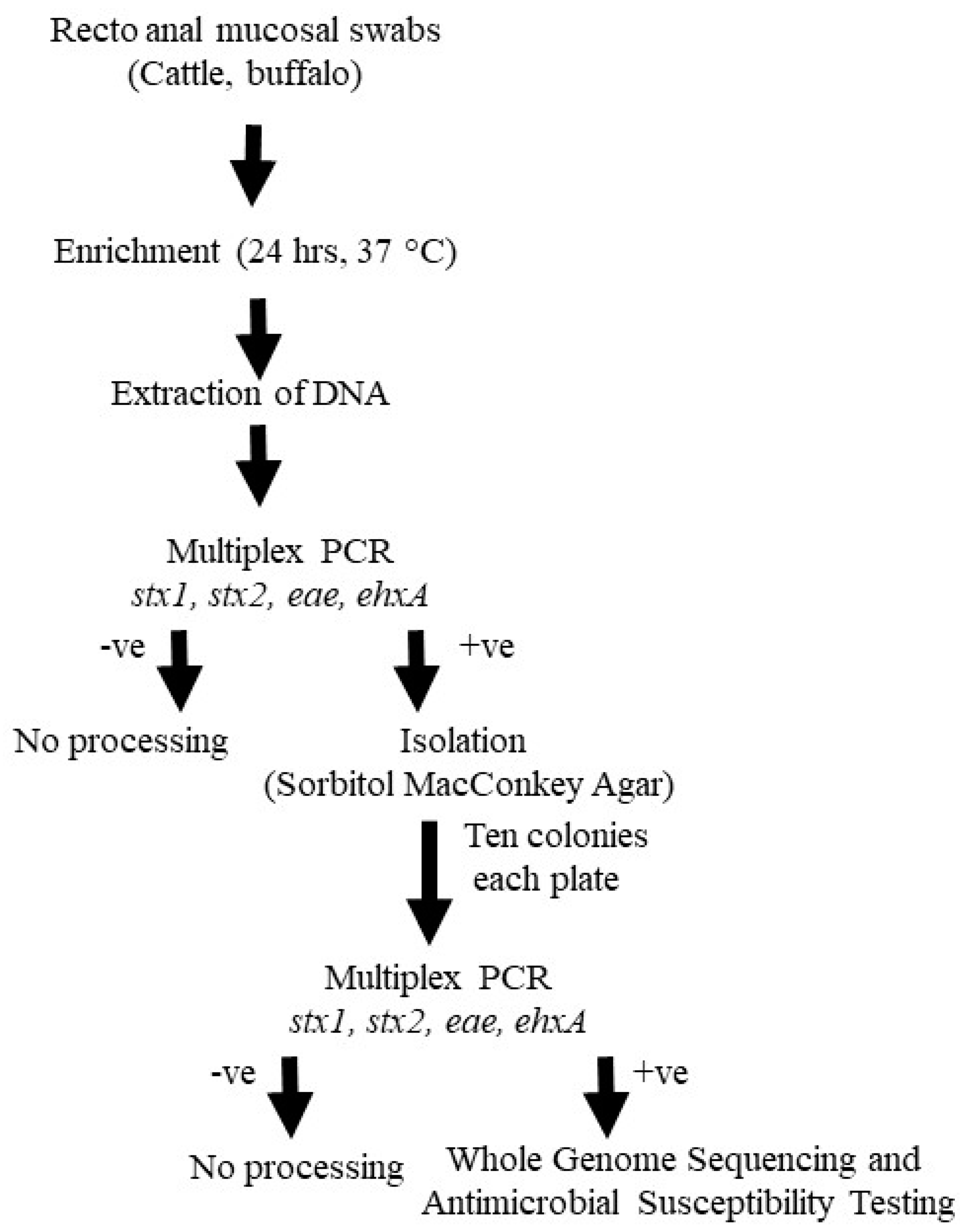
2.1. Study Population and Sample Collection

Cattle and buffaloes from various farms in the Punjab province were transported together to the slaughterhouse. Most of the facility’s 700–800 animals slaughtered daily were culled dairy animals. Two hundred recto-anal mucosal swabs (RAMSs) were collected from cattle (n = 104) and buffaloes (n = 96) in 2016 and 2017 during the slaughter process in a slaughterhouse in Islamabad, Pakistan. The RAMSs were taken by rotating a plastic cotton-tipped swab inside the first 2–5 cm of the rectal mucosa of the selected carcass, then placing it into the transport medium (Amies) and keeping it chilled. Samples were transported to the laboratory in a cool box containing ice gel packs under cold conditions. Samples were collected from a single slaughterhouse visited once a month for eight months. Twenty-five animals were selected systematically (every 10th animal) during each visit for RAMS collection.

2.2. Isolation and Identification of STEC

The samples of RAMSs were enriched in buffered peptone water (BPW) in the laboratory at 37 °C for 24 h and then processed for DNA extraction [28]. Briefly, 1 mL of enriched broth was centrifuged at 12,000× g for 2 min. After discarding the supernatant, the pellet was re-suspended in 500 mL of sterile distilled water and DNA was extracted at 95 °C for 10 min. After cooling at 4 °C, the bacterial suspension was re-centrifuged at 12,000× g for 2 min. The supernatant containing the DNA was transferred to another tube. The DNA was analyzed using multiplex PCR for the presence of four virulence genes, including stx1, stx2, eae, and ehxA [29,30]. Positive samples for one or more virulence genes were subjected to the isolation of STEC using Sorbitol MacConkey agar (SMAC). Ten isolates from each plate (sorbitol fermenting and sorbitol non-fermenting isolates) were analyzed for the presence of stx1, stx2, eae, and ehxA using multiplex PCR. The positive isolates for one or more virulence genes were confirmed as E. coli using biochemical tests, including indole, methyl red, Voges Proskauer, the citrate utilization test, and triple sugar iron (Oxoid, UK). The isolates found positive for indole, methyl red and triple sugar iron and negative for Voges Proskauer and citrate were identified as E. coli [31]. The DNA of the isolates was extracted using a GeneJet Genomic DNA purification kit (Thermoscientific, Vilnius, Lithuania). The summary of methodology used for the processing of samples for isolation and identification of STEC is given in Figure 1.

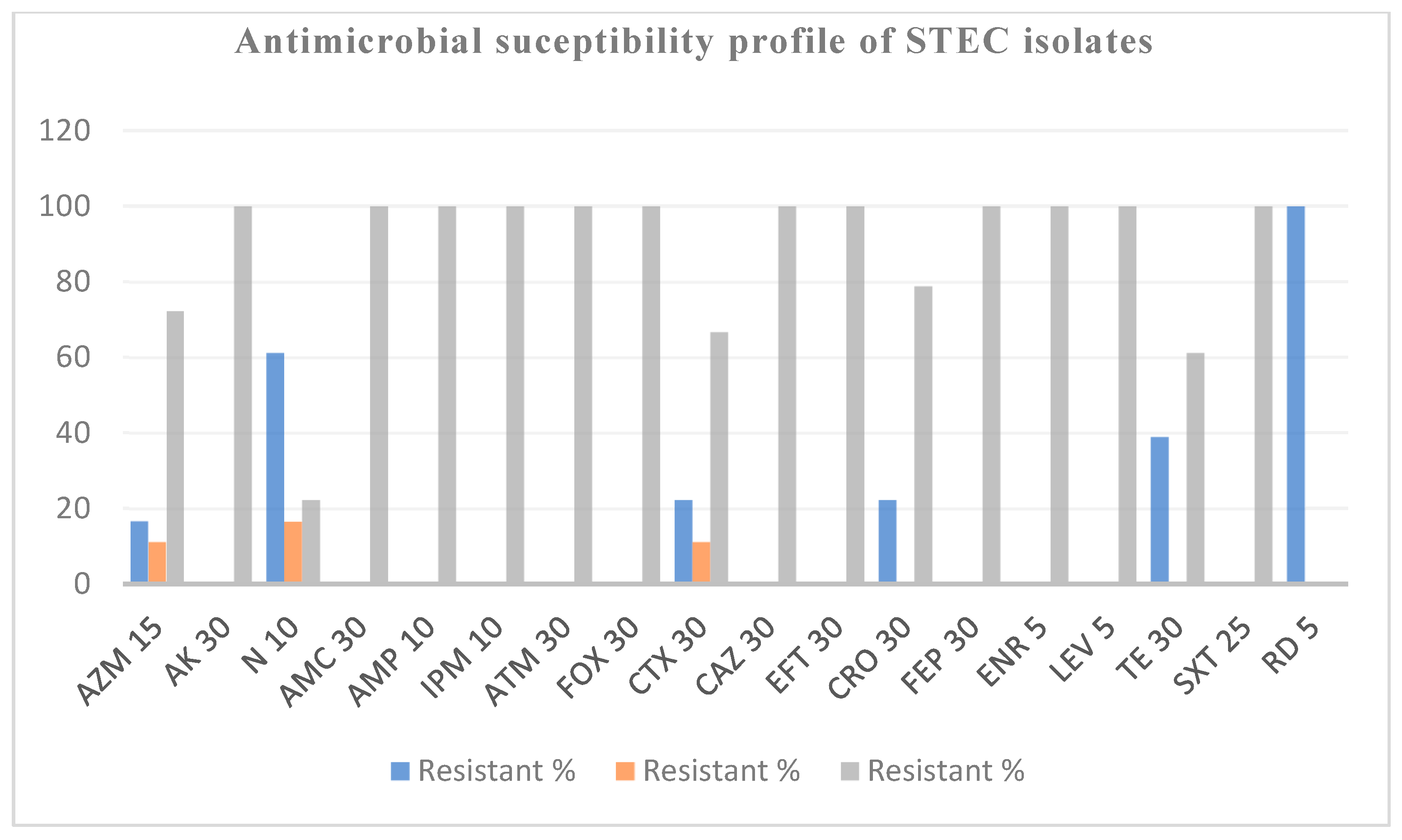
2.3. Antimicrobial Susceptibility Testing of STEC

STEC isolates were subjected to antimicrobial susceptibility testing using the Kirby–Bauer disk diffusion test following the recommendations of the Clinical Laboratory Standard Institute (CLSI) [32]). A total of 18 different antibiotics from 10 different antibiotic classes were tested for STEC isolates during the study. These antibiotics were selected due to their clinical relevance in veterinary and human health [33]. The details of the antibiotic panel used for STEC isolates is given in the Supplementary Materials (Supplementary Table S1). Briefly, 2–3 well-isolated colonies from the identified pure STEC cultures were emulsified into 5 mL of sterile normal saline solution, and the turbidity of the solution was adjusted to that of the 0.5 McFarland standard. A sterile cotton swab was immersed in normal saline suspension and uniformly streaked onto a Mueller–Hinton agar medium (Oxoid, Hampshire, UK) plate. Following this, antibiotic disks (Oxoid, Hampshire, UK) containing a specific concentration of antibiotics were placed on the agar surface, and the plates were incubated at 37 °C for 24 h. The interpretation of the zone of inhibition was carried out following the guidelines of CLSI [32]). The exact breakpoints used for the analysis of AST data are mentioned in Supplementary Table S2. Additionally, the antimicrobial susceptibility testing (AST) data were comprehensively analyzed to characterize the STEC isolates as multidrug resistant (MDR: resistant to ≥one agent in ≥3 antimicrobial classes) and extensively drug-resistant (XDR: resistant to one or more antibiotics in all tested classes, except 1 or 2 classes), as previously described [34]. Moreover, a multiple antibiotic resistance index (MAR) was calculated using (MAR = No. of antibiotics resistant/No. of antibiotics tested), an already established method [35,36].

2.4. Whole Genome Sequencing and Sequence Analysis

The DNA samples were analyzed at Omics Sciences and Bioinformatics Center, Bangkok, Thailand, using whole genome sequencing (WGS). The library preparation was carried out with QIAGEN FX kit (QIAGEN, Hilden, Germany) using 100 ng of the DNA. Briefly, DNA was fragmented using an enzymatic reaction and cleaned with magnetic beads. An adaptor index was ligated to the fragmented DNA. The quality and quantity of the indexed libraries were measured using Agilent 2100 Bioanalyzer (Agilent Technologies, Santa Clara, CA, USA) and a Denovix fluorometer and pooled in an equimolar quantity. Cluster generation and paired-end sequencing were performed with the 150 bp short read length using the Illumina MiSeq sequencer (Illumina, Inc., San Diego, CA, USA).
The quality of raw reads was checked using FASTQC software (0.12.0) [37]. Adaptors and poor-quality reads were removed using Trim Galore [38]. Trim Galore uses a default Phred score threshold of 20 for trimming low-quality bases from the 3′ ends of reads. This parameter is critical as it determines the minimum quality score required for bases to be retained, thereby ensuring that only high-quality reads are used for downstream analysis. Sequences shorter than 20 bp (default threshold) are discarded to prevent issues with alignment tools. Trim Galore auto-detects common adapters such as Nextera XT DNA library preparation kit used by Illumina (Illumina, Inc., San Diego, CA, USA) but users can also specify custom adapters. A stringent overlap criterion is applied by default for accuracy. The filtered reads were used as an input to the genome assembly by Velvet Version 1.2.10 [39]. The assembled genomes were annotated using Prokka Version 1.12. The homology was profiled using HMMER and BLAST, prodigal version 2.6 was used for annotation, and tRNA genes were identified using ARAGORN. The whole genome shotgun data were used for the determination of O and H serotypes using online SerotypeFinder 2.0 (https://cge.cbs.dtu.dk/services/SerotypeFinder/; accessed on 3 February 2023) with 85% identity, based on the mapping of reads to at least 60% of genes [40,41,42]. The virulence genes were identified from whole genome shotgun data using VirulenceFinder 2.0 (https://cge.cbs.dtu.dk/services/VirulenceFinder/; accessed on 3 February 2023) tools with 90% identity and with a minimum 60% overlapping along gene length [43]. The cgMLSTFinder 1.1 (https://cge.cbs.dtu.dk/services/cgMLSTFinder/; accessed on 3 February 2023) was employed on the whole genome shotgun data to check the variation in the genes and the number of alleles in the core genome of E. coli [44]. A cgMLST-based dendrogram was made using cano-wgMLST_BacCompare (http://baccompare.imst.nsysu.edu.tw/; accessed on 20 June 2023). It annotates contigs using Prokka and generates files in the. gff3 format for further processing. Pan-genome statistics and a list of highly discriminatory loci were also derived using the same tool [45]. It differentiates loci using the Discriminatory Loci Refinement (DLR) process, employing a random forest algorithm to evaluate the significance of each genetic locus in distinguishing strains. The algorithm integrates this information with a binary tree-traversal method to accurately identify highly discriminatory loci, refining the set of core genes to focus on those with the greatest discriminatory power.
ResFinder version 4 was used to predict acquired antimicrobial resistance (AMR) genes with thresholds of 90% identity and 60% coverage [46]. Potential virulence genes were predicted by VirulenceFinder [47] using default thresholds and scoring of subject gene sequences with more than 95% identity. Since ResFinder is limited in scope regarding AMR gene profiling, we also used CARD database 3.2.9 (https://card.mcmaster.ca/; accessed on 18 April 2024). The annotated protein file was uploaded, and RGI 6.0.3 module was selected, with the selection criteria being kept as strict (at least bitscore of 500 for matched proteins), loose (bit score below 500 for matched proteins), and perfect (all amino acids of a protein matching within the one in database) hits for AMR proteins. Loose criteria were accounted for (as some of the proteins might be missed if the criteria are kept very strict). The perfect algorithm identifies exact matches to curated reference sequences in CARD, while the strict algorithm finds new variants of known AMR genes with specific cut-offs to confirm their functionality. The loose algorithm detects new AMR threats by identifying distant homologs and less precise hits beyond standard cut-offs.

3. Results

Of the 200 RAMS, 118 (59%; 95% CI = 55–62%) were positive for one or more virulence genes. Among 96 sampled buffaloes, 61.4% (95% CI = 56–65%) exhibited the presence of virulence genes, whereas of 104 sampled cattle, the comparable rate was 56.7% (95% CI = 51–60%). On initial screening using multiplex PCR, six combinations of virulence genes were observed in samples collected from cattle. Four combinations of virulence genes were observed in samples collected from buffaloes. The most common combination of virulence genes observed in positive samples collected from cattle and buffaloes was stx1, eae, ehxA (Table 1). In total, 18 E. coli isolates with one or more virulence genes were recovered from 118 positive samples. Of the 18 E. coli isolates, 14 isolates were from cattle, and four were from buffaloes.

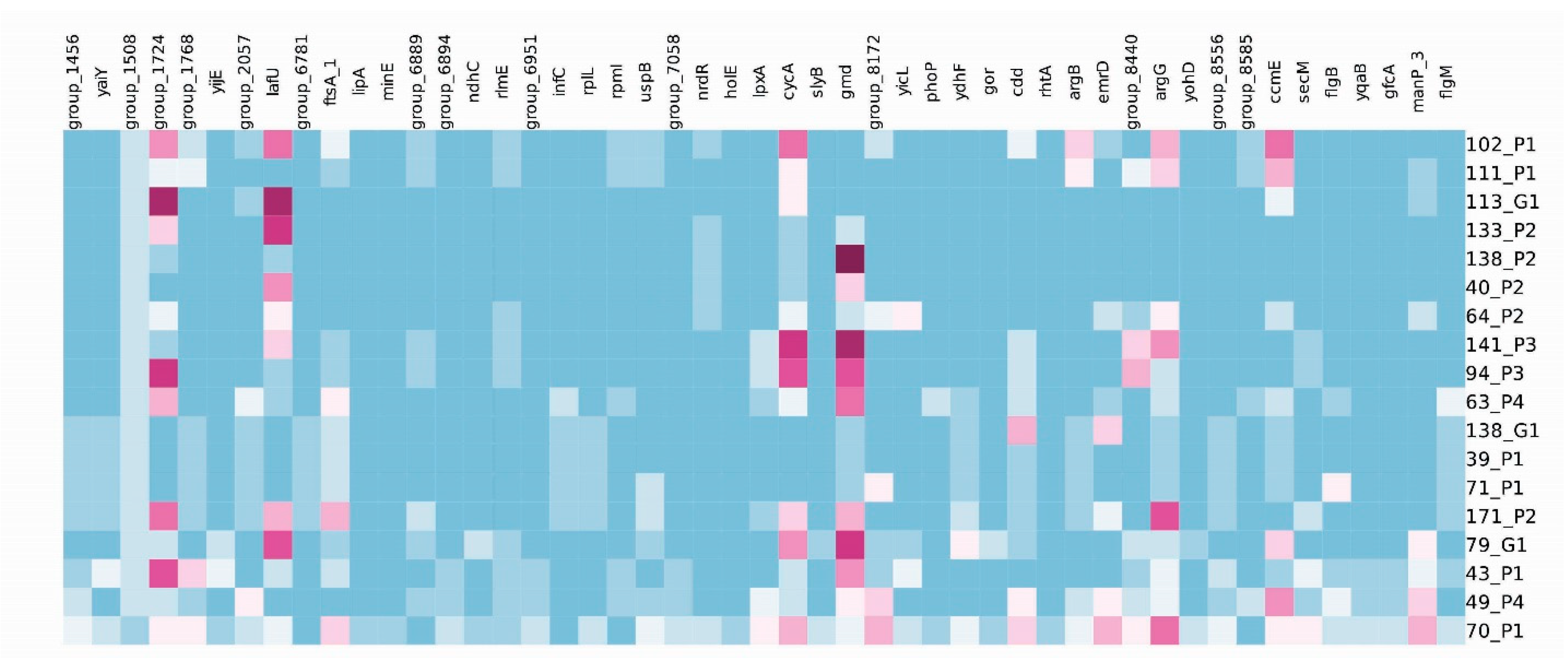
3.1. Identification of E. coli Serotypes and Virulence Gene Profiling

The E. coli isolates (n = 18) were categorized into 11 serotypes by in silico determination of O and H antigens. The serotypes identified were O8:H19, O8:H21, O10:H45, O22:H16, O74:H42, O87:H16, O88:H25, O91:H8, O150:H20, O172:H49 and O181:H8. The serogroup of five E. coli isolates could not be identified (Table 2). The detailed in silico analysis of 102 virulence genes showed the presence of 18 virulence genes in E. coli isolates. The identified virulence genes were stx1 (stx1a and stx1b), stx2 (stx2a and stx2b), espI, iha, ireA, iss, IpfA, espP, exhA, epeA, mcmA, mch, ast, celB, eilA, gad, katP and capU (Table 2). Of 18 E. coli isolates, 13 were STEC. The majority of the STEC isolates (10/13; 76.9%) carried both stx1 and stx2 genes. The remaining three isolates carried stx1 (n = 2) and stx2 genes (n = 1). There were 94.4% (17/18) isolates that carried three or more virulence genes. The most detected virulence genes in E. coli isolates were ipfA (17/18 isolates, 94.4%), followed by iss (15/18 isolates, 83.3%), iha (12/18 isolate, 66.6%), ehxA (8/18 isolates, 44.4%), espP (4/18 isolates, 22.2%), espI (3/18 isolates, 16.6%), mch (3/18 isolates, 16.6%), celB (3/18 isolates, 16.6%), and epeA (2/18 siolates, 11.1%). The least commonly observed virulence genes in E. coli isolates were ire (1/18 isolates; 0.05%), mcmA (1/18 isolates; 0.05%), ast (1/18 isolates; 0.05%), eilA (1/18 isolates; 0.05%), katP (1/18 isolates; 0.05%), and capU (1/18 isolates; 0.05%).

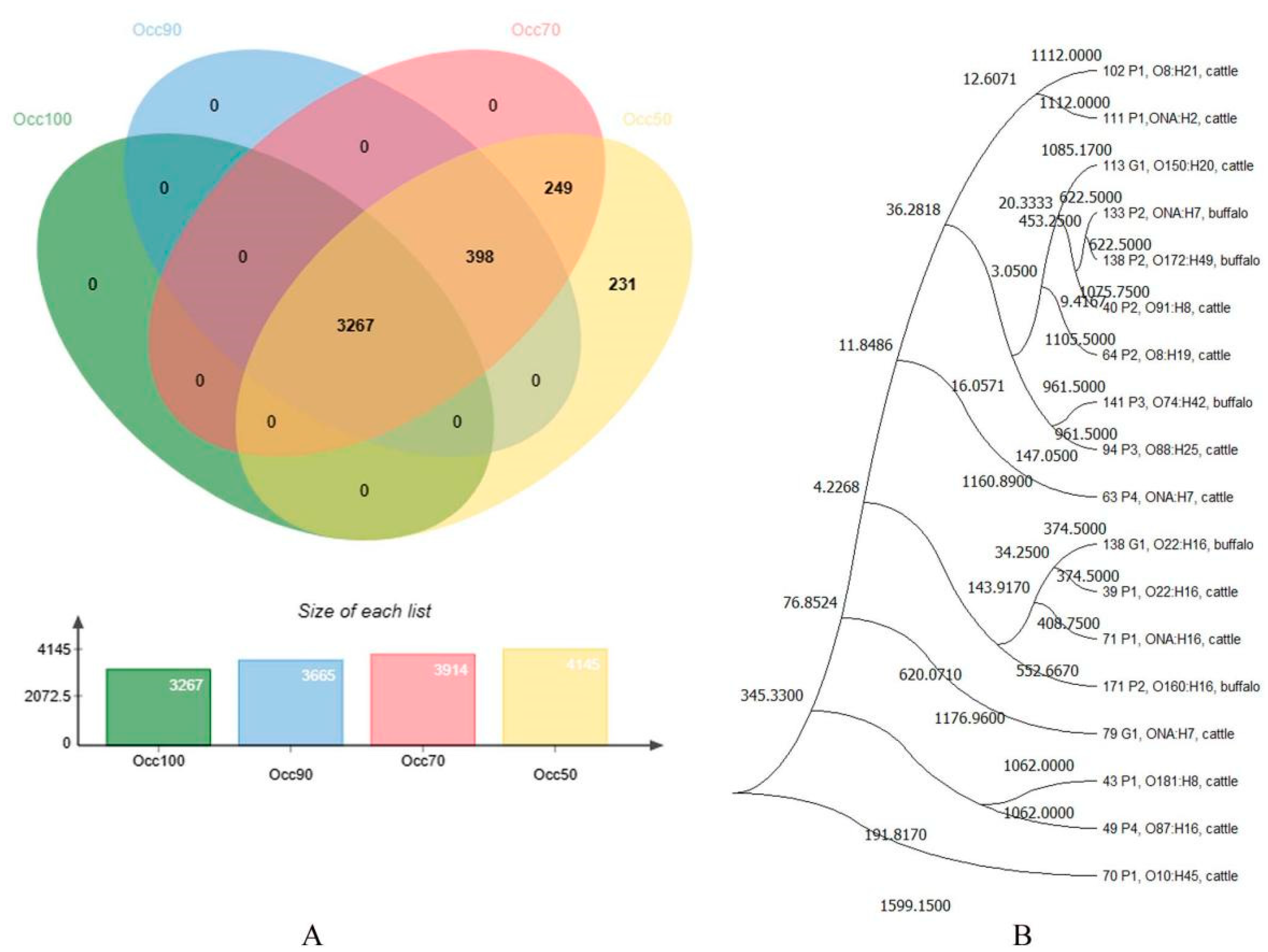
3.2. cgMLST Analyses

The cgMLST analyses provided insight into the core genome of the E. coli strain. We searched for 2513 loci in the online database (cgMLSTFinder 1.1). We found 2163 (86.07%) to 2439 (97.06%) alleles in our analyzed isolates. The percentage of allele matches also showed a high level of variation, ranging from 76.04% to 96.02%.
The total number of genes in the sequenced 18 isolates (Table 3) was 9231. Among these, core or shared genes were 35.4% (n = 3267), occurring in all genomes (Figure 2A). The number of shared genes increased as the number of strains decreased due to the overlap of common genes among them becoming more pronounced. When there are many strains, the gene pool is more diverse, and each strain may have a unique set of genes, leading to less overlap. However, with fewer strains, there is less diversity, so the remaining shared genes become more prominent. This increased proportion of shared genes reflects a higher similarity among the remaining strains. Overall, the accessory genes (occurring in two or more genomes) were 32.8% (n = 3030), while unique genes were 31.8% (n = 2934). A whole genome-based phylogenetic tree was also constructed (Figure 2B).
Interestingly, the core loci contained genes encoded with Shiga toxin subunit A and B precursors. Additionally, the core loci also harbored genes encoded with antibiotic efflux pump outer membrane protein ArpC precursor, Peptide antibiotic transporter SbmA, and multiple antibiotic resistance protein MarA and MarR. These findings suggest that the discriminatory loci not only contribute to genetic differentiation but may also play a role in antibiotic resistance mechanisms.
A refinement process was carried out to identify discriminatory loci within the core genes of the 18 genomes under investigation. The discriminatory loci are genetic variations that can effectively differentiate between different strains or groups. The refinement process resulted in a Robinson–Foulds (RF) distance of 2, indicating two discrepancies between the discriminatory loci sets. These loci were selected based on their ability to effectively differentiate between the genomes and to visualize the discriminatory patterns across the genomes, a heatmap (Figure 3) was generated. A functional annotation was carried out to gain insights into the functional aspects of these loci, and some of the genes were allied with energy metabolism (formate dehydrogenase, lipA, ndhC), chemotaxis (IafU), cell division (minE, FtsA), stress response (uspB, gor), and the transport of molecules across membranes (yijE). Some hypothetical proteins were obtained, with their functions yet to be elucidated. Variations in these genes may affect key cellular processes and contribute to strain-specific characteristics.
Group_1456, group_1508, group_1724, group_6781, group_6889, group_7058, group_8172, group_8440. group_8556,group_8585 = hypothetical protein; Inner membrane protein YaiY; group_1768 = Lumazine-binding domain protein; yijE = putative inner membrane transporter yiJE; group_2057 = formate dehydrogenase accessory protein; IafU = Chemotaxis protein LafU; ftsA = Cell division protein FtsA; lipA = Lipoyl synthase; minE = Cell division topological specificity factor; Group_6894 = GnsA/GnsB family protein; ndhC = NAD(P)H-quinone oxidoreductase subunit 3; rlmE = Ribosomal RNA large subunit methyltransferase E; group_6951 = DNA-binding transcriptional regulator Nlp; infC = Translation initiation factor IF-3; rpIL = 50S ribosomal protein L7/L12; rpmI = 50S ribosomal protein L35; uspB = Universal stress protein B; nrdR = transcriptional repressor NrdR; hole = DNA polymerase III subunit theta; lpxA = Acyl-[acyl-carrier-protein]--UDP-N-acetylglucosamine O-acyltransferase; cycA = D-serine/D-alanine/glycine transporter; slyB = Outer membrane lipoprotein SlyB precursor; gmd = GDP-mannose 4,6-dehydratase;yicL = putative inner membrane transporter YicL; phoP = Transcriptional regulatory protein PhoP; YdhF = Oxidoreductase YdhF; gor = Glutathione reductase; cdd = Cytidine deaminase; rhtA = Threonine/homoserine exporter RhtA; argB = Acetylglutamate kinase; emrD = Multidrug resistance protein D;argG = Argininosuccinate synthase;yohD = Inner membrane protein YohD; ccmE = Cytochrome c-type biogenesis protein CcmE; secM = Secretion monitor precursor; flgB = Flagellar basal body rod protein FlgB; yqaB = Fructose-1-phosphate phosphatase YqaB; gfcA = Threonine-rich inner membrane protein GfcA precursor;manP_3 = PTS system mannose-specific EIIBCA component; flgM = Negative regulator of flagellin synthesis.

3.3. Phenotypic and Genotypic Characterization of STEC Isolates

Of 18 STEC isolates, 18 (100%), 11 (61.11%), 7 (38.88%), 4 (22.22%), 4 (22.22%) and 3 (16.66%) were resistant to rifampicin, neomycin, tetracycline, ceftriaxone, cefotaxime, and azithromycin, respectively (Figure 4). In contrast, all the isolates were susceptible to the rest of the 12 antibiotics tested during the study, including ampicillin, amoxicillin with clavulanic acid, ceftazidime, cefoxitin, ceftiofur, cefepime, aztreonam, imipenem, amikacin, enrofloxacin, levofloxacin, and trimethoprim/sulfamethoxazole (Figure 4). A relatively low MAR index was discovered for the study isolates ranging from 0.06 to 0.32 (Table 4). A total of 9/18 (50%) isolates were identified as being multidrug resistant (MDR); however, none of the isolates were identified as XDR. MDR isolates were resistant to four classes, and 7/9 (77.78%) MDR isolates carried rpoB, ramA, evgA, adeL, emrY, acrAB-TolC, mexR genes. However, we could not detect the antibiotics resistance genes for 2/9 (22.22%) isolates. As such, the dataset encompassed a wide array of ARGs for each strain of the studied E. coli isolates and antibiotic classes (Table 4).

4. Discussion

The study aimed to explore a practical approach to collecting samples from important sources of red meat in Islamabad, Pakistan, and characterized STEC isolates. Accurate and quick identification and typing of food-borne pathogens of zoonotic significance are highly important in limiting the spread of these pathogens. Current identification and characterization methods, such as agglutination assays, panels of phenotypic tests, panels of PCRs, and pulse field gel electrophoresis (PFGE), are expensive, time-consuming, and require specialized training. To overcome these drawbacks, scientists have started using WGS to characterize STEC [25,48]. For example, a study carried out in the US to identify and characterize STEC indicated that serotyping and virulence profiling of STEC isolates using WGS was simple, reliable, economical, and provided more detailed information than conventional methods [48]. Therefore, we used WGS to serotype and characterize STEC isolates in this study.
In the current study, E. coli isolated from the ruminants were serotyped based on “O” and “H” antigens. The genetic diversity of these isolates was determined using cgMLST and the presence of various virulent genes. Previous data have shown that the most recovered serogroups from human cases are O157, O26, O45, O103, O111, O121, and O145 [49]. Previous studies have shown that the virulence of STEC depends upon a number of factors including serotype and virulence genes [50,51,52,53]. Boerlin et al. (1999) [54] reported that STEC serotypes isolated from human cases, which carried both stx2 and eae genes, were more frequently associated with human disease. However, in this study, none of the E. coli isolates belonged to these serogroups, and a high diversity of serotypes was observed in E. coli isolates. Most of the serotypes in this study have also been isolated from human patients, indicating their zoonotic potential [49]. The zoonotic significance of less commonly isolated serotypes from human patients should not be underestimated. The unavailability of standardized isolation protocols could be a reason for the lower amounts of recovery of these serotypes from human patients. In addition, an outbreak caused by STEC O104: H4 in Europe, affecting 3222 individuals, indicated that the acquisition of additional virulence factors by virulent STEC might lead to the establishment of highly virulent STEC [55]. The O serogroup of five E. coli isolates could not be identified, and the zoonotic potential of these serogroups is unknown. However, the presence of various virulence genes, especially the stx gene, may indicate that these isolates could represent emerging serogroups of zoonotic importance.
Shiga toxins (Stx1 and Stx2) are the main virulent determinants of STEC, causing gastroenteritis, HC, and HUS [56,57]. Numerous studies have classified Stx1 into three subtypes, Stx1a, Stx1c, and Stx1d, while classifying Stx2 into seven subtypes, from Stx2a to Stx2g (10). The stx usually exists in bacteriophages, where horizontal gene transfer could be a prime means of new incidence of stx-subtypes/variants or Stx-producing pathogens [58]. Studies have indicated a relationship between STEC virulence and stx genotype. STEC isolates with the stx2 gene are considered more virulent than STEC isolates with stx1 genes, as previous studies have shown a high rate of isolation of STEC with stx2 genes from HUS and HC patients [10,59]. Previous studies have also indicated an association between virulence and the stx2 subtype [9]. In this study, most STEC carried the stx2 gene, indicating their zoonotic potential. Previously, Irshad et al. (2020) [60] reported an occurrence of around 43% for virulence genes (stx1, stx2, eae and ehxA) in STEC isolated from goat and cattle meat from retail meat shops in the twin cities, Rawalpindi and Islamabad. However, Shahzad et al. 2021 [61] recently reported a negligible quantity of STEC from the RAMSs of sheep, goat, cattle, and buffalo from slaughterhouses in Islamabad and Rawalpindi. The contrasting results could be attributed to differences in sampling methods, sampling periods, or protocol for gene amplification/detection.
Apart from stx, 17 other virulence genes were identified in this study. The most observed virulent gene in these E. coli isolates was the lpfA gene. This gene is associated with forming large-sized fimbriae and then adhesion to the surface of targeted cells [62]. A recent study highlighted the importance of the lpfA gene in the mechanism of pathogenesis, indicating that lpfA may play a role in the interaction with Peyer’s patches and M-cells and could contribute to intestinal colonization [63].
The increased serum survival gene (iss) is an important virulence marker in extra-intestinal pathogenic E. coli [10]. However, the iss gene is associated with STEC isolates recovered from stool samples of patients with non-bloody diarrhea [64,65], indicating the low virulence potential of STEC isolates carrying the iss gene. Our study reported a high prevalence of the iss gene in STEC isolates, indicating that STEC isolates recovered from cattle and buffaloes slaughtered in Islamabad may have low virulence potential.
The iha (iron-regulated gene) is an important adherence-conferring gene detected commonly in STEC strains recovered from HUS cases [12,64]. In our study, 92.3% (12/13) of the STEC isolates carried the iha gene. Like our study, another study reported a high prevalence of the iha gene (91%: 127/139 isolates) in STEC isolates [66]. However, a previous study carried out to understand the distribution of eight adhesin genes that are not encoded in the locus of enterocyte effacement (LEE) indicated that non-pathogenic E. coli also carried the iha gene, suggesting that the presence of iha is essential but insufficient for the development of infection [62]. This also highlights that iha could be a candidate for the development of vaccines. Targeting adhesins with vaccines to inhibit colonization in humans or animal reservoirs could be an effective strategy for controlling STEC infections, given that adhesion is the initial stage in disease development.
The EspI (encoded by espI gene) is an important type III secreted protein not encoded in the LEE region [67]. A previous study has shown an association between the presence of the espI gene and the eae gene in STEC isolates (χ2 = 67.9; df = 1; p < 0.0001) [67]. In contrast, in our study, all three isolates carrying the espI gene were negative for the eae gene. A previous study reported that the espI gene was more frequently present in STEC isolates recovered from patients with HUS and diarrhea (47.4%; 37/78) compared to STEC isolates without the espI gene recovered from asymptomatic individuals (100%; 15/15), indicating the potential role of espI in the virulence of STEC isolates [67].
Enterohemolysin (ehxA) is considered an important virulence marker of STEC. A large plasmid of 94 kb called pO157 harbors the ehx operon in STEC O157:H7 [68]. The role of ehxA in pathogenesis is not clearly defined; however, the presence of ehxA in many STEC isolates from HUS and HC patients [66,67], the presence of antibodies against enterohemolysin in the sera of HUS patients [67] and the hemolysis of erythrocytes [68,69,70] all indicate the potential role of ehxA in disease mechanisms. In our study, 61.5% of STEC (8/13 isolates) carried the ehxA gene, indicating the zoonotic potential of the STEC isolated from the slaughtered animals of Pakistan.
Only one serotype of E. coli O22:H16 harbored the ireA gene. The exact role of the ireA gene is not known, but it is believed to be responsible for adhesion to the targeted cells and acquisition of iron in the blood of humans, and it has been identified in the strains isolated from urine and blood samples [15,71]. Only three serotypes, O91:H8, ONA:H7, and O8:H21, carried the espP gene. This gene codes serine protease, which may help in the cleavage of pepsin and human-coagulating factor V, and, therefore, is believed to be important for the survival and dissemination of the pathogenic E. coli in the host’s body [10,12]. The ast gene (Aspartate transaminases) was present in only two serotypes, ONA:H7 and O8:H19. A study reported the role of E. coli’s gene in the tolerance of acid-based stress during infection [10]. Four serotypes carried the celB gene. The exact function of this gene is still unknown. The celB gene has recently been reported to be in necrotizing E. coli [72]. Only one serotype, O10:H45, harbored the eilA gene, a HilA-like regulator which is mostly present in enteroaggregative E. coli [10]. katP, which is responsible for hydrogen peroxide resistance, was found in one E. coli isolate analyzed in this study [73]. Interestingly, the most common STEC of zoonotic significance are termed the “gang of seven” (O157, O26, O45, O103, O111, O121, and O145) and were not isolated from the RAMSs of slaughtered animals in this study. This may be because no preliminary screening method was used for these serogroups (O157, O26, O45, O103, O111, O121, and O145). More specific screening tests such as PCR and immuno-magnetic separation will be needed to explicitly test for their presence. Finally, larger-scale studies are required to understand the epidemiology and distribution of clinically important STEC more clearly, especially O157, O26, O45, O103, O111, O121, and O145, to devise on-farm and pre-slaughter mitigation strategies to reduce the occurrence of STEC in cattle and buffaloes in Pakistan. Large-scale studies can provide valuable insights to develop on-farm and pre-slaughter mitigation strategies for STEC infections. These studies can help to evaluate the impact of different farming practices, such as hygiene measures, feed management, and animal housing conditions, upon the occurrence and transmission of STEC. This information may be helpful in devising practices that reduce the risk of STEC contamination at the farm level. Similarly, studies focusing on the pre-slaughter phase, such as assessing the effectiveness of carcass decontamination methods, hygiene protocols for slaughterhouse workers, and monitoring the presence of STEC throughout the slaughter process, can be helpful in evaluating the interventions at the slaughterhouse to minimize the STEC contamination of meat.
The eae gene encodes intimin, which is involved in the causation of A/E lesions [13]. Previous studies suggest that distinct E. coli serogroups have unique intimin types [74]. The different eae types may be responsible for host specificity, and the tissue tropism shown by various STEC may be influenced by different eae types [75,76]. All the isolates recovered in this study did not possess eae. The STEC without eae have also been recovered from diarrhea and HUS patients [77]. Moreover, STEC O104 isolated from the European outbreak lacked eae, highlighting the virulence potential of STEC strains without eae [55].
The isolates of E. coli sequenced in the current study showed a high level of variations, as shown by cgMLST analysis. The cgMLSTFinder 1.1 was based on the 2513 core loci and provided information not only on the similarities and variation in the number of core loci but also on the percentage of allele matches. The variations we found in the number and types of virulent genes also agreed with cgMLST analyses and detected alleles in the core genome from 2163 alleles (86.07%) to 2439 (97.06%), whereas allele matches were found from 76.04% to 96.02%. These data showed high variations in E. coli isolates, as reported previously [15,78,79]. Additionally, the BacCompare tool was utilized to generate a wgMLST tree based on 3267 core genes. This tree provides a comprehensive overview of the genetic relatedness and evolutionary relationships among the E. coli isolates. Overall, the cgMLST analysis, in conjunction with the detection of virulent genes and the wgMLST tree, highlights the substantial genetic variations present within the E. coli isolates examined in this study. These findings contribute to our understanding of the genomic diversity and evolutionary dynamics of E. coli populations.
Regarding the antimicrobial resistance pattern, the E. coli isolates recovered during the study showed a remarkable resistance to rifampicin, aminoglycosides, tetracyclines, macrolids, and β lactam antibiotics. These findings were nearly consistent to [19,80]. During the current study, E. coli isolates were found to be resistant to only six different antibiotic agents among the 18 antibiotic agents tested. Interestingly, E. coli isolates were 100% resistant to only one antibiotic, namely rifampicin. This high resistance may be attributed to the excessive usage of rifampicin against tuberculosis, as it is a highly effective first line antituberculosis drug [81]. From well-known resistance mechanisms, such as efflux pumps and target alterations, to less-studied variants and mutations, the diversity of ARGs underscores the adaptability of bacteria in evading antibiotic action. The wide array of multidrug resistance genes was present in the study isolates, representing various mechanisms involved in the emergence of antimicrobial resistance. Multidrug resistance is recognized as a significant threat to public health globally. This phenomenon arises primarily from the inappropriate use of antibiotics in both veterinary and medical settings, leading to the acquisition of antimicrobial resistance genes through mobile genetic elements [36,82]. Some antibiotic resistance genes (ARGs) for isolates that exhibited phenotypic resistance to certain antibiotics in disk diffusion tests could not be identified through CARD. Although the antibiotic resistance ontology in the CARD database listed proteins associated with resistance to these antibiotics, the ARG sequences might have been missed during assembly, were below the threshold of similarity, or the strains may employ alternative mechanisms to evade the antibiotics.
This study provides useful information regarding the distribution of various E. coli, especially STEC serogroups of zoonotic importance in cattle and buffaloes slaughtered in Islamabad, Pakistan, indicating that these slaughtered animals may represent an important source of infection for humans. There were several limitations to this pilot study. It was a small-scale study. The samples were collected from a single slaughterhouse in Islamabad. The animals slaughtered came from different production systems and various geographical areas of the Punjab province of Pakistan. Unfortunately, the data regarding production systems and geographical location was not available. The production systems can vary widely in terms of farming practices, hygiene measures, and biosecurity protocols. Therefore, the occurrence of STEC strains in different production systems and geographical areas may vary in their virulence, resistance profiles, and prevalence; thus, a control strategy that is effective in one system may not be effective in another due to these variations. Due to the small number of isolates and the unavailability of data regarding production systems and geographical areas, it was impossible to associate the distribution of virulence factors in the isolates with the geographical area or production system. This study has provided an approach that can be used by the public health and animal health community to better understand the type and diversity of STEC in livestock populations. Demographic and spatial factors associated with the isolates are essential for addressing the role of STEC in public and animal health planning. Thus, such a problem should be addressed before the execution of similar studies in the future.

5. Conclusions

In conclusion, this study focused on investigating the distribution of virulence gene markers and antimicrobial susceptibility profiles associated with STEC within E. coli isolates obtained from the RAMSs of slaughtered cattle and buffaloes in Islamabad, Pakistan. Around 59% of samples tested positive for one or more virulence genes. Subsequently, E. coli isolates (18 isolates) carrying one or more virulence genes were subjected to WGS and differentiated into 11 serotypes, indicating genetic diversity among the isolates. The study revealed a spectrum of antibiotic resistance among the STEC isolates, with varying degrees of susceptibility to different antibiotics. Notably, the highest resistance was observed against rifampicin. The MDR profile was observed in a significant proportion of isolates. However, none of the isolates were classified as XDR. The study also highlighted the complexity of AMR mechanisms, with a diverse array of ARGs being identified across the isolates.
These findings have important implications for public health and food safety. The detection of genetically varied STEC strains with zoonotic significance that are resistant to various antibiotics in cattle and buffaloes slaughtered in Islamabad suggests that these animals can serve as carriers of potential pathogens. This raises concerns about the transmission of STEC-associated diseases to humans, emphasizing the need for effective measures to mitigate the risk. Implementing rigorous food safety protocols, including proper animal husbandry practices, hygiene standards, and thorough meat inspection procedures, is crucial for preventing the transmission of STEC infections. Additionally, promoting awareness among both producers and consumers about the risks associated with STEC-contaminated food can help reduce the incidence of related diseases.

Supplementary Materials

The following supporting information can be downloaded at: https://www.mdpi.com/article/10.3390/agriculture14091537/s1, Table S1: Antibiotic agents, their abbreviations and concentration and breakpoints used for the interpretation of AST results of E. coli isolates recovered during the study period. Table S2: Age and sex of the animals from which E. coli isolates were recovered during study period.

Author Contributions

Conceptualization, H.I. and A.Y.; data curation, A.A., I.A., Z.B. and M.N.; methodology, A.A., N.K., T.P., W.S., P.P., I.A., Z.B., M.N., A., H.A., M.I.K. and M.U.Z.; project administration, H.I., A.A. and A.D.T.; resources, H.I. and N.K.; software, P.P. and I.A.; supervision, A.Y., N.K. and M.S.; validation, H.I., A.Y. and W.S.; visualization, T.P.; writing—original draft, H.I., A.A. and Z.B; writing—review and editing, A.A., A.Y., Z.B., M.U.Z., S.R. and M.S. All authors have read and agreed to the published version of the manuscript.

Funding

This research received no external funding.

Institutional Review Board Statement

Not applicable.

Data Availability Statement

All data generated or analyzed during this study are included in this submitted draft. Additional information/datasets generated during and/or analyzed during the current study are available from the corresponding author on reasonable request.

Acknowledgments

We thank Katie Steneroden, affiliated faculty, CVMBS, Colorado State University, for providing the English language editing of this manuscript.

Conflicts of Interest

It is declared that authors have no competing interests or other interests that might be perceived to influence the results and/or discussion reported in this paper.

References

  1. Bettelheim, K.A. The non-O157 Shiga-toxigenic (verocytotoxigenic) Escherichia coli; under-rated pathogens. Crit. Rev. Microbiol. 2007, 33, 67–87. [Google Scholar] [CrossRef] [PubMed]
  2. Hunt, J.M. Shiga toxin-producing Escherichia coli (STEC). Clin. Lab. Med. 2010, 30, 21–45. [Google Scholar] [CrossRef]
  3. Mcpherson, M.; Kirk, M.D.; Raupach, J.; Combs, B.; Butler, J.R.G. Economic Costs of Shiga Toxin-Producing Escherichia coli Infection in Australia. Foodborne Pathog. Dis. 2011, 8, 55–62. [Google Scholar] [CrossRef] [PubMed]
  4. Heredia, N.; García, S. Animals as sources of food-borne pathogens: A review. Anim. Nutr. 2018, 4, 250–255. [Google Scholar] [CrossRef] [PubMed]
  5. Kanayama, A.; Yahata, Y.; Arima, Y.; Takahashi, T.; Saitoh, T.; Kanou, K.; Kawabata, K.; Sunagawa, T.; Matsui, T.; Oishi, K. Enterohemorrhagic Escherichia coli outbreaks related to childcare facilities in Japan, 2010–2013. BMC Infect. Dis. 2015, 15, 539. [Google Scholar] [CrossRef]
  6. Rangel, J.M.; Sparling, P.H.; Crowe, C.; Griffin, P.M.; Swerdlow, D.L. Epidemiology of Escherichia coli O157 outbreaks, United States, 1982–2002. Emerg. Infect. Dis. 2005, 11, 603–609. [Google Scholar] [CrossRef] [PubMed]
  7. Riley, L.W.; Remis, R.S.; Helgerson, S.D.; McGee, H.B.; Wells, J.G.; Davis, B.R.; Hebert, R.J.; Olcott, E.S.; Johnson, L.M.; Hargrett, N.T.; et al. Hemorrhagic Colitis Associated with a Rare Escherichia coli Serotype. N. Engl. J. Med. 1983, 308, 681–685. [Google Scholar] [CrossRef] [PubMed]
  8. Naseer, U.; Løbersli, I.; Hindrum, M.; Bruvik, T.; Brandal, L.T. Virulence factors of Shiga toxin-producing Escherichia coli and the risk of developing haemolytic uraemic syndrome in Norway, 1992–2013. Eur. J. Clin. Microbiol. Infect. Dis. 2017, 36, 1613–1620. [Google Scholar] [CrossRef]
  9. Melton-Celsa, A.R. Shiga Toxin (Stx) Classification, Structure, and Function. Microbiol. Spectr. 2014, 2, 10–1128. [Google Scholar] [CrossRef]
  10. Smith, J.L.; Fratamico, P.M.; Gunther, N.W. Shiga toxin-producing Escherichia coli. In Advances in Applied Microbiology; Academic Press Inc.: Cambridge, MA, USA, 2014; pp. 145–197. [Google Scholar]
  11. González-Escalona, N.; Kase, J.A. Virulence gene profiles and phylogeny of Shiga toxin-positive Escherichia coli strains isolated from FDA regulated foods during 2010–2017. PLoS ONE 2019, 14, e0214620. [Google Scholar] [CrossRef]
  12. Fan, R.; Shao, K.; Yang, X.; Bai, X.; Fu, S.; Sun, H.; Xu, Y.; Wang, H.; Li, Q.; Hu, B.; et al. High prevalence of non-O157 Shiga toxin-producing Escherichia coli in beef cattle was detected by combining four selective agars. BMC Microbiol. 2019, 19, 213. [Google Scholar] [CrossRef] [PubMed]
  13. Donnenberg, M.S.; Tzipori, S.; McKee, M.L.; O’Brien, A.D.; Alroy, J.; Kaper, J.B. The role of the eae gene of enterohemorrhagic Escherichia coli in intimate attachment in vitro and in a porcine model. J. Clin. Investig. 1993, 92, 1418–1424. [Google Scholar] [CrossRef] [PubMed]
  14. Thomas, R.R.; Brooks, H.J.L.; O’Brien, R. Prevalence of Shiga toxin-producing and enteropathogenic Escherichia coli marker genes in diarrhoeic stools in a New Zealand catchment area. J. Clin. Pathol. 2017, 70, 81–84. [Google Scholar] [CrossRef] [PubMed]
  15. Ferdous, M.; Friedrich, A.W.; Grundmann, H.; de Boer, R.F.; Croughs, P.D.; Islam, M.A.; Kluytmans-van den Bergh, M.F.; Kooistra-Smid, A.M.; Rossen, J.W. Molecular characterization and phylogeny of Shiga toxin–producing Escherichia coli isolates obtained from two Dutch regions using whole genome sequencing. Clin. Microbiol. Infect. 2016, 22, 642.e1–642.e9. [Google Scholar] [CrossRef]
  16. Savarino, S.J.; Fasano, A.; Watson, J.; Martin, B.M.; Levine, M.M.; Guandalini, S.; Guerry, P. Enteroaggregative Escherichia coli heat-stable enterotoxin 1 represents another subfamily of E. coli heat-stable toxin. Proc. Natl. Acad. Sci. USA 1993, 90, 3093–3097. [Google Scholar] [CrossRef]
  17. Lim, J.Y.; Yoon, J.W.; Hovde, C.J. A brief overview of Escherichia coli O157 and its plasmid O157. J. Microbiol. Biotechnol. 2010, 20, 5. [Google Scholar] [CrossRef]
  18. Pan, Y.; Hu, B.; Bai, X.; Yang, X.; Cao, L.; Liu, Q.; Sun, H.; Li, J.; Zhang, J.; Jin, D.; et al. Antimicrobial resistance of non-o157 shiga toxin-producing Escherichia coli isolated from humans and domestic animals. Antibiotics 2021, 10, 74. [Google Scholar] [CrossRef]
  19. Algammal, A.M.; Hashem, M.E.A.; Alfifi, K.J.; Al-Otaibi, A.S.; Alatawy, M.; Eltarabili, R.M.; Abd El-Ghany, W.A.; Hetta, H.F.; Hamouda, A.M.; Elewa, A.A.; et al. Sequence Analysis, Antibiogram Profile, Virulence and Antibiotic Resistance Genes of XDR and MDR Gallibacterium anatis Isolated from Layer Chickens in Egypt. Infect. Drug Resist. 2022, 15, 4321–4334. [Google Scholar] [CrossRef]
  20. Algammal, A.M.; El-Kholy, A.W.; Riad, E.M.; Mohamed, H.E.; Elhaig, M.M.; Al Yousef, S.A.; Hozzein, W.N.; Ghobashy, M.O. Genes encoding the virulence and the antimicrobial resistance in enterotoxigenic, and shiga-toxigenic E. coli isolated from diarrheic calves. Toxins 2020, 12, 383. [Google Scholar] [CrossRef]
  21. Mahanti, A.; Samanta, I.; Bandopaddhay, S.; Joardar, S.N.; Dutta, T.K.; Batabyal, S.; Sar, T.K.; Isore, D.P. Isolation, molecular characterization and antibiotic resistance of Shiga Toxin-Producing Escherichia coli (STEC) from buffalo in India. Lett. Appl. Microbiol. 2013, 56, 291–298. [Google Scholar] [CrossRef]
  22. Conrad, C.; Stanford, K.; Narvaez-Bravo, C.; Neumann, N.; Munns, K.; Tymensen, L.; Jokinen, C.; McAllister, T.A. Zoonotic Fecal Pathogens and Antimicrobial Resistance in Canadian Petting Zoos. Microorganisms 2018, 6, 70. [Google Scholar] [CrossRef] [PubMed]
  23. Fathi Sharafa, E.; Shabanaa, I.I. Prevalence and molecular characterization of Shiga toxin-producing Escherichia coli isolates from human and sheep in Al-Madinah Al-Munawarah. Infectio 2017, 21, 81–87. [Google Scholar] [CrossRef]
  24. Jacob, M.E.; Foster, D.M.; Rogers, A.T.; Balcomb, C.C.; Shi, X.; Nagaraja, T.G. Evidence of Non-O157 Shiga toxin-producing Escherichia coli in the feces of meat goats at a U.S. slaughter plant. J. Food Prot. 2013, 76, 1626–1629. [Google Scholar] [CrossRef] [PubMed]
  25. Bumunang, E.W.; McAllister, T.A.; Zaheer, R.; Ortega Polo, R.; Stanford, K.; King, R.; Niu, Y.D.; Ateba, C.N. Characterization of Non-O157 Escherichia coli from Cattle Faecal Samples in the North-West Province of South Africa. Microorganisms 2019, 7, 272. [Google Scholar] [CrossRef]
  26. Samad, A.; Abbas, F.; Ahmad, Z.; Tanveer, Z.; Ahmad, I.; Patching, S.G.; Nawaz, N.; Asmat, M.T.; Raziq, A.; Asadullah; et al. Multiplex polymerase chain reaction detection of Shiga toxin genes and antibiotic sensitivity of Escherichia coli O157 isolated from beef meat in Quetta, Pakistan. J. Food Saf. 2018, 38, e12540. [Google Scholar] [CrossRef]
  27. Erickson, M.C.; Doyle, M.P. Food as a vehicle for transmission of Shiga toxin-producing Escherichia coli. J. Food Prot. 2007, 70, 2426–2449. [Google Scholar] [CrossRef] [PubMed]
  28. Irshad, H.; Cookson, A.L.; Hotter, G.; Besser, T.E.; On, S.L.W.; French, N.P. Epidemiology of Shiga toxin-producing Escherichia coli O157 in very young calves in the North Island of New Zealand. N. Z. Vet. J. 2012, 60, 21–26. [Google Scholar] [CrossRef]
  29. Sharma, V.K.; Dean-Nystrom, E.A. Detection of enterohemorrhagic Escherichia coli O157 by using a multiplex real-time PCR assay for genes encoding intimin and Shiga toxins. Vet. Microbiol. 2003, 93, 247–260. [Google Scholar] [CrossRef] [PubMed]
  30. Paton, A.W.; Paton, J.C. Detection and characterization of Shiga toxigenic Escherichia coli by using multiplex PCR Assays for stx1, stx2, eaeA, Enterohemorrhagic E. coli hlyA, rfbO111, and rfbO157. J. Clinic. Microbiol. 1998, 36, 598–602. [Google Scholar] [CrossRef]
  31. Mahe, A. Effect of Citric Acid at Different pH on the Survival of Escherichia coli. Bayero J. Pure Appl. Sci. 2021, 14, 79–84. [Google Scholar] [CrossRef]
  32. CLSI. Performance Standards for Antimicrobial Susceptibility Testing, 33rd ed.; CLSI Supplement M 100; Clinical and Laboratory Standards Institute: Wayne, PA, USA, 2023. [Google Scholar]
  33. WHO. Integrated Surveillance of Antimicrobial Resistance—Guidance from WHO Advisory Group; WHO: Geneva, Switzerland, 2013; pp. 1–100. [Google Scholar]
  34. Magiorakos, A.P.; Srinivasan, A.; Carey, R.B.; Carmeli, Y.; Falagas, M.E.; Giske, C.G.; Harbarth, S.; Hindler, J.F.; Kahlmeter, G.; Olsson-Liljequist, B. Multidrug-resistant, extensively drug-resistant and pandrug-resistant bacteria: An international expert proposal for interim standard definitions for acquired resistance. Clin. Microbiol. Infect. 2012, 18, 268–281. [Google Scholar] [CrossRef]
  35. Siddique, A.; Azim, S.; Ali, A.; Andleeb, S.; Ahsan, A.; Imran, M.; Rahman, A. Antimicrobial resistance profiling of biofilm forming non typhoidal salmonella enterica isolates from poultry and its associated food products from Pakistan. Antibiotics 2021, 10, 785. [Google Scholar] [CrossRef] [PubMed]
  36. Algammal, A.M.; Ibrahim, R.A.; Alfifi, K.J.; Ghabban, H.; Alghamdi, S.; Kabrah, A.; Khafagy, A.R.; Abou-Elela, G.M.; Abu-Elala, N.M.; Donadu, M.G. A First Report of Molecular Typing, Virulence Traits, and Phenotypic and Genotypic Resistance Patterns of Newly Emerging XDR and MDR Aeromonas veronii in Mugil seheli. Pathogens 2022, 11, 1262. [Google Scholar] [CrossRef] [PubMed]
  37. Andrews, S.; Krueger, F.; Segonds-Pichon, A.; Biggins, L.; Krueger, C., 3rd; Wingett, S.; Babraham Bioinformatics; FastQC. A Quality Control Tool for High Throughput Sequence Data (Online). 2019. Available online: https://www.bioinformatics.babraham.ac.uk/projects/fastqc/ (accessed on 22 January 2022).
  38. Krueger, F. Trim Galore: A wrapper tool around Cutadapt and FastQC to consistently apply quality and adapter trimming to FastQ files, with some extra functionality for MspI-digested RRBS-type (Reduced Representation Bisufite-Seq) libraries. 2012. Available online: http://www.bioinformatics.babraham.ac.uk/projects/trim_galore/ (accessed on 22 January 2022).
  39. Zerbino, D.R.; Birney, E. Velvet: Algorithms for de novo short read assembly using de Bruijn graphs. Genome Res. 2008, 18, 821–829. [Google Scholar] [CrossRef]
  40. Joensen, K.G.; Tetzschner, A.M.M.; Iguchi, A.; Aarestrup, F.M.; Scheutz, F. Rapid and easy in silico serotyping of Escherichia coli isolates by use of whole-genome sequencing data. J. Clin. Microbiol. 2015, 53, 2410–2426. [Google Scholar] [CrossRef] [PubMed]
  41. Clausen, P.T.L.C.; Aarestrup, F.M.; Lund, O. Rapid and precise alignment of raw reads against redundant databases with KMA. BMC Bioinform. 2018, 19, 307. [Google Scholar] [CrossRef] [PubMed]
  42. Camacho, C.; Coulouris, G.; Avagyan, V.; Ma, N.; Papadopoulos, J.; Bealer, K.; Madden, T.L. BLAST+: Architecture and applications. BMC Bioinform. 2009, 10, 421. [Google Scholar] [CrossRef]
  43. Joensen, K.G.; Scheutz, F.; Lund, O.; Hasman, H.; Kaas, R.S.; Nielsen, E.M.; Aarestrup, F.M. Real-time whole-genome sequencing for routine typing, surveillance, and outbreak detection of verotoxigenic Escherichia coli. J. Clin. Microbiol. 2014, 52, 1501–1510. [Google Scholar] [CrossRef]
  44. Alikhan, N.F.; Zhou, Z.; Sergeant, M.J.; Achtman, M. A genomic overview of the population structure of Salmonella. PLoS Genet. 2018, 14, e1007261. [Google Scholar] [CrossRef]
  45. Liu, Y.Y.; Lin, J.W.; Chen, C.C. Cano-wgMLST_BacCompare: A bacterial genome analysis platform for epidemiological investigation and comparative genomic analysis. Front. Microbiol. 2019, 10, 1687. [Google Scholar] [CrossRef]
  46. Bortolaia, V.; Kaas, R.S.; Ruppe, E.; Roberts, M.C.; Schwarz, S.; Cattoir, V.; Philippon, A.; Allesoe, R.L.; Rebelo, A.R.; Florensa, A.F. ResFinder 4.0 for predictions of phenotypes from genotypes. J. Antimicrob. Chemother. 2020, 75, 3491–3500. [Google Scholar] [CrossRef] [PubMed]
  47. Malmberg, J.L.; O’Toole, D.; Creekmore, T.; Peckham, E.; Killion, H.; Vance, M.; Philippon, A.; Allesoe, R.L.; Rebelo, A.R.; Florensa, A.F. Mycoplasma bovis Infections in Free-Ranging Pronghorn, Wyoming, USA. Emerg. Infect. Dis. 2020, 26, 2807–2814. [Google Scholar] [CrossRef] [PubMed]
  48. Lindsey, R.L.; Pouseele, H.; Chen, J.C.; Strockbine, N.A.; Carleton, H.A. Implementation of whole genome sequencing (WGS) for identification and characterization of Shiga toxin-producing Escherichia coli (STEC) in the United States. Front. Microbiol. 2016, 7, 766. [Google Scholar] [CrossRef] [PubMed]
  49. Bettelheim, K.A.; Kuzevski, A.; Gilbert, R.A.; Krause, D.O.; McSweeney, C.S. The diversity of Escherichia coli serotypes and biotypes in cattle faeces. J. Appl. Microbiol. 2005, 98, 699–709. [Google Scholar] [CrossRef]
  50. Karmali, M.A. Infection by Verocytotoxin-Producing Escherichia coli. Clin. Microbiol. Rev. 1989, 2, 15–38. [Google Scholar] [CrossRef]
  51. Pikrard, D.; Stevens, D.; Moriau, L.; Lior, H.; Lauwers, S. Isolation and virulence factors of vero cyto toxin-producing Escherichia coli in human stool samples. Clin. Microbiol. Infect. 1997, 3, 531–540. [Google Scholar] [CrossRef]
  52. Schmidt, H.; Karch, H. Enterohemolytic Phenotypes and Genotypes of Shiga Toxin-Producing Escherichia coli O111 Strains from Patients with Diarrhea and Hemolytic-Uremic Syndrome. J. Clin. Microbiol. 1996, 34, 2364–2367. [Google Scholar] [CrossRef]
  53. Scotland, S.M.; Willshaw, G.A.; Smith, H.R.; Rowe, B. Properties of strains of Escherichia coli belonging to serogroup O 157 with special reference to production of Vero cytotoxins VT1 and VT2. Epidemiol. Infect. 1987, 99, 613–624. [Google Scholar] [CrossRef]
  54. Boerlin, P.; McEwen, S.A.; Boerlin-Petzold, F.; Wilson, J.B.; Johnson, R.P.; Gyles, C.L. Associations between Virulence Factors of Shiga Toxin-Producing Escherichia coli and Disease in Humans. J. Clin. Microbiol. 1999, 37, 497–503. [Google Scholar] [CrossRef]
  55. Frank, C.; Werber, D.; Cramer, J.P.; Askar, M.; Faber, M.; an der Heiden, M.; Bernard, H.; Fruth, A.; Prager, R.; Spode, A.; et al. Epidemic Profile of Shiga-Toxin–Producing Escherichia coli O104 outbreak in Germany. N. Engl. J. Med. 2011, 365, 1771–1780. [Google Scholar] [CrossRef]
  56. Brandal, L.T.; Tunsjø, H.S.; Ranheim, T.E.; Løbersli, I.; Lange, H.; Wester, A.L. Shiga Toxin 2a in Escherichia albertii. J. Clin. Microbiol. 2015, 53, 1454–1455. [Google Scholar] [CrossRef]
  57. Carter, M.Q.; Pham, A.; Huynh, S.; He, X. Complete genome sequence of a shiga toxin-producing Enterobacter cloacae clinical isolate. Genome Announc. 2017, 5, 10–1128. [Google Scholar] [CrossRef] [PubMed]
  58. Khalil, R.; Skinner, C.; Patfield, S.; He, X. Phage-mediated Shiga toxin (Stx) horizontal gene transfer and expression in non-Shiga toxigenic Enterobacter and Escherichia coli strains. Pathog. Dis. 2016, 74, ftw037. [Google Scholar] [CrossRef] [PubMed]
  59. Pradel, N.; Bertin, Y.; Martin, C.; Livrelli, V. Molecular analysis of Shiga toxin-producing Escherichia coli strains isolated from hemolytic-uremic syndrome patients and dairy samples in France. Appl. Environ. Microbiol. 2008, 74, 2118–2128. [Google Scholar] [CrossRef] [PubMed]
  60. Irshad, H.; Binyamin, I.; Ahsan, A.; Riaz, A.; Shahzad, M.A.; Qayyum, M.; Kanchanakhan, N.; Siriwong, W.; Pumpaibool, T.; Hussain, M.; et al. Occurrence and molecular characterization of shiga toxin-producing Escherichia coli isolates recovered from cattle and goat meat obtained from retail meat shops in Rawalpindi and Islamabad, Pakistan. Pak. Vet. J. 2020, 40, 295–300. [Google Scholar]
  61. Shahzad, A.; Ullah, F.; Irshad, H.; Ahmed, S.; Shakeela, Q.; Mian, A.H. Molecular detection of Shiga toxin-producing Escherichia coli (STEC) O157 in sheep, goats, cows and buffaloes. Mol. Biol. Rep. 2021, 48, 6113–6121. [Google Scholar] [CrossRef]
  62. Toma, C.; Espinosa, E.M.; Song, T.; Miliwebsky, E.; Chinen, I.; Iyoda, S.; Iwanaga, M.; Rivas, M. Distribution of putative adhesins in different seropathotypes of Shiga toxin-producing Escherichia coli. J. Clin. Microbiol. 2004, 42, 4937–4946. [Google Scholar] [CrossRef]
  63. Cordonnier, C.; Etienne-Mesmin, L.; Thévenot, J.; Rougeron, A.; Rénier, S.; Chassaing, B.; Darfeuille-Michaud, A.; Barnich, N.; Blanquet-Diot, S.; Livrelli, V. Enterohemorrhagic Escherichia coli pathogenesis: Role of long polar fimbriae in Peyer’s patches interactions. Sci. Rep. 2017, 7, 44655. [Google Scholar] [CrossRef]
  64. Bai, X.; Zhang, J.; Ambikan, A.; Jernberg, C.; Ehricht, R.; Scheutz, F.; Xiong, Y.; Matussek, A. Molecular Characterization and Comparative Genomics of Clinical Hybrid Shiga Toxin-Producing and Enterotoxigenic Escherichia coli (STEC/ETEC) Strains in Sweden. Sci. Rep. 2019, 9, 5619. [Google Scholar] [CrossRef]
  65. Baschera, M.; Cernela, N.; Stevens, M.J.A.; Liljander, A.; Jores, J.; Corman, V.M.; Nüesch-Inderbinen, M.; Stephan, R. Shiga toxin-producing Escherichia coli (STEC) isolated from fecal samples of African dromedary camels. One Health 2019, 7, 100087. [Google Scholar] [CrossRef]
  66. Colello, R.; Krüger, A.; Velez, M.V.; Del Canto, F.; Etcheverría, A.I.; Vidal, R.; Padola, N.L. Identification and detection of iha subtypes in LEE-negative Shiga toxin-producing Escherichia coli (STEC) strains isolated from humans, cattle and food. Heliyon 2019, 5, e03015. [Google Scholar] [CrossRef] [PubMed]
  67. Mundy, R.; Jenkins, C.; Yu, J.; Smith, H.; Frankel, G. Distribution of espI among clinical enterohaemorrhagic and enteropathogenic Escherichia coli isolates. J. Med. Microbiol. 2004, 53, 1145–1149. [Google Scholar] [CrossRef]
  68. Beutin, L.; Prada, J.; Zimmermann, S.; Stephan, R.; Ørskov, I.; Ørskov, F. Enterohemolysin, a new type of hemolysin produced by some strains of enteropathogenic E. coli (EPEC). Zentralbl. Bakteriol. Mikrobiol. Hyg. 1988, 267, 576–588. [Google Scholar] [CrossRef] [PubMed]
  69. Schmidt, H.; Beutin, L.; Karch, H. Molecular analysis of the plasmid-encoded hemolysin of Escherichia coli O157 strain EDL 933. Infect. Immun. 1995, 63, 1055–1061. [Google Scholar] [CrossRef]
  70. Karch, H.; Janetzki-Mittmann, C.; Aleksic, S.; Datz, M. Isolation of enterohemorrhagic Escherichia coli O157 strains from patients with hemolytic-uremic syndrome by using immunomagnetic separation, DNA-based methods, and direct culture. J. Clin. Microbiol. 1996, 34, 516–519. [Google Scholar] [CrossRef] [PubMed]
  71. Pilarczyk-Zurek, M.; Chmielarczyk, A.; Gosiewski, T.; Tomusiak, A.; Adamski, P.; Zwolinska-Wcislo, M.; Mach, T.; Heczko, P.B.; Strus, M. Possible role of Escherichia coli in propagation and perpetuation of chronic inflammation in ulcerative colitis. BMC Gastroenterol. 2013, 13, 61. [Google Scholar] [CrossRef]
  72. Feng, Y.; Mannion, A.; Madden, C.M.; Swennes, A.G.; Townes, C.; Byrd, C.; Marini, R.P.; Fox, J.G. Cytotoxic Escherichia coli strains encoding colibactin and cytotoxic necrotizing factor (CNF) colonize laboratory macaques. Gut Pathog. 2017, 9, 71. [Google Scholar] [CrossRef]
  73. Djoko, K.Y.; Phan, M.D.; Peters, K.M.; Walker, M.J.; Schembri, M.A.; McEwan, A.G. Interplay between tolerance mechanisms to copper and acid stress in Escherichia coli. Proc. Natl. Acad. Sci. USA 2017, 114, 6818–6823. [Google Scholar] [CrossRef]
  74. Zhang, W.L.; Köhler, B.; Oswald, E.; Beutin, L.; Karch, H.; Morabito, S.; Caprioli, A.; Suerbaum, S.; Schmidt, H. Genetic diversity of intimin genes of attaching and effacing Escherichia coli strains. J. Clin. Microbiol. 2002, 40, 4486–4492. [Google Scholar] [CrossRef]
  75. Fitzhenry, R.J.; Pickard, D.J.; Hartland, E.L.; Reece, S.; Dougan, G.; Phillips, A.D. Intimin type influences the site of human intestinal mucosal colonisation by enterohaemorrhagic Escherichia coli O157. Gut. 2002, 50, 180–185. [Google Scholar] [CrossRef]
  76. Phillips, A.D.; Frankel, G. Intimin-Mediated Tissue Specificity in Enteropathogenic Escherichia coli Interaction with Human Intestinal Organ Cultures. J. Infect. Dis. 2000, 181, 1496–1506. Available online: https://academic.oup.com/jid/article/181/4/1496/865392 (accessed on 22 January 2022). [CrossRef] [PubMed]
  77. Beutin, L.; Martin, A. Outbreak of shiga toxin-producing Escherichia coli (STEC) O104 infection in Germany causes a paradigm shift regarding human pathogenicity of STEC strains. J. Food Prot. 2012, 75, 408–418. [Google Scholar] [CrossRef] [PubMed]
  78. Pasquali, F.; Palma, F.; Trevisani, M.; Parisi, A.; Lucchi, A.; De Cesare, A.; Manfreda, G. Whole genome sequencing based typing and characterisation of shiga-toxin producing Escherichia coli strains belonging to O157 and O26 serotypes and isolated in dairy farms. Ital. J. Food Saf. 2018, 7, 181–188. [Google Scholar] [CrossRef] [PubMed]
  79. Browne, A.S.; Biggs, P.J.; Wilkinson, D.A.; Cookson, A.L.; Midwinter, A.C.; Bloomfield, S.J.; Hranac, C.R.; Rogers, L.E.; Marshall, J.C.; Benschop, J. Use of genomics to investigate historical importation of shiga toxin-producing Escherichia coli serogroup O26 and nontoxigenic variants into New Zealand. Emerg. Infect. Dis. 2019, 25, 489–500. [Google Scholar] [CrossRef]
  80. Elbehiry, A.; Marzouk, E.; Aldubaib, M.; Moussa, I.; Abalkhail, A.; Ibrahem, M.; Hamada, M.; Sindi, W.; Alzaben, F.; Almuzaini, A.M. Pseudomonas species prevalence, protein analysis, and antibiotic resistance: An evolving public health challenge. AMB Express 2022, 12, 53. [Google Scholar] [CrossRef]
  81. Zhang, Q.; An, X.; Liu, H.; Wang, S.; Xiao, T.; Liu, H. Uncovering the Resistance Mechanism of Mycobacterium tuberculosis to Rifampicin Due to RNA Polymerase H451D/Y/R Mutations from Computational Perspective. Front. Chem. 2019, 7, 819. [Google Scholar] [CrossRef]
  82. Shafiq, M.; Zeng, M.; Permana, B.; Bilal, H.; Huang, J.; Yao, F.; Algammal, A.M.; Li, X.; Yuan, Y.; Jiao, X. Coexistence of blaNDM–5 and tet(X4) in international high-risk Escherichia coli clone ST648 of human origin in China. Front. Microbiol. 2022, 13, 1031688. [Google Scholar] [CrossRef]
Figure 1. The flow diagram showing the methodology of the study.
Agriculture 14 01537 g001
Figure 2. (A). Venn diagram showing common genes in studied 18 STEC strains obtained from cattle and buffaloes. Occ100 depicts occurrence in all genomes, Occ90 in 90% of genomes, Occ70 in 70% of genomes, and Occ50 in half of the studied genomes. Size of each list depicts total genes pertaining to the similarity criteria. (B) Whole genome MLST tree-based tree topology of the studied strains with an SBL of 18556.19.
Agriculture 14 01537 g002
Figure 3. Heatmap of highly discriminatory loci for the sequences STEC genomes. Colors represent the abundance of the specific gene in the given isolate and it is a standard format to represent genes presence or absence in a sample/isolate.
Agriculture 14 01537 g003
Figure 4. Antibiotic susceptibility profile of STEC isolates featuring 18 different antibiotics of veterinary and human clinical relevance obtained using the disk diffusion method. The isolates were recovered from RAMSs samples collected from slaughtered animals of the Islamabad Capital Territory during August 2016 to March 2017.
Agriculture 14 01537 g004
Table 1. Distribution of virulence genes (stx1, stx2, eae, and ehxA) upon initial screening of RAMSs from slaughterhouses in Islamabad (2016–2017). The RAMSs were analyzed using multiplex PCR for stx1, stx2, eae, and ehxA.
Species of Animal
Cattle Buffalo
Virulence GenesNo. of Positive SamplesVirulence GenesNo. of Positive Samples
stx1, stx2, ehxA18stx1, stx2, ehxA29
stx117stx1, ehxA20
stx1, ehxA15stx17
stx1, stx25stx1, stx23
ehxA3ehxA0
stx21stx20
Total59/104 (56.7%)Total59/96 (61.4%)
Table 2. Serotyping and distribution of virulence genes in E. coli isolates (n = 18) recovered from cattle (n = 14) and buffaloes (n = 4) slaughtered in Islamabad, Pakistan. Whole genome sequencing of E. coli isolates was carried out using Illumina MiSeq sequencer.
Isolate IDSerotypeSpecieShiga Toxin TypeVirulence Genes
Stx1Stx2espIIhaireAissIpfAespPehxAgadepeAmcmAmchastcelBeilAkatPcapU
E. coli 39P1O22:H16CattleStx1aStx2a+++++ +
E. coli 40P2O91:H8CattleStx1aStx2c++++++
E. coli 43P1O181:H8CattleStx1aStx2c+++++++
E. coli 49P4O87:H16CattleStx1aStx2c++++++
E. coli 63P4ONA:H7CattleStx1aStx2c++++++++++
E. coli 64P2O8:H19Cattle--+++
E. coli 70P1O10:H45Cattle--+++
E. coli 71P1ONA:H16CattleStx1aStx2c++++
E. coli 79G1ONA:H7CattleStx1aStx2c+++
E. coli 94P3O88:H25CattleStx1aStx2a+++++
E. coli 102P1O8:H21Cattle-Stx2a++++++
E. coli 111P1ONA:H2Cattle--++
E. coli 113G1O150:H20Cattle--+++
E. coli 133P2ONA:H7Buffalo--+++
E. coli 138P2O172:H49BuffaloStx1aStx2b+++++
E. coli 138G1O22:H16BuffaloStx1a-+++++
E. coli 141P3O74:H42BuffaloStx1a-++++++
E. coli 171P2O160:H16CattleStx1aStx2a++++++
Total 121131211517481621313111
“+” means gene present. “−” means gene absent.
Table 3. Genome accession identifiers of whole genome-sequenced STEC isolates.
Isolate IDSerotypeSpecieGenBank Accession NumberNo. of ContigsCoverageTotal no. of Genes
39P1O22:H16CattleDAERGV000000000.1402108×5392
40P2O91:H8CattleDAERHR000000000.1180152×4959
43P1O181:H8CattleDAERHL000000000.1112136×4834
49P4O87:H16CattleDAERGQ000000000.1123127×4792
63P4ONA:H7CattleDAERHQ000000000.1142129×4861
64P2O8:H19CattleDAERGG000000000.18690×4509
70P1O10:H45CattleDAERGZ000000000.1174138×4762
71P1ONA:H16CattleDAERHF000000000.1211142×5122
79G1ONA:H7CattleDAERHH000000000.1161106×4939
94P3O88:H25CattleDAERGJ000000000.1135135×4982
102P1O8:H21CattleDAERHO000000000.1246163×5055
111P1ONA:H2CattleDAERHG000000000.1126131×4481
113G1O150:H20CattleDAERHA000000000.192160×4598
133P2ONA:H7BuffaloDAERHQ000000000.1142129×4861
138P2O172:H49BuffaloDAERHS000000000.11322544749
138G1O22:H16BuffaloDAERGN000000000.11891174963
141_P3O74:H42BuffaloDAERGW000000000.1158126×5136
171_P2O160:H16CattleDAERGS000000000.1218105×4972
Table 4. The distribution of phenotypic and genotypic resistance of STEC isolates recovered from slaughtered animals in Islamabad, Pakistan.
Isolate IDSerotypePhenotypic Resistance ProfileMAR IndexResistance Genes
39P1O22:H16RIF0.06rpoB
40P2O91:H8NEO, AZM, TCY, RIF0.25ramA, evgA, adeL, emrY, acrAB-TolC, mexR, tet (M), tetA, tetB, rpoB
43P1O181:H8NEO, TCY, RIF, CTX0.25ramA, evgA, adeL, emrY, acrAB-TolC, mexR, tetA, tetB, rpoB
49P4O87:H16RIF0.06rpoB
63P4ONA:H7RIF0.06rpoB
64P2O8:H19RIF0.12rpoB
70P1O10:H45TCY, RIF, NEO0.12ramA, evgA, adeL, emrY, acrAB-TolC, mexR, tetA, tetB, rpoB
71P1ONA:H16TCY, RIF0.12ramA, evgA adeL, emrY, acrAB-TolC, tetA, tet (M), tetB, rpoB
79G1ONA:H7NEO, RIF0.12rpoB
94P3O88:H25NEO, RIF, CRO0.18rpoB
102P1O8:H21NEO, RIF0.12rpoB
111P1ONA:H2RIF0.06rpoB
113G1O150:H20NEO, RIF, CRO0.18rpoB
133P2ONA:H7NEO, TCY, RIF, CTX, CRO0.31ramA, evgA, adeL, emrY, acrAB-TolC, mexR, tetA, tetB, rpoB
138P2O172:H49NEO, AZM, TCY, RIF, CRO0.31ramA, evgA adeL, emrY, acrAB-TolC, tetA, tet (M), tetB, rpoB
138G1O22:H16AZM, RIF, CTX0.18ramA, evgA, adeL, emrY, acrAB-TolC, mexR, rpoB
141P3O74:H42RIF0.06rpoB
171P2O160:H16NEO, RIF, CRO0.18rpoB
NEO = Neomycin, AZM = Azithromycin, TCY = Tetracycline, RIF = Rifampicin, CTX = Cefotaxime, CRO = Ceftriaxone.
Disclaimer/Publisher’s Note: The statements, opinions and data contained in all publications are solely those of the individual author(s) and contributor(s) and not of MDPI and/or the editor(s). MDPI and/or the editor(s) disclaim responsibility for any injury to people or property resulting from any ideas, methods, instructions or products referred to in the content.

