Article

Fungal Colonization of the Anatomical Parts of Soybean Seeds Supplied with Different Nitrogen Rates and Inoculated with Bradyrhizobium japonicum

1
Experiment and Education Station, Faculty of Agriculture and Forestry, University of Warmia and Mazury in Olsztyn, Pl. Łódzki 1, 10–727 Olsztyn, Poland
2
Department of Entomology, Phytopathology and Molecular Diagnostics, Faculty of Agriculture and Forestry, University of Warmia and Mazury in Olsztyn, Pl. Łódzki 5, 10–727 Olsztyn, Poland
*
Author to whom correspondence should be addressed.
Agriculture 2025, 15(8), 857; https://doi.org/10.3390/agriculture15080857
Submission received: 19 February 2025 / Revised: 10 April 2025 / Accepted: 12 April 2025 / Published: 15 April 2025
(This article belongs to the Section Crop Protection, Diseases, Pests and Weeds)

Abstract

:
The soybean [Glycine max (L.) Merr.] plays an important role in human and animal nutrition due to its high protein and oil content. The present study was undertaken to determine the effect of different mineral nitrogen (N) rates and inoculation with Bradyrhizobium japonicum bacteria on fungal colonization of the anatomical parts of seeds (APSS) of two soybean cultivars (Aldana and Annushka). Fungi were identified with the use of the macroscopic method and the polymerase chain reaction (PCR) assay. The study demonstrated that fungal colonization was higher on soybeans cv. Annushka than cv. Aldana. The obtained results indicate that fungal colonization intensity was highest in the cotyledons, lower in the seed coat, and lowest in the embryonic axis. The APSS were colonized by pathogenic fungi belonging mostly to the genus Fusarium, as well as saprotrophic fungi represented by Alternaria alternata, Cladosporium cladosporioides, Penicillium spp., and Rhizopus nigricans. Fungal colonization intensity was highest in soybean seeds inoculated with HiStick®Soy and in control seeds, whereas the number of fungal isolates obtained from the APSS was lower in the remaining treatments: 60 kg N ha−1 + HiStick®Soy, 30 kg N ha−1 + HiStick®Soy, Nitragina, and 60 kg N ha−1. In addition, the statistical analysis revealed that fungal abundance and the biodiversity indicators of fungal communities, including relative frequency (Rf), dominance (Y), and species richness (S), differed across the analyzed APSS and years of the study, which indicates that these parameters were significantly influenced by weather conditions. The abundance of pathogenic and saprotrophic fungal species did not differ significantly between the examined soybean cultivars. Spearman’s rank correlation coefficients were calculated to assess the strength of the relationship between weather conditions and the diversity of fungal communities colonizing soybean seeds. The analysis revealed that the development of pathogenic fungi on soybean seeds was determined by temperature and precipitation on 11–30 June and 1–10 August, whereas the prevalence of saprotrophic fungi was influenced only by precipitation on 1–10 and 21–30 July and 1–10 August. The qPCR analysis demonstrated that the colonization of soybean seeds by F. graminearum and P. verrucosum was affected by all experimental factors.

1. Introduction

Soybean [Glycine max (L.) Merr.] is an important legume crop grown throughout the world for the high biological value of its protein, which plays a key role in human and animal nutrition [1]. Soybeans are a rich source of protein and oil, and the growing demand for soybean protein has contributed to an increase in the area under soybean cultivation [2,3]. The growth, development, and yield of soybeans are affected by numerous factors, including nitrogen (N) fertilization and seed inoculation with B. japonicum. Nitrogen is an essential component of biomolecules such as proteins, nucleic acids, phospholipids, chlorophyll, hormones, vitamins, and alkaloids that influence living organisms. The stress associated with low soil N levels inhibits plant growth and reduces crop yield and quality. Most plant species utilize various forms of N, including nitrate (NO3) and ammonium (NH4+), and this nutrient may affect physiological processes such as enzyme activity, respiration rate, water balance, and rate of photosynthesis [4,5,6,7]. Szpunar-Krok et al. [8] demonstrated that N significantly influences vegetative growth, biomass yields, and seed yields in soybeans, and concluded that these parameters are optimized in response to an N rate of 30 kg ha−1 and seed inoculation with B. japonicum. Rhizobium bacteria promote plant growth by fixing atmospheric N and suppressing the growth of pathogens as a biocontrol agent [9]. These bacteria also exert a beneficial influence on plant growth by producing growth hormones, vitamins, and siderophores, making insoluble phosphate available to plants, inducing systemic resistance to disease, and increasing resistance to stress [9,10]. Egamberdieva et al. [11] found that the symbiotic relationship between B. japonicum and soybeans can be modulated by soil nutrient levels. The interactions between N, P, and Mg affect the growth of soybean plants by improving the symbiotic effectiveness of B. japonicum and promoting root growth. The cited authors also reported positive correlations between the number of root nodules, N content of plant tissues, and root growth parameters in soybean plants supplied with Mg and N, which indicates that the availability of N and Mg influences root architecture and nodule formation [11]. New soybean cultivars have a high yield potential. The main factors that decrease soybean yields include inappropriate agricultural practices, weeds, pests, and fungal diseases. In recent years, soybean production in Central Europe has been significantly compromised by the high prevalence of fungal diseases [12]. Nitrogen availability/fertilization [6] and inoculation with B. japonicum can affect resistance to disease in soybean plants. Various forms of N can regulate disease tolerance by affecting the adaptive mechanisms and metabolism of pathogens, and by initiating signaling cascades responsible for the production of virulence factors [5,6,13]. The prevalence of plant diseases can increase in response to low N rates, which suggests that these factors are bound by a complex relationship. Nitrogen plays an important role in regulating signaling pathways that are activated under exposure to various biotic and abiotic stresses. Nitrogen decreases the strength of physical barriers that protect plants against infection, and affects the synthesis of antimicrobial phytoalexins as well as enzymes and proteins that participate in both local and systemic immune responses [6]. Nandi et al. [9] found that Rhizobium japonicum and B. japonicum isolates inhibited the growth of the following fungal species: Asprillus nigier, Alternaria alternata, and Fusarium oxysporum. Soybean production losses are caused by diseases such as damping-off in seedlings, Ascochyta blight, anthracnose, Septoria brown spot, Fusarium root rot and basal rot, Fusarium wilt, Cercospora leaf blight, and Fusarium pod rot [12,14]. Seed-borne fungi also pose a considerable threat to soybean cultivation. Infected seeds often appear asymptomatic, and precision diagnostic techniques are required to detect these pathogens and prevent their spread. Fungi colonizing soybean seeds, in particular pathogenic Fusarium spp. [15,16], can inhibit germination and root development, cause root and seedling diseases, and decrease seed quality and quantity [17]. Fungi of the genera Alternaria, Cladosporium, Cercospora, Diaporthe, and Fusarium can infect seeds in the field during the growing season. However, the prevalence of fungal pathogens can be limited during seed storage [18]. The presence and spread of pathogens are determined mainly by environmental conditions and cultivar susceptibility [3]. The most prevalent fungal genera were Alternaria, Aspergillus, Fusarium, and Penicillium [19,20,21,22,23,24,25]. Rao et al. [26] analyzed the health status of soybean seeds produced in India and found that soybeans were colonized mainly by Macrorophomina phaseolina, Colletotrichum dematium, Aspergillus flavus, Aspergillus niger, Rhizopus spp. Curvularia spp. Alternaria spp., Cladosporium spp., and Fusarium spp. In turn, Sobczak et al. [27] evaluated the purity of feed ingredients and complete feeds and identified the following fungal species in soybean meal: Aspergillus candidus, Aspergillus fumigatus, Epicoccum spp., Penicillium nordicum, and Penicillium solitum. Many fungal species of the genus Fusarium (F. culmorum, F. graminerum, F. sporotrichioides, F. verticillioides, F. equiseti, F. semitectum, F. avenaceum, F. cerealis, and F. fujikuroi) produce mycotoxins such as trichothecenes, zearalenone, fumonisins, and enniatins [24,28,29,30,31]. In turn, fungal species of the genera Asperillus (A. fumigatus, A. ochraceus, and A. parasiticus) and Penicillium (mainly P. verrucosum) are responsible for the production of ochratoxin A (OTA) [19,20]. These mycotoxins pose a health threat to humans and animals. Mycotoxins exert phytotoxic and zootoxic effects and can contaminate feed raw materials [30,31]. Depending on their type and concentration, mycotoxins exhibit different levels of activity and can exert carcinogenic, cytotoxic, embryotoxic, teratogenic, and mutagenic effects [32,33]. In both humans and animals, exposure to mycotoxins can cause acute or chronic disease, or may even lead to death in some cases. Mycotoxins are undesirable and harmful compounds with nephrotoxic, hepatotoxic, carcinogenic, teratogenic, immunotoxic, neurotoxic, genotoxic, cytotoxic, and mutagenic effects that can lead to reproductive and gastrointestinal disorders, and skin toxicity [34,35,36]. Okorski et al. [30] reported that the prevalence of fungi producing DON and 3-AcDON as well as other mycotoxins was associated with the proportion of soybeans in animal feed. Cegielska-Radziejewska et al. [37] also identified mycotoxins in all feed samples, including those containing soybean meal. Soybean seeds and plant tissues are colonized by undesirable microorganisms during the growing season, which is why effective diagnostic methods are needed to determine the species composition of microflora. In the traditional approach, seeds are incubated on culture media, and microorganisms are identified based on culture morphology. Pathogenic fungi are also identified with the use of molecular methods that are highly sensitive and specific tools for identifying fungal DNA (polymerase chain reaction—PCR and Real-Time PCR) [3,38,39].
The prevalence of fungi on the anatomical parts of soybean seeds (APSS) and their impact on agricultural production remain insufficiently investigated. The present study was undertaken to fill this research gap, and its objectives were to evaluate the effect of different mineral N rates and inoculation with Bradyrhizobium japonicum bacteria on fungal colonization of the APSS. Aldana and Annushka, and to identify the isolated fungi using microscopic and PCR methods. Selected indicators of the biodiversity of pathogenic and saprotrophic fungi colonizing soybean seeds were calculated. The influence of temperature and precipitation during the growing season on the diversity of fungal communities was also evaluated, which has important practical implications due to climate change.

2. Materials and Methods

2.1. Field Experiment

A small-area field experiment was conducted at the Agricultural Experiment Station (AES) in Bałcyny in 2016–2018. The two-factor experiment had a split-plot design with three replications. The experimental factors were: (I) soybean cultivar: Aldana (early maturing (000+++); Hodowla Roślin Strzelce Sp. z o.o., IHAR group, Strzelce, Poland) and Annushka (very early maturing (0000); AgeSoya Center for Soybean Research and Development, Poland; (II) rate of N fertilizer and seed inoculation: (a) control, (b) 30 kg N ha−1, (c) 60 kg N ha−1, (d) inoculation with HiStick®Soy, (e) inoculation with Nitragina, (f) 30 kg N ha−1 + HiStick®Soy, (g) 30 kg N ha−1 + Nitragina, (h) 60 kg N ha−1 + HiStick®Soy, and (i) 60 kg N ha−1 + Nitragina. Soybean plants were supplied with different rates of N fertilizer (from 0 kg ha−1 (control) to 60 kg ha−1), and constant rates of phosphorus and potassium fertilizers, and were inoculated (before sowing) with different preparations containing Bradyrhizobium japonicum root nodule bacteria (Nitragina—Department of Microbiology IUNG-PIB, Puławy, Poland; HiStick®Soy— BASF Agricultural Specialities Limited, GB). At the beginning of the experiment, at a depth of 0–30 cm, the soil layer contaimed phosphorus (P) 83.8–179.4 mg·kg−1 soil, potassium (K) 134.0–190.8 mg·kg−1 soil, magnesium (Mg)71.2–103.0 mg·kg−1 soil and mineral N content in the profile of 0–60 cm was 49–59 kg·ha−1. The soil pH was 5.9–6.6, measured in 1 M of potassium chloride (KCl). Before sowing, mineral fertilizers were applied at: P2O5—60 kg ha−1 (Fos Dar 40 enriched superphosphate, 40%), K2O—120 kg ha−1 (potash salt, 60%), and N—at the rates specified above (ammonium nitrate, 34%). The fertilizers were incorporated into the soil. The studied soybean cultivars were sown on 11 May 2016, 15 May 2017, and 11 May 2018, at 90 live seeds per m2, to a depth of 3–4 cm, with 12.5 cm spacing. Weeds, pests, and pathogens were controlled in line with Integrated Crop Protection guidelines. The experimental plots had a harvested area of 15 m2.

2.2. Laboratory Analyses

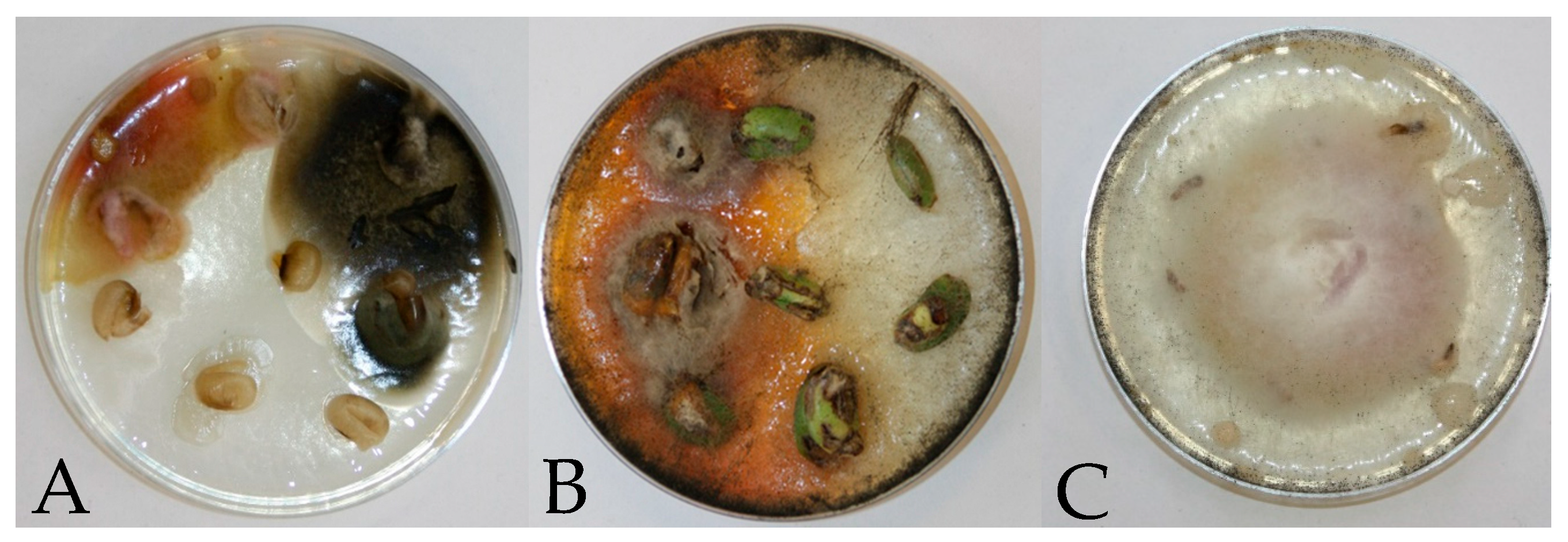
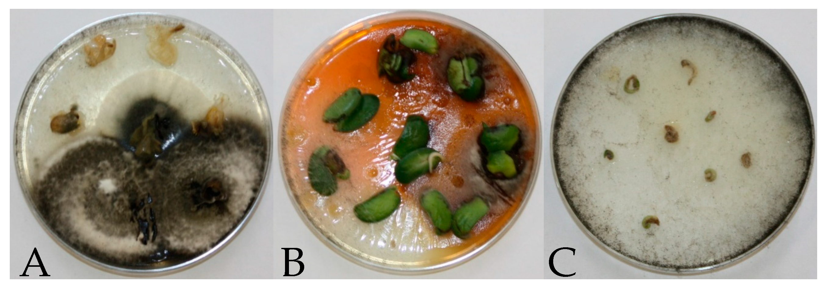
2.2.1. Mycobiome Analysis

In each year of the study, fungi colonizing the anatomical parts of mature soybean seeds (seed coat, cotyledons, embryonic axis) were identified in the samples collected from each plot using the culture-based method. Composite samples were prepared by pooling seeds from three plots in the field experiment (three replicates of 34 seeds each, a total of 102 seeds per treatment). The seeds were rinsed under running water for 15–20 min and disinfected with 70% ethanol and 1% sodium hypochlorite to remove impurities. Disinfected seeds were rinsed in sterile distilled water, dried, and separated into anatomical parts (seed coat, cotyledons, embryonic axis) under a laminar flow hood. The prepared material was spread on Petri plates (six anatomical parts per plate) containing solidified potato dextrose agar (PDA) and placed under a laminar flow hood. Each treatment consisted of 17 Petri plates. The plates were incubated at a temperature of 20–23 °C for 7–10 days. Mycelial fragments were transplanted to sterile Petri plates filled with PDA. The emerged fungal isolates were identified to the genus and species level based on their morphological features under an optical microscope (Olympus CX40, Tokyo, Japan), using the keys described by [40,41,42,43].

2.2.2. Molecular Analysis

In each year of the study, toxin-producing fungi colonizing mature soybean seeds were identified with the use of PCR methods. DNA was isolated from soybean seeds based on the method proposed by Kulik et al. [44] with some modifications. The isolated DNA was analyzed in the PCR assay to identify selected toxin-producing fungi with the use of primers specific for the genus Fusarium: P58SL—5′AGTATTCTGGCGGGCATGCCTGT3′; P28SL—5′ACAAATTACAACTCGGGCCCGAGA3′ [45]. The PCR reaction mix consisted of: Fail SafeTM PCR 2X Premix E, 0.2 U of Fail SafeTM Enzyme Mix Only polymerase (Epicentre Biotechnologies, Madison, WI, USA), 10 pM of each primer, 5.75–7.5 μL of deionized water, and 5 μL of matrix DNA. The PCR reaction was carried out with the use of the reagents supplied by Epicentre Biotechnologies (Madison, WI, USA) in the Mastercycler Gradient thermal cycler (Eppendorf, Germany) under the following conditions: 30 cycles at 94 °C for 5 min (94 °C—1 min, 68 °C—1 min, 72 °C—1 min), and 72 °C for 5 min.
The PCR products were visualized by electrophoresis on 1.5% agarose gel with the addition of ethidium bromide. The reaction products were compared against the M100-1000 DNA ladder (DNA Gdańsk, Gdańsk, Poland).
Quantitative PCR (qPCR) was carried out to identify F. graminerum with the use of the following primers: Tri5F—5′TCT TAA CAC TAG CGT GCG CCT TC3′; Tri5R—3′CAT GCC AAC GAT TGT TTG GAG GGA; a FGTri5 probe: Fam—AAC AAG GCT GCC CAC CAC TTT GCT CAG CCT—Tamra; primers specific for P. verrucosum—rRNAF TAA GGT GCC GGA ATA CAC GCT CAT; rRNAR—TAG TTC ATT CGG CCC GTG AGT TGT; and a PV rRNA probe: Fam—TCT AGA CAG CCC GAC GGT GGC CAT GG AAG T—Tamra [46]. The reaction was conducted in 25 µL of the reaction mix composed of: 12.5 µL of the TaqMan Universal PCR Master Mix [Applied Biosystems, Waltham, MA, USA], 10 pM of each primer, 10 pM of the FAM probe attached to the 5′ end and 10 pM of the TAMRA probe attached to the 3′ end as the quencher, 4.5 µL of deionized water, and 5 µL of DNA. The amplification reaction was conducted under the following conditions: 40 cycles at 95 °C for 3 min (95 °C—15 s, 60 °C—15 s, 72 °C—60 s). The analysis was conducted with the use of Applied Biosystems reagents in the 7500 FAST Real-Time PCR system [Applied Biosystems, Waltham, MA, USA]. Fungal DNA was quantified based on standard curves that were generated by serial dilutions of F. graminearum and P. verrucosum gDNA [30] using the methods described by Livak and Schmittgen [47] and Pfaffl [48] with some modifications. The results were expressed in pg of fungal DNA for each biological sample.

2.3. Statistical Analysis

The mean fungal counts were analyzed statistically using Tukey’s HSD test. The significance of differences between the mean values was determined at α = 0.05 and α = 0.01. All analyses were conducted with the use of Statistica v. 14.0 software (Tibco Software Inc., Palo Alto, CA, USA). Spearman’s rank correlation coefficients were calculated to determine the strength of the relationships between the biodiversity indicators of saprotrophic and pathogenic fungi vs. mean daily temperature and average precipitation in ten-day periods of each month (P-0.05). The biodiversity of fungal communities was assessed by calculating relative frequency (RF), dominance (Y), and species richness (S) with the following formulas: RF [%] = (ni/Ni) × 100%, Y = (ni/Ni)f, and S = number of species in each variant, where N i and n i denote the number of isolates belonging to the i­th species and genus, respectively, and f i denotes the frequency of the identified fungal genera.

2.4. Weather Conditions
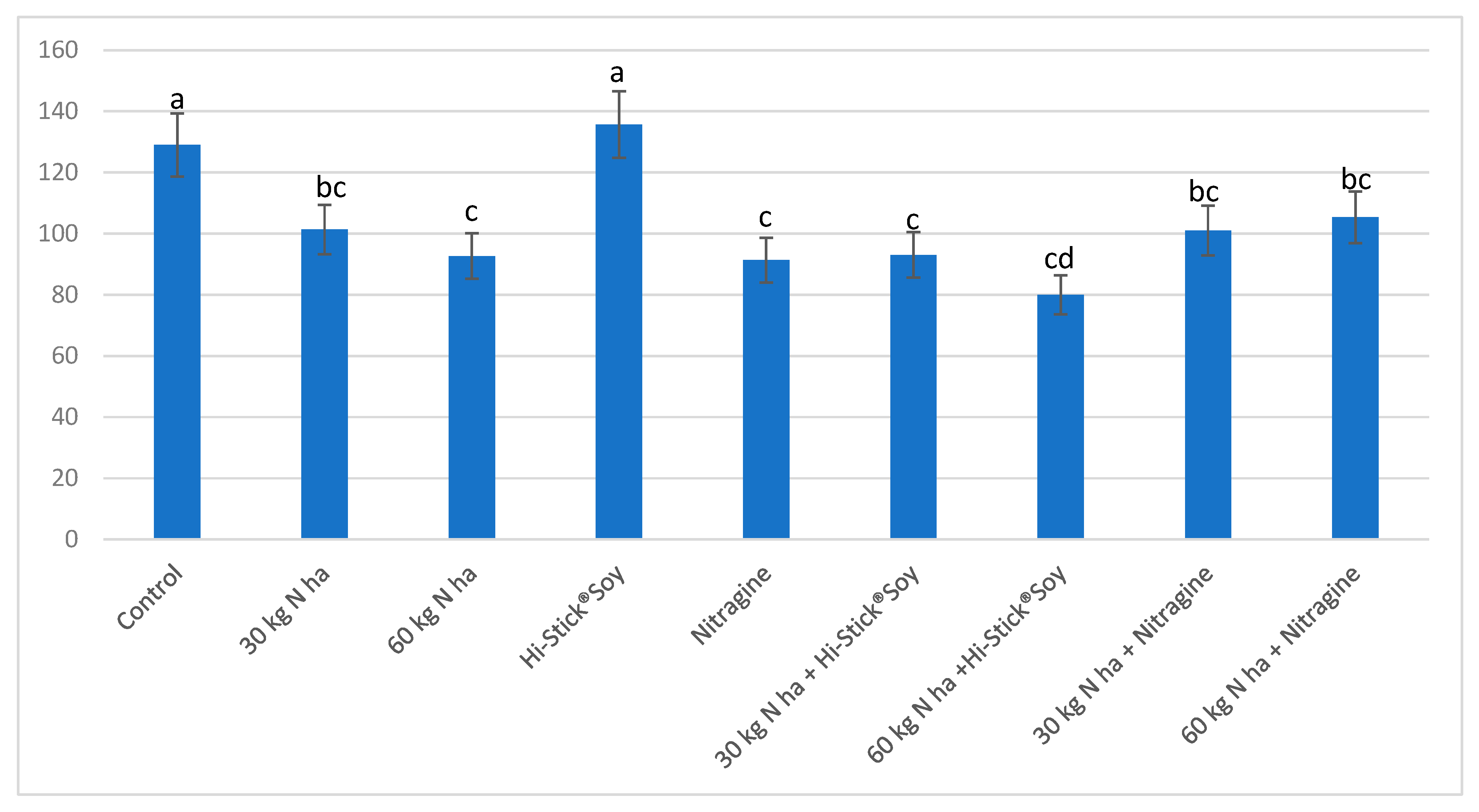
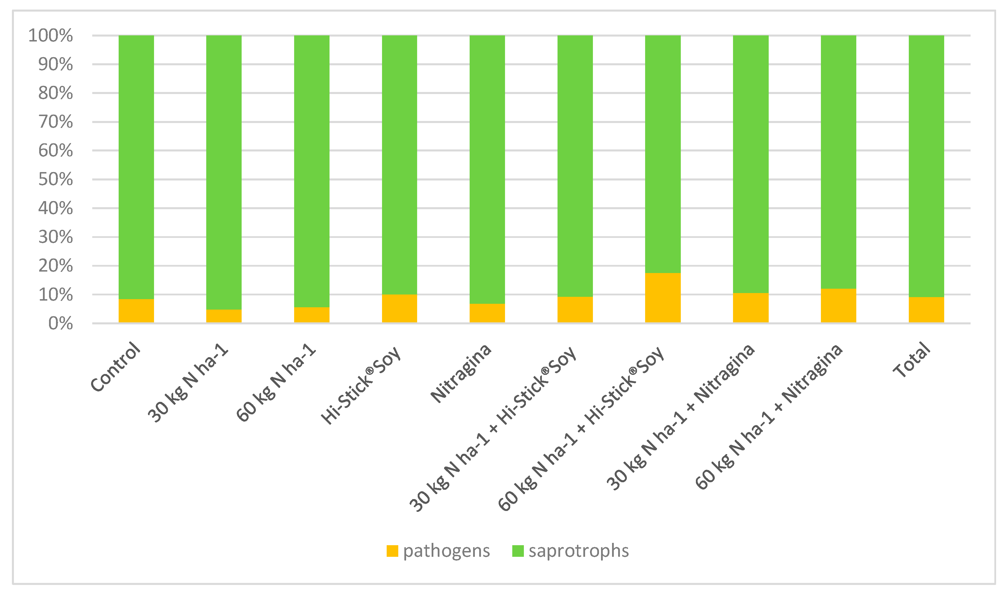
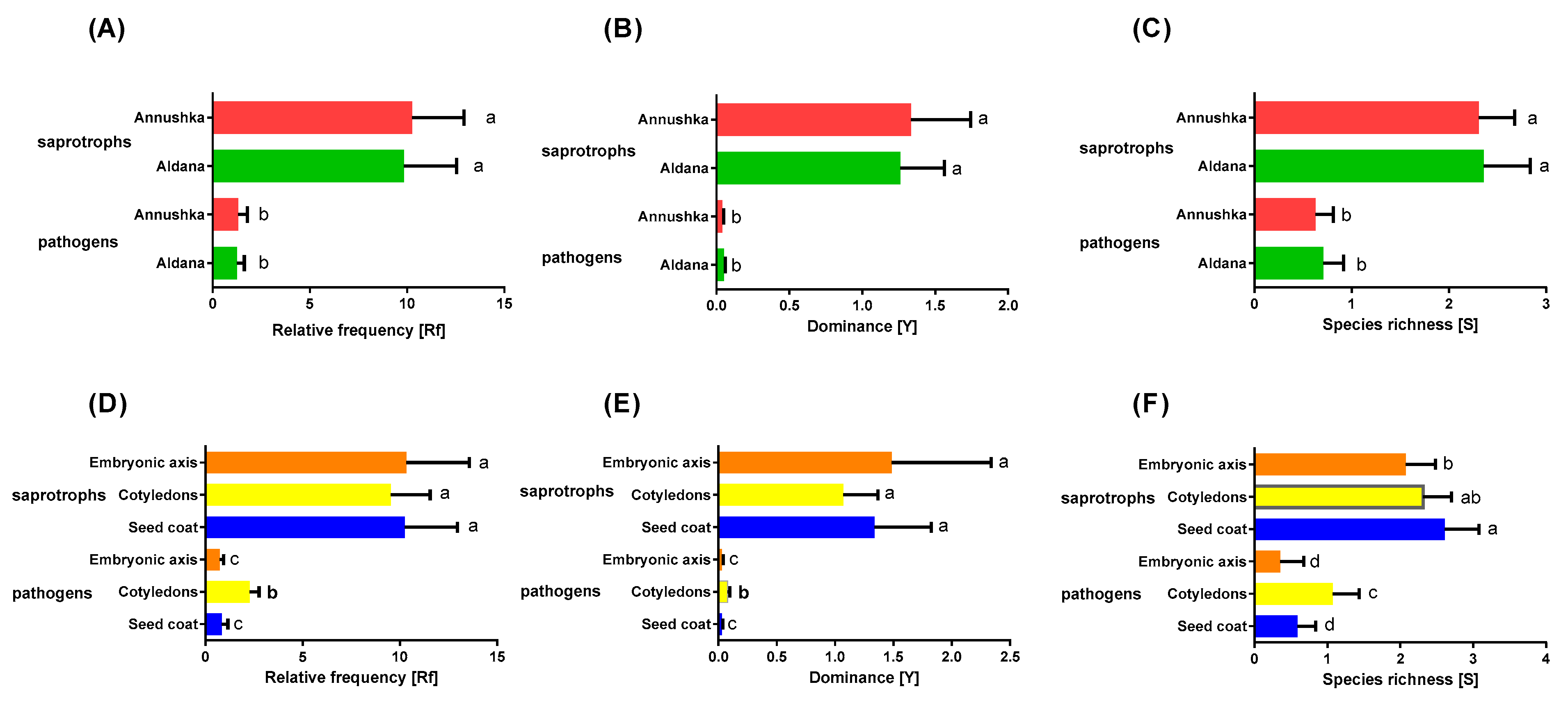
The weather conditions in Bałcyny (Region of Warmia and Mazury) in 2016–2018 are presented in Table 1. Precipitation levels were high in 2017, in particular in June (109.9 mm), July (106.1 mm), and September (211.6 mm), which delayed seed harvest. Rainfall distribution was optimal in June, July, and August of 2016 and 2018. In each year of the experiment, the temperature in June, July, and August ranged from 13 °C and 20 °C and was conducive to soybean development.

3. Results

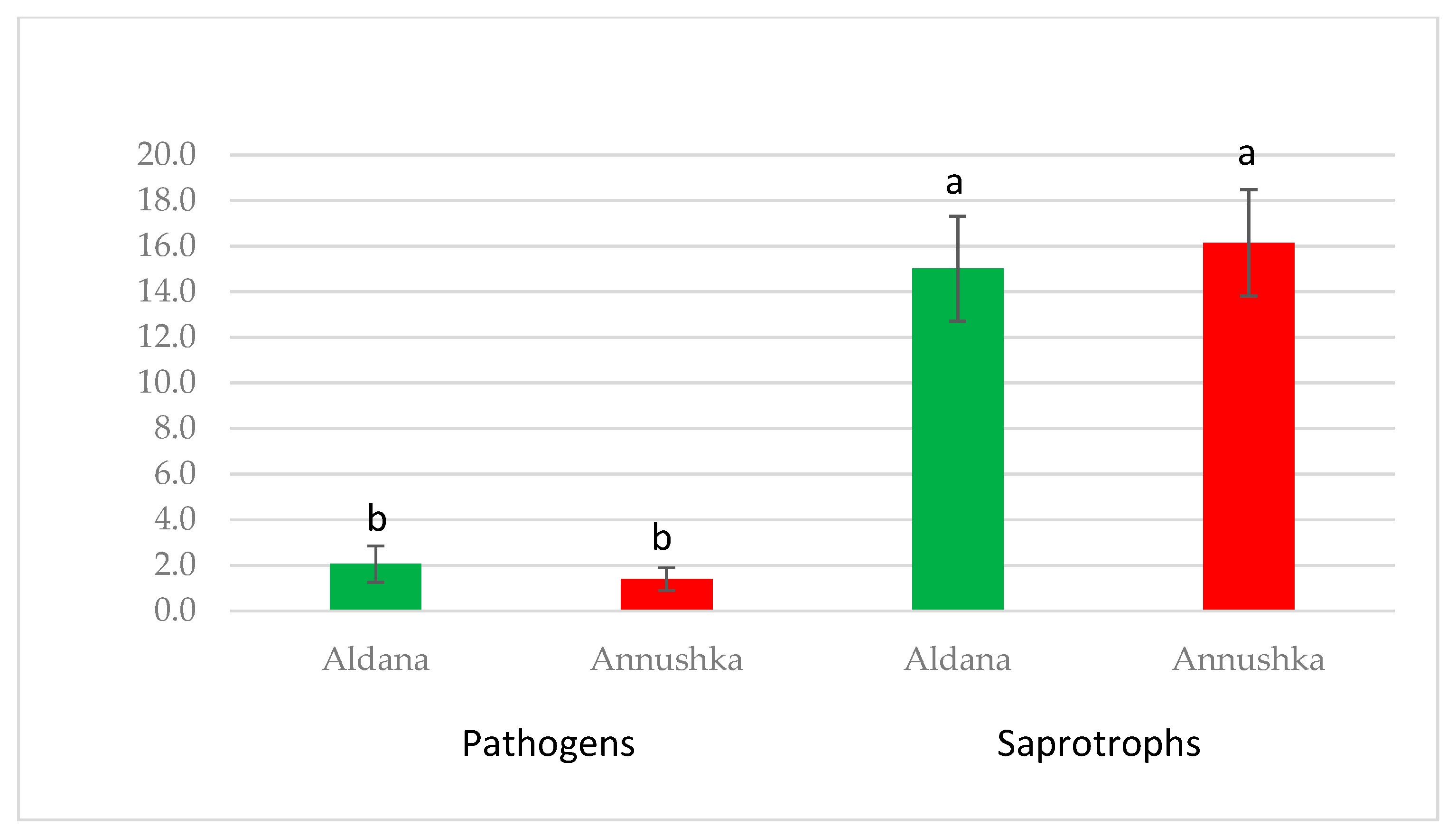
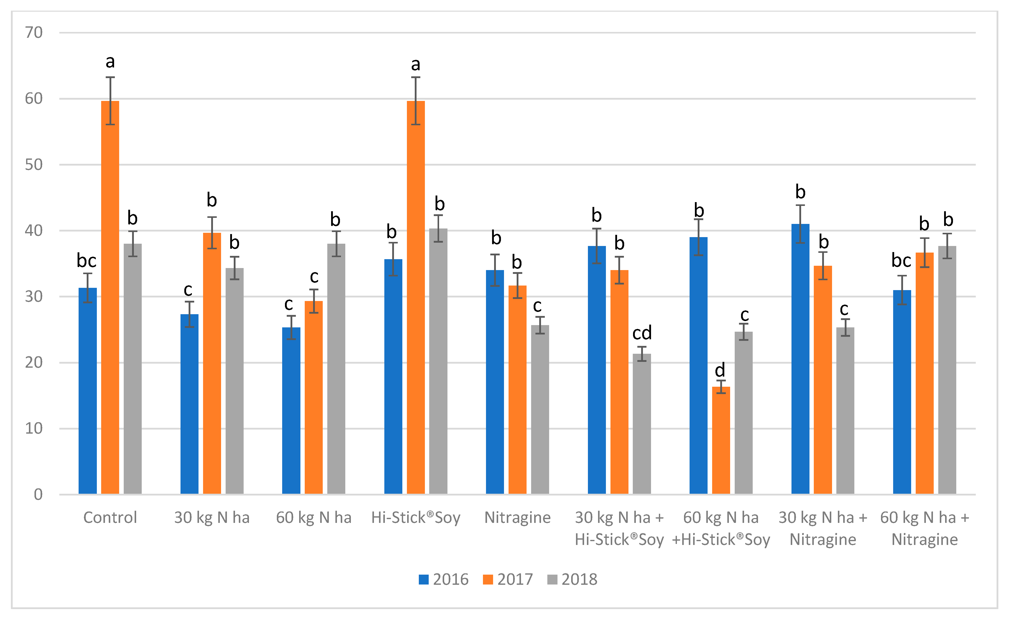
3.1. Mycobiome Analysis

Fungal colonization of the APSS (seed coat, cotyledons, embryonic axis) of soybean cvs. Aldana and Annushka was analyzed in each year of the study. A total of 2788 fungal isolates were isolated from the examined samples (Table 2, Table 3, Table 4, Table 5, Table 6, Table 7 and Table 8). The number of fungal isolates was highest (1025) in 2017 and lowest (856) in 2018. Fungal colonization intensity was higher on soybean cv. Annushka (1420 isolates) than cv. Aldana (1368 isolates) (Table 2).
The number of fungal isolates was highest on soybean seeds cv. Annushka in 2017. The fungal isolates isolated from the APSS were represented mainly by saprotrophic genera and species: Alternaria alternata, Cladosporium cladosporioides, Penicillium spp., and Rhizopus nigricans, whereas most fungal pathogens belonged to the genus Fusarium (Table 3, Table 4, Table 5, Table 6, Table 7 and Table 8, Figure 1 and Figure 2).
In 2016, a total of 454 fungal isolates were obtained from the APSS in cv. Aldana. The highest number of isolates (171) was obtained from the cotyledons, whereas the lowest number of isolates (116) was obtained from the seed coat (Table 2). The number of fungal isolates obtained from the seed coat was highest in the 60 kg N ha−1 treatment (17 isolates) and lowest in the control treatment and the 30 kg N ha−1 treatment (11 isolates each). Rhizopus nigricans was the dominant fungal species, and it was isolated from all treatments. Penicillium spp. and C. cladospirioides were identified in seven out of nine treatments. Pathogenic fungi of the genera Fusarium and Colletotrichum were isolated from the seed coat in two treatments (HiStick®Soy, Nitragina). The number of fungal isolates identified on the cotyledons was highest in the Nitragina treatment (23 isolates) and lowest in the 60 kg N ha−1 treatment (12 isolates). Rhizopus nigricans was identified in all treatments. Fungi of the genus Penicillium were detected in eight out of nine treatments. Pathogens of the genus Fusarium were found in the following treatments: 30 kg N ha−1 + HiStick®Soy, 60 kg N ha−1 + HiStick®Soy, and 30 kg N ha−1 + Nitragina, and they accounted for 50%, 31.8%, and 25% of all isolates, respectively. The number of fungal isolates identified on the embryonic axis was highest in the 30 kg N ha−1 + HiStick®Soy treatment (33 isolates) and lowest in the 60 kg N ha−1 treatment (5 isolates). Fungal pathogens were present in four treatments: 60 kg N ha−1, HiStick®Soy, Nitragina, and 60 kg N ha−1 + Nitragina. In soybean cv. Aldana, the total number of fungal isolates obtained from all seed parts was highest in 30 kg N ha−1 + HiStick®Soy and 60 kg N ha−1 + HiStick®Soy treatments (61 isolates each) and lowest in the 60 kg N ha−1 treatment (34 isolates) (Table 3).
In soybean cv. Annushka, a total of 453 fungal isolates were isolated from the analyzed seed parts in 2016 (Table 2 and Table 4). Fungi were most prevalent on the cotyledons (165 isolates), followed by the embryonic axis (155), and were least prevalent on the seed coat (133 isolates). The number of fungal isolates identified on the seed coat was highest in the 30 kg N ha−1 + Nitragina treatment (18 isolates) and lowest in the 60 kg N ha−1 + Nitragina treatment (11 isolates). Rhizopus nigricans was the dominant fungal species in all treatments. Penicillium spp. were identified in eight out of nine treatments. Potentially pathogenic fungi were isolated only from the 60 kg N ha−1 + Nitragina treatment and were identified as Colletotrichum spp. The number of fungal isolates on the cotyledons was highest in the 60 kg N ha−1 + HiStick®Soy treatment (26 isolates) and lowest in the 60 kg N ha−1 + Nitragina treatment (14 isolates). The saprotrophic R. nigricans was identified in all treatments, and Penicillium spp. were detected in eight out of nine treatments. Pathogenic fungi were identified in six treatments, and the percentage of infected seeds ranged from 9.5% to 23.1%. The majority of potentially pathogenic fungi were identified as Fusarium spp. The number of fungal isolates obtained from the embryonic axis was highest in the 30 kg N ha−1 + Nitragina treatment (33 isolates) and lowest in the 60 kg N ha−1 treatment (10 isolates). The presence of R. nigricans was confirmed in all treatments, and fungi of the genus Penicillium were detected in eight out of nine treatments. Pathogenic fungi, represented by F. culmorum, were identified only in the 60 kg N ha−1 + Nitragina treatment (Table 4). In soybean cv. Annushka, the total number of fungi colonizing all seed parts was highest in the 30 kg N ha−1 + Nitragina treatment (67 isolates) and lowest in the 60 kg N ha−1 + Nitragina treatment (41 isolates) (Table 4).
An analysis of fungal isolates isolated from both soybean cultivars in all treatments revealed that the total number of isolates increased in six treatments, but not in 30 kg N ha−1, 60 kg N ha−1, and 60 kg N ha−1 + Nitragina treatments, relative to the control treatment (Figure 1). In comparison with the control treatment, the number of potentially pathogenic species also increased in treatments with inoculation and in treatments with both N fertilizer and inoculation. It should be noted that soybean seeds were colonized mostly by saprotrophic species, whereas pathogenic fungi accounted for 1.32% to 11.11% of all isolates.
In 2017, a total of 443 fungal isolates were isolated from the examined seed parts of soybean cv. Aldana (Table 2 and Table 5). The highest number of fungal isolates (208) was obtained from the cotyledons, followed by the seed coat (173 isolates), whereas the lowest number of isolates (62) was obtained from the embryonic axis. The number of fungal isolates obtained from the seed coat was highest in the control treatment (53 isolates) and lowest in the Nitragina treatment (4 isolates).
Rhizopus nigricans was identified in six treatments. Potentially pathogenic species were isolated from seed coat in four treatments: control, HiStick®Soy, Nitragina, and 60 kg N ha−1 + Hi Stick, and they accounted for 22.6%, 40%, 50%, and 33.3% of all isolates, respectively. These fungi were represented mainly by members of the genus Fusarium as well as individual isolates of B. cinerea and Diaporthe spp. (Table 5).
The number of fungal isolates obtained from the cotyledons was also highest in the control treatment (43 isolates) and lowest in the 30 kg N ha−1 + HiStick®Soy treatment (4 isolates). Alternaria alternata was isolated from all treatments, whereas R. nigricans and Penicillium spp. were identified in five treatments (Table 5). Pathogenic fungi represented by Fusarium spp., B. cinerea, and Diaporthe spp. were detected in six treatments.
In 2017, the number of fungal isolates obtained from the embryonic axis of soybean seeds cv. Aldana was highest in the 60 kg N ha−1 + Nitragina treatment (16 isolates) and lowest in the 30 kg N ha−1 treatment (2 isolates). Fungi of the genus Penicillium were most frequently identified. Pathogenic fungi were detected in two treatments: control and 30 kg N ha−1. The total number of fungal isolates obtained from all seed parts of soybean cv. Aldana was highest in the control treatment (108 isolates) and lowest in the 30 kg N ha−1 + HiStick®Soy treatment (13 isolates) (Table 5).
In 2017, a total of 582 fungal isolates were isolated from all APSS in cv. Annushka. The number of fungal isolates was highest on the cotyledons (248 isolates), followed by the seed coat (204 isolates), and lowest on the embryonic axis (130 isolates) (Table 2 and Table 6). The number of fungal isolates obtained from the seed coat was highest in the HiStick®Soy treatment (51 isolates) and lowest in the 30 kg N ha−1 + Nitragina treatment (8 isolates). Fungi of the genus Penicillium and A. alternata were most frequently identified. Pathogenic fungi represented by F. avenaceum, B. cinerea, and Diaporthe spp. were detected in three treatments. The number of fungal isolates obtained from the cotyledons was highest in the control treatment (48 isolates) and lowest in the 60 kg N ha−1 + HiStick®Soy treatment (8 isolates). Alternaria alternata was detected in all treatments, whereas R. nigricans was identified in six out of nine treatments. Potentially pathogenic fungi of the genus Fusarium were isolated from seven treatments, and F. avenaceum was the dominant species. The number of fungal isolates obtained from the embryonic axis was highest in the HiStick®Soy treatment (23 isolates) and lowest in the 60 kg N ha−1 + HiStick®Soy treatment (7 isolates). Penicillium spp. were most frequently detected (in 8 out of 9 treatments). Potentially pathogenic fungi were identified in three treatments: control, HiStick®Soy, and 30 kg N ha−1 + HiStick®Soy. The total number of fungal isolates obtained from all APSS in cv. Annushka was highest in the HiStick®Soy treatment (107 isolates) and lowest in the 60 kg N ha−1 + HiStick®Soy treatment (26 isolates) (Table 6).
An analysis of seed colonization in both soybean cultivars and in all treatments revealed that the number of fungal isolates was lower in seven treatments than in the control treatment. The number of fungal isolates in the examined seed parts was comparable in the HiStick®Soy treatment and the control treatment. In comparison with the control treatment, the number of potentially pathogenic fungi was higher in the HiStick®Soy treatment and lower in the remaining treatments. In 2017, pathogenic fungi were detected in all treatments, and the number of pathogens ranged from 4 to 25 isolates in different treatments.
In 2018, a total of 471 fungal isolates were isolated from the APSS in cv. Aldana. The number of fungal isolates was highest on the cotyledons (205 isolates), followed by the seed coat (162 isolates), and lowest on the embryonic axis (104 isolates) (Table 2 and Table 7). The number of fungal isolates obtained from the seed coat was highest in the 60 kg N ha−1 treatment (33 isolates) and lowest in the 60 kg N ha−1 + Nitragina treatment (3 isolates). The seed coat was colonized mainly by fungi of the genus Penicillium. Pathogenic fungi of the genus Fusarium were identified in six treatments, and the percentage of infected seeds ranged from 6.1% (60 kg N ha−1) to 53.3% (60 kg N ha−1 + HiStick). The number of fungal isolates isolated from the cotyledons was highest in the control treatment (35 isolates) and lowest in the 30 kg N ha−1 + Nitragina treatment (13 isolates). Pathogens were detected in seven treatments. Penicillium spp. were identified in eight out of nine treatments, whereas A. alternata was present in seven treatments. The number of fungal isolates isolated from the embryonic axis was highest in the HiStick®Soy treatment (25 isolates) and lowest in the control treatment, 30 kg N ha−1 + HiStick®Soy, and 60 kg N ha−1 + Nitragina treatments (4 isolates each). Penicillium spp. were most prevalent. Pathogenic fungi represented by F. culmorum, F. oxysporum, and F. solani were identified in three treatments. The total number of fungal isolates obtained from all seed parts of soybean cv. Aldana was highest in the 60 kg N ha−1 treatment (87 isolates) and lowest in the 60 kg N ha−1 + Nitragina treatment (30 isolates) (Table 7).
In 2018, the APSS in cv. Annushka were colonized by 386 fungal isolates. The number of fungal isolates was highest on the seed coat (169 cultures), followed by the cotyledons (141 cultures), and lowest on the embryonic axis (76 cultures) (Table 2 and Table 8). The number of fungal isolates obtained from the seed coat of soybean cv. Annushka was highest in the 60 kg N ha−1 + Nitragina treatment (39 isolates) and lowest in the 60 kg N ha−1 treatment (9 isolates).
Fungi of the genus Penicillium were detected in all treatments, whereas A. alternata was identified in six out of nine treatments. Potentially pathogenic fungi of the genus Fusarium were present in six treatments. The number of fungal isolates isolated from the cotyledons was highest in the 30 kg N ha−1 + Nitragina treatment (26 isolates) and lowest in the 60 kg N ha−1 + HiStick®Soy treatment (4 isolates).
Fungi of the genus Penicillium were identified in eight out of nine treatments, whereas A. alternata was present in six treatments. Fusarium spp. were detected in five treatments. The number of fungi isolated from the embryonic axis was highest in the 60 kg N ha−1 + Nitragina treatment (26 cultures) and lowest in 30 kg N ha−1 and Nitragina treatments (1 culture each). Penicillium spp. were most frequently isolated, whereas the prevalence of pathogenic fungi was low. The total number of fungal isolates obtained from all APSS in cv. Annushka was highest in the 60 kg N ha−1 + Nitragina treatment (87 isolates) and the lowest in the 60 kg N ha−1 + HiStick®Soy treatment (21 isolates) (Table 8).
The statistical analysis demonstrated that the number of fungal isolates varied across treatments, years of the study, and the analyzed APSS (Table 9). The abundance of pathogenic and saprotrophic fungi did not differ significantly between soybean cultivars (Figure 3).
The statistical analysis revealed that the N rates of 30 kg N ha−1 and 60 kg N ha−1 decreased the number of fungal isolates isolated from the examined seed parts of both soybean cultivars in 2016. In turn, in 2017, the prevalence of fungi on the APSS was highest in the control treatment and the treatment where seeds were inoculated with the HiStick®Soy preparation. In the remaining treatments, the number of fungal isolates was significantly lower than in the control treatment (Figure 4). In 2018, the mean number of fungal isolates was lower in Nitragina, 30 kg N ha−1 + HiStick®Soy, 60 kg N ha−1 + HiStick®Soy, and 30 kg N ha−1 + Nitragina treatments than in the control treatment. In the remaining treatments, the number of isolated fungal isolates was similar to that noted in the control treatment (Figure 4).
The statistical analysis revealed the highest levels of fungal colonization in seeds inoculated with HiStick®Soy and in control seeds (Figure 5). Fewer fungal isolates were isolated from the remaining treatments (Figure 5).
However, it should be stressed that the APSS of both soybean cultivars were colonized mainly by saprotrophic species that accounted for 90.8% of all isolates, whereas the proportion of pathogens was 9.2% (Figure 6). The prevalence of potentially pathogenic fungi was highest in 60 kg N ha−1 + HiStick®Soy (17.5%), 60 kg N ha−1 + Nitragina (12%), and 30 kg N ha−1 + Nitragina (10.6%) treatments (Figure 6).
The fungal colonization of the APSS (seed coat, cotyledons, and the embryonic axis) was examined in the study. The analyzed seed parts were colonized by saprotrophic and pathogenic fungi (Table 10, Table 11 and Table 12; Figure 7). The prevalence of fungi was highest on the cotyledons (40.81%), followed by the seed coat (34.32%), and lowest on the embryonic axis (28.84%) (Table 2, Figure 7). These observations were made in all years of the study (2016–2018) and in both soybean cultivars.
The seed coat and the embryonic axis were colonized mainly by fungi of the genus Penicillium, whereas the cotyledons were characterized by the highest prevalence of fungi belonging to the genus Alternaria (Table 10, Table 11 and Table 12). Fusarium species were the predominant pathogens that accounted for 7.21% of all fungal isolates on the seed coat, 12.56% on the cotyledons, and 4.18% on the embryonic axis. Fusarium species were most prevalent on the cotyledons.
The biodiversity of pathogenic and saprophytic fungi was evaluated by comparing the mycobiomes of the examined APSS (Table 9 and Tables S1–S3). The values of the calculated biodiversity indicators—relative frequency (Rf), dominance (Y), and species richness (S)—imply that the proportions of saprotrophic and pathogenic fungi were not affected by treatment or soybean cultivar (Table 9, Figure 8). The analyzed APSS were characterized by a similar diversity of saprotrophic fungi (Figure 8D,E). The species richness (R) of saprotrophic fungi was higher on the seed coat and significantly lower on the embryonic axis (Figure 8F). The values of the biodiversity indicators of pathogenic fungi exhibited greater differentiation across the examined APSS (Figure 8D–F).
The values of relative frequency (Rf), dominance (Y), and species richness (S) were significantly higher (p-0.05) on the cotyledons than on the seed coat or in the embryonic axis.
The statistical analysis revealed the effect of years of the study on fungal communities (Table 9). Therefore, Spearman’s rank correlation coefficients were calculated to assess the strength of the relationship between weather conditions and fungal diversity in each year of the study (Table 13). It was found that total precipitation on 21–30 April contributed to a greater diversity of pathogenic species (Table 13), and weather conditions on 11–30 June and 1–10 August promoted the growth of fungal pathogens. Positive correlations (R) were noted between the biodiversity indicators of pathogenic species vs. mean daily temperature and total precipitation in ten-day periods of each month. Precipitation levels on 1–10 July, 21–31 July, and 1–10 August enhanced the biodiversity of saprotrophic fungi, but reduced the biodiversity of pathogenic fungi (Table 13).

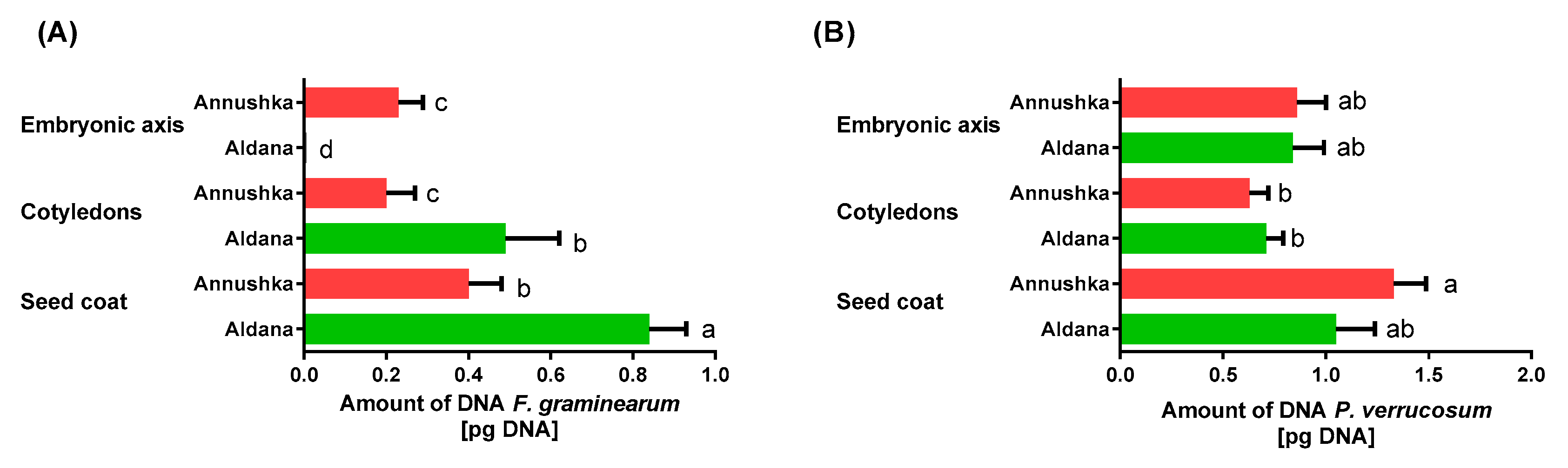
3.2. Molecular Analysis

Penicillium verrucosum was identified in the molecular analysis, and the qPCR assay confirmed that this fungal species was present in most treatments, where different inoculants and N rates were applied in the cultivation of soybean cvs. Aldana and Annushka in 2016–2018 (Table 14 and Table 15). The prevalence of Penicillium spp. (determined using culture-based methods) and the prevalence of P. verrucosum, the primary producer of ochratoxin A (OTA) (determined using molecular (qPCR) methods), are presented in Table 14 and Table 15, respectively. These analyses confirmed that P. verrucosum colonized all APSS in cvs. Aldana and Annushka in all treatments and years of the study, excluding the HiStick®Soy + 30 kg N ha−1 treatment in 2018 (Table 14 and Table 15).
Quantitative PCR revealed that the prevalence of F. graminearum and P. verrucosum on soybean seeds was influenced by all analyzed factors (cultivar [C], anatomical part [APSS], treatment [T] and year [Y]) (Table 9). The results of qPCR are presented in detail in the Supplementary Materials (Tables S4 and S5).
Fusarium species and the toxin-producing F. graminearum (which harbors the tri5 gene encoding trichothecene synthesis) were also directly identified in PCR and qPCR assays in ground APSS in both cultivars (Table 16 and Table 17). Fungi of the genus Fusarium were most frequently identified in 2017 on different seed parts in all treatments. The PCR analysis revealed that Fusarium spp. were less prevalent on the seeds of soybean cv. Aldana in 2016 and 2018, and were absent on the seeds of soybean cv. Annushka in 2018 (Table 16 and Table 17). Fusarium graminearum was identified sporadically based on a fragment of the tri5 gene. In the seeds of soybean cv. Aldana, F. graminearum was detected in the following years and samples: 2016—cotyledons in the 60 kg N ha−1 treatment; 2017—seed coat in 30 kg N ha−1, HiStick®Soy, and 60 kg N ha−1 + Nitragina treatments, and cotyledons in the 30 kg N ha−1 + Nitragina treatment; 2018—seed coat in the 30 kg N ha−1 + Nitragina treatment, and cotyledons in the 30 kg N ha−1 treatment (Table 16). In the seeds of soybean cv. Annushka, F. graminearum was detected in the following years and samples: 2016—embryonic axis in the control treatment, seed coat and embryonic axis in the 60 kg N ha−1 + HiStick®Soy treatment; 2017—embryonic axis in the control treatment, cotyledons—in the 30 kg N ha−1 treatment, seed coat in HiStick®Soy and Nitragina treatments, and cotyledons in the 30 kg N ha−1 + HiStick®Soy treatment (Table 17). The seed coat of soybean seeds cv. Aldana was most severely infected by F. graminearum (Figure 9A). Fungal colonization intensity, expressed by the amount of fungal gDNA, was somewhat lower on the cotyledons in cv. Aldana and on the seed coat in cv. Annushka. The embryonic axis was more severely infected by F. graminearum in cv. Annushka than in cv. Aldana (Figure 9A). The qPCR analysis revealed the presence of P. verrucosum DNA in all APSS of both soybean cultivars (Figure 9B). The amount of P. verrucosum DNA was highest on the seed coat, and similar values of this parameter were noted on the seed coat in cv. Aldana and in the embryonic axis in both soybean cultivars. The smallest amount of P. verrucosum DNA was determined on the cotyledons in both soybean cultivars (Figure 9B).

4. Discussion

Weather conditions that promote the growth of pathogens significantly influence the prevalence of infections in legume plants [49]. The presence and spread of pathogens are determined mainly by environmental conditions and cultivar susceptibility. Seed-borne pathogens include dormant mycelia, conidia/spores on the seed surface, or sclerotia mixed with seeds [3]. According to Vidić et al. [17], leaf and pod diseases are caused by fungal pathogens that compromise both the quality and quantity of seeds. Fungi and oomycetes are the most important soybean pathogens. Infected seeds often appear asymptomatic, which is why precise detection techniques are needed to prevent the spread of pathogens and minimize the negative effects of the resulting infections [3]. In the traditional approach, the isolated pathogens are incubated on culture media and classified under a microscope based on their morphological features. In turn, PCR-based methods are highly specific and accurate tools for differentiating between pathogenic species and their genetic variants. Pathogens colonizing plant tissues and environmental samples (such as soil or water) can be effectively quantified with the use of qPCR techniques [3,50,51,52,53,54,55].
The conducted analyses demonstrated that anatomical seed parts (seed coat, cotyledons, embryonic axis) in the tested soybean cultivars were colonized by pathogenic and saprotrophic fungi. Alternaria alternata, C. cladosporioides, Penicillium spp., and R. nigricans were the most prevalent saprotrophs, whereas pathogens were represented mainly by Fusarium spp. and individual isolates of B. cinerea, Colletotrichum spp., and Diaporthe spp. These fungi had been previously identified on soybean seeds by many researchers [20,25,26,54,56,57,58]. In a study by Escamilla et al. [59], seven species and six genera were identified in the total number of 55 fungal isolates obtained from the seeds of “MFS-561”, a sprout soybean cultivar. Alternaria, Diaporthe, and Fusarium were the dominant fungal genera. In the current study, a total of 2788 fungal isolates were isolated from soybean seed samples. The number of fungal isolates was highest in 2017 (1025) and lowest in 2018 (856). The seeds of soybean cv. Annushka were colonized by a higher number of fungi (1420) than the seeds of soybean Aldana (1368). The highest number of fungal isolates was obtained from the seeds of soybean cv. Annushka in 2017. Fungal colonization was influenced by weather conditions in each growing season. Precipitation was higher in 2017 than in 2016 and 2018. Total precipitation reached 215.1 mm in June and July, and 211.1 mm in September, which promoted fungal growth. High precipitation in September delayed soybean harvest and contributed to fungal colonization. Pszczółkowska et al. [60] reported that weather conditions were directly related to seed colonization by various fungal species and the presence of mycotoxins, i.e., undesirable secondary metabolites that are synthesized by these microorganisms. The cited authors found that the prevalence of saprotrophic, pathogenic, and toxin-producing fungi was affected mainly by temperature at the beginning of flowering (BBCH 61) and by precipitation in BBCH stages 63–64. In the work of Marcinkowska [61], fungal colonization of seeds was influenced not only by weather conditions during the growing season, but also by cultivar and environmental conditions at a given site. Precipitation was a particularly important factor that increased the prevalence of fungi on seeds. In the present study, the APSS were colonized by Penicillium spp. and R. nigricans. The highest number of Penicillium spp. isolates was noted in 2018, whereas R. nigricans was most prevalent in 2016. These fungi had been previously isolated from soybean seeds by Janda and Wolska [56] and Escamilla et al. [59]. In the work of Escamilla et al. [59], Penicillium citrinum was identified only on soybeans grown in North Carolina, and it accounted for 15.4% of all isolates. In the current study, A. alternata was isolated from all APSS in both cultivars in all years of the field experiment. Alternaria alternata was first identified on soybean seeds in the 1970s. In a study by Mengistu and Sinclair [62], this fungal species caused brown spots on the seed coat, cotyledons, and embryos. Alternaria alternata was isolated from soybean seeds by Broggi et al. [20], Rao et al. [26], and Janda and Wolska [40]. In the work of Escamilla et al. [59], the prevalence of A. alternata on soybean seeds produced in northeastern North Carolina (NC), southern Virginia (SV) and eastern Virginia (EV) in the United States was 7.7%, 34.8%, and 15.8%, respectively. Jędryczka and Kaczmarek [63] reported that lupin seeds were severely infected by fungi of the genera Penicillium and Alternaria. In the cited study, Penicillium spp. were isolated from 46.8% of non-disinfected seeds and from 35.4% of disinfected seeds. In 11.3% of the cases, these fungi were detected only on the seed coat, and in around 30% of the cases—under the seed coat. In the current experiment, C. cladosporioides was identified in all APSS in both soybean cultivars and in all years. Rao et al. [26] and Janda and Wolska [56] also found that C. cladosporioides was abundant in soybean seeds. Aureobasidium spp. exert antagonistic effects against R. solani [64], and in the present study, these fungal species were detected on the seed coat in soybean cv. Annushka in 2018. The mycoparasitic fungi of the genus Trichoderma were identified in the seed coat of cv. Aldana in 2016 and 2017, and in the seed coat and the embryonic axis of cv. Annushka in 2017. Trichoderma spp. had been previously isolated from soybean seeds by Janda and Wolska [56]. Lupin seeds were also moderately colonized by Trichoderma spp. in the work of Jędryczka and Kaczmarek [63]. In this study, potentially pathogenic fungi were represented by Fusarium spp., B. cinerea, Colletotrichum spp., Diaporthe spp., and Stemphylium botryosum. These pathogens had been previously identified on soybean seeds by Rao et al. [26], Janda and Wolska [56], and Escamilla et al. [59]. In the present experiment, most of the potentially pathogenic fungi belonged to the genus Fusarium and were represented by F. avenaceum, F. culmorum, F. equiseti, F. oxysporum, F. poae, F. solani, and F. tricinctum. Fusarium avenaceum, F. oxysporum, and F. culmorum were most prevalent and accounted for 22.4%, 22.4%, and 17.3% of Fusarium isolates, respectively. Fusarium spp. were most frequently isolated in 2018. Fusarium fungi are important pathogens that cause root rot, Fusarium wilt, sudden death syndrome (SDS), seed rot, and damping-off in soybean seedlings [39,65,66]. Petrović et al. [67] conducted pathogenicity tests and found that F. sporotrichioides, F. graminearum, F. incarnatum-F. equiseti species complex (FIESC), and F. avenaceum were the most aggressive species that caused soybean seed rot. In addition, F. subglutinans, F. solani species complex (FSSC), and F. proliferatum were characterized by high pathogenicity (80–100%) on soybean seeds, and variability in pathogenicity was observed within F. tricinctum isolates. The F. oxysporum species complex (FOSC), F. commune, and F. acuminatum were characterized by the lowest pathogenicity. Fusarium fungi had been previously isolated from soybean seeds by Żelechowski et al. [24], Janda and Wolska [56], Escamilla et al. [59], and Petrović et al. [67]. In the work of Escamilla et al. [59], Fusarium species were isolated from 38.46% and 26.32% of soybean seeds grown in North Carolina and eastern Virginia, respectively, whereas F. equiseti was identified on 30.77% of the seeds from North Carolina. Diaporthe spp. accounted for 7.7%, 34.8%, and 5.3% of all isolates from North Carolina, southern Virginia, and eastern Virginia, respectively [59]. In research studies conducted in Poland, saprotrophic and pathogenic fungi were also detected on other legume species. Alternaria alternata, B. cinerea, C. cladosporioides, E. nigrum, Fusarium spp., F. avenaceum, F. poae, Penicillium spp., R. nigricans, and Trichoderma spp. were identified in lupin by Pszczółkowska et al. [68], in peas by Pszczółkowska et al. [69], and in faba beans by Pszczółkowska et al. [60]. In other countries, these saprotrophs and pathogens were isolated from legumes by Ozgonen and Gulcu [70], and Alomran et al. [71]. Cladosporium cladosporioides was detected on soybean seeds at low frequencies (8.7–10.5%) [59]. The prevalence of fungi on different APSS has been rarely investigated in the literature, and the present study was undertaken to fill this research gap. Fungal colonization intensity was highest in the cotyledons (40.81%), lower on the seed coat (34.32%), and lowest in the embryonic axis (24.87%). Similar observations were made by Ahmed et al. [72] in soybean seed components and by Ahmed et al. [73] in cowpea seeds, where the cotyledons were most severely infected. According to Ahmed et al. [72], soybean cotyledons store food reserves, which could explain the higher degree of fungal colonization in this seed component. In turn, Aiswarya et al. [74] relied on the ISTA technique to analyze fungal colonization of groundnut seeds and reported the highest intensity of fungal infection in the seed coat (25.84%), followed by the cotyledons (16.36%), and the embryo (9.98%). They observed that the prevalence of saprophytic fungi, including A. niger, A. flavus, Rhizophus spp., and Penicillium spp., was gradually reduced from the seed coat to the embryo, whereas the occurrence of deep-seated pathogenic fungi such as Fusarium spp. and M. phaseolina increased. These findings partially corroborate the results of the present study, where the prevalence of Fusarium spp. was also highest in the cotyledons, but the highest number of Penicillium spp. isolates was obtained from the embryo (49.07%). Aiswarya et al. [74] also found that the percentage of seeds infected by Fusarium spp. and Penicillium spp. varied significantly (p < 0.05) between soybean cultivars. The seeds of cultivars MSJ and FSSBu were most abundantly colonized by Fusarium spp. (15.20%) and Penicillium spp. (8.54%), respectively. In the present study, the prevalence of Fusarium spp. also differed between the examined soybean cultivars and was determined at 9.22% in cv. Aldana and 6.85% in cv. Annushka. Seed-borne fungi pose a widespread problem in agriculture. Pathogenic fungi are identified both inside and on the surface seeds, and they can be transferred between plant generations. The spread of seed-borne fungi is influenced by various factors, including crop species, environmental conditions, agricultural practices, and seed quality. These pathogens decrease the viability of seeds, inhibit germination, infect seedlings, and pose a risk during plant growth and storage [75,76]. In the current study, the embryonic axis was colonized mainly by fungi of the genera Penicillium, Rhizopus, Alternaria, and Fusarium. These fungal species can inhibit germination and compromise the development and health status of seedlings and plants in successive stages of growth. According to Vidić et al. [17], fungi colonizing soybean seeds can inhibit germination and root development in seedlings, and they can cause root diseases and damping-off in seedlings.
The species composition and biodiversity of the seed microbiome play an important role in the protection of seeds and the maintenance of optimal soil conditions. According to Klaedtke et al. [77], the structure of seed-associated fungal microbiota is more strongly affected by the local environment than the host genotype. This observation is supported by the results of the present study, which confirmed that the applied treatments and weather conditions influenced the biodiversity of fungi colonizing soybean seeds in all years of the experiment. In contrast, Liu et al. [78] found that the species composition of fungal communities was largely determined by the soybean genotype. However, the cited authors also acknowledged the impact of environmental conditions on the biodiversity of fungal communities. In the current study, the biodiversity of pathogenic and saprotrophic fungi was affected by temperature and precipitation in different growth stages of soybeans. Water availability, temperature, and intergranular gas composition affect the germination, growth, and sporulation of fungi on seeds [79]. According to Majewska-Sawka and Nakashima [80], infections with endophytes take place in the final stage of seed development. However, other researchers identified fungal filaments in the embryonic root and the plumule, and concluded that seeds are colonized by endophytic fungi already during embryo maturation. The hyphae remain inactive at this stage, and fungi, especially molds, infect germinating seeds by colonizing the shoot tip meristem [81]. In the present study, APSS were colonized by both pathogens and saprotrophs. The abundance of both types of fungi was highest on the seed coat, whereas pathogens, in contrast to saprotrophs, colonized the cotyledons more frequently than the embryonic axis.
Fewer fungal isolates were obtained from treatments fertilized with 30 kg N∙ha−1 and 60 kg N∙ha−1 than from the control treatment. According to Wang et al. [82], N plays a key role in plant disease management, but its regulatory mechanism remains insufficiently investigated. In the cited study, nitrate was significantly more effective than ammonium in inhibiting cucumber wilt caused by Fusarium oxysporum f. sp. cucumerinum in both pot and hydroponic experiments. In the current experiment, N combined with B. japonicum (Nitragina) and B. japonicum applied alone also exerted a protective effect on soybean seeds, whereas seed inoculation with HiStick®Soy only increased fungal abundance on soybean seeds. According to many authors, Rhizobium bacteria effectively suppress fungal pathogens. In the work of Arora et al. [83], Rhizobium bacteria reduced the severity of soybean root rot caused by Phytophthora megasperma. Deshwal et al. [84] also found that various Rhizobium and Bradyrhizobium strains inhibited the growth of Macrophomina phaseolina. Bradyrhizobium japonicum effectively eliminated F. oxysporum in a study by Mariastuti et al. [85]. In turn, Nandi et al. [9] reported on the antimicrobial activity of R. japonicum and B. japonicum against F. oxysporum, A. alternata, and A. nigier.
In recent years, numerous research studies have relied on molecular methods to identify plant pathogens. Most of these studies were undertaken to analyze fungi of the genus Fusarium [3,24,30,44,54,60,68,86,87]. In the present study, P. verrucosum, the main OTA producer in Poland, was identified using a molecular method (qPCR). This toxin-producing species was detected in various APSS in cvs. Aldana and Annushka in all treatments and years, excluding the HiStick®Soy + 30 kg N∙ha−1 treatment in 2018. Fungi of the genus Fusarium and the toxin-producing F. graminearum (where trichothecene synthesis is encoded by the tri5 gene) were identified in the APSS in the studied cultivars with the use of PCR and qPCR assays. Fusarium spp. were most frequently detected in 2017, whereas F. graminearum was identified sporadically based on a fragment of tri5 gene. Żelechowski et al. [24] analyzed 104 colonies of Fusarium spp. isolated from soybean seeds in Poland in 2017–2020 based on the results of species-specific PCR and found that F. avenaceum (n = 40) F. equiseti (n = 22), and F. sporotrichioides (n = 11) were the most prevalent pathogens. The cited authors also identified F. graminearum (6 isolates) and F. culmorum (1 isolate). The isolates that were not identified in the PCR assay were subjected to whole gene sequencing. Multiple sequencing analyses based on tef-1a, top1, rpb1, rpb2, tub2, pgk, cam, and lsu genes revealed that most of these isolates belonged to the Equiseti clade. Of those, three cryptic species—F. clavum, F. flagelliforme, and FIESC31—had not been previously identified on soybeans in Poland [24]. According to Wu et al. [54], Hafez et al. [65], and van Diepeningen et al. [88], Fusarium spp. are most widely and effectively identified by DNA sequencing of PCR amplification products with the use of primers specific for selected genes or genomic regions. This method delivers rapid, accurate, sensitive, and reliable results. Chang et al. [39] examined the seeds of 12 soybean cultivars in south-western China. Based on an analysis of the internal transcribed spacer region of ribosomal DNA (rDNA ITS), the cited authors identified 148 isolates belonging to 13 fungal genera. Most of these isolates (55.0%) belonged to the genus Fusarium, followed by the genus Colletotrichum. Based on the sequencing analysis of the translation elongation factor 1-alpha (EF-1 alpha) and the DNA-directed RNA polymerase II subunit (RPB2), Fusarium isolates were classified as five distinct species: F. fujikuroi, F. proliferatum, F. verticillioides, F. asiaticum, and F. incarnatum.
Fungi not only decrease crop yields and compromise crop quality, but may also be a source of toxic metabolites in food and feed [30]. Food and feed are often contaminated by toxin-producing fungi of the genera Aspergillus, Fusarium, and Penicillium [32,89]. Aspergillus ochraceus, P. verrucosum and other Penicillium spp. synthesize OTA, the most toxic fungal metabolite [32,88,90]. Penicillium verrucosum produces OTA in cool and temperate climates [91], and it could have played an important role in the present experiment, which was conducted in north-eastern Poland. Fusarium species infect plants, cause diseases, and produce mycotoxins that contaminate the end products, including feed. Zearalenone, trichothecenes, and fumonisins are the most ubiquitous mycotoxins [87,92,93,94]. Okorski et al. [30] examined the prevalence of toxin-producing fungi and their metabolites in pig diets containing soybean meal and reported different levels of contamination in feed samples. Based on the results of qPCR assays, the cited authors concluded that the presence of Fusarium spp. producing DON, 3-AcDON, and other mycotoxins was associated with the proportion of soybean meal in the analyzed diets.

5. Conclusions

A higher number of fungal isolates was obtained from the seeds of soybean cv. Annushka (1420) than cv. Aldana (1368). The number of fungal isolates was highest on the seeds of soybean cv. Annushka in 2017. Fungal colonies on the examined APSS were represented mainly by saprotrophic genera and A. alternata, C. cladosporioides, Penicillium spp., and R. nigricans, which accounted for 90.8% of all isolates. In turn, most pathogenic fungi belonged to the genus Fusarium and accounted for 9.2% of all isolates. Fungal colonization intensity was highest in the cotyledons, lower in the seed coat, and lowest in the embryonic axis. The biodiversity of saprotrophic fungi was similar in the analyzed APSS, whereas the values of the biodiversity indicators of pathogenic fungi were higher on the cotyledons than on the seed coat and on the embryonic axis. Deep-seated infections can suppress seed germination and seedling emergence, thus decreasing yields. The phytopathological analyses conducted over a period of three years revealed that fungal colonization intensity was highest in soybean seeds inoculated with HiStick®Soy and in control seeds. The prevalence of potentially pathogenic fungal species on soybean seeds was highest in 60 kg N ha−1 + HiStick®Soy (17.5%), 60 kg N ha−1 + Nitragina (12%), and 30 kg N ha−1 + Nitragina (10.6%) treatments. The present study confirmed that fungal colonization of seeds is largely influenced by precipitation and temperature. The development of pathogenic fungi on soybean seeds was determined by temperature and precipitation on 11–30 June and 1–10 August, whereas the prevalence of saprotrophic fungi was influenced only by precipitation on 1–10 and 21–30 July and 1–10 August.

Supplementary Materials

The following supporting information can be downloaded at: https://www.mdpi.com/article/10.3390/agriculture15080857/s1, Table S1. Diversity index of fungi isolated from seeds coat soybean seeds selected cultivars: Aldana and Annushka in years of research 2016–2018; Table S2. Diversity index of fungi isolated from cotyledons soybean seeds selected cultivars: Aldana and Annushka in years of research 2016–2018; Tables S3. Diversity index of fungi isolated from embryonic axis soybean seeds selected cultivars: Aldana and Annushka in years of research 2016–2018; Table S4. Quantification of DNA F. culmorum/F. graminearum genotype by qPCR on the anatomical parts of soybean seeds in years 2016–2018 (pg DNA); Table S5. Quantification of DNA P. verrucosum by qPCR on the anatomical parts of soybean seeds in years 2016–2018 (pg DNA).

Author Contributions

Conceptualization: A.P. and G.D.; methodology, G.D., A.O., J.O. and A.P.; software, A.O. and A.P.; validation, G.D., A.O., W.G. and A.P.; formal analysis, J.O. and A.P.; investigation, G.D., A.O., J.O. and A.P.; data curation, G.D., A.O., J.O., W.G. and A.P.; writing—original draft preparation, G.D., A.O., J.O., W.G. and A.P.; writing—review and editing, G.D., J.O., A.P., A.O. and W.G.; visualization, A.O. and A.P.; supervision, A.P.; project administration, A.P. All authors have read and agreed to the published version of the manuscript.

Funding

The results presented in this paper were obtained as part of the Grant of the Polish Ministry of Agriculture and Rural Development, Project No. HOR 3.4/2016–2020, comprehensive study financed by the University of Warmia and Mazury in Olsztyn Grant No. 30.610.009-110 and funded by the Minister of Science under “the Regional Initiative of Excellence Program”.

Institutional Review Board Statement

Not applicable.

Data Availability Statement

Details are available on request from the Department of Entomology, Phytopathology and Molecular Diagnostics, Faculty of Agriculture and Forestry University of Warmia and Mazury in Olsztyn Plac Łódzki 5, 10-727 Olsztyn, Poland.

Acknowledgments

The authors express their gratitude to Aleksandra Poprawska for language editing. We would also like to thank the staff of the Agricultural Experiment Station in Bałcyny for technical support during the performance of the experiment.

Conflicts of Interest

The authors declare no conflicts of interest.

References

  1. Sudarić, A.; Kočar, M.M.; Duvnjak, T.; Zdunić, Z.; Kulundžić, A.M. Improving seed quality of soybean suitable for growing in Europe. In Soybean for Human Consumption and Animal Feed; IntechOpen: London, UK, 2019; Available online: https://www.intechopen.com/online-first/improving-seed-quality-of-soybean-suitable-for-growing-in-europe (accessed on 28 April 2024).
  2. Niwińska, B.; Witaszek, K.; Niedbała, G.; Pilarski, K. Seeds of n-GM Soybean Varieties Cultivated in Poland and Their Processing Products as High-Protein Feeds in Cattle Nutrition. Agriculture 2020, 10, 174. [Google Scholar] [CrossRef]
  3. Hosseini, B.; Voegele, R.T.; Link, T.I. Diagnosis of Soybean Diseases Caused by Fungal and Oomycete Pathogens: Existing Methods and New Developments. J. Fungi 2023, 9, 587. [Google Scholar] [CrossRef] [PubMed]
  4. Ueda, Y.; Konishi, M.; Yanagisawa, S. Molecular basis of the nitrogen response in plants. Soil. Sci. Plant Nutr. 2017, 4, 329–341. [Google Scholar] [CrossRef]
  5. Horchani, F.; Hajri, R.; Aschi-Smiti, S. Effect of ammonium or nitrate nutrition on photosynthesis, growth, and nitrogen assimilation in tomato plants. J. Plant Nutr. Soil. Sci. 2010, 173, 610–617. [Google Scholar] [CrossRef]
  6. Sharma, S. Impacts of nitrogen on plant disease severity and plant defense mechanism. Fundam. Appl. Agric. 2020, 5, 303–314. [Google Scholar] [CrossRef]
  7. Wang, Q.; Li, S.; Li, J.; Huang, D. The Utilization and Roles of Nitrogen in Plants. Forests 2024, 15, 1191. [Google Scholar] [CrossRef]
  8. Szpunar-Krok, E.; Bobrecka-Jamro, D.; Pikuła, W.; Janńczak-Pieniążek, M. Effect of Nitrogen Fertilization and Inoculation with Bradyrhizobium japonicum on Nodulation and Yielding of Soybean. Agronomy 2023, 13, 1341. [Google Scholar] [CrossRef]
  9. Nandi, R.G.; Bara, J.K.; Shrivastava, P. Antimicrobial activity of Rhizobium japonicum and Bradyrhizobium japonicum on different plant pathogenic fungal strains. Microbiol. Commun. Biosci. Biotech. Res. Comm. 2019, 12, 435–439. [Google Scholar] [CrossRef]
  10. Mabrouk, Y.; Hemissi, I.; Salem, I.B.; Mejri, S.; Saidi, M.; Belhadj, O. Potential of Rhizobia in Improving Nitrogen Fixation and Yields of Legumes. In Symbiosis; Rigobelo, E.C., Ed.; IntechOpen: Rijeka, Croatia, 2018; Chapter 6. [Google Scholar] [CrossRef]
  11. Egamberdieva, D.; Jabborova, D.; Wirth, S.J.; Alam, P.; Alyemeni, M.N.; Ahmad, P. Interactive Effects of Nutrients and Bradyrhizobium japonicum on the Growth and Root Architecture of Soybean (Glycine max L.). Front. Microbiol. 2018, 9, 1000. [Google Scholar] [CrossRef]
  12. Lin, F.; Chhapekar, S.S.; Vieira, C.C.; Da Silva, M.P.; Rojas, A.; Lee, D.; Liu, N.; Pardo, E.M.; Lee, Y.-C.; Dong, Z.; et al. Breeding for disease resistance in soybean: A global perspective. Theor. Appl. Genet. 2022, 135, 3773–3872. [Google Scholar] [CrossRef]
  13. Zhou, J.; Wang, M.; Sun, Y.; Gu, Z.; Wang, R.; Saydin, A.; Shen, Q.; Guo, S. Nitrate increased cucumber tolerance to fusarium wilt by regulating fungal toxin production and distribution. Toxins 2017, 9, 100. [Google Scholar] [CrossRef] [PubMed]
  14. Upadhyay, P.; Thakur, S.; Nagre, S.P.; Anand, K.; Saraswat, S.; Mohare, S. Review of soybean breeding for disease and insect resistance. Front. Crop Improv. 2022, 10, 541–548. [Google Scholar]
  15. Chiotta, M.L.; Alaniz Zanon, M.S.; Palazzini, J.M.; Scandiani, M.M.; Formento, A.N.; Barros, G.G.; Chulze, S.N. Pathogenicity of Fusarium graminearum and F. meridionale on soybean pod blight and trichothecene accumulation. Plant Pathol. 2016, 65, 1492–1497. [Google Scholar] [CrossRef]
  16. Zhao, L.; Wei, X.; Zheng, T.; Guo, Y.-N.; Wang, J.; Deng, J.-X.; Li, M.-J. Evaluation of pathogenic Fusarium spp. associated with soybean seed (Glycine max) in Hubei Province, China. Plant Dis. 2022, 106, 3178–3186. [Google Scholar] [CrossRef]
  17. Vidić, M.; Đorđević, V.; Petrović, K.; Miladinović, J. Review of soybean resistance to pathogens. Ratar. Povrt. 2013, 50, 52–61. [Google Scholar] [CrossRef]
  18. Carvalho, E.R.; Reis, L.V.; Rocha, D.K.; Penido, A.C.; da Rosa Mavaieie, D.P.; de Fátima Ferreira, V.; Oliveira, J.A. Incidence of fungal species in stored soybean seeds in relation to cooling before packing and to packing material. SCAP 2021, 44, 193–202. [Google Scholar] [CrossRef]
  19. Bhattacharya, K.; Raha, S. Deteriorative changes of maize, groundnut and soybean seeds by fungi in storage. Mycopathologia 2002, 155, 135–141. [Google Scholar] [CrossRef]
  20. Broggi, L.E.; González, H.H.; Resnik, S.L.; Pacin, A. Alternaria alternata prevalence in cereal grains and soybean seeds from Entre Ríos, Argentina. Rev. Iberoam. Micol. 2007, 24, 47–51. [Google Scholar] [CrossRef] [PubMed]
  21. Haikal, N.Z. Effect of filtrates of pathogenic fungi of soybean on seed germination and seedling parameters. J. Appl. Sci. Res. 2008, 4, 48–52. [Google Scholar]
  22. Medić-Pap, S.; Milošević, M.; Jasnić, S. Soybean seed-borne fungi in the Vojvodina province. Phytopathol. Pol. 2007, 45, 55–65. [Google Scholar]
  23. Nasir, N. Detecting seed borne fungi of soybean by different incubation methods. Plant Pathol. J. 2003, 2, 114–118. [Google Scholar] [CrossRef]
  24. Żelechowski, M.; Molcan, T.; Bilska, K.; Myszczyński, K.; Olszewski, J.; Karpiesiuk, K.; Wyrębek, J.; Kulik, T. Patterns of Diversity of Fusarium Fungi Contaminating Soybean Grains. Toxins 2021, 13, 884. [Google Scholar] [CrossRef] [PubMed]
  25. Olszak-Przybyś, H.; Korbecka-Glinka, G. The Diversity of Seed-Borne Fungi Associated with Soybean Grown in Southern Poland. Pathogens 2024, 13, 769. [Google Scholar] [CrossRef] [PubMed]
  26. Rao, T.V.; Rajeswari, B.; Keshavulu, K.; Varma, V.S. Studies on seedborne fungi of soybean. SSRG Int. J. Agric. Environ. Sci. (SSRG-IJAES) 2015, 2, 16–24. [Google Scholar]
  27. Sobczak, P.; Zawiślak, K.; Żukiewicz-Sobczak, W.; Mazur, J.; Nadulski, R.; Kozak, M. The assessment of microbiological purity of selected components of animal feeds and mixtures which underwent thermal processing. J. Cent. Eur. Agric. 2016, 17, 303–314. [Google Scholar] [CrossRef]
  28. Garcia, D.; Barros, G.; Chulze, S.; Ramos, A.J.; Sanchis, V.; Marín, S. Impact of cycling temperatures on Fusarium verticillioides and Fusarium graminnearum growth and mycotoxins production in soybean. J. Sci. Food Agric. 2012, 92, 2952–2959. [Google Scholar] [CrossRef]
  29. Hartman, G.L.; McCormick, S.P.; O’Donnell, K. Trichothecene-Producing Fusarium Species Isolated from Soybean Roots in Ethiopia and Ghana and their Pathogenicity of Soybean. Plant Dis. 2019, 103, 2070–2075. [Google Scholar] [CrossRef]
  30. Okorski, A.; Polak-Śliwińska, M.; Karpiesiuk, K.; Pszczółkowska, A.; Kozera, W. Real time pcr: A good tool to estimate mycotoxin contamination in pig diets. World Mycotoxin J. 2017, 10, 219–228. [Google Scholar] [CrossRef]
  31. Jedziniak, P.; Panasiuk, Ł.; Pietruszka, K.; Posyniak, A. Multiple mycotoxins analysis in animal feed with LC-MS/MS: Comparison of extract dilution and immunoaffinity clean-up. J. Sep. Sci. 2019, 42, 1240–1247. [Google Scholar] [CrossRef]
  32. Alshannaq, A.; Yu, J.-H. Occurrence, Toxicity, and Analysis of Major Mycotoxins in Food. Int. J. Environ. Res. Public. Health. 2017, 14, 632. [Google Scholar] [CrossRef]
  33. Zhang, C.; Qu, Z.; Hou, J.; Yao, Y. Contamination and Control of Mycotoxins in Grain and Oil Crops. Microorganisms 2024, 12, 567. [Google Scholar] [CrossRef] [PubMed]
  34. Pleadin, J.; Frece, J.; Markov, K. Mycotoxins in food and feed. In Advances in Food and Nutrition Research; Toldra, F., Ed.; Elsevier: Cambridge, UK, 2019; pp. 297–345. [Google Scholar]
  35. El-Sayed, R.A.; Jebur, A.B.; Kang, W.; El-Demerdash, F.M. An overview on the major mycotoxins in food products: Characteristics, toxicity, and analysis. J. Future Foods. 2022, 2, 91–102. [Google Scholar] [CrossRef]
  36. Qu, Z.; Ren, X.; Du, Z.; Hou, J.; Li, Y.; Yao, Y.; Yi, A. Fusarium mycotoxins: The major food contaminants. mLIfe 2024, 3, 176–206. [Google Scholar] [CrossRef] [PubMed]
  37. Cegielska–Radziejewska, R.; Szablewski, T.; Karolczak, K.; Kaczmarek, A.; Kijowski, J. An immunoenzymatic method for the determination of mycotoxins contents in cereals and feeds. Nauka Przyr. Technol. 2009, 3, 114. [Google Scholar]
  38. Costa, J.H.; Saraiva, K.D.C.; Morais, V.D.; Oliveira, J.T.A.; Sousa, D.O.B.; Fernandes de Melo, D.; Morais, J.K.S.; Vasconcelos, I.M. Reference gene identification for real-time PCR analyses in soybean leaves under fungus (Cercospora kikuchii) infection and treatments with salicylic and jasmonic acids. Australas. Plant Pathol. 2016, 45, 191–199. [Google Scholar] [CrossRef]
  39. Chang, X.; Li, H.; Naeem, M.; Wu, X.; Yong, T.; Song, C.; Liu, T.; Chen, W.; Yang, W. Diversity of the Seedborne Fungi and Pathogenicity of Fusarium Species Associated with Intercropped Soybean. Pathogens 2020, 9, 531. [Google Scholar] [CrossRef]
  40. Gilman, J.C. A Manual of Soil Fungi; Oxford And Ibh Publishing, Co.: New Delhi, India, 1957. [Google Scholar]
  41. Gerlach, W.; Nirenberg, H. The Genus Fusarium—A Pictorial Atlas; Biologische Bundesanstalt für Land- und Forstwirtschaft: Berlin, Germany, 1982. [Google Scholar]
  42. Leslie, J.F.; Summerell, B.A. The Fusarium Laboratory Manual; Blackwell Publishing: Hoboken, NY, USA, 2006; pp. 81–159. [Google Scholar]
  43. Watanabe, I.; Kakishima, M.; Adachi, Y.; Nakajima, H. Potential mycotoxin productivity of Alternaria alternata isolated from garden trees. Mycotoxins 2007, 57, 34–45. [Google Scholar] [CrossRef]
  44. Kulik, T.; Pszczółkowska, A.; Fordoński, G.; Olszewski, J. PCR approach based on the esyn1 gene for the detection of potential enniatin–producing Fusarium species. Int. J. Food Microbiol. 2007, 116, 319–324. [Google Scholar] [CrossRef]
  45. Hue, F.X.; Huerre, M.; Rouffault, M.A.; Bievre, C. Specific Detection of Fusarium Species in Blood and Tissues by PCR Technique. J. Clin. Microbiol. 1999, 37, 2434–2438. [Google Scholar] [CrossRef]
  46. Vegi, A.; Wolf-Hall, C. Multiplex real-time pcr marhod for detection and quantification of mycotoxigenic fungi belong to three different genera. J. Food Sci. 2013, 78, 70–76. [Google Scholar] [CrossRef]
  47. Livak, K.J.; Schmittgen, T.D. Analysis of Relative Gene Expression Data Using Real-Time Quantitative PCR and the 2−ΔΔCT Method. Methods 2001, 25, 402–408. [Google Scholar] [CrossRef]
  48. Pfaffl, M.W. A new mathematical model for relative quantification in real-time RT–PCR. Nucleic Acids Res. 2001, 29, e45. [Google Scholar] [CrossRef] [PubMed]
  49. Podleśny, J.; Podleśna, A.; Nędzi, M. Occurrence of fungal diseases caused by fungi on faba bean (Vicia faba L. var. minor Harz.) plants in different regions of Poland. Prog. Plant Prot. 2017, 57, 190–195. [Google Scholar] [CrossRef]
  50. Rocha, L.F.; Srour, A.Y.; Pimentel, M.; Subedi, A.; Bond, J.P.; Fakhoury, A.; Ammar, H.A. A panel of qPCR assays to detect and quantify soybean soil-borne pathogens. Lett. Appl. Microbiol. 2022, 76, ovac023. [Google Scholar] [CrossRef] [PubMed]
  51. Rojas, J.A.; Miles, T.D.; Coffey, M.D.; Martin, F.N.; Chilvers, M.I. Development and application of qPCR and RPA genus- and species-specific detection of Phytophthora sojae and P. sansomeana root rot pathogens of soybean. Plant Dis. 2017, 101, 1171–1181. [Google Scholar] [CrossRef]
  52. Wang, J.; Jacobs, J.L.; Byrne, J.M.; Chilvers, M.I. Improved diagnoses and quantification of Fusarium virguliforme, causal agent of soybean sudden death syndrome. Phytopathology 2015, 105, 378–387. [Google Scholar] [CrossRef]
  53. Kim, T.G.; Knudsen, G.R. Quantitative real-time PCR effectively detects and quantifies colonization of sclerotia of Sclerotinia sclerotiorum by Trichoderma spp. Appl. Soil. Ecol. 2008, 40, 100–108. [Google Scholar] [CrossRef]
  54. Wu, L.; Hwang, S.-F.; Strelkov, S.E.; Fredua-Agyeman, R.; Oh, S.-H.; Bélanger, R.R.; Wally, O.; Kim, Y.-M. Pathogenicity, Host Resistance, and Genetic Diversity of Fusarium Species under Controlled Conditions from Soybean in Canada. J. Fungi 2024, 10, 303. [Google Scholar] [CrossRef]
  55. Ciampi-Guillardi, M.; Ramiro, J.; de Mores, M.H.D.; Barbieri, C.G.; Massola, N.S., Jr. Multiplex qPCR assay for direct detection and quantification of Colletotrichum, Corynespora cassiicola and Sclerotninia sclerotiorum in soybean seeds. Plant Dis. 2020, 104, 3002–3009. [Google Scholar] [CrossRef]
  56. Janda, K.; Wolska, J. Study on the quantitative and qualitative of fungi colonizing soybeans (Glycine max L.). Pomeranian J. Life Sci. 2015, 61, 426–432. [Google Scholar] [PubMed]
  57. Kinnikar, A.; Desai, P.; Jahagirdar, S. Identification and Detection of Seed Borne Diseases of Soybean Using Image Processing—A Survey. Int. J. Emerg. Technol. Comput. Sci. Electron. 2015, 14, 363. [Google Scholar]
  58. Olszak-Przybyś, H.; Korbecka-Glinka, G.; Patkowska, E. Identification and Pathogenicity of Fusarium Isolated from Soybean in Poland. Pathogens 2023, 12, 1162. [Google Scholar] [CrossRef] [PubMed]
  59. Escamilla, D.; Rosso, M.L.; Zhang, B. Identification of fungi associated with soybeans and effective seed disinfection treatments. Food Sci. Nutr. 2019, 7, 3194–3205. [Google Scholar] [CrossRef]
  60. Pszczółkowska, A.; Okorski, A.; Fordoński, G.; Kotecki, A.; Kozak, M.; Dzienis, G. Effect of weather conditions on yield and health status of Faba bean seeds in Poland. Agronomy 2020, 10, 48. [Google Scholar] [CrossRef]
  61. Marcinkowska, J. Fungi occurrence on seeds of field pea. Acta Mycol. 2008, 43, 77–89. [Google Scholar] [CrossRef]
  62. Mengistu, A.; Sinclair, J. Seed borne microorganisms of Ethiopian-grown soybean and chickpea seeds. Plant Diesease. Rep. 1979, 63, 616–619. [Google Scholar]
  63. Jędryczka, M.; Kaczmarek, J. Infestation of commercial seed lots of narrow-leafed lupin by pathogenic and saprotrophic fungi. Fragm. Agron. 2012, 29, 63–69. [Google Scholar]
  64. Di Francesco, A.; Di Foggia, M.; Corbetta, M.; Baldo, D.; Ratti, C.; Baraldi, E. Biocontrol Activity and Plant Growth Promotion Exerted by Aureobasidium pullulans Strains. J. Plant Growth Regul. 2021, 40, 1233–1244. [Google Scholar] [CrossRef]
  65. Hafez, M.; Abdelmagid, A.; Aboukhaddour, R.; Adam, L.R.; Daayf, F. Fusarium Root Rot Complex in Soybean: Molecular Characterization, Trichothecene Formation, and Cross-Pathogenicity. Phytopathology 2021, 111, 2287–2302. [Google Scholar] [CrossRef]
  66. Diaz-Arias, M.M.; Leandro, L.F.S.; Munkvold, G. Aggressiveness of Fusarium species and impact of root infection on growth and yield of soybean. Phytopathology 2013, 103, 822–832. [Google Scholar] [CrossRef]
  67. Petrović, K.; Orzali, L.; Krsmanović, S.; Valente, M.T.; Tolimir, M.; Pavlov, J.; Riccioni, L. Genetic Diversity and Pathogenicity of the Fusarium Species Complex on Soybean in Serbia. Plant Dis. 2024, 108, 1851–1860. [Google Scholar] [CrossRef] [PubMed]
  68. Pszczółkowska, A.; Okorski, A.; Fordoński, G.; Prusiński, J.; Faligowska, A.; Borowska, M. Fungal colonization of seeds of three lupine species in different regions of Poland. Acta Agrobot. 2017, 70, 1714. [Google Scholar] [CrossRef]
  69. Pszczółkowska, A.; Okorski, A.; Fordoński, G.; Faligowska, A.; Kaszkowiak, E.; Olszewski, J.; Chareńska, A. The frequency of occurrence of pathogenic and saprotrophic fungi in pea seeds in different regions of Poland. Legume Res.-Int. J. 2019, 42, 270–276, 118:2357149008. [Google Scholar] [CrossRef]
  70. Ozgonen, H.; Gulcu, M. Determination of mycoflora of pea (Pisum sativum) seeds and the effects of Rhizobium leguminosorum on fungal pathogens of peas. Afr. J. Biotechnol. 2011, 10, 6235–6240. [Google Scholar] [CrossRef]
  71. Alomran, M.M.; Lupien, S.L.; Coyne, C.J.; Dugan, F.M. Mycobiota of Lupinus albus seed from a public germplasm collection. N. Am. Fungi 2013, 8, 1–15. [Google Scholar] [CrossRef]
  72. Ahmed, O.; Balogun, O.S.; Fawole, O.B.; Fabiyi, O.A.; Aliyu, T.H.; Kassoum, K.O. Seed-borne fungi of soybeans (Glycine max [L.] Merr) in the Guinea savannah agroecology of Nigeria. J. Agric. Sci. 2016, 61, 57–68. [Google Scholar] [CrossRef]
  73. Ahmed, O.; Fawole, O.B.; Balogun, O.S. Component analysis for seed mycoflora in four cultivars of cowpea (Vigna unguiculata [L.] Walp). Cent. Point (Sci. Ed.) 2006, 14, 71–77. [Google Scholar]
  74. Aiswarya, N.; Bhattiprolu, S.L.; Bayyapu Reddy, K.; Rama Rao, G. Location of seedborne fungi in farmers saved samples of groundnut variety tag 24. Int. J. Chem. Stud. 2019, 7, 3103–3105. [Google Scholar]
  75. Martín, I.; Gálvez, L.; Guasch, L.; Palmero, D. Fungal Pathogens and Seed Storage in the Dry State. Plants 2022, 11, 3167. [Google Scholar] [CrossRef]
  76. Tayyab, M.; Ali, Q.; Sultana, M.; Iqbal, A.; Ali Khan, R.M.; Hiza Noor, H.; Ahmad, K.; Aleem, M.; Anmol, S.; Faisal, M.; et al. Detection of Seed Borne Fungi Associated with Glycine max and its Management Through Physio–Chemical Treatments. J. Xi’an Shiyou Univ. Nat. Sci. Ed. 2024, 20, 543–603. [Google Scholar]
  77. Klaedtke, S.; Jacques, M.A.; Raggi, L.; Préveaux, A.; Bonneau, S.; Negri, V.; Chablem, V.; Barret, M. Terroir is a key driver of seed-associated microbial assemblages. Environ. Microbiol. 2016, 18, 1792–1804. [Google Scholar] [CrossRef] [PubMed]
  78. Liu, J.; Deng, J.C.; Yang, C.Q.; Huang, N.; Chang, X.L.; Zhang, J.; Yang, F.; Liu, W.G.; Wang, X.C.; Yong, T.W.; et al. Fungal Diversity in Field Mold-Damaged Soybean Fruits and Pathogenicity Identification Based on High-Throughput rDNA Sequencing. Front. Microbiol. 2017, 8, 779. [Google Scholar] [CrossRef] [PubMed]
  79. Zhang, S.-B.; Zhai, H.-C.; Huang, S.-X.; Cai, J.-P. A site-directed CO2 detection method for monitoring the spoilage of stored grains by insects and fungi in Chinese horizontal warehouses. J. Stored Prod. Res. 2014, 59, 146–151. [Google Scholar] [CrossRef]
  80. Majewska-Sawka, A.; Nakashima, H. Endophyte transmission via seeds of Lolium perenne L. immunodetection of fungal antigens. Fungal Genet. Biol. 2004, 41, 534–541. [Google Scholar] [CrossRef]
  81. Ngugi, H.K.; Scherm, H. Biology of flower-infecting fungi. Annu. Rev. Phytopathol. 2006, 44, 261–282. [Google Scholar] [CrossRef]
  82. Wang, M.; Sun, Y.; Gu, Z.; Wang, R.; Sun, G.; Zhu, C.; Guo, S.; Shen, Q. Nitrate Protects Cucumber Plants Against Fusarium oxysporum by Regulating Citrate Exudation. Plant Cell Physiol. 2016, 57, 2001–2012. [Google Scholar] [CrossRef]
  83. Arora, N.K.; Kang, S.C.; Maheshwari, D.K. Isolation of siderophore-producing strains of Rhizobium meliloti and their biocontrol potential against Macrophomina phaseolina that causes charcoal rot of groundnut. Curr. Sci. 2001, 81, 673–677. [Google Scholar]
  84. Deshwal, V.K.; Dubey, R.C.; Maheshwari, D.K. Isolation of plant growth-promoting strains of Bradyrhizobium (Arachis) spp. with biocontrol potential against Macrophomina phaseolina causing charcoal rot of peanut. Curr. Sci. 2003, 84, 443. [Google Scholar]
  85. Mariastuti, H.D.; Listiyowati, S.; Wahyudi, A.T. Antifungal activity of soybean rhizosphere actinomycetes producing bioactive compounds against Fusarium oxysporum. Biodiversitas 2018, 19, 2127–2133. [Google Scholar] [CrossRef]
  86. Mishra, P.K.; Fox, R.T.; Culham, A. Development of a PCR-based assay for rapid and reliable identification of pathogenic Fusaria. FEMS Microbiol. Lett. 2003, 218, 329–332. [Google Scholar] [CrossRef]
  87. Nicholson, P.; Chandler, E.; Draeger, R.C.; Gosman, N.E.; Simpson, D.R.; Thomsett, M.; Wilson, A.H. Molecular tools to study epidemiology and toxicology of Fusarium head blight of cereals. Eur. J. Plant Pathol. 2003, 109, 691–703. [Google Scholar] [CrossRef]
  88. van Diepeningen, A.D.; Brankovics, B.; Iltes, J.; van der Lee, T.A.J.; Waalwijk, C. Diagnosis of Fusarium Infections: Approaches to Identification by the Clinical Mycology Laboratory. Curr. Fungal Infect. Rep. 2015, 9, 135–143. [Google Scholar] [CrossRef] [PubMed]
  89. Sweeney, M.J.; Dobson, D.W. Mycotoxin production by Aspergillus, Fusarium and Penicillium species. Int. J. Food Microbiol. 1998, 43, 141–158. [Google Scholar] [CrossRef] [PubMed]
  90. Bennett, J.W.; Klich, M. Mycotoxins. Clin. Microbiol. Rev. 2003, 16, 497–516. [Google Scholar] [CrossRef]
  91. Duarte, S.C.; Pena, A.; Lino, C.M. A review on ochratoxin A occurrence and effects of processing of cereal and cereal derived food products. Food Microbiol. 2010, 27, 187–198. [Google Scholar] [CrossRef]
  92. Perkowski, J.; Stuper, K.; Buśko, M.; Góral, T.; Kaczmarek, A.; Jeleń, H. Differences in metabolomic profiles of the naturally contaminated grain of barley, oats and rye. J. Cereal Sci. 2012, 56, 544–551. [Google Scholar] [CrossRef]
  93. Dawidziuk, A.; Koczyk, G.; Popiel, D.; Kaczmarek, J.; Buśko, M. Molecular diagnostics on the toxigenic potential of Fusarium spp. plant pathogens. J. Appl. Microbiol. 2014, 116, 1607–1620. [Google Scholar] [CrossRef]
  94. Perincherry, L.; Lalak-Kańczugowska, J.; Stępień, Ł. Fusarium-produced mycotoxins in plant-pathogen interactions. Toxins 2019, 11, 664. [Google Scholar] [CrossRef]
Figure 1. Colonization of the APSS in cv. Aldana by microscopic fungi. (A)—seed coat; (B)—cotyledons; (C)—embryonic axis.
Agriculture 15 00857 g001
Figure 2. Colonization of the APSS in cv. Annushka by microscopic fungi. (A)—seed coat; (B)—cotyledons; (C)—embryonic axis.
Agriculture 15 00857 g002
Figure 3. Average number of isolates of pathogenic and saprotrophic fungi obtained from the seeds of the evaluated soybean cultivars in 2016–2018, (a, b, c…—mean values marked with the same letters do not differ significantly at p = 0.05).
Agriculture 15 00857 g003
Figure 4. Mean number of fungal isolates obtained from the anatomical parts of soybean seeds (APSS) in different treatments in 2016, 2017, and 2018 (a, b, c…—mean values marked with the same letters do not differ significantly at p = 0.05).
Agriculture 15 00857 g004
Figure 5. Mean number of fungal isolates obtained from the anatomical parts of soybean seeds(APSS) in different treatments in 2016–2018 (a, b, c…—mean values marked with the same letters do not differ significantly at p = 0.05).
Agriculture 15 00857 g005
Figure 6. Proportions (%) of pathogenic and saprotrophic fungi in the total number of fungal isolates obtained from the anatomical parts of soybean seeds (APSS) in different treatments.
Agriculture 15 00857 g006
Figure 7. Proportions (%) of anatomical parts of soybean seeds (APS) by fungi.
Agriculture 15 00857 g007
Figure 8. Biodiversity indicators (Rf, Y and S) of pathogenic and saprotrophic fungi colonizing the examined soybean cultivars (AC) and anatomical seed parts (DF) (p-0.05; ±SD) in 2016–2018, a, b, c…—mean values marked with the same letters do not differ significantly at p = 0.05).
Agriculture 15 00857 g008
Figure 9. Quantification of fungal DNA by qPCR in the anatomical parts of soybean seeds APSS in 2016–2018: (A) F. graminearum genotype; (B) P. verrucosum, a, b, c…—mean values marked with the same letters do not differ significantly at p = 0.05).
Agriculture 15 00857 g009
Table 1. Weather conditions in Bałcyny (Region of Warmia and Mazury) in 2016–2018.
Year/Month2016201720181981–2015 *
Precipitation (mm)
April33.152.128.630.0
May70.834.041.059.0
June66.3109.964.772.0
July138.6106.1140.785.0
August71.954.831.266.0
September17.1211.629.155.0
Temperature (°C)
April8.86.711.97.8
May14.81316.513.3
June13.016.717.915.9
July18.517.219.918.3
August17.518.820.517.9
September14.813.315.313.1
* Long-term average (1981–2015).
Table 2. Total fungal isolates obtained from the anatomical parts of soybean seeds (APSS).
Anatomical Part/Year201620172018Total
Aldana
Seed coat116 *173162451
Cotyledons171208205584
Embryonic axis16762104333
Total isolates4544434711368
Annushka
Seed coat133204169506
Cotyledons165248141554
Embryonic axis15513075360
Total isolates4535823861420
Total fungal isolates90710258562788
*—number of fungal isolates.
Table 3. Fungi colonizing the anatomical parts of soybean seeds (APSS) in cv. Aldana in treatments supplied with different nitrogen rates and different inoculants in 2016.
Fungal Genus/SpeciesTreatment
123456789
Seed coat
Alternaria alternata (Fr.) Keissler1 * 2 2353
Cladosporium cladosporioides (Fries.) de Vries223324 2
Colletotrichum spp. 1
Fusarium spp. 2
Penicillium spp.337 3 342
Rhizopus nigricans Ehrenb.565366766
Trichoderma spp. 4
Total111117121212131513
% pathogens0.00.00.016.78.30.00.00.00.0
% saprotrophs100.0100.0100.0083.391.7100.0100.0100.0100.0
Cotyledons
Alternaria alternata (Fr.) Keissler 7
Cladosporium cladosporioides (Fries.) de Vries 33 2
Fusarium culmorum (W.G. Smith) Sacc. 8 4
Fusarium spp. 7
Penicillium spp.141271512 2611
Rhizopus nigricans Ehrenb.785785656
Verticillium spp. 1
Total212012222316221619
% pathogens0.000.000.00.00.050.031.831.250.0
% saprotrophs100.0100.0100.0100.0100.050.068.268.75100.0
Embryonic axis
Alternaria alternata (Fr.) Keissler 1 7 1
Cladosporium cladosporioides (Fries.) de Vries3 121321
Colletotrichum spp. 3
Fusarium culmorum (W.G. Smith) Sacc. 2
Fusarium spp. 11
Penicillium spp.84 942481811
Rhizopus nigricans Ehrenb.832756966
Total1975191333262520
% pathogens0.00.020.05.323.10.00.00.010.0
% saprotrophs100.0100.080.094.776.9100.0100.0100.090.0
Total number of fungal isolates obtained from seeds513834534861615652
(1) Control; (2) 30 kg N ha−1; (3) 60 kg N ha−1; (4) HiStick®Soy; (5) Nitragina; (6) 30 kg N ha−1 + HiStick®Soy; (7) 60 kg N ha−1 + HiStick®Soy; (8) 30 kg N ha−1 + Nitragina; (9) 60 kg N ha−1 + Nitragina. *—number of fungal isolates.
Table 4. Fungi colonizing the anatomical parts of soybean seeds (APSS) in cv. Annushka in treatments supplied with different nitrogen rates and different inoculants in 2016.
Fungal Genus/SpeciesTreatment
123456789
Seed coat
Alternaria alternata (Fr.) Keissler 5 * 2
Cladosporium cladosporioides (Fries.) de Vries 3
Colletotrichum spp. 2
Epicoccum nigrum Ehrenb. 1
Penicillium spp.93241339
Rhizopus nigricans Ehrenb.61014612101189
Total151316151316161811
% pathogens0.00.00.00.00.00.00.00.018.1
% saprotrophs100.0100.0100.0100.0100.0100.0100.0100.081.8
Cotyledons
Alternaria alternata (Fr.) Keissler24 3
Colletotrichum spp. 1
Fusarium avenaceum (Corda ex Fr.) Sacc. 1
Fusarium culmorum (W.G. Smith) Sacc.1 1 3 2
Fusarium spp.2 2223
Penicillium spp.4373 7824
Rhizopus nigricans Ehrenb.69911181212147
Total151616192221261614
% pathogens20.00.00.010.518.29.523.10.021.4
% saprotrophs80.0100.0100.089.581.890.576.9100.078.5
Embryonic axis
Alternaria alternata (Fr.) Keissler 31
Cladosporium cladosporioides (Fries.) de Vries1 1 1
Fusarium culmorum (W.G. Smith) Sacc. 2
Penicillium spp.643576521
Rhizopus nigricans Ehrenb.6117151298912
Total131510201915143316
% pathogens0.00.00.00.00.00.00.00.012.5
% saprotrophs100.0100.0100.0100.0100.0100.0100.0100.087.5
Total number of fungal isolates obtained from seeds434442545452566741
(1) Control; (2) 30 kg N ha−1; (3) 60 kg N ha−1; (4) HiStick®Soy; (5) Nitragina; (6) 30 kg N ha−1 + HiStick®Soy; (7) 60 kg N ha−1 + HiStick®Soy; (8) 30 kg N ha−1 + Nitragina; (9) 60 kg N ha−1 + Nitragina. *—number of fungal isolates.
Table 5. Fungi colonizing the anatomical parts of soybean seeds (APSS) in cv. Aldana in treatments supplied with nitrogen rates and different inoculants in 2017.
Fungal Genus/SpeciesTreatment
123456789
Seed coat
Alternaria alternata (Fr.) Keissler41 *7176 5278
Botrytis cinerea Pers ex Pers2
Cladosporium cladosporioides (Fries.) de Vries 72 52
Diaporthe spp.1
Epicoccum nigrum Ehrenb. 2
Fusarium avenaceum (Corda ex Fr.) Sacc.6 5
Fusarium tricinctum (Corda) Sacc. 4
Fusarium spp.3 32
Penicillium spp. 4 23
Rhizopus nigricans Ehrenb. 6 42 447
Total5317242045122117
% pathogens22.60.00.040.050.00.033.30.00.0
% saprotrophs77.4100.0100.060.050.0100.066.7100.0100.0
Cotyledons
Alternaria alternata (Fr.) Keissler383618271346119
Botrytis cinerea Pers. ex Pers.1
Cladosporium cladosporioides (Fries.) de Vries 4 1
Diaporthe spp.1
Fusarium avenaceum (Corda ex Fr.) Sacc. 2 2 2
Fusarium equiseti (Corda) Sacc. 24 2
Fusarium spp. 2
Penicillium spp. 31 222
Rhizopus nigricans Ehrenb.3 33 31
Total4338184121482114
% pathogens4.75.30.09.819.10.00.019.114.3
% saprotrophs95.394.7100.090.280.9100.0100.080.985.7
Embryonic axis
Alternaria alternata (Fr.) Keissler7 45
Botrytis cinerea Pers. ex Pers.4
Cladosporium cladosporioides (Fries.) de Vries 2
Diaporthe spp.1
Fusarium avenaceum (Corda ex Fr.) Sacc. 2
Penicillium spp. 2412249
Rhizopus nigricans Ehrenb. 2117
Trichoderma spp. 2
Total122611343516
% pathogens41.7100.00.00.00.00.00.00.00.0
% saprotrophs58.30.0100.0100.0100.0100.0100.0100.0100.0
Total number of fungal isolates obtained from seeds1085748722813234747
(1) Control; (2) 30 kg N ha−1; (3) 60 kg N ha−1; (4) HiStick®Soy; (5) Nitragina; (6) 30 kg N ha−1 + HiStick®Soy; (7) 60 kg N ha−1 + HiStick®Soy; (8) 30 kg N ha−1 + Nitragina; (9) 60 kg N ha−1 + Nitragina. *—number of fungal isolates.
Table 6. Fungi colonizing the anatomical parts of soybean seeds (APSS) in cv. Annushka in treatments supplied with different nitrogen rates and different inoculants in 2017.
Fungal Genus/SpeciesTreatment
123456789
Seed coat
Alternaria alternata (Fr.) Keissler9 *494321924
Aureobasidium spp.4
Btrytis cinerea Pers. ex Pers. 1
Cladosporium cladosporioides (Fries.) de Vries 3
Diaporthe spp. 1
Fusarium avenaceum (Corda ex Fr.) Sacc. 24 2
Penicillium spp. 222521729
Rhizopus nigricans Ehrenb.16 142223
Trichoderma spp. 15
Total14121351244411827
% pathogens0.00.015.411.80.04.50.00.00.0
% saprotrophs100.0100.084.688.2100.095.5100.0100.0100.0
Cotyledons
Alternaria alternata (Fr.) Keissler3121102918224213
Fusarium avenaceum (Corda ex Fr.) Sacc. 26422
Fusarium equiseti (Corda) Sacc. 2
Fusarium poae (Peck) Wollenw. 2
Fusarium tricinctum (Corda) Sacc.22
Fusarium spp. 2
Penicillium spp.82 24512
Phoma glomerata (Corda) Wollenw.1
Rhizopus nigricans Ehrenb.642 54 2
Stemphylium botryosum Wallr. 6
Total48311833253283419
% pathogens4.212.933.312.18.012.50.00.021.1
% saprotrophs95.887.166.787.992.087.5100.0100.078.9
Embryonic axis
Alternaria alternata (Fr.) Keissler5 201282
Botrytis cinerea Pers. ex Pers. 1
Fusarium avenaceum (Corda ex Fr.) Sacc. 2
Fusarium spp.2 2
Penicillium spp.289 2351512
Rhizopus nigricans Ehrenb. 4
Trichoderma spp. 11 5
Total919923181371517
% pathogens44.40.00.013.00.015.40.00.00.0
% saprotrophs55.6100.0100.087.0100.084.6100.0100.0100.0
Total number of fungal isolates obtained from seeds7162401076789265763
(1) Control; (2) 30 kg N ha−1; (3) 60 kg N ha−1; (4) HiStick®Soy; (5) Nitragina; (6) 30 kg N ha−1 + HiStick®Soy; (7) 60 kg N ha−1 + HiStick®Soy; (8) 30 kg N ha−1 + Nitragina; (9) 60 kg N ha−1 + Nitragina. *—number of fungal isolates.
Table 7. Fungi colonizing the anatomical parts of soybean seeds (APSS) in cv. Aldana in treatments supplied with different nitrogen rates and different inoculants in 2018.
Fungal Genus/SpeciesTreatment
123456789
Seed coat
Alternaria alternata (Fr.) Keissler3 *55 5 9
Cladosporium cladosporioides (Fries.) de Vries 18
Fusarium culmorum(W.G. Smith) Sacc.2 2 2
Fusarium oxysporum Schltdl. 22 4
Fusarium solani (Mart.) Sacc. 22
Penicillium spp.922231015 4 3
Periconia spp. 2
Rhizopus nigricans Ehrenb.413 3
Trichoderma spp.
Total18303312221815113
% pathogens11.10.06.116.79.10.053.318.20.0
% saprotrophs88.9100.095.983.390.9100.046.781.8100.0
Cotyledons
Alternaria alternata (Fr.) Keissler2444 17 355
Cladosporium cladosporioides (Fries.) de Vries2 4 2 2
Fusarium avenaceum (Corda ex Fr.) Sacc. 2
Fusarium culmorum (W.G. Smith) Sacc.2 2 2
Fusarium oxysporum Schltdl. 3 34427
Fusarium solani (Mart.) Sacc.4 1 4 1
Penicillium spp. 182414481145
Rhizopus nigricans Ehrenb. 2 2
Total352233182418221320
% pathogens25.70.015.10.012.533.336.415.440.0
% saprotrophs74.3100.084.9100.087.566.763.684.660.0
Embryonic axis
Alternaria alternata (Fr.) Keissler4 53 2
Cladosporium cladosporioides (Fries.) de Vries 442
Fusarium culmorum (W.G. Smith) Sacc. 2
Fusarium oxysporum Schltdl. 3 2 2
Fusarium solani (Mart.) Sacc. 2
Penicillium spp. 1615177 64
Rhizopus nigricans Ehrenb. 11 2
Total4172125741664
% pathogens0.00.00.020.000.00.025.00.050.0
% saprotrophs100.0100.0100.080.0100.0100.075.0100.050.0
Total number of fungal isolates obtained from seeds576987555340533027
(1) Control; (2) 30 kg N ha−1; (3) 60 kg N ha−1; (4) HiStick®Soy; (5) Nitragina; (6) 30 kg N ha−1 + HiStick®Soy; (7) 60 kg N ha−1 + HiStick®Soy; (8) 30 kg N ha−1 + Nitragina; (9) 60 kg N ha−1 + Nitragina. *—number of fungal isolates.
Table 8. Fungi colonizing the anatomical parts of soybean seeds (APSS) in cv. Annushka in treatments supplied with different nitrogen rates and different inoculants in 2018.
Fungal Genus/SpeciesTreatment
123456789
Seed coat
Alternaria alternata (Fr.) Keissler18 *4 10 53 2
Cladosporium cladosporioides (Fries.) de Vries 2 2 3
Fusarium oxysporum Schltdl. 2 2252
Fusarium solani (Mart.) Sacc.2 3
Penicillium spp.46427821831
Rhizopus nigricans Ehrenb. 3 11411
Total24129371110101739
% pathogens8.316.70.00.00.020.020.047.15.1
% saprotrophs91.783.3100.0100.0100.080.080.052.994.9
Cotyledons
Alternaria alternata (Fr.) Keissler5 2 25
Aspergillus spp. 1
Cladosporium cladosporioides (Fries.) de Vries 1 3
Fusarium oxysporum Schltdl. 24 223
Fusarium solani (Mart.) Sacc. 2 25
Penicillium spp.141621194 2211
Rhizopus nigricans Ehrenb.2 2 22 3
Total252141412892622
% pathogens16.019.00.00.00.00.022.215.436.4
% saprotrophs84.081.0100.0100.0100.0100.077.884.663.6
Embryonic axis
Alternaria alternata (Fr.) Keissler4 3
Cladosporium cladosporioides (Fries.) de Vries 3
Fusarium oxysporum Schltdl. 1 2
Fusarium solani (Mart.) Sacc. 2
Penicillium spp. 915161119
Rhizopus nigricans Ehrenb.4 2 12
Total811415162325
% pathogens0.0100.00.00.00.00.050.00.016.0
% saprotrophs100.00.0100.0100.0100.0100.050.0100.084.0
Total number of fungal colonies isolated from seeds573427662424214686
(1) Control; (2) 30 kg N ha−1; (3) 60 kg N ha−1; (4) HiStick®Soy; (5) Nitragina; (6) 30 kg N ha−1 + HiStick®Soy; (7) 60 kg N ha−1 + HiStick®Soy; (8) 30 kg N ha−1 + Nitragina; (9) 60 kg N ha−1 + Nitragina. *—number of fungal isolates.
Table 9. The effects of the main factors (cultivar [C], anatomical part [APSS], treatment [T], and year [Y]) and their interactions on the fungal colonization of soybean seeds determined by ANOVA.
ParameterCultivar (C)Anatomical part (APSS)Treatment (T)Year (Y)C × APSSC × TC × YAPSS × TAPSS × YT × YC × APSS × TC × APSS × YC × T × YAPSS × T × YC × APSS × T × Y
Number of fungal isolates ns***********************
Relative frequency [Rf]pathogensns*ns**ns*********
saprotrophsns*ns**ns*********
Dominance [Y]pathogensns*ns**ns*********
saprotrophsns*ns**ns*********
Species richness [S]pathogensns*ns**ns*********
saprotrophsns**ns****ns*****************
F. graminearum DNA****************************
P. verrucosum DNA*****************************
* Significant at p ≤ 0.05; ** significant at p ≤ 0.01; ns—not significant.
Table 10. Fungal genera identified on the seed coat in both soybean cultivars.
Fungal GeneraSeed Coat
201620172018Total%201620172018Total%Total%
AldanaAnnushka
Alternaria16 *932713630.167924214127.8627728.95
Aureobasidium 0 4 40.7940.42
Botrytis 2 20.44 1 10.2030.31
Cladosporium1816185211.53337132.57656.79
Colletotrichum1 10.222 20.4030.31
Diaporthe 1 10.22 1 10.2020.21
Epicoccum 2 20.441 10.2030.31
Fusarium22318439.53 818265.14697.21
Penicillium2598612026.6134509117534.5829530.82
Periconia 220.44 020.21
Rhizopus5027118819.5286301112725.1021522.48
Trichoderma4 40.89 15 152.96191.98
Total116173162451100133204169506100957100
*—number of fungal isolates.
Table 11. Fungal genera identified on the cotyledons in both soybean cultivars.
Fungal GeneraCotyledons
201620172018Total%201620172018Total%Total%
AldanaAnnushka
Alternaria7 *1626223139.5591591418232.8541336.29
Aspergillus 0 110.1810.09
Cladosporium8510233.94 440.72272.37
Penicillium79108817730.3138338916028.8833729.61
Rhizopus571347412.6798231113223.8420618.10
Stemphylium 0 6 61.0860.53
Botrytis 1 10.17 010.09
Colletotrichum 01 10.1810.09
Diaporthe 1 10.17 010.09
Fusarium1916417613.021926226712.0914312.56
Phoma 0 1 10.1810.09
Verticillium1 10.17 010.09
Total1712082055841001652481415541001138100
*—number of fungal isolates.
Table 12. Fungal genera identified on the embryonic axis in both soybean cultivars.
Fungal GeneraEmbryonic axis
201620172018Total%201620172018Total%Total%
AldanaAnnushka
Alternaria9 *16143911.7144775816.129714.00
Cladosporium13210257.513 361.67314.47
Penicillium86246517552.5557565216545.8334049.07
Rhizopus521146720.12894910228.3416924.39
Trichoderma 2 20.60 16 164.44182.60
Botrytis 4 41.20 1 10.2750.72
Colletotrichum3 30.90 030.43
Diaporthe 1 10.30 010.14
Fusarium4211175.11264123.33294.18
Total1676210433310015513075360100693100
*—number of fungal isolates.
Table 13. Coefficients of Spearman’s rank correlation between mean daily temperature and total precipitation in ten-day periods of each month vs. diversity of pathogenic and saprotrophic fungi (R*—significant at p-0.05).
Ten-Day Periods PathogensSaprotrophs
Relative Frequency [Rf]Dominance [Y]Species Richness [S]Relative Frequency [Rf]Dominance [Y]Species Richness [S]
1–10 AprilMean daily temperaturensnsnsnsns0.21 *
Total precipitationnsnsnsnsnsns
11–20 AprilMean daily temperaturensnsnsnsnsns
Total precipitationnsnsnsnsnsns
21–30 AprilMean daily temperaturensnsnsnsnsns
Total precipitation0.24 *0.23 *0.18 *nsns−0.22 *
1–10 MayMean daily temperaturensnsnsnsnsns
Total precipitationnsnsnsnsnsns
11–20 MayMean daily temperaturensnsnsnsnsns
Total precipitationnsnsnsnsnsns
21–31 MayMean daily temperaturensnsnsnsnsns
Total precipitationnsns−0.19 *nsns0.23 *
1–10 JuneMean daily temperaturensnsnsnsnsns
Total precipitationnsns0.16 *nsns−0.17 *
11–20 JuneMean daily temperaturensnsnsnsns−0.24 *
Total precipitation0.29 *0.26 *0.19 *nsnsns
21–30 JuneMean daily temperature0.31 *0.20 *0.29 *nsns0.24
Total precipitation0.19 *0.21 *0.16 *nsns−0.17 *
1–10 JulyMean daily temperaturensnsnsnsnsns
Total precipitation−0.24 *−0.23 *−0.18 *0.31 *0.28 *0.27 *
11–20 JulyMean daily temperaturensnsnsnsnsns
Total precipitationnsnsnsnsnsns
21–31 JulyMean daily temperaturensnsnsnsnsns
Total precipitation−0.28 *−0.26 *−0.19 *0.25 *0.21 *0.24 *
1–10 AugustMean daily temperature0.31 *0.26 *0.23 *nsns−0.24 *
Total precipitation−0.25 *−0.23 *−0.27 *0.270.230.29
11–20 AugustMean daily temperaturensnsnsnsnsns
Total precipitationnsnsnsnsnsns
21–31 AugustMean daily temperaturensnsnsnsnsns
Total precipitationnsnsnsnsnsns
Table 14. Identification of Penicillium spp. in the anatomical parts of soybean seeds (APSS) in cv. Aldana by culture-based and qPCR methods in 2016–2018 in treatments supplied with different nitrogen rates and different bacterial inoculants.
TreatmentPenicillium spp.—Culture-Based MethodP. verrucosum—qPCR (rRNA) Assay
201620172018201620172018
O *LZOLZOLZOLZOLZOLZ
Control+++___+__+_+__++__
30 kg N ha−1++++_+++++_++_++++
60 kg N ha−1++___+++++_+++++++
HiStick®Soy_++_++++++++_+_+++
Nitragina+++_++++++_+__++++
30 kg N ha−1 + HiStick®Soy__+__+_+-+_+__+___
60 kg N ha−1 + HiStick®Soy++++++++++_+++++++
30 kg N ha−1 + Nitragina++++++_+++_++++++_
60 kg N ha−1 + Nitragina+++_++++_+_+++++_+
* Anatomical seed parts: O—seed coat, L—cotyledons, Z—embryonic axis.
Table 15. Identification of Penicillium spp. in thein the anatomical parts of soybean seeds (APSS) in cv. Annushka by culture-based and qPCR methods in 2016–2018 in treatments supplied with different nitrogen rates and different bacterial inoculants.
TreatmentPenicillium spp.—Culture-Based MethodP. verrucosum—qPCR (rRNA) Assay
201620172018201620172018
O *LZOLZOLZOLZOLZOLZ
Control+++_++++_+_++__+_+
30 kg N ha−1++++++++_+++++++__
60 kg N ha−1++++_+++++_+++++++
HiStick®Soy++++__++++_++_++++
Nitragina++++_++++++++_++++
30 kg N ha−1 + HiStick®Soy++++++++++_+++++++
60 kg N ha−1 + HiStick®Soy+++++++_++++++++++
30 kg N ha−1 + Nitragina++++++++++++++++++
60 kg N ha−1 + Nitragina_+_+++++++++++_++_
* Anatomical seed parts: O—seed coat, L—cotyledons, Z—embryonic axis.
Table 16. Identification of Fusarium spp. in the anatomical parts of soybean seeds (APSS) in cv. Aldana by culture-based and qPCR methods in 2016–2018 in treatments supplied with different nitrogen rates and different bacterial inoculants.
TreatmentFusarium spp.—Culture-Based MethodFusarium spp.—PCR AssayF. graminearum—qPCR (Tri5 gene) Assay
201620172018201620172018201620172018
O *LZOLZOLZOLZOLZOLZOLZOLZOLZ
Control___+__++____+______________
30 kg N ha−1____++_-__+__+_______+_____
60 kg N ha−1__+___++___+_+__+__+_____+_
HiStick®Soy+_++__+-+__++________+_____
Nitragina___++_++__+__+_____________
30 kg N ha−1 + HiStick®Soy_+_____+___+__+_______+____
60 kg N ha−1 + HiStick®Soy_+_+__+++_+_+___+_____+____
30 kg N ha−1 + Nitragina_+__+_++___+_+_+________+__
60 kg N ha−1 + Nitragina__+_+__++___+________+_____
* Anatomical seed parts: O—seed coat, L—cotyledons, Z—embryonic axis.
Table 17. Identification of Fusarium spp. in the anatomical parts of soybean seeds (APSS) in cv. Annushka by culture-based and qPCR methods in 2016–2018 in treatments supplied with different nitrogen rates and different bacterial inoculants.
TreatmentFusarium spp.—Culture-Based MethodFusarium spp.—PCR AssayF. graminearum—qPCR (Tri5 gene) Assay
201620172018201620172018201620172018
O *LZOLZOLZOLZOLZOLZOLZOLZOLZ
Control_+__++++__++_+______+__+___
30 kg N ha−1____+_+++___+_________+____
60 kg N ha−1___+_______+__+____________
HiStick®Soy_+_+_+____+_+________+_____
Nitragina_+________+_+________+_____
30 kg N ha−1 + HiStick®Soy_+_++++___+__+________+____
60 kg N ha−1 + HiStick®Soy______++__+__+____+_+______
30 kg N ha−1 + Nitragina_+____++_+++_+_____________
60 kg N ha−1 + Nitragina_++_+_+++_+__+__-___________
* Anatomical seed parts: O—seed coat, L—cotyledons, Z—embryonic axis.
Disclaimer/Publisher’s Note: The statements, opinions and data contained in all publications are solely those of the individual author(s) and contributor(s) and not of MDPI and/or the editor(s). MDPI and/or the editor(s) disclaim responsibility for any injury to people or property resulting from any ideas, methods, instructions or products referred to in the content.

