Article

Description, Identification, and Growth of Ectomycorrhizae in Tuber sinense-Mycorrhized Castanea mollissima Seedlings

1
College of Horticulture, Xinjiang Agricultural University, Urumqi 830052, China
2
College of Plant Science and Technology, Beijing University of Agriculture, Beijing 102206, China
*
Authors to whom correspondence should be addressed.
Agriculture 2025, 15(8), 868; https://doi.org/10.3390/agriculture15080868
Submission received: 16 March 2025 / Revised: 11 April 2025 / Accepted: 13 April 2025 / Published: 16 April 2025
(This article belongs to the Section Crop Production)

Abstract

:
The synthesis and symbiotic mechanisms of truffle ectomycorrhizae have attracted considerable scientific interest in recent decades. Although previous research has successfully identified the symbiotic partners of truffles (Tuber spp.) and characterized their mature morphological features, the dynamic processes involved in truffle ectomycorrhizal formation remain insufficiently understood. In this study, we established an ectomycorrhizal synthesis system using Castanea mollissima seedlings inoculated with Tuber sinense spore suspensions under controlled greenhouse conditions, followed by an eight-month observation period. To systematically characterize and model the morphological changes during ectomycorrhizal development, we employed an innovative approach integrating resin sectioning with confocal microscopy. Ectomycorrhizal formation was initially observed two months post inoculation, with a colonization rate reaching 24.4 ± 5.3% by the third month. The ectomycorrhizae displayed a distinct color progression from light brown through ochre and finally dark brown, typically manifesting either monopodial or branched structures. Early developmental stages (2–3 months) were characterized by a thin mycelial membrane enveloping the root surface, accompanied by limited hyphal penetration into the root system. By the eighth month, the colonization rate stabilized at 45.2 ± 8.6%, with enhanced organization and density of the fungal mantle and extended Hartig nets reaching the periphery of outer cortical cells. The continuous growth and differentiation of mycorrhizal root tips generated repetitive root architectures, significantly enhancing symbiotic efficiency. These findings provide critical insights into the morphological development and symbiotic effectiveness of truffle ectomycorrhizae while establishing a methodological framework for investigating ectomycorrhizal associations in other economically significant plant–fungal systems.

1. Introduction

Truffles are renowned for their distinctive aroma and exceptional culinary value, standing as some of the most expensive edible fungi globally. The genus Tuber comprises subterranean ascomycetes, with the truffle serving as the ascocarp of these fungi. To facilitate economically viable truffle cultivation, these fungi must form mycorrhizal symbiosis with specific host trees such as chestnut (Castanea spp.), oak (Quercus spp.), and pine (Pinus spp.). Consequently, investigating the cultivation of inoculated truffle seedlings and the establishment of truffle plantations has become imperative for boosting commercialization [1].
The inoculation of host saplings with Tuber melanosporum, specifically, has been notably successful, contributing to over 80% of black truffle production in France from cultivated plantations [2]. Despite China’s substantial annual truffle export volume of 31 tons, this largely results from wild truffle harvesting, presenting environmental challenges linked to excessive exploitation. In an effort to alleviate environmental harm, diminish dependence on wild resources, and advance sustainable forestry practices, China has launched numerous truffle plantation initiatives.
Although these endeavors have commenced yielding truffles, widespread commercial production continues to evade realization. The establishment of a truffle plantation is dependent upon the planting of saplings that are colonized by truffles, necessitating complex processes of inoculation and mycorrhizal formation. A multitude of studies has investigated the symbiotic relationships between truffles and their host plants, in addition to examining the effects of inoculation [3,4]. The intricate mechanisms governing truffle mycorrhizal formation are inadequately documented, revealing a knowledge gap that necessitates in-depth exploration.
The formation of ectomycorrhizae constitutes an essential prerequisite for the growth of high-value edible fungi, such as truffles, and represents a critical component of their life cycle [5]. In this symbiotic relationship, fungi acquire essential nutrients, such as phosphorus and nitrogen, from the soil in exchange for carbon fixed by plants through photosynthesis. Beyond nutrient exchange, ectomycorrhizal fungi confer substantial benefits to host plants, including enhanced tolerance to both biotic and abiotic stresses [6]. This mutualistic interaction integrates both ecological and economic values, playing a vital role in sustainable forestry practices. There is a growing recognition of the substantial research value and application potential of ectomycorrhizal symbioses in production practices [7]. Understanding the mechanisms underlying mycorrhizal formation and function is essential for developing effective inoculation techniques and optimizing cultivation methods. Advances in this field not only support sustainable agricultural practices but also contribute to the mitigation of the environmental impacts associated with wild harvesting.
To adapt to ectomycorrhizal symbiosis, fungi and plants have co-evolved, mutually influencing and adapting to one another, resulting in a unique symbiotic structure. The formation of ectomycorrhizae can be broadly categorized into three distinct stages: recognition, colonization, and maturation [8,9,10]. During this process, fungal hyphae in the soil make contact with and envelop plant root tips, forming a symbiotic association characterized by structures such as extraradical hyphae, a mantle, a Hartig net, and intercellular spaces [11]. This structural characteristic is also commonly observed in truffle mycorrhizae. Ectomycorrhizae formed by gymnosperms, such as pine trees, in association with truffles typically exhibit clustered dichotomous structures, ranging in color from yellow to brown to black [4]. Huang et al. [12] observed that the mantle of T. sinense associated with Pinus pinaster consists of five to seven layers of epidermoid cells arranged in an irregular, puzzle-like pattern. In contrast, the mycorrhizae formed by Tuber aestivum and silver fir (Abies alba Mill.) exhibit abundant monopodial–pinnate to monopodial–pyramidal branching [13]. The ectomycorrhizae formed by Tuber melanosporum and T. sinense on Pinus armandii displayed morphologies characteristic of monopodial structures, binary branching, and coral- or cluster-like forms [1]. Notably, differences were observed in the mantle thickness and the size of the epidermal cells between the ectomycorrhizae of the two Tuber species.
Ectomycorrhizae formed by truffles in association with angiosperms typically exhibit convergent morphological characteristics while displaying diverse variations in finer details. Specifically, ectomycorrhizae associated with angiosperm hosts, such as chestnut, oak, and hazel (Corylus spp.), frequently display rod-shaped or forked structures and exhibit a color range from pale yellow to brown [4]. Huang et al. [12] observed that the mantle of T. sinense associated with Quercus consists of two to four layers of epidermoid cells arranged in a manner resembling a puzzle. The ectomycorrhizal systems of T. melanosporum with Quercus species and those of T. sinense share similar morphological characteristics and coloration. Both typically exhibit simple, club-shaped structures with a yellowish-brown ochre color. The mantle surface is smooth to hairy or slightly woolly, characterized by abundant emanating hyphae [14]. Additionally, the mantle surface in both cases is jigsaw-puzzle-like. The ectomycorrhizal colonization of Castanopsis rockii by T. sinense and Tuber lijiangense exhibited simple or monopodial structures with lateral branches, predominantly ramified in a monopodial–pinnate pattern. The mantle surface of T. sinense mycorrhizae ranged from smooth to hairy or slightly woolly, with some woolly emanating hyphae present. In contrast, the mantle surface of T. lijiangense mycorrhizae was characterized by an abundance of spiky emanating hyphae [3]. Unlike root nodules and arbuscular mycorrhizae, fungal hyphae in ectomycorrhizal symbiosis extend exclusively through intercellular spaces, without penetrating plant cells [6]. These structures facilitate nutrient exchange and signaling communication between truffles and plants, thereby enhancing symbiotic efficiency and playing a crucial role in sustainable development.
In recent years, extensive research has been conducted on symbiotic processes such as legume–rhizobium symbiosis and arbuscular mycorrhizal symbiosis; however, relatively little attention has been devoted to ectomycorrhizal symbiosis [15,16]. In this study, ectomycorrhizae were synthesized under greenhouse conditions using truffle ascocarps and chestnut trees, with systematic sampling and analysis conducted over an eight-month period. Through regular sampling, methods such as resin sectioning and vibrating sectioning were employed for microscopic structural observation. Additionally, the formation processes of truffle and chestnut mycorrhizae, as well as the morphological changes in roots, were characterized using modeling and other techniques. This research aims to elucidate the intricate mechanisms governing the growth of mycorrhizae of truffles. By elucidating these processes, this research seeks to enhance the understanding of the interactions between mycorrhizal fungi and their host plants. This knowledge will provide valuable insights into the changes occurring within mycorrhizal associations and will offer new perspectives and strategies for promoting sustainable agriculture and ecological conservation.

2. Materials and Methods

2.1. Materials

The C. mollissima seeds utilized in this study were of the ‘Yanshan Hongli’ variety and were procured from Huairou District, Beijing, China. The chestnuts were cleaned and sorted, with mature and pest-free nuts selected for storage at −4 °C in a controlled-atmosphere cold room for one month prior to use. The fresh ascomata of T. sinense were obtained from the Mushuihua market in Kunming, Yunnan, China. Following morphological identification, these ascocarps were thoroughly washed to eliminate surface soil contamination and were subsequently preserved at −20 °C.

2.2. Preparation of Seedlings

The surface of the chestnut seeds was sterilized using sodium hypochlorite (containing 2% available chlorine) for a duration of 15 min. Following sterilization, the seeds were meticulously rinsed with distilled water and then placed in a large plastic container on a vermiculite substrate to facilitate germination. Germination was conducted in a controlled-environment growth chamber (25 ± 0.5 °C) under complete darkness. At 3 days post germination, the distal 1 cm segment of the primary root was aseptically excised using sterile surgical scissors [17,18]. Subsequently, the seeds were transplanted into 32-cell nursery trays specifically designed for forestry applications. Seedling cultivation was conducted in a controlled-environment growth chamber, where the daytime temperature was maintained at 25 °C and adjusted to 22 °C at night. Humidity levels were controlled between 40% and 65%, while light intensity was set within the range of 15,000 to 20,000 lux. The photoperiod was established at 16 h of light followed by 8 h of darkness. Throughout the experiment, the seedlings were watered once or twice a week, without the application of fertilizer. After 20 days of growth, chestnut seedlings with a uniform height of approximately 30 cm and fully expanded true leaves (4–6 leaves) were selected for inoculation.

2.3. Inoculum Production and Seedling Inoculation

The preparation of seedlings and truffle inoculum was conducted according to the methods described by Huang et al. [3]. The ascocarps were initially rinsed with sterile water and subsequently placed in a blender with sterile water incorporated at a weight-to-volume ratio of 5:1. The mixture was subsequently homogenized at a rotational speed of 15,000 rpm for 30 s. Each of the 130 seedlings was inoculated with a 10 mL aliquot of the ascocarp suspension, with a concentration of 0.2 g/mL, at the base. In contrast, each of the 20 non-inoculated control seedlings was administered 10 mL of sterile water at its base [19]. Both treated and control seedlings were transplanted into a sterilized substrate composed of vermiculite, sphagnum peat moss, and river sand, with a volumetric ratio of 10:1:1 (v/v/v). Both inoculated and control seedlings were maintained under the identical growth condition mentioned above in a controlled-environment growth chamber. During the experiment, the seedlings were watered two to three times per week, without the addition of fertilizer. Starting from the second week, the plant root systems were examined under a stereomicroscope to check for mycorrhizal formation, and any potential truffle mycorrhizae were confirmed through molecular identification.

2.4. Molecular Identification of Truffle Ectomycorrhizae

To verify the presence of truffle mycorrhizal fungi and to determine their identity, five colonized root tips were collected from seedlings for DNA extraction. The internal transcribed spacer (ITS) region of ribosomal DNA was amplified using ITS1F and ITS4 primers [20,21]. DNA was amplified in 50 μL reactions containing DNA template 1 μL, primer (10 μM) 2 μL each, and 2 × Master Mix 25 μL as follows: an initial denaturation at 95 °C for 3 min, followed by 36 cycles at 95 °C for 30 s, 55 °C for 30 s, 72 °C for 1 min, and a final extension at 72 °C for 10 min. Following PCR analysis, 3 μL of each PCR product was electrophoresed on a 1% (w/v) agarose gel containing GelRed and subsequently visualized using a gel documentation system. Additionally, bidirectional Sanger sequencing of the PCR products was conducted by Sangon Biotech (Shanghai, China) Co., Ltd., and the sequences were aligned with sequences published in the GenBank database using MEGA X [22].

2.5. Statistical Analysis of Ectomycorrhizal Colonization Rates

The colonization rates of ectomycorrhizae on each plant were assessed in accordance with the methods and criteria established by Murat [23] and Donnini et al. [24]. In summary, the colonization density of ectomycorrhizae across the entire root system was visually assessed and documented. The roots of control plants were randomly analyzed using the same methodology to verify the absence of ectomycorrhizae. When deemed necessary, hand-prepared cross-sections were created and examined under an optical microscope to elucidate the characteristics of the roots [3,25].

2.6. Morphological Observations of Truffle Ectomycorrhizae

2.6.1. Macroscopic Morphology Under Stereomicroscopy

The morphological characteristics, including the shape, color, and texture, of plant root systems and ectomycorrhizae were analyzed utilizing a Zeiss V20 stereomicroscope (Carl Zeiss AG, Oberkochen, Germany). Chinese chestnut seedlings were carefully handled to remove the substrate through gentle shaking. They were then lightly rinsed with distilled water to ensure the removal of any residual soil particles while preserving the integrity of the delicate root structures. The cleaned root systems were then placed in Petri dishes containing distilled water, which facilitated optimal positioning under the microscope for observation. To capture detailed images of the roots and associated ectomycorrhizal structures, the magnification was adjusted within a range of 20× to 80×, based on the specific features of interest. A CCD camera attached to the microscope was employed to capture high-resolution images, with settings optimized for the clear visualization of structural details. Systematic observations were conducted across multiple fields of view for each sample to ensure thorough coverage and precise representation of morphological traits.

2.6.2. Preparation and Light Microscopic Examination of Resin-Embedded Sections

The morphological characterization of Tuber ectomycorrhizal colonization was conducted via the light microscopic analysis of resin-embedded cross-sections. Toluidine blue staining was utilized to examine modifications in epidermal cells and the apical meristematic region of the root tips. The samples were initially subjected to vacuum degassing for 30 min in a fixation solution, followed by storage overnight at 4 °C. Subsequently, the fixed materials were washed with sterile water and dehydrated through a graded series of ethanol. Embedding was conducted in accordance with the instructions provided for Technovit 7100, utilizing a volume ratio of 15:1 for Solution A and Hardener II. Sections were prepared using a Leica RM2255 microtome (Leica Microsystems GmbH, Wetzlar, Germany) and subsequently stained with 0.05% toluidine blue for 2 min, followed by washing with distilled water to eliminate excess stain. Images were acquired using an Olympus BX51 microscope (Olympus Corporation, Tokyo, Japan) [26].

2.6.3. Preparation and Laser Confocal Microscopy Examination of Vibratome Sections

Vibratome sections were employed for the investigation of root–fungal associations and the assessment of the depth of the Hartig net using confocal microscopy. In vibratome sections, mycorrhizae excised from plants were fixed in a solution comprising 4% paraformaldehyde (w/v) and 0.25% glutaraldehyde (v/v) in 0.01 M phosphate buffer (pH 7.2–7.4). The samples underwent vacuum degassing for 1–2 h to eliminate air and were subsequently stored at 4 °C. The fixed materials were washed twice with phosphate buffer. The fixed samples were embedded in 6% low-melting-point agarose. Sections were cut using a Leica vibratome with a vibration frequency of 80 Hz and a thickness of 50–75 μm. The sections were subsequently incubated in a working solution of WGA488 at a concentration of 10 μg/mL for 2 h, followed by three washes with phosphate-buffered saline (PBS). Observations were conducted using a confocal microscope with excitation at 488 nm and detection wavelengths ranging from 500 to 550 nm for WGA-488 imaging [27]. Settings (laser intensity, gain, and offset) for observations were maintained consistently among all samples.

2.7. Morphological Statistics and Statistical Analysis

To evaluate the structural characteristics of mycorrhizal roots using imaged root sections, the digital images must first be imported into ImageJ (version 1.46). Following this, the freehand selection tool can be utilized to measure various morphometric features of the mycorrhizae, including the hyphal width, mantle thickness, size of epidermal cells, and penetration depth of the Hartig net. To determine the width of the mycorrhizae, the distance between the dense mantle layers is measured. Four measurements should be taken per root, and the average should be calculated to obtain the value for each biological replicate. In a similar manner, the thickness of the mantle is measured from the outer edge of the epidermal cells to the outer edge of the fungal mantle. The characteristics of the epidermal cells encompass the maximum length and mid-width of the root epidermis. The penetration depth of the Hartig net is quantified from the inner edge of the dense mantle to the deepest point of hyphal penetration [28].
In our experiment, the statistical significance of differences was assessed using the Student’s t-test and one-way ANOVA. Specifically, a Student’s t-test was employed to compare the colonization rates between the early and mature stages of mycorrhizal development (n = 18). To evaluate the morphological characteristics of mycorrhizae—specifically, the length, width, and area—at different time points, we employed one-way ANOVA. This analysis aimed to determine whether significant differences existed in these characteristics across the various time periods. For each time period, at least nine representative root samples were selected and analyzed. When the one-way ANOVA indicated significant differences, we performed post hoc tests using Tukey’s Honestly Significant Difference (HSD) method to identify the specific time points that differed from one another. For all analyses, differences were deemed statistically significant at a probability level of p < 0.05.

2.8. Mycorrhizal Model Construction Based on Microstructure

To conduct two-dimensional modeling of mycorrhizal morphology utilizing Icy (version 2.5.2.0) [29], it is essential to import representative images into the software. The Polygon tool is utilized to delineate and analyze the root images by tracing the boundaries of each Region of Interest (ROI) while systematically naming and categorizing each ROI. Subsequently, the mycorrhizal models are constructed and mapped utilizing ggPlantmap [30]. The method for constructing the mycorrhizal model follows the general procedures outlined in previous studies. Specifically, plant images are manually traced using Icy, a bioimage analysis software. The polygon-type ROI marking tool in Icy is used to label different cells within the images. These labeled cells are then grouped based on cell type and location to facilitate subsequent mapping and analysis. The traced polygons are stored as Extensible Markup Language (XML) files, which can later be converted into map table objects using the XML.to.ggPlantmap() function. Finally, the ggPlantmap.plot() function is employed to visualize the processed data by generating detailed plots that represent the spatial distribution and grouping of the labeled cells.

3. Results and Discussion

3.1. ITS Identification of Truffle Ectomycorrhizae

Molecular identification techniques were utilized to determine the exact identity of the mycorrhizae. The ITS sequences obtained from the formed ectomycorrhizae confirmed their identity, showing high similarity to T. sinense. The ITS sequences obtained in this study have been deposited in GenBank (accession number PQ394081). This was also supported by phylogenetic analysis and is consistent with the results of Fan et al. [31] (Figure 1). This further confirms that the inoculated T. sinense successfully formed ectomycorrhizae with the chestnut seedlings and that these mycorrhizae are highly specific.
The use of ITS sequences is effective in distinguishing morphologically similar truffle mycorrhizae. The amplification and sequencing of ITS regions have been widely applied in the detection and identification of truffle mycorrhizal seedlings after inoculation and play a crucial role in truffle taxonomy [3,31,32]. However, the effectiveness of ITS-based identification largely depends on the completeness and accuracy of existing databases. Within the Tuber genus, there is considerable controversy regarding the distribution and taxonomic relationships between T. sinense and T. indicum [31,33]. One reason for this controversy is the lack of relevant species information in early samples or subsequent erroneous identifications and annotations. These issues can affect the reliability of ITS-based identification. To enhance the reliability of identification, additional molecular markers such as the large subunit ribosomal RNA (LSU) and the second largest subunit of RNA polymerase II (rpb2) can be introduced [31,34]. These supplementary molecular markers provide additional genetic information, which aids in more accurately resolving complex taxonomic relationships and addressing controversies. In our study, we focused on two key aspects: confirming that the mycorrhizae formed were specifically those of the inoculated T. sinense and detecting any contamination of the seedlings by other ectomycorrhizal fungi. Utilizing ITS sequencing allowed us to simultaneously identify the mycorrhizae and assess potential contamination. The truffle fruiting bodies used for inoculation were exclusively sourced from the Kunming region in Yunnan, China, and were identified as T. sinense through morphological screening. Furthermore, according to the findings of Fan et al. [33], T. indicum and T. himalayense do not occur in China, and ITS sequencing is adequate for distinguishing T. sinense from other species. In recent years, the use of metabolomics and other analytical methods has provided more efficient and comprehensive approaches for identifying differences between various truffle species [35].

3.2. Tuber Ectomycorrhizal Root Colonization Rates

The inoculated truffle seedlings were monitored every two weeks to assess the timing and mechanisms of truffle mycorrhizal formation. No significant mycorrhizal colonization was detected during visual inspections performed between one and two months post inoculation. However, at 2.5 months post inoculation, early signs of mycorrhizal colonization were observed in a limited number of plants. By three months (T3) after inoculation, all inoculated seedlings exhibited colonization by truffles, resulting in a colonization rate of 24.4 ± 5.3%. Eight months (T8) after inoculation, the root colonization rate reached a colonization rate of 45.2 ± 8.6%. The time elapsed since inoculation had a significant effect on mycorrhizal colonization (p < 0.05), with a significantly higher colonization rate at eight months (Table 1). No ectomycorrhizal fungi were detected in the control seedlings (NI), thereby confirming the absence of natural colonization and any contamination by other ectomycorrhizal fungi. Furthermore, only Tuber ectomycorrhizae were observed in the tested seedlings, thereby underscoring the specificity and efficacy of the inoculation procedure. This observation further corroborates the absence of contamination by other ectomycorrhizal fungi and substantiates the successful establishment of Tuber ectomycorrhizae.
According to Geng et al. [36], mycorrhizae were formed five months after inoculation in the case of T. sinense in association with C. mollissima. In contrast, both Pinus and Quercus form mycorrhizae relatively quickly after inoculation, typically within a timeframe of 2.5 to 3 months [32,36]. This observation aligns with the timeframe documented for C. mollissima in the present study. In previous studies, the colonization rate of T. borchii has been reported to range from 36.5 ± 2.75% to 48.0 ± 7.40% at nine months after inoculation [37]. Quercus seedlings inoculated with T. melanosporum exhibited a colonization rate ranging from 32.6% to 45.4% twelve months following inoculation [38]. The mycorrhization of pecan (Carya illinoinensis) with T. melanosporum and T. brumale led to the formation of well-developed ectomycorrhizae, with root colonization levels of 37.3% and 34.5% in the first year, respectively [39]. In this study, the colonization rate reached 45.2 ± 8.6% eight months post inoculation, which is comparable to or exceeds the colonization rate observed in other studies involving truffle inoculations. These findings suggest that our symbiotic system possesses distinct advantages. This success can be attributed to the strong mycorrhizal compatibility between truffles and host plants. This enhanced mycorrhizal compatibility likely facilitates a quicker establishment of symbiosis and increases colonization rates. In contrast, interactions with incompatible hosts typically result in lower colonization rates [40,41]. Mycorrhization was carried out in a greenhouse, providing plants with more favorable growth conditions than those found in natural environments. Although our cultivation environment did not significantly differ from those used in other studies, one notable difference was the use of a lower peat content in our substrate mix. While other studies [36,38] commonly utilize 30–50% humus or peat, our substrate contained less than 10% peat. Relatively nutrient-poor conditions may have promoted the formation of mutualistic ectomycorrhizal symbiosis, as these conditions favor the development of beneficial associations between symbiotic partners rather than relying solely on the plant’s root uptake capacity [5]. However, this hypothesis requires additional validation and investigation.
The colonization rate of truffles is influenced by multiple factors, including the symbiotic duration, environment, substrate, growth conditions, and even differences in genotypes [6,16]. In this study, the primary influencing factors may be plant growth variability and the randomness in the mycorrhization process. This enhanced mycorrhizal compatibility likely facilitates a more rapid establishment of symbiosis and increases colonization rates. Despite selecting chestnut seedlings that share similar growth conditions for inoculation, variations in root growth among individual plants are inevitable. Even when using cuttings or tissue-cultured seedlings, controlling root growth and its interaction with fungal hyphae remains a challenge. The randomness inherent in the process of T. sinense hyphae contacting roots and forming mycorrhizae results in variability among samples, making it challenging to achieve uniform colonization rates. Additionally, recent advancements in the controlled monitoring of mycorrhizal formation often involve in vitro culture methods. These methods typically employ sterile seedlings grown on sterile plates, with fungal hyphae inoculated under controlled conditions. This approach has been successfully applied in symbiotic combinations, such as PopulusLaccaria and EucalyptusPisolithus [42,43]. However, this method requires stringent conditions for both plant and fungal materials, as well as their compatibility, making it challenging to apply universally—particularly for slower-growing fungi such as Tuber spp. and Tricholoma matsutake.

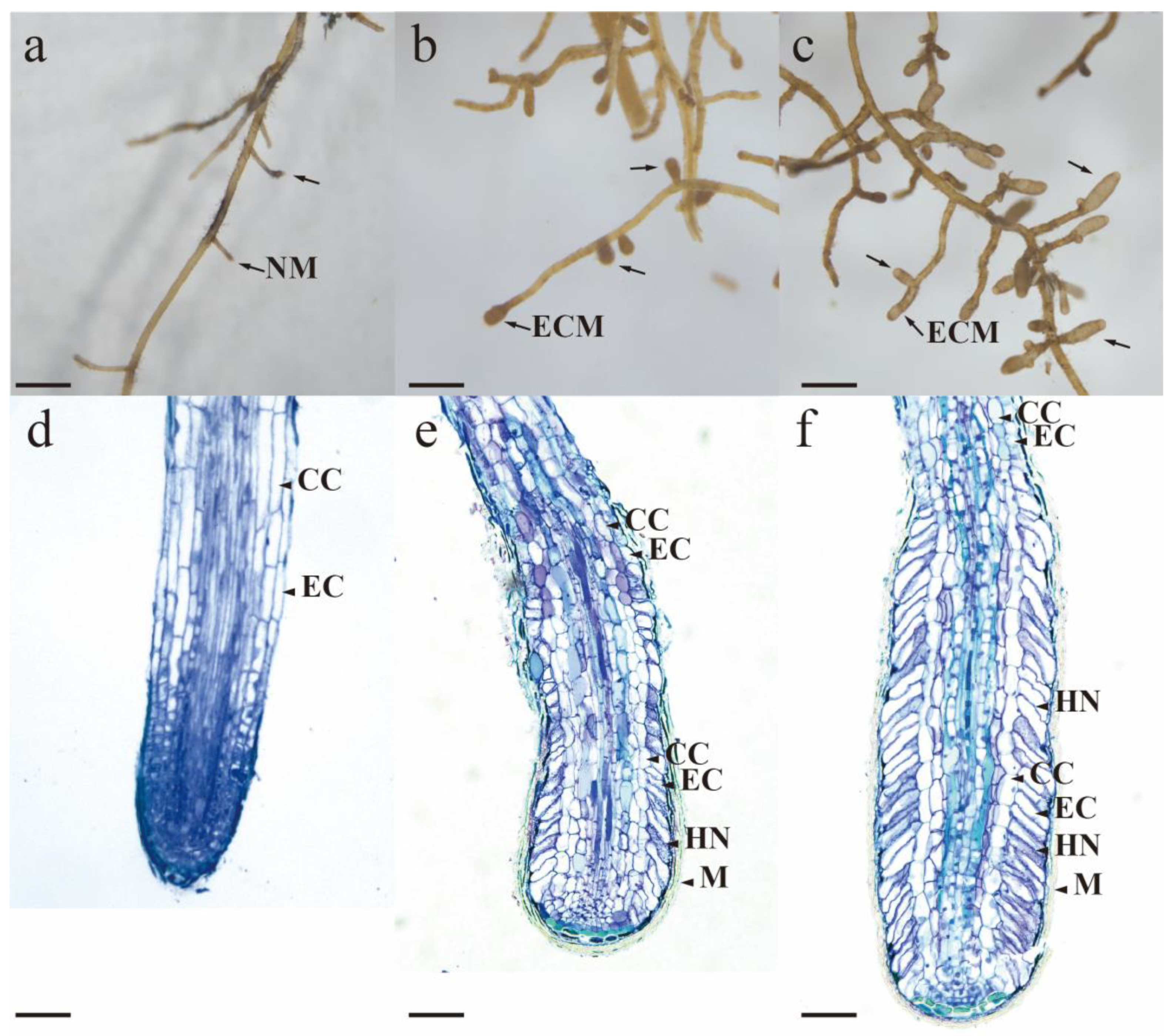
3.3. Macroscopic Morphology Under Stereomicroscopy

The appearance and morphology of the mycorrhizae were examined through both visual inspection and microscopic observation (Figure 2a–c). Visual assessments of the inoculated plants indicated a singular ectomycorrhizal morphology, exhibiting morphological and anatomical features that are characteristic of T. sinense [17,36]. Mature truffle–chestnut symbionts exhibit greater thickness than non-symbiotic roots, exhibiting an overall appearance and coloration that ranges from pale brown to dark brown. The unbranched ends are predominantly straight, occasionally exhibiting slight bends, and are typically cylindrical or slightly tapering. The surface of the unbranched ends exhibits a jigsaw-puzzle-like pattern composed of outer mantle cells. The emanating hyphae are relatively sparse, predominantly single or branched, with certain hyphae exhibiting right-angle ramification. These features suggest that T. sinense have formed ectomycorrhizal associations with chestnut. No ectomycorrhizae were observed in the control plant roots, indicating the effectiveness of the inoculation process.
The morphological characteristics of ectomycorrhizae constitute the fundamental basis for the investigation of ectomycorrhizal fungi. The initial identification predominantly depended on morphological classification and microscopic characteristics. The diversity of ectomycorrhizal fungi and their host plants results in substantial variations in the morphology, pigmentation, and texture of ectomycorrhizae, which serve as critical criteria for preliminary classification. Ectomycorrhizae exhibit a range of morphologies, including rod-like, clustered, feather-like, or tower-shaped structures; mantle pigmentation may vary from white to yellow, brown, or black; and surface textures may be smooth and dense, rough and cottony, or irregularly granular [44]. The German scientist Agerer integrated microscopic structural analysis with the macroscopic characteristics of mycorrhizae to offer comprehensive descriptions and distinctions and established the DEEMY website (www.deemy.de) as a resource for the classification of ectomycorrhizae [45]. However, morphological identification exhibits inherent limitations regarding accuracy and timeliness. These limitations stem from the diversity of fungal species, the absence of distinct individual characteristics, the variability in mycorrhizal features produced by the same fungus–host combination under varying conditions, and discrepancies in observations made by different individuals. Our experiments were conducted under controlled greenhouse conditions, which may not fully capture the variability present in natural ecosystems. Another significant limitation is that we did not observe the decline and regrowth of ectomycorrhizae. Addressing this aspect necessitates the long-term monitoring of mycorrhizal seedlings to fully understand the dynamics of mycorrhizal colonization across multiple growing seasons.

3.4. Characterization of Mycorrhizal Colonization Morphology by Light Microscopy

To conduct a comprehensive morphological and anatomical characterization, we prepared resin sections of the synthesized mycorrhizae and examined them using optical microscopy (Figure 2d–f). Based on previous studies on the growth and anatomy of ectomycorrhizae [10], the development of ectomycorrhizae can be categorized into distinct stages based on the maturation of the mantle and Hartig net. To identify the different stages of truffle mycorrhizal development, ectomycorrhizae at various maturity stages were selected for detailed anatomical observation. A comparative analysis was performed between uninoculated roots and mycorrhizal roots. Non-mycorrhizal root tips display a well-defined root cap structure, prominent meristematic and elongation zones, and epidermal cells elongated along the longitudinal axis. In the early stages of mycorrhizal symbiosis, a higher density of hyphae is observed at the root tip, forming 2–5 layers. Surrounding the epidermal cells, the roots are enveloped by 2–3 layers of relatively loose hyphae. The epidermal cells of the mycorrhizae are radially elongated. In mature mycorrhizae, fungal hyphae densely surround the epidermal cells, forming a well-defined and compact mantle composed of interlocking pseudoparenchymatous cells resembling a ‘jigsaw puzzle’. This mantle typically comprises 3–5 layers of hyphae. The symbiotic associations between various truffle species and their respective host plants exhibit analogous structural characteristics. When various truffle species coexist with chestnut trees, they consistently exhibit elongated modified epidermal cells [46]. The mycorrhizal structure resulting from the symbiotic relationship between truffles and Quercus species also demonstrates analogous structural modifications, which represent a defining characteristic of the symbiosis between truffles and angiosperms [12,14]. Kinoshita et al. [19] compared the microscopic characteristics among various symbiotic associations involving Japanese truffles and concluded that the morphology and anatomical features of Japanese black truffles closely resemble those of T. melanosporum and T. sinense. In certain specific mycorrhizal associations, truffles can also form arbutoid mycorrhizae, which may allow them to play unexpected roles in natural ecosystems [47,48]. These differences reflect the diversity in the degree of dependence on Tuber and the symbiotic methods among different plant species.

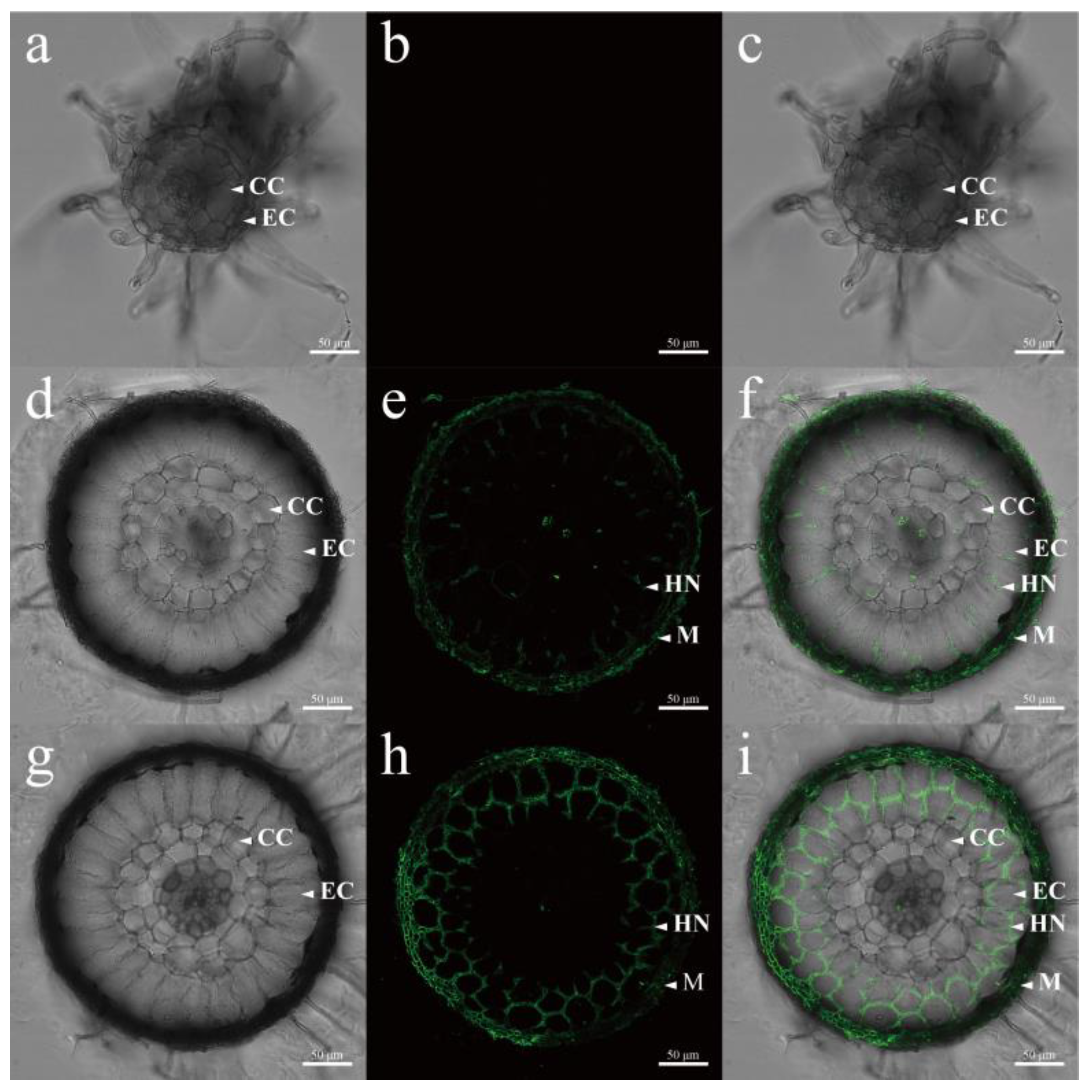
3.5. Laser Confocal Microscopy Analysis of Hyphal Growth

To more clearly determine the formation progress of the Hartig net, we used WGA-488 to specifically label the fungal cell walls and observed them using confocal laser scanning microscopy (Figure 3). In the mycorrhizae in the early stage of symbiosis, the average thickness is 4.09 ± 0.62 μm, and the hyphae are relatively loose. Meanwhile, small numbers of hyphae enter the intercellular spaces of the epidermal cells (Figure 3e). In mature mycorrhizae, characterized by an average mantle thickness of 5.23 ± 0.56 μm, the mantle exhibits a significantly greater thickness compared to that of early-stage mycorrhizae (p = 0.0013, Figure 3h). The epidermal cells of the root undergo radial elongation, subsequently allowing the fungi to extend within the intercellular spaces until their growth is obstructed by cortical cells.
The epidermal cells of roots are integral in mediating various biotic interactions, encompassing localized immune responses and modifications in cell wall chemistry [49]. Epidermal cells serve as the principal interface through which plants engage in direct interactions with fungi. In ectomycorrhizal symbiosis, hyphae adhere to the root tips and epidermal cells of developing lateral roots. The hyphae adhere to the root surface, where they proliferate and differentiate into a pseudoparenchymatous mantle, which incorporates air and water channels, thereby facilitating nutrient transport and storage [50]. As the mycorrhizae mature, the mantle thickens, thereby facilitating increased storage capacity for substances and providing enhanced mechanical strength [51]. Consequently, this increased thickness augments the mycorrhizae’s resistance to environmental stresses and bolsters overall resilience.
The hyphal network, referred to as the Hartig net, forms around the epidermal root cells of angiosperms, as well as both the epidermal and cortical root cells of gymnosperms. This intricate, maze-like structure, characterized by extensive hyphal branching and a substantial surface area, functions as an efficient interface for bidirectional nutrient transport across the cell walls of host cells [52]. The colonization of the host’s apoplastic space is primarily facilitated by the mechanical force generated through hyphal extension. Nevertheless, the secretion of fungal polysaccharide lyases, including symbiotically upregulated GH5 endoglucanases and GH28 polygalacturonases, significantly enhances the invasion process [5].

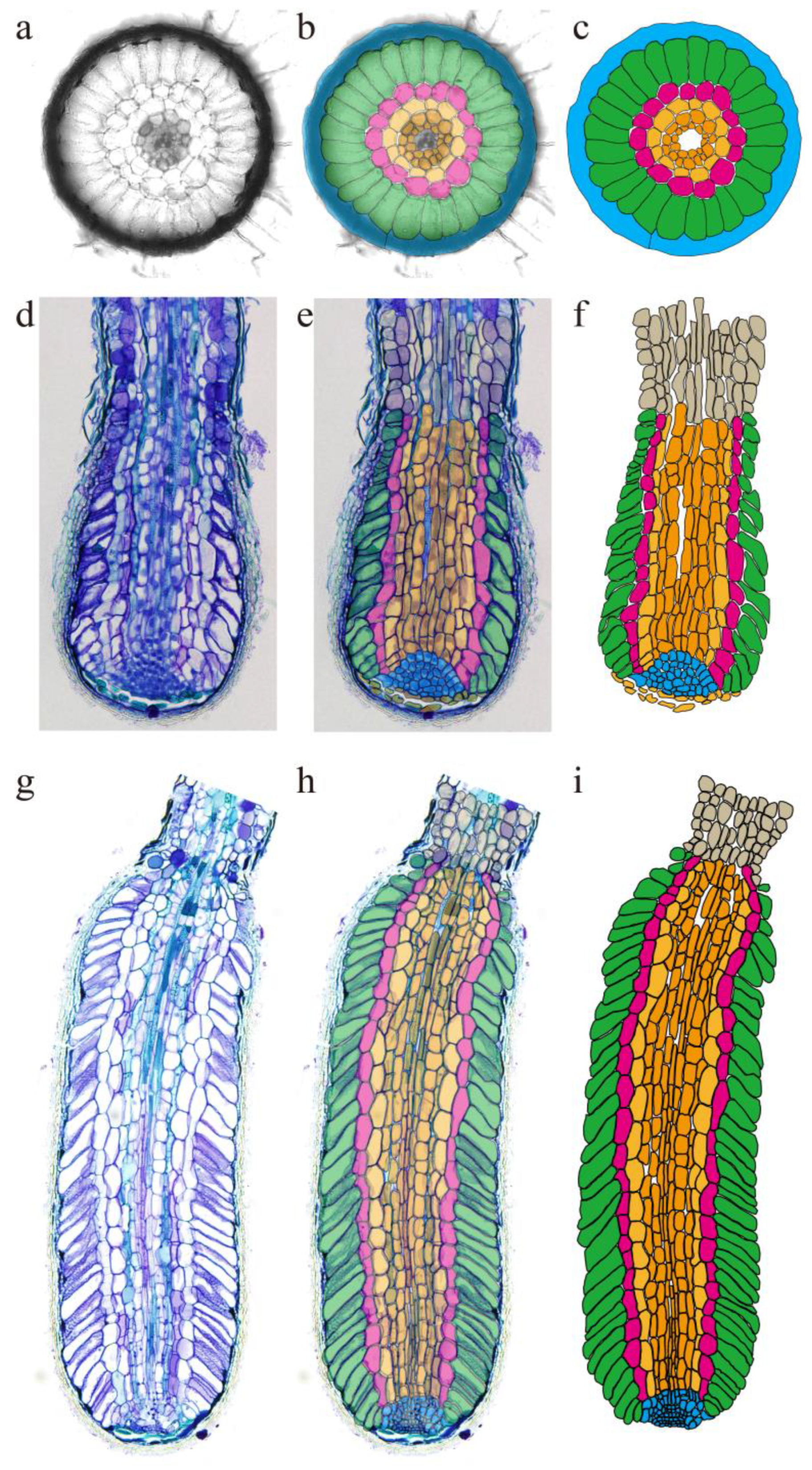
3.6. Mycorrhizal Model Construction Based on Microstructure

To more effectively illustrate the growth and development of ectomycorrhizae, a model was constructed to represent their growth. Icy was utilized to illustrate the root morphology, while Regions of Interest (ROIs) were employed to monitor cellular growth. Subsequently, ggPlantmap was employed to annotate and color the ROIs (Figure 4). The extraction of multiple key features from the original optical images enables a more precise characterization of the changes occurring in truffle mycorrhizae at various developmental stages. Following the encapsulation of the root tip by the mycelium and the establishment of a symbiotic relationship, the root tip cells of the mycorrhizae undergo continuous division, growth, and differentiation. As the newly formed epidermal cells progressively elongate, the mycelium concurrently invades the intercellular spaces among these cells. This process facilitates the penetration of the Hartig net into the root tissue, ultimately extending to the cortical cells. The progressive elongation of epidermal cells further promotes the expansion of the Hartig net, thereby enhancing nutrient exchange efficiency within the symbiotic interface.
Notably, the analysis revealed that epidermal cells constitute 58.3 ± 3.4% of the entire root in the transverse direction, underscoring their significant role in symbiosis, particularly in expanding the area of the Hartig net to enhance nutrient exchange efficiency. During the initial stage of mycorrhizal development, the perimeter of infected cells was measured at 44.30 ± 7.12 μm, accompanied by a cross-sectional area of 1551.88 ± 558.33 μm2. In mature mycorrhizae, a repetitive arrangement of cells is observed, with the morphology and area of these cells displaying similarities. Specifically, the cell perimeter was measured at 31.88 ± 3.79 μm, and the cross-sectional area was 941.56 ± 261.57 μm2, both significantly higher than those of early mycorrhizae (p < 0.01). Although it may seem surprising that the average size of epidermal cells with no mycorrhizae is smaller than that with early-stage mycorrhizae, this reduction in cell volume provides a functional advantage. As the volume of a cell decreases, the surface-area-to-volume ratio increases, thereby enhancing the efficiency of substance exchange, which is vital for the symbiotic relationship between the plant and the fungus.
The analysis demonstrated that epidermal cells constitute over 50% of the total cross-sectional area of the root, underscoring their pivotal role in the symbiotic relationship. This substantial proportion indicates their critical involvement in the expansion of the Hartig net interface and the enhancement in nutrient exchange efficiency. Recent advancements in imaging technologies have fundamentally transformed the spatial–temporal analysis of biological systems. These sophisticated platforms facilitate the precise identification and differentiation of diverse ROIs across various tissues and developmental stages, thereby enabling quantitative visualization and analysis based on experimental inputs [29,30]. The integration of these technologies with optical microscopy has yielded unprecedented resolution in characterizing the growth dynamics and developmental patterns of truffle mycorrhizae, providing novel insights into their adaptive mechanisms under varying environmental conditions.
Although the existing two-dimensional (2D) feature models have provided valuable quantitative data, their extension to three-dimensional (3D) or multi-dimensional analyses would significantly enhance the understanding of truffle mycorrhizal development. For example, confocal laser scanning microscopy has been effectively utilized for the high-resolution imaging of herbaceous plant roots, such as Arabidopsis and Medicago. Nevertheless, the application of 3D imaging techniques to woody plant roots poses significant challenges owing to their increased thickness, limited light penetration, and pronounced chromatic dispersion. Despite the attempts to utilize various clearing techniques, acquiring comprehensive 3D structural information on truffle mycorrhizae through confocal microscopy continues to pose technical challenges [53].
Future research directions ought to prioritize the optimization of image processing algorithms and the exploration of the potential for extending two-dimensional (2D) feature models to three-dimensional (3D) or multi-dimensional data analyses. The integration of gene expression profiling and spatial transcriptomics presents considerable potential for elucidating the molecular mechanisms that underpin mycorrhizal symbiosis [42,54]. Leveraging multi-omics approaches (e.g., genomics, transcriptomics, proteomics, and metabolomics) will facilitate a more comprehensive understanding of the recognition, response, and maintenance processes involved in mycorrhizal formation. By enhancing our knowledge and techniques in these areas, we aim to broaden the application of ectomycorrhizal symbiosis to non-mycorrhizal plant and fungal species. These multidisciplinary approaches are likely to yield novel insights into the understanding and conservation of these ecologically and economically significant fungi, potentially informing sustainable management strategies and conservation initiatives.

4. Conclusions

In conclusion, this study provides a comprehensive characterization of the ectomycorrhizal symbiosis established between Tuber species and C. mollissima, significantly enhancing our understanding of the potential for mycorrhizal formation within this symbiotic relationship. Our findings underscore the persistence and efficacy of these associations, offering novel insights into the life cycle of truffles, particularly focusing on the development of Tuber ectomycorrhizae. One limitation of this study is that different environments or symbiotic combinations may lead to variations in the morphological characteristics of mycorrhizae, which are adapted to specific cultivation conditions. Therefore, more effort is needed to maintain consistency and control to promote the efficient formation and maintenance of mycorrhizae, in order to better uncover both unique and common traits in ectomycorrhizal symbioses. Future research should prioritize comparative analyses across various symbiotic combinations to elucidate both conserved mechanisms and species-specific adaptations. Ultimately, the goal is to achieve the controlled artificial production of mycorrhizal edible fungi, such as truffles. Such advancements could pave the way for innovative agricultural practices and ecosystem restoration efforts.

Author Contributions

Conceptualization, G.Z.; data curation, Y.W. and W.Z.; formal analysis, Y.W. and Q.C.; funding acquisition, G.Z. and Q.C.; investigation, Y.W., W.Z. and R.Y.; methodology, Y.W., Y.Q. and G.Z.; project administration, Y.W., Y.Q. and G.Z.; resources, G.Z.; software, Y.W. and W.Z.; supervision, Y.Q. and G.Z.; visualization, Y.W.; writing—original draft, Y.W.; writing—review and editing, G.Z. All authors have read and agreed to the published version of the manuscript.

Funding

This work was financially supported by the Beijing Innovation Consortium of Agriculture Research System (BAIC03) and R&D Program of Beijing Municipal Education Commission (KZ20231002035).

Institutional Review Board Statement

Not applicable.

Informed Consent Statement

Not applicable.

Data Availability Statement

The data presented in the present work are available upon reasonable request to the corresponding author.

Conflicts of Interest

The authors declare no conflicts of interest.

References

  1. Zhang, X.P.; Li, X.L.; Ye, L.; Huang, Y.; Kang, Z.J.; Zhang, B.; Zhang, X.P. Colonization by Tuber melanosporum and Tuber indicum affects the growth of Pinus armandii and phoD alkaline phosphatase encoding bacterial community in the rhizosphere. Microbiol. Res. 2020, 239, 126520. [Google Scholar] [CrossRef] [PubMed]
  2. Zambonelli, A.; Iotti, M.; Murat, C. (Eds.) True Truffle (Tuber spp.) in the World: Soil Ecology, Systematics and Biochemistry; Springer International Publishing: Cham, Switzerland, 2016; ISBN 978-3-319-31436-5. [Google Scholar]
  3. Huang, L.; Li, Y.; Yuan, J.; Wan, S.; Colinas, C.; He, X.; Shi, X.; Wang, Y.; Yu, F. Tuber indicum and T. lijiangense colonization differentially regulates plant physiological responses and mycorrhizosphere bacterial community of Castanopsis rockii seedlings. Front. Plant Sci. 2023, 14, 1134446. [Google Scholar] [CrossRef] [PubMed]
  4. Huang, L.L.; Wang, Y.L.; Guerin-Laguette, A.; Wang, R.; Zhang, P.; Li, Y.M.; Yu, F.Q. Ectomycorrhizal synthesis between two Tuber species and six tree species: Are different hostfungus combinations having dissimilar impacts on host plant growth? Mycorrhiza 2022, 32, 341–351. [Google Scholar] [CrossRef]
  5. Martin, F.; Kohler, A.; Murat, C.; Veneault-Fourrey, C.; Hibbett, D.S. Unearthing the roots of ectomycorrhizal symbioses. Nat. Rev. Microbiol. 2016, 14, 760–773. [Google Scholar] [CrossRef]
  6. Genre, A.; Lanfranco, L.; Perotto, S.; Bonfante, P. Unique and common traits in mycorrhizal symbioses. Nat. Rev. Microbiol. 2020, 18, 649–660. [Google Scholar] [CrossRef] [PubMed]
  7. Wang, Y.; Yu, F.Q.; Zhang, C.; Liu, C.; Yang, M.; Li, S. Edible ectomycorrhizal fungi and their cultivation in China. In Mushrooms, Humans and Nature in a Changing World: Perspectives from Ecological, Agricultural and Social Sciences; Pérez-Moreno, J., Guerin-Laguette, A., Flores Arzú, R., Yu, F.Q., Eds.; Springer: Cham, Switzerland, 2020; pp. 31–60. ISBN 9783030373788. [Google Scholar]
  8. Tschaplinski, T.J.; Plett, J.M.; Engle, N.L.; Deveau, A.; Cushman, K.C.; Martin, M.Z.; Doktycz, M.J.; Tuskan, G.A.; Brun, A.; Kohler, A.; et al. Populus trichocarpa and Populus deltoides exhibit different metabolomic responses to colonization by the symbiotic fungus Laccaria bicolor. Mol. Plant-Microbe Interact. 2014, 27, 546–556. [Google Scholar] [CrossRef]
  9. Daguerre, Y.; Levati, E.; Ruytinx, J.; Tisserant, E.; Morin, E.; Kohler, A.; Montanini, B.; Ottonello, S.; Brun, A.; Veneault-Fourrey, C.; et al. Regulatory networks underlying mycorrhizal development delineated by genome-wide expression profiling and functional analysis of the transcription factor repertoire of the plant symbiotic fungus Laccaria bicolor. BMC Genom. 2017, 18, 737. [Google Scholar] [CrossRef]
  10. Tang, N.; Lebreton, A.; Xu, W.; Dai, Y.; Yu, F.; Martin, F.M. Transcriptome profiling reveals differential gene expression of secreted proteases and highly specific gene repertoires involved in Lactarius-Pinus symbioses. Front. Plant Sci. 2021, 12, 714393. [Google Scholar] [CrossRef]
  11. Tedersoo, L.; Bahram, M.; Zobel, M. How mycorrhizal associations drive plant population and community biology. Science 2020, 367, eaba1223. [Google Scholar] [CrossRef]
  12. Huang, L.L.; Guerin-Laguette, A.; Wang, R.; Yu, F.Q. Characterization of Tuber indicum (Pezizales, Tuberaceae) mycorrhizae synthesized with four host trees exotic to China. Symbiosis 2020, 82, 215–224. [Google Scholar] [CrossRef]
  13. Nahberger, T.U.; Benucci, G.M.N.; Kraigher, H.; Grebenc, T. Effect of earthworms on mycorrhization, root morphology and biomass of silver fir seedlings inoculated with black summer truffle (Tuber aestivum Vittad.). Sci. Rep. 2021, 11, 6167. [Google Scholar] [CrossRef] [PubMed]
  14. Wang, R.; Guerin Laguette, A.; Butler, R.; Huang, L.; Yu, F. The European delicacy Tuber melanosporum forms mycorrhizae with some indigenous Chinese Quercus species and promotes growth of the oak seedlings. Mycorrhiza 2019, 29, 649–661. [Google Scholar] [CrossRef] [PubMed]
  15. Zhang, X.; Wu, J.; Kong, Z. Cellular basis of legume-rhizobium symbiosis. Plant Commun. 2024, 5, 101045. [Google Scholar] [CrossRef] [PubMed]
  16. Shi, J.; Wang, X.; Wang, E. Mycorrhizal symbiosis in plant growth and stress adaptation: From genes to ecosystems. Annu. Rev. Plant Biol. 2022, 74, 569–607. [Google Scholar] [CrossRef]
  17. Wan, S.P.; Yu, F.Q.; Tang, L.; Wang, R.; Wang, Y.; Liu, P.G.; Wang, X.H.; Zheng, Y. Ectomycorrhizae of Tuber huidongense and T. liyuanum with Castanea mollissima and Pinus armandii. Mycorrhiza 2016, 26, 249–256. [Google Scholar] [CrossRef]
  18. Li, X.; Ye, L.; Zhang, X.; Tan, H.; Li, Q. Root-tip cutting and uniconazole treatment improve the colonization rate of Tuber indicum on Pinus armandii seedlings in the greenhouse. Microb. Biotechnol. 2020, 13, 535–547. [Google Scholar] [CrossRef]
  19. Kinoshita, A.; Obase, K.; Yamanaka, T. Ectomycorrhizae formed by three Japanese truffle species (Tuber japonicum, T. longispinosum, and T. himalayense) on indigenous oak and pine species. Mycorrhiza 2018, 28, 679–690. [Google Scholar] [CrossRef]
  20. White, T.J.; Bruns, T.; Lee, S.; Taylor, J. Amplification and direct sequencing of fungal ribosomal RNA genes for phylogenetics. In PCR Protocols: A Guide to Methods and Applications; Academic Press: Cambridge, MA, USA, 1990. [Google Scholar]
  21. Gardes, M.; Bruns, T.D. ITS primers with enhanced specificity for basidiomycetes-application to the identification of mycorrhizae and rusts. Mol. Ecol. 1993, 2, 113–118. [Google Scholar] [CrossRef]
  22. Kumar, S.; Stecher, G.; Li, M.; Knyaz, C.; Tamura, K. MEGA X: Molecular Evolutionary Genetics Analysis across Computing Platforms. Mol. Biol. Evol. 2018, 35, 1547–1549. [Google Scholar] [CrossRef]
  23. Murat, C. Forty years of inoculating seedlings with truffle fungi: Past and future perspectives. Mycorrhiza 2015, 25, 77–81. [Google Scholar] [CrossRef]
  24. Donnini, D.; Benucci, G.M.N.; Bencivenga, M.; Baciarelli-Falini, L. Quality assessment of truffle-inoculated seedlings in Italy: Proposing revised parameters for certification. For. Syst. 2014, 23, 385–393. [Google Scholar] [CrossRef]
  25. Sillo, F.; Brunetti, C.; Marroni, F.; Vita, F.; Santos Nascimento, L.B.; Vizzini, A.; Mello, A.; Balestrini, R.; dos Santos Nascimento, L.B. Systemic effects of Tuber melanosporum inoculation in two Corylus avellana genotypes. Tree Physiol. 2022, 42, 1463–1480. [Google Scholar] [CrossRef] [PubMed]
  26. Xiao, T.T.; Schilderink, S.; Moling, S.; Deinum, E.E.; Kondorosi, E.; Franssen, H.; Kulikova, O.; Niebel, A.; Bisseling, T. Fate map of Medicago truncatula root nodules. Development 2014, 141, 3517–3528. [Google Scholar] [CrossRef] [PubMed]
  27. Leng, J.; Wei, X.; Jin, X.; Wang, L.; Fan, K.; Zou, K.; Zheng, Z.; Saridis, G.; Zhao, N.; Zhou, D.; et al. ARBUSCULAR MYCORRHIZA-INDUCED KINASES AMK8 and AMK24 associate with the receptor-like kinase KINASE3 to regulate arbuscular mycorrhizal symbiosis in Lotus japonicus. Plant Cell 2023, 35, 2006–2026. [Google Scholar] [CrossRef]
  28. Hill, R.A.; Wong-Bajracharya, J.; Anwar, S.; Coles, D.; Wang, M.; Lipzen, A.; Ng, V.; Grigoriev, I.V.; Martin, F.; Anderson, I.C.; et al. Abscisic acid supports colonization of Eucalyptus grandis roots by the mutualistic ectomycorrhizal fungus Pisolithus microcarpus. New Phytol. 2021, 233, 966–982. [Google Scholar] [CrossRef]
  29. de Chaumont, F.; Dallongeville, S.; Chenouard, N.; Hervé, N.; Pop, S.; Provoost, T.; Meas-Yedid, V.; Pankajakshan, P.; Lecomte, T.; Le Montagner, Y.; et al. Icy: An open bioimage informatics platform for extended reproducible research. Nat. Methods 2012, 9, 690–696. [Google Scholar] [CrossRef]
  30. Jo, L.; Kajala, K. ggPlantmap: An open-source R package for the creation of informative and quantitative ggplot maps derived from plant images. J. Exp. Bot. 2024, 75, 5366–5376. [Google Scholar] [CrossRef]
  31. Fan, L.; Li, T.; Xu, Y.Y.; Yan, X.Y. Species diversity, phylogeny, endemism and geography of the truffle genus Tuber in China based on morphological and molecular data. Persoonia-Mol. Phylogeny Evol. Fungi 2022, 48, 175–202. [Google Scholar] [CrossRef]
  32. Gwon, J.H.; Park, H.; Eom, A.H. Mycorrhization of Quercus spp. with Tuber huidongense and T. himalayense collected in Korea. Mycobiology 2022, 50, 104–109. [Google Scholar] [CrossRef]
  33. Fan, L.; Zhang, J.L.; Li, T.; Sun, H.J.; Xiong, W.P.; Li, Y. Chinese black truffles: Tuber yigongense sp. nov., taxonomic reassessment of T. indicum s.l., and re-examination of the T. sinense isotype. Mycotaxon 2018, 133, 183–196. [Google Scholar] [CrossRef]
  34. Liu, F.; Wang, S.H.; Cheewangkoon, R.; Zhao, R.L. Uneven distribution of prokaryote-derived horizontal gene transfer in fungi: A lifestyle-dependent phenomenon. mBio 2025, 16, e0285524. [Google Scholar] [CrossRef] [PubMed]
  35. Tejedor-Calvo, E.; Marco, P.; Fischer, M.; Creydt, M. Mass spectrometry-based non-targeted lipidome analysis and extraction of markers for the authentication of white and black truffle species and their origin determination. Agriculture 2024, 14, 2350. [Google Scholar] [CrossRef]
  36. Geng, L.; Wang, X.; Yu, F.; Deng, X.; Tian, X.; Shi, X.; Xie, X.; Liu, P.; Shen, Y. Mycorrhizal synthesis of Tuber indicum with two indigenous hosts, Castanea mollissima and Pinus armandii. Mycorrhiza 2009, 19, 461–467. [Google Scholar] [CrossRef] [PubMed]
  37. Mrak, T.; Grebenc, T.; Friedrich, S.; Münzenberger, B. Description, identification, and growth of Tuber borchii Vittad. mycorrhized Pinus sylvestris L. seedlings on different lime contents. Mycorrhiza 2024, 34, 85–94. [Google Scholar] [CrossRef] [PubMed]
  38. Garcia-Barreda, S.; Molina-Grau, S.; Reyna, S. Fertilisation of Quercus seedlings inoculated with Tuber melanosporum: Effects on growth and mycorrhization of two host species and two inoculation methods. Iforest-Biogeosci. For. 2017, 10, 267–272. [Google Scholar] [CrossRef]
  39. Marozzi, G.; Sánchez, S.; Benucci, G.M.N.; Bonito, G.; Falini, L.B.; Albertini, E.; Donnini, D. Mycorrhization of pecan (Carya illinoinensis) with black truffles: Tuber melanosporum and Tuber brumale. Mycorrhiza 2017, 27, 303–309. [Google Scholar] [CrossRef]
  40. Miyauchi, S.; Kiss, E.; Kuo, A.; Drula, E.; Kohler, A.; Sánchez-García, M.; Morin, E.; Andreopoulos, B.; Barry, K.W.; Bonito, G.; et al. Large-scale genome sequencing of mycorrhizal fungi provides insights into the early evolution of symbiotic traits. Nat. Commun. 2020, 11, 5125. [Google Scholar] [CrossRef]
  41. Liao, H.-L.; Chen, Y.; Vilgalys, R. Metatranscriptomic study of common and host-specific patterns of gene expression between pines and their symbiotic ectomycorrhizal fungi in the genus Suillus. PLoS Genet. 2016, 12, e1006348. [Google Scholar] [CrossRef]
  42. Basso, V.; Kohler, A.; Miyauchi, S.; Singan, V.; Guinet, F.; Šimura, J.; Novák, O.; Barry, K.W.; Amirebrahimi, M.; Block, J.; et al. An ectomycorrhizal fungus alters sensitivity to jasmonate, salicylate, gibberellin, and ethylene in host roots. Plant Cell Environ. 2020, 43, 1047–1068. [Google Scholar] [CrossRef]
  43. Plett, K.L.; Buckley, S.; Plett, J.M.; Anderson, I.C.; Lundberg-Felten, J.; Jämtgård, S. Novel microdialysis technique reveals a dramatic shift in metabolite secretion during the early stages of the Interaction between the ectomycorrhizal fungus Pisolithus microcarpus and its host Eucalyptus grandis. Microorganisms 2021, 9, 1817. [Google Scholar] [CrossRef]
  44. Agerer, R. Fungal relationships and structural identity of their ectomycorrhizae. Mycol Prog. 2006, 5, 67–107. [Google Scholar] [CrossRef]
  45. Rambold, G.; Agerer, R. DEEMY—The concept of a characterization and determination system for ectomycorrhizae. Mycorrhiza 1997, 7, 113–116. [Google Scholar] [CrossRef]
  46. Álvarez-Lafuente, A.; Benito-Matías, L.F.; Peñuelas-Rubira, J.L.; Suz, L.M. Multi-cropping edible truffles and sweet chestnuts: Production of high-quality Castanea sativa seedlings inoculated with Tuber aestivum, its ecotype T. uncinatum, T. brumale, and T. macrosporum. Mycorrhiza 2018, 28, 29–38. [Google Scholar] [CrossRef]
  47. Ori, F.; Leonardi, M.; Faccio, A.; Sillo, F.; Iotti, M.; Pacioni, G.; Balestrini, R. Synthesis and ultrastructural observation of arbutoid mycorrhizae of black truffles (Tuber melanosporum and T. aestivum). Mycorrhiza 2020, 30, 715–723. [Google Scholar] [CrossRef]
  48. Lancellotti, E.; Iotti, M.; Zambonelli, A.; Franceschini, A. Characterization of Tuber borchii and Arbutus unedo mycorrhizas. Mycorrhiza 2014, 24, 481–486. [Google Scholar] [CrossRef]
  49. Tedersoo, L.; Bahram, M.; Põlme, S.; Kõljalg, U.; Yorou, N.S.; Wijesundera, R.; Villarreal Ruiz, L.; Vasco-Palacios, A.M.; Thu, P.Q.; Suija, A.; et al. Fungal biogeography. Global diversity and geography of soil fungi. Science 2014, 346, 1256688. [Google Scholar] [CrossRef]
  50. Martin, F.M.; van der Heijden, M.G.A. The mycorrhizal symbiosis: Research frontiers in genomics, ecology, and agricultural application. New Phytol. 2024, 242, 1486–1506. [Google Scholar] [CrossRef]
  51. Ruytinx, J.; Miyauchi, S.; Hartmann-Wittulsky, S.; de Freitas Pereira, M.; Guinet, F.; Churin, J.-L.; Put, C.; Le Tacon, F.; Veneault-Fourrey, C.; Martin, F.; et al. A transcriptomic atlas of the ectomycorrhizal fungus Laccaria bicolor. Microorganisms 2021, 9, 2612. [Google Scholar] [CrossRef]
  52. Peterson, R.L.; Massicotte, H.B. Exploring structural definitions of mycorrhizas, with emphasis on nutrient-exchange interfaces. Can. J. Bot. 2004, 82, 1074–1088. [Google Scholar] [CrossRef]
  53. Ursache, R.; Andersen, T.G.; Marhavý, P.; Geldner, N. A protocol for combining fluorescent proteins with histological stains for diverse cell wall components. Plant J. 2018, 93, 399–412. [Google Scholar] [CrossRef]
  54. Larsen, P.E.; Sreedasyam, A.; Trivedi, G.; Desai, S.; Dai, Y.; Cseke, L.J.; Collart, F.R. Multi-omics approach identifies molecular mechanisms of plant-fungus mycorrhizal interaction. Front. Plant Sci. 2015, 6, 1061. [Google Scholar] [CrossRef] [PubMed]
Figure 1. The phylogeny of Tuber ectomycorrhizal roots was inferred from the ITS sequences using maximum likelihood analysis. Bootstrap values are indicated above the individual branches. The Tuber ectomycorrhizal roots identified in this study are highlighted in red for clarity.
Agriculture 15 00868 g001
Figure 2. Macro-morphology and micro-morphology of Tuber ectomycorrhiza. (a,d) The morphology and micro-morphology of non-mycorrhizae; (b,e) the morphology and micro-morphology of early mycorrhizae; (c,f) the morphology and micro-morphology of mature mycorrhizae. Numerous ectomycorrhizae are visible in panels (b,c) (arrowheads). NM, non-mycorrhizal root; ECM, ectomycorrhizal root; EC, epidermal cell; CC, cortical cell; M, mantle; HN, Hartig net. Scale bar in (ac) = 10 mm, in (df) = 50 μm.
Agriculture 15 00868 g002
Figure 3. Cross-section of Tuber ectomycorrhizae observed under confocal laser scanning microscopy. (ac) The morphology and micro-morphology of non-mycorrhizae; (df) the morphology and micro-morphology of early mycorrhizae; (gi) the morphology and micro-morphology of mature mycorrhizae. Vibratome sections were stained with WGA-488 (green color, fungal hyphae). NM, non-mycorrhizal root; ECM, ectomycorrhizal root; EC, epidermal cell; CC, cortical cell; M, mantle; HN, Hartig net. Scale bar = 20 μm.
Agriculture 15 00868 g003
Figure 4. Two-dimensional growth model of Tuber ectomycorrhizae. (ac) The cross-sectional morphology of mycorrhizae; (df) longitudinal section morphology of early mycorrhizae; (gi) longitudinal section morphology of mature mycorrhizae.
Agriculture 15 00868 g004
Table 1. Mycorrhizal development and structural characteristics at different time points.
SampleColonization Rate (%)Mantle Cell LayersMantle Thickness (μm)Epidermal Cell Perimeter (μm)Epidermal Cell Cross-Sectional Area (μm2)
NI---152.70 ± 22.51 a2295.21 ± 336.81 b
T324.4 ± 5.3 b2–34.09 ± 0.62 b44.30 ± 7.12 c1551.88 ± 558.33 c
T845.2 ± 8.6 a3–55.23 ± 0.56 a79.70 ± 9.48 b2353.89 ± 653.93 a
Different letters represent significant differences (p < 0.05) in the same column. Abbreviations: NI: control seedlings (no inoculation); T3: seedlings three months post inoculation; T8: seedlings eight months post inoculation.
Disclaimer/Publisher’s Note: The statements, opinions and data contained in all publications are solely those of the individual author(s) and contributor(s) and not of MDPI and/or the editor(s). MDPI and/or the editor(s) disclaim responsibility for any injury to people or property resulting from any ideas, methods, instructions or products referred to in the content.

