Article

Impacts of Warming and Acidification on Coral Calcification Linked to Photosymbiont Loss and Deregulation of Calcifying Fluid pH

1
Department of Marine and Environmental Sciences, Northeastern University, Nahant, MA 01908, USA
2
Leibniz Centre for Tropical Marine Research (ZMT), 28359 Bremen, Germany
3
Geocoastal Research Group, School of Geosciences, The University of Sydney, Camperdown, Sydney, NSW 2006, Australia
4
Alfred Wegener Institute, Helmholtz Center for Polar and Marine Research, Am Handelshafen 12, 27570 Bremerhaven, Germany
5
GEOMAR Helmholtz Centre for Ocean Research Kiel, 24148 Kiel, Germany
6
Max Planck Institute for Marine Microbiology, 28359 Bremen, Germany
7
Atmospheric and Oceanic Sciences Department, Institute of the Environment and Sustainability, Earth, Planetary and Space Sciences, University of California, Los Angeles, CA 90095, USA
8
Department for Geosciences, University of Bremen, 28359 Bremen, Germany
9
Physical Science and Engineering Division, King Abdullah University of Science and Technology (KAUST), Thuwal, Makkah 23955-6900, Saudi Arabia
*
Author to whom correspondence should be addressed.
J. Mar. Sci. Eng. 2022, 10(8), 1106; https://doi.org/10.3390/jmse10081106
Submission received: 20 July 2022 / Revised: 5 August 2022 / Accepted: 6 August 2022 / Published: 12 August 2022
(This article belongs to the Special Issue The Effect of Ocean Acidification on Skeletal Structures)

Abstract

:
Corals are globally important calcifiers that exhibit complex responses to anthropogenic warming and acidification. Although coral calcification is supported by high seawater pH, photosynthesis by the algal symbionts of zooxanthellate corals can be promoted by elevated pCO2. To investigate the mechanisms underlying corals’ complex responses to global change, three species of tropical zooxanthellate corals (Stylophora pistillata, Pocillopora damicornis, and Seriatopora hystrix) and one species of asymbiotic cold-water coral (Desmophyllum pertusum, syn. Lophelia pertusa) were cultured under a range of ocean acidification and warming scenarios. Under control temperatures, all tropical species exhibited increased calcification rates in response to increasing pCO2. However, the tropical species’ response to increasing pCO2 flattened when they lost symbionts (i.e., bleached) under the high-temperature treatments—suggesting that the loss of symbionts neutralized the benefit of increased pCO2 on calcification rate. Notably, the cold-water species that lacks symbionts exhibited a negative calcification response to increasing pCO2, although this negative response was partially ameliorated under elevated temperature. All four species elevated their calcifying fluid pH relative to seawater pH under all pCO2 treatments, and the magnitude of this offset (Δ[H+]) increased with increasing pCO2. Furthermore, calcifying fluid pH decreased along with symbiont abundance under thermal stress for the one species in which calcifying fluid pH was measured under both temperature treatments. This observation suggests a mechanistic link between photosymbiont loss (‘bleaching’) and impairment of zooxanthellate corals’ ability to elevate calcifying fluid pH in support of calcification under heat stress. This study supports the assertion that thermally induced loss of photosymbionts impairs tropical zooxanthellate corals’ ability to cope with CO2-induced ocean acidification.

1. Introduction

Anthropogenic emissions are predicted to cause sea-surface warming [1] and ocean acidification (OA)—a process that lowers seawater pH and aragonite saturation state [2] (ΩA). OA increases both the dissolution rate of CaCO3 shell/skeleton [3] and the rate at which new shell/skeleton is formed [4]. Tropical scleractinian corals are carbonate producers [5] that acquire nourishment via symbiotic photosynthetic zooxanthellae and from heterotrophic feeding [6]. They are vulnerable to warming as many exist near the upper end of their thermal tolerance limits [7]. Warming beyond a coral’s thermal tolerance may cause the loss of photosymbionts—a process known as bleaching.
Coral calcification occurs in the coral’s calcifying fluid, which is influenced by both external seawater chemistry and the coral itself [8]. Corals elevate saturation state of this fluid through a combination of pH elevation via the active removal of protons using membrane-bound Ca2+-ATPase proton pumps [8,9,10,11,12,13,14] and DIC elevation [15,16]. Photosynthesis may aid pH elevation by supplying the necessary ATP required to drive Ca2+-ATPase proton pumps. Under ocean acidification, the formation of CaCO3 is more energetically costly [11,17,18], although this energetic cost is negligible compared to the total proportion of energy produced through photosynthesis [8]. Photosymbionts may therefore play a crucial role in mitigating the impacts of OA on corals by providing enough ATP to increase ion pumping rates in support of calcification. Photosynthesis may also be responsible for metabolic DIC elevation at the site of calcification, as implied by long-term seasonal variations in the concentration of DIC in the calcifying fluid [16]. Under warming, bleaching may impair coral calcification by reducing the amount of photosynthate that is translocated to the coral host, thereby increasing the coral’s reliance on heterotrophic feeding [6], decreasing the metabolically derived DIC pool, and, potentially, reducing the amount of ATP available for proton-regulation in support of calcification.
Although tropical scleractinian corals generally exhibit parabolic calcification responses to ocean warming that are centered on their thermal optima [19], their calcification responses to OA are more nuanced. Many species exhibit reduced calcification rates [20,21], while others exhibit threshold responses [22], parabolic responses [19,21], or no calcification response to acidification [13,23,24].
This highlights the complexity of coral biomineralization, which can be biologically mediated by the secretion of skeletal organic molecules (SOM) such as adhesion, signaling, and structural proteins (e.g., calmodulin and sulphated acidic proteoglycans) [25,26,27]. The abundance of these SOMs in the calcifying fluid has been shown to alter the rate and morphology of aragonite precipitated [28,29,30,31,32].
Cold-water corals that inhabit deeper aphotic environments generally lack zooxanthellae and acquire nourishment exclusively through heterotrophic feeding [33]. The systems for regulating calcifying fluid pH (pHCF) within azooxanthellate corals may therefore differ from zooxanthellate corals. Azooxanthellate corals exhibit reduced skeletal density [34], reduced calcification rate [35], and altered rates of respiration and feeding [36] in response to OA, but increased calcification rates under elevated temperature [35,37].
Numerous studies have investigated the isolated effects of ocean acidification (e.g., [11,24,38,39]) and thermal stress (e.g., [40]) on coral pHCF. However, the present study—along with its companion paper [41]—are the first to investigate both the independent and combined effects of warming and acidification on coral pHCF. These empirical constraints, combined with photosymbiont and calcification data for the three tropical coral species, yield insight into the mechanism by which warming and acidification interact to so negatively impact coral growth. Examining the roles that pHCF regulation and symbiont abundance play in the coral calcification response to OA and warming should improve understanding and prediction of how different species of corals will respond to future global change.
We investigate these relationships by culturing three species of tropical zooxanthellate corals (Stylophora pistillata, Pocillopora damicornis, Seriatopora hystrix) and one azooxanthellate cold-water species (Desmophyllum pertusum, syn. Lophelia pertusa) under control (tropical = 28 °C, cold-water = 9 °C) and elevated (tropical = 31 °C, cold-water = 12 °C) temperatures at near-present-day (451–499 ppm), year 2100 (885–1096 ppm), and year 2500 (2807–3194 ppm) pCO2 scenarios [1]. Coral calcifying fluid pH was measured with proton-sensitive microelectrodes. Symbiont abundance of the zooxanthellate corals was estimated based on coral color to investigate the role of photosymbionts in the coral calcification response to warming and acidification.
This work is a companion paper to Guillermic et al. [41], which investigated the impacts of pCO2 and temperature on the calcifying fluid chemistry and calcification rate of two tropical species of scleractinian corals, S. pistillata and P. damicornis. The present study builds upon its companion paper by investigating these relationships for two additional species of scleractinian corals—the tropical species S. hystrix and the cold-water species L. pertusa. The present study also includes photosymbiont data for all three tropical coral species cultured under all experimental treatments.

2. Materials and Methods

2.1. Overview of Experimental Design

Three species of tropical scleractinian corals (S. pistillata, P. damicornis, and S. hystrix) were cultured under three ocean acidification (OA) scenarios established by modification of pCO2 at control and elevated temperatures (control acidification: 28.29 °C ± 0.01 s.e./466 ppm ± 8; 31.72 °C ± 0.06/499 ppm ± 9; moderate acidification: 27.88 °C ± 0.02/925 ppm ± 15; 30.83 °C ± 0.02/885 ppm ± 12; high acidification: 28.17 °C ± 0.02/2807 ppm ± 119; 30.93 °C ± 0.02/3194 ppm ± 135) in four replicate tanks at each treatment for 62 days in April–June 2016 (Table 1). The control temperature treatment was assigned to fall within the natural temperature range of coral reefs in Fiji (26–29 °C), where these corals were collected [42]. Likewise, the elevated temperature was assigned to be just above the bleaching threshold (30–30.5 °C) for corals in Fiji [42]. Simultaneously, the cold-water coral L. pertusa was cultured under three OA scenarios at control and elevated temperatures (control acidification: 8.86 °C ± 0.02/451 ppm ± 24; 12.17 °C ± 0.03/494 ppm ± 16; moderate acidification: 9.07 °C ± 0.03/1096 ppm ± 99; 12.52 °C ± 0.04/1079 ppm ± 60; high acidification: 8.83 °C ± 0.02/2864 ppm ± 222; 12.67 °C ± 0.03/3167 ppm ± 202) in four replicate tanks at each treatment for 33 days in April–May 2016 (Table 2). Corals were acclimated to laboratory conditions for one week and then to experimental conditions for an additional two weeks prior to the start of the experiment. Calcification rates, pHCF, and relative photosymbiont abundance were quantified during the experiment (details below).

2.2. Coral Husbandry

Experiments were carried out in the MAREE marine experimental facility at the Leibniz Centre for Tropical Marine Research (ZMT). Specimens of the tropical scleractinian coral species S. pistillata and P. damicornis were obtained from DeJong MarineLife (Netherlands). Colony-level information was not available for these specimens. Specimens of the tropical scleractinian coral species S. hystrix were obtained from an experimental stock colony provided by the ZMT. Fragments of the cold-water coral L. pertusa were obtained from four colonies of an experimental stock culture provided by the Marine Biogeochemistry Department of the Helmholtz Centre for Ocean Research in Kiel (GEOMAR), previously collected at a depth of ca. 200 m at Nord-Leksa Reef in Trondheimsfjord, Norway. Coral fragments (S. pistillata = 65, P. damicornis = 63, S. hystrix = 52, and L. pertusa = 43) were mounted onto 3 cm x 3 cm plastic egg-crate stands using cyanoacrylate epoxy and assigned a unique identifier. Each treatment was replicated in four tanks. Equivalent size ranges of specimens were maintained across treatments. Coral specimens obtained from larger colonies were randomly distributed amongst pCO2 and temperature treatments and replicate tanks. Coral specimens that died before the completion of the experiment were promptly removed from the tanks so as not to impact the remaining live corals in the experiment. Additional details about the number and weight of individuals in each treatment are provided in Section S1 of the Supplementary Online Material.
After 1-week of acclimation at control conditions, temperature and pCO2 were then incrementally increased to treatment levels over an additional week, after which time coral specimens were acclimated to treatment conditions for an additional week prior to the experiment. All coral specimens were cultured in 10-L replicate tanks supplied with seawater from 244-L sumps, where water filtration, temperature control, and pCO2 control occurred. Seawater was filtered with protein skimmers, mechanical filters, and activated charcoal. All tropical coral aquaria were illuminated with 150 lux using actinic blue and white aquarium lights on a 12-h light/dark cycle. Aquaria holding the cold-water coral L. pertusa were not illuminated as this species lives below the photic zone. Each experimental treatment containing L. pertusa specimens (comprised of 4 replicate tanks) shared a water source with a separate reservoir containing five specimens of the king scallop Pecten maximus cultured as part of a separate experiment [43]. Seawater was filtered with protein skimmers, mechanical filters, and activated charcoal before returning to the L. pertusa tanks.
During acclimation and experimental periods, the tropical corals were fed 1-day old Artemia salina nauplii hatched from ca. 40 mg of eggs. Approximately 10 mL of concentrated live nauplii were introduced into each replicate tank every second day. The food mixture was pipetted directly adjacent to each coral specimen. Specimens of L. pertusa were fed the same diet supplemented with 20 mL of Calanus finmarchicus concentrate (Goldpods) suspended in the initial aliquot of Artemia salina. Corals were fed at the end of the day, and all filtration material was cleaned the following morning.

2.3. Seawater Chemistry Manipulation and Measurement

Measured and calculated carbonate system parameters are summarized in Table 1 and Table 2. Experimental tank pCO2 was maintained by vigorously bubbling mixtures of CO2-free air and CO2 into the 244-L treatment sumps with microporous sparging tubes. The pCO2 of the bubbled gases was achieved by mixing compressed CO2-free air and compressed CO2 with solenoid-valve mass flow controllers at flow rates proportional to the target pCO2 conditions. Natural seawater, originally collected from Spitsbergen, Norway, was continuously added to each of the 244-L sumps at a rate of 0.6 L/hour. Temperature (s.e.) for the tropical corals was maintained at 28 (0.02) °C and 31 (0.06) °C using 125-watt aquarium heaters (EHEIM), controlled with a programmable thermostat. Temperature for L. pertusa was maintained at 9 °C (0.03) and 12 °C (0.04) using aquarium chillers (Aqua Medic).
Temperature, pH, and salinity of all replicate tanks were measured three times per week using a multi-electrode probe (WTW Multi 3430 Set K). Samples for the analysis of dissolved inorganic carbon (DIC) and total alkalinity (TA) were collected weekly from each of the replicate tanks at midday and used to calculate other carbonate system parameters using the program CO2SYS [44]. Nutrient concentrations ([NO3], [PO43−], and [NH4+]) of all replicate tanks were measured weekly. Additional details about the methods used to measure the carbonate system and nutrient concentrations of replicate tanks are provided in Section S2 of the Supplementary Online Materials.

2.4. Calcification Rates

Calcification rates were calculated from the change in estimated dry weight of all coral fragments over the experimental period. Dry weights were estimated from buoyant weight measurements taken at the start and end of the experimental period according to the following empirically derived relationships:
Stylophora pistillata: Dry weight (g) = 1.919 × Buoyant Weight (g) + 7.677;
Pocillopora damicornis: Dry weight (g) = 1.662 × Buoyant Weight (g) + 8.777;
Seriatopora hystrix: Dry weight (g) = 1.668 × Buoyant Weight (g) + 8.493;
Lophelia pertusa: Dry weight (g) = 1.594 × Buoyant Weight (g) − 0.206;
where the precision of this relationship is equivalent to the standard error of the regression (S. pistillata: 0.060 g; P. damicornis: 0.053 g; S. hystrix: 0.068 g; L. pertusa: 0.023 g). Additional details about the methods used to calculate calcification rate via buoyant weights are provided in Section S3 of the Supplementary Online Materials.
The number of days between the start and end buoyant weight measurements was then used to standardize %-calcification to a daily rate. Coral skeletons were also labeled with the fluorescent dye calcein (30 mg Se-Mark liquid calcein/kg-seawater) for 5 days prior to the initial buoyant weighing to identify skeletal material produced exclusively under the experimental conditions. Although all four species of corals recorded the calcein marker in their coral skeleton, rates of linear extension could not be reliably measured from the calcein marker because the dye was not incorporated into the skeletons in a consistent manner (see Section S4 of the Supplementary Online Materials for images of coral uptake of the calcein dye).

2.5. Estimating Coral Photosymbiont Index

The tropical coral specimens were photographed alongside the Coral Watch Coral Health Chart color scale [45,46,47] (Section S5 of the Supplementary Online Materials) under 150 lux (i.e., equivalent lighting to their experimental treatments) at the end of the experimental period to estimate relative photosymbiont abundance (a proxy for bleaching) of the coral specimens. This method involved extracting red-band color of the live coral tissue and the color scale and then assigning each pixel within the coral tissue image a discrete score (1–5) relative to the red-band values of the color scale (Figure 1). Additional details about the methods used to process photographs used for the estimation of photosymbiont index are provided in Section S5 of the Supplementary Online Materials.

2.6. Measurement of Calcifying Fluid pH

Calcifying fluid pH was measured using proton-sensitive liquid ion-exchanger (LIX) microelectrodes produced at the Max Planck Institute for Marine Microbiology (MPIMM) using a modified version of the technique described in De Beer et al. [48]. In brief, green soda lime glass microcapillary tubes (Schott model 8516) were held in a heated coil and pulled to a target tip diameter of ca. 10 µm, yielding final diameters of 8–20 µm. These were then silanized to produce a hydrophobic surface that allowed the adhesion of the LIX membrane. The microcapillary tubes were filled with ca. 300 µm of degassed, filtered electrolyte (300 mM KCl, 50 mM sodium phosphate adjusted to pH 7.0) using a plastic syringe with a 0.1-mm tip. The microcapillary tubes were then backfilled with LIX containing a polyvinyl chloride (PVC) epoxy to prevent leakage of electrolyte by submerging the tips of the microcapillary tubes in LIX and apply suction to the other end of the tube until the PVC-containing LIX was drawn into the tip of the microcapillary by 100–200 µm. Microcapillary tubes were encased in a Pasteur pipette for shielding, with the pulled tip of the microcapillary tube protruding ca. 2 cm beyond this casing. This casing was filled with a 0.3 M KCl solution and connected to the reference electrode with an Ag/AgCl wire to minimize electrical noise. Microelectrodes were left for 24 h after construction to allow for stabilization of the LIX membranes.
All microelectrode equipment (millivolt meter, National Instruments DAQ Pad 6020E, laptop, cables, micromanipulator, VT80 Micos motor arm, lab stands, Zeiss Stemi SV6 binocular microscope) was set up adjacent to the experimental tanks to minimize transport stress for the corals. Two reservoirs of seawater, sourced from the corresponding experimental treatment tanks, were established next to the microelectrode system. These reservoirs were bubbled with the corresponding treatment gases and maintained at the corresponding treatment temperature using aquarium heaters or chillers. The seawater was circulated between the two reservoirs through two 5.4 L flow-through chambers (30 × 12 × 15 cm). All pH microelectrode measurements were performed within these smaller flow-through chambers.
Measurements of calcifying fluid pH were made in the flow-through chambers filled with treatment seawater. Light levels in these chambers were measured using a digital lux-meter positioned next to the target coral polyp and were maintained at 150 lux. All corals were acclimated to the microelectrode chamber until polyp extension was observed prior to measurements (minimum of 10 min). Measurements of calcifying fluid pH were performed on three replicate individuals per treatment, with one measurement obtained for each individual. Calcifying fluid pH measurements were obtained for all species in all pCO2 treatments under the control temperature treatment. Due to constraints on time and resources available for the experiment, calcifying fluid pH measurements under the high temperature treatment were only obtained for one species (S. pistillata).
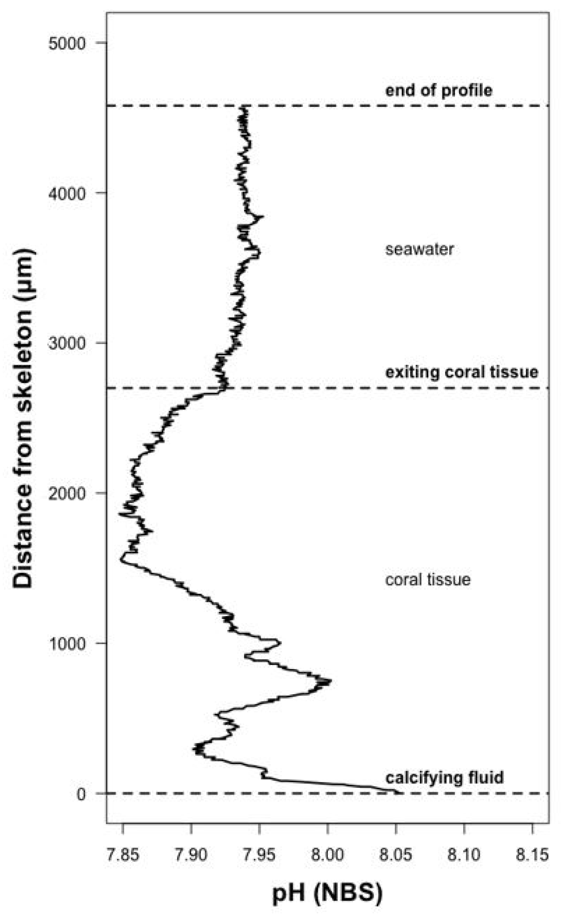
The proton-sensitive LIX microelectrodes were used to measure both seawater and calcifying fluid pH. Before and after measurement of calcifying fluid pH, all microelectrodes were calibrated at the treatment temperature with pH 7 and 9 NBS buffers. The vertical position of the microelectrode was controlled with one-micron precision using a motorized micromanipulator. The microelectrodes were slowly inserted with a micromanipulator through the coral tissue into the upper portion of the coral calyx, between septal ridges and proximal to the thecal wall, until the skeleton was reached. This positioning of the electrode was verified by a shift in the pH profile [14] (Figure 2). A vertical pH profile (Figure 2) was then obtained by moving the microelectrode out of the calyx into the adjacent seawater. This profile was obtained in 1 µm steps for the first 20 µm, followed by 5 µm steps out into the surrounding seawater.
The 1-µm spatial resolution of the micromanipulator allowed for the positioning of the electrode within the thin calcifying fluid immediately adjacent to the coral skeleton. If the skeleton was inadvertently contacted during this positioning, it is possible that the tip of the microelectrode would break and render it dysfunctional. It was visually evident if the microelectrode tip broke upon contact with the skeleton, and this would also result in an abrupt voltage anomaly, often followed by a drift in the voltage even while the electrode was in a fixed position. The pH profile was aborted if there was evidence of any of these issues and then reinitiated with a new microelectrode.
The calibration and microelectrode pH data were processed by parsing scatter-plots of the data into three zones, which were annotated at the time of data collection. The calibration data were parsed as pH 7 buffer and pH 9 buffer. The microelectrode pH data were parsed as calcifying fluid, tissue, and seawater. Notes recorded during the original measurements were used to assist in identifying boundaries of adjacent zones. Measured mV within each zone of the calcifying fluid measurements were converted to pH using the calibration regression produced for each microelectrode. The ∆[H+] was calculated for each measured coral as the difference between the proton concentration ([H+]) of the coral’s surrounding seawater and the [H+] of the coral’s calcifying fluid, both measured with the calibrated, proton-sensitive LIX microelectrodes.

2.7. Statistical Analysis

Statistical analyses were carried out in R. Corals that did not survive the experimental period (see Section S1 of the Supplementary Online Material) were excluded from analyses. A series of linear mixed effects models (lmers) were performed to investigate the influence of seawater pCO2 and temperature on coral physiology (calcification rate, calcifying fluid pH, Δ[H+], photosymbiont index), with treatment tank specified as a blocking factor [49]. Akaike information criterion (AIC) was used to estimate the relative amount of information lost by any given model [50]. The final model was chosen based on the lowest AIC score (whereby a lower score reflects a better fitting model) and highest R2, which reflects the goodness of fit (from 0 to 1, 1 being a perfect fit) (see Section S6 of the Supplementary Online Material for AIC model selection tables). The normality and homoscedasticity of the chosen tests were then analyzed using diagnostic plots (QQ-plot, residuals vs. fitted plot), and normality was tested using a Shapiro–Wilk test. Color scores were square-root transformed to meet the assumption of normality. If an interaction term was significant, the individual levels of that interaction were examined in their own linear mixed effects models to interpret main effects.
Analysis of co-variance (ANCOVA) was used to investigate the impacts of seawater pCO2, temperature, and species on calcification rate, thereby allowing interspecific comparisons of the impacts of OA and warming on coral calcification rate. An ANCOVA was also used to make interspecific comparisons of the impact of OA on calcifying fluid pH and Δ[H+] of different coral species. The latter analyses excluded individuals from the elevated temperature treatment, as calcifying fluid pH was only obtained for one of the three coral species in this treatment.
Linear mixed effects models were used to examine the impact of photosymbiont index, as a proxy for photosymbiont abundance, on calcification rate. Multiple linear models were generated, starting with the model that contained the most terms (i.e., modeling calcification rate as a function of seawater pCO2, temperature, and photosymbiont index). Final interpretations of the data were based upon the models that maximized R2 and minimized AIC (see Table S8). An alpha of 0.05 was used for all models, whereby any relationship with a p-value of <0.05 was deemed statistically significant.

3. Results

3.1. Predictors of Calcification Rate

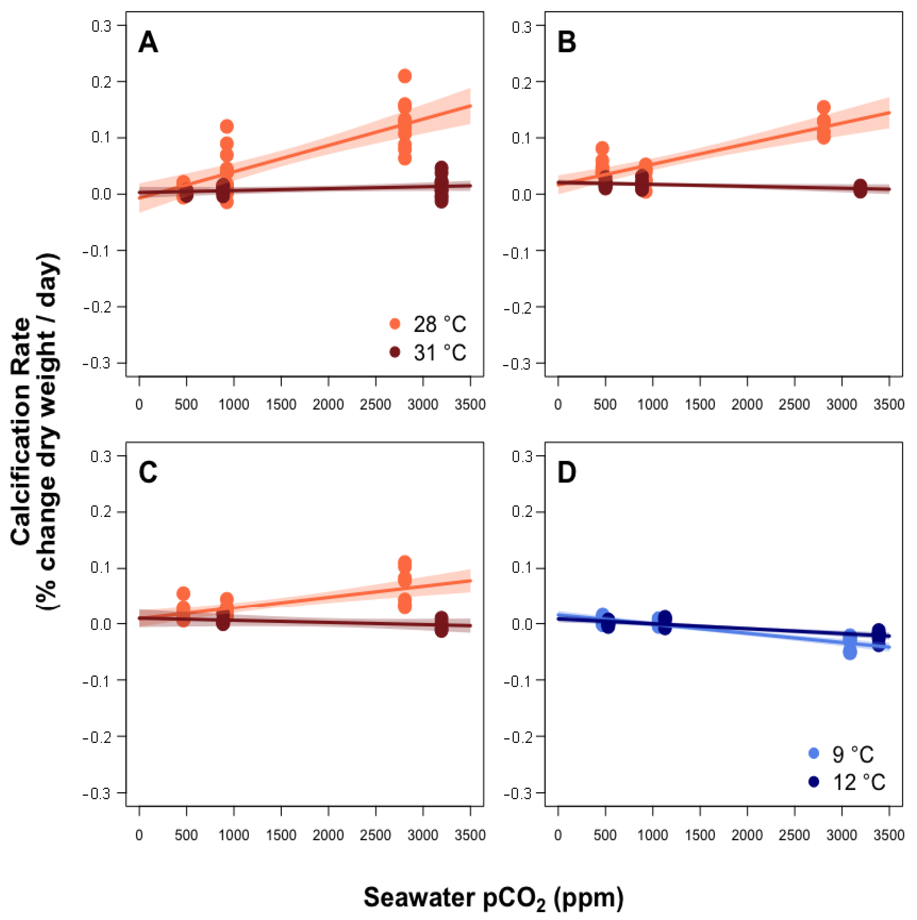
The calcification rate of all coral species (Figure 3) was significantly impacted by the interaction between seawater pCO2 and temperature (statistical significance indicated by p-value ≤ 0.05; Table S4). Calcification rate significantly increased with pCO2 under control temperature for all three tropical species, but showed no change across pCO2 treatments under elevated temperature for S. pistillata or P. damicornis, and decreased with increasing pCO2 under elevated temperature for S. hystrix. Temperature had a significant negative effect on calcification rate in all tropical species (Table S4. Calcification rate of the cold-water azooxanthellate coral L. pertusa declined significantly with increasing pCO2 at both temperatures, but showed a significantly stronger response to pCO2 under the control temperature (Figure 3; Table S4).

3.2. Calcifying Fluid Chemistry

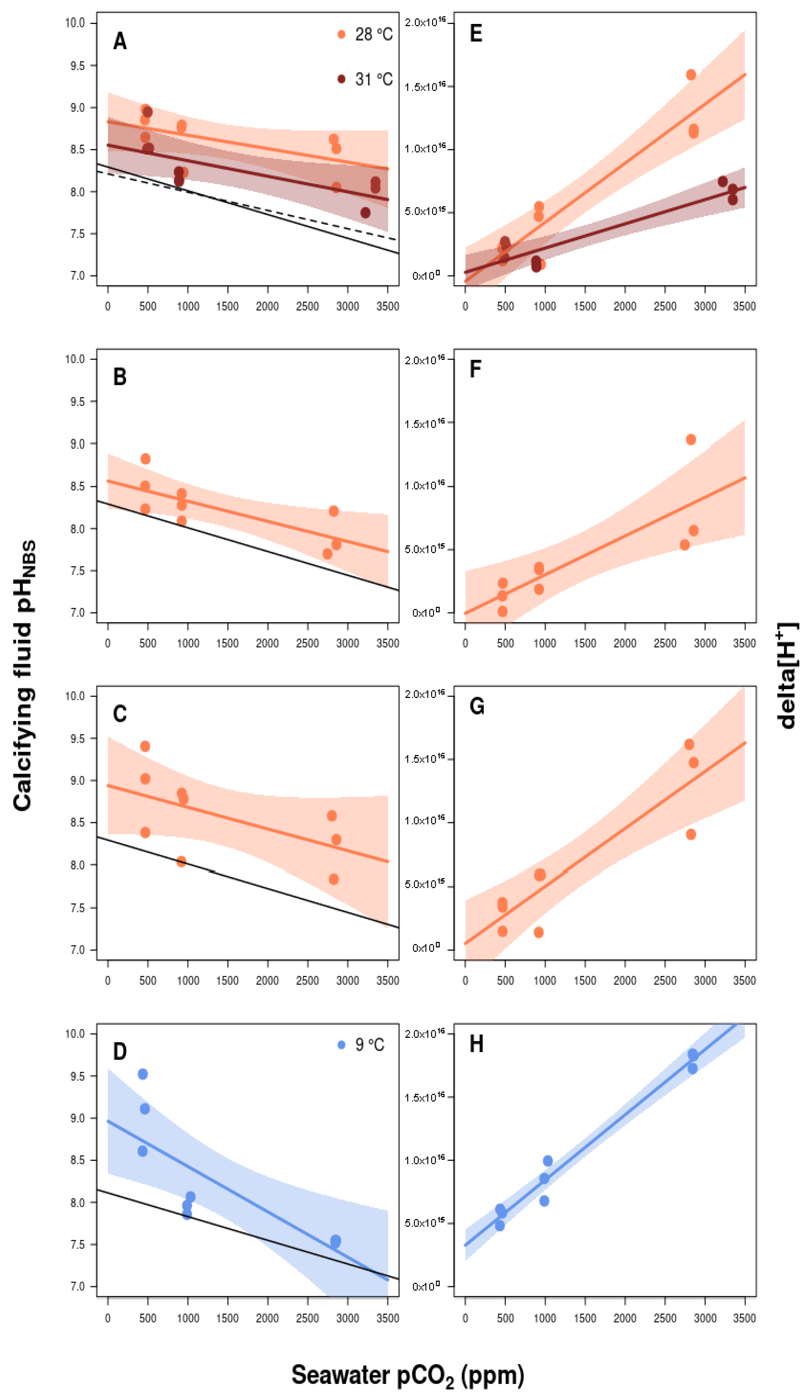
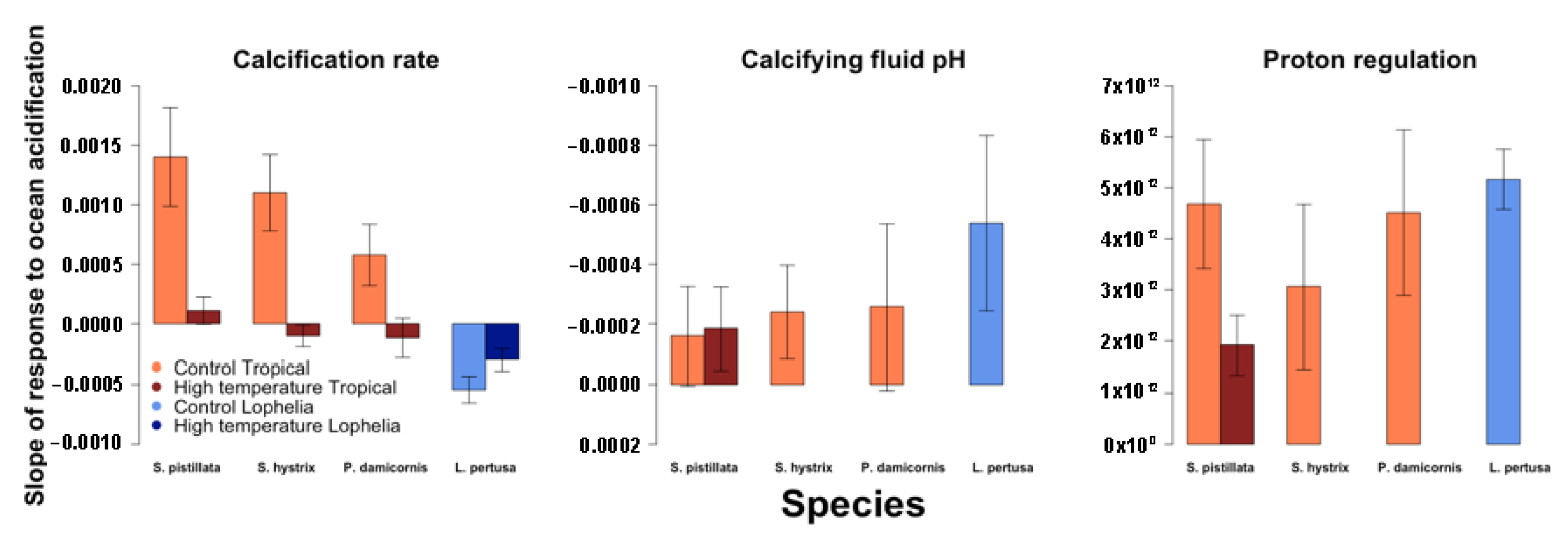
The pHCF of all four coral species was significantly greater than the pH of the corals’ surrounding seawater (pHSW) after 30 days of exposure to ocean acidification and warming (Figure 4A–D; lmer, S. pistillata: p < 0.001; P. damicornis: p = 0.002; S. hystrix: p = 0.013; L. pertusa: p = 0.002). The pHCF of all four species declined significantly with increasing pCO2 under control temperatures (Table S5), and, also, for S. pistillata under elevated temperature (S. pistillata was the only species for which pHCF was measured at both control and elevated temperature; Figure 4). Under the elevated temperature treatment, pHCF of S. pistillata remained higher than pHSW, but was significantly lower than pHCF at the control temperature (Table S5). The cold-water coral Lophelia pertusa exhibited the steepest decline in pHCF with increasing pCO2 (Figure 5).
All four coral species increased their ∆[H+] (i.e., seawater [H+]—calcifying fluid [H+]) in response to increasing seawater pCO2 (Figure 4E–H, Table S6). The ∆[H+] of S. pistillata was significantly influenced by the interaction between pCO2 and temperature (Table S6). In this species, there was no difference in ∆[H+] between the two temperature treatments under control pCO2. The ∆[H+] increased with pCO2 under both temperature treatments, but this increase was significantly greater in the control temperature treatment.

3.3. Estimated Coral Photosymbiont Index

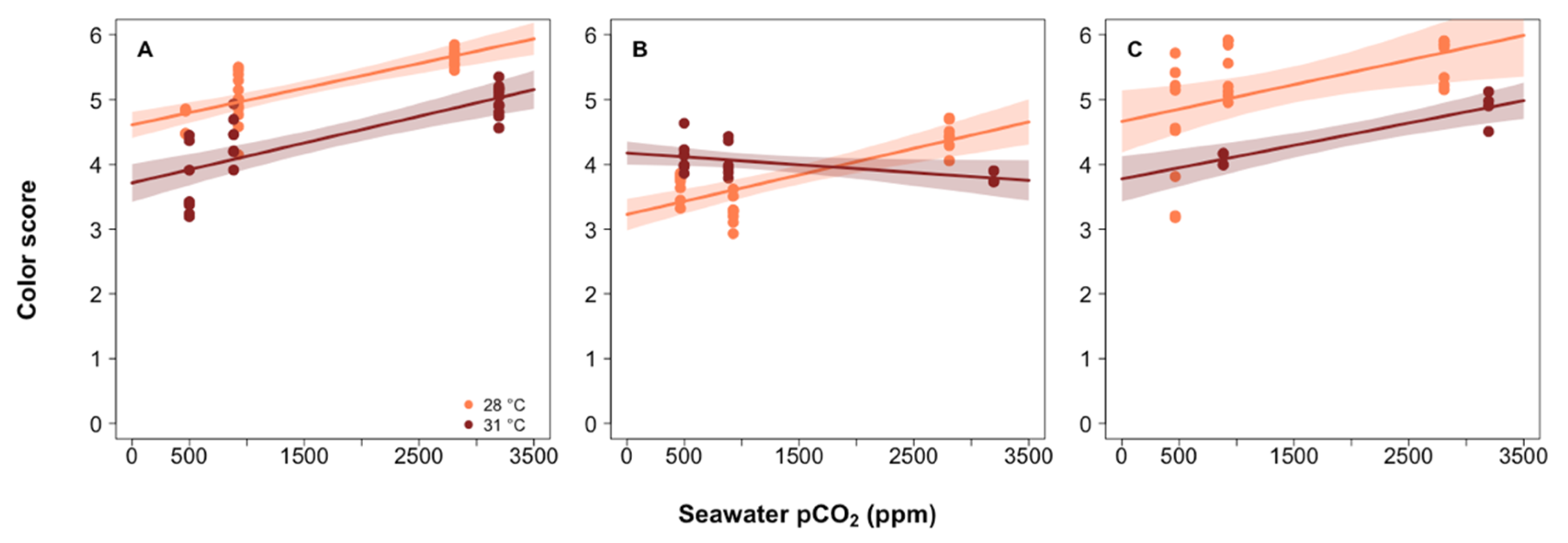
Both S. pistillata and P. damicornis exhibited a lower photosymbiont index (i.e., lower estimated photosymbiont abundance) under the elevated temperature treatments (Figure 6, Table S7). Photosymbiont index significantly increased in both S. pistillata and P. damicornis when pCO2 was elevated from control conditions (Figure 6, Table S7). The photosymbiont index of S. hystrix was significantly negatively correlated with the interaction between temperature and pCO2, whereby photosymbiont index increased significantly with increasing pCO2 under the control temperature treatment, but showed no change in response to pCO2 in the elevated temperature treatment.

3.4. Investigating the Role of Photosynthesis in Calcification

The calcification rate of S. pistillata was best predicted by the significant interaction between photosymbiont index and temperature, whereby photosymbiont index was positively correlated with calcification rate under both temperatures, but the slope of this correlation was significantly greater under the control temperature treatment (Figure 7A, Table S8). The calcification rate of S. hystrix was best predicted by a model that included both photosymbiont index and temperature independently. Photosymbiont index had a positive linear relationship with calcification rate under both temperatures, but the slope of this relationship significantly decreased in the high temperature treatment (Figure 7B, Table S8). The calcification rate of P. damicornis was best predicted by a model that contained photosymbiont index, seawater pCO2, and temperature (Table S8). The only significant predictor of calcification rate of P. damicornis was the interaction between photosymbiont index, temperature, and pCO2 (Figure 7C, Table S8).

4. Discussion

4.1. Calcification Response to pCO2 and Thermal Stress

Prior studies have shown that S. pistillata and P. damicornis both exhibit resilience in their calcification response to ocean acidification [13,51,52], while S. hystrix [53] tends to exhibit more negative responses. The present study, however, shows that, in the absence of thermal stress, these three common species of tropical zooxanthellate corals exhibit increased rates of calcification in response to a one-month exposure to CO2-acidified conditions. Although the disparity between the results of the past and present studies on these species could be due to differences in experimental design, such as experimental duration, the method used to measure calcification rates, temperature treatments, and/or light levels, these findings provide compelling evidence that, under certain circumstances (e.g., absence of thermal stress), some tropical zooxanthellate coral species can tolerate OA over at least one-month intervals.
Under the elevated temperature treatment (31 °C), the calcification rates of all three tropical coral species were reduced compared to the control temperature, but were unchanged by increasing pCO2, showing that thermal stress effectively impaired the zooxanthellate corals’ calcification response to CO2-induced OA. However, it should be noted that warming can have either positive or negative effects on coral calcification rate, depending on whether the warming causes temperatures to approach or exceed, respectively, the coral’s thermal optimum [19,21,54,55].
Few studies have investigated the calcification response of corals to combined ocean acidification and warming. While some of the studies show a negative response to these combined stressors [55], others contrast the results of the present study by showing no interactive effects of ocean acidification and warming [21,56,57]. The results presented here show that the impacts of ocean acidification on the calcification rates of three Pocilloporid coral species are exacerbated by warming. Because OA and global warming typically occur in tandem during major perturbations to the Earth’s carbon cycle, both throughout Earth history [58] and as a consequence of anthropogenic CO2 emissions [59], future CO2-induced global change poses a substantial threat to these coral species.
The calcification rate of the cold-water azooxanthellate coral L. pertusa declined under elevated pCO2 at both temperatures. This negative calcification response to pCO2 was weaker under the elevated temperature. Notably, the direction of the pCO2-temperature interaction for the cold-water azooxanthellate species was opposite that of the three tropical species. Lophelia pertusa exhibited net dissolution when seawater pCO2 reached ca. 1000 ppm, although low levels of calcification have been previously observed for L. pertusa under similar conditions [34,35,60]. These conditions are predicted to occur in the surface open ocean by the end of the 21st century [1] and earlier in high-latitude cold-water environments [61]—suggesting that this cold-water ecosystem engineer, whose reef-systems function as nursery ground for a number of commercially important fish species [62], may be unable to form reefs beyond this century. However, the observation that increased temperature partially mitigates the impact of OA agrees with the results of longer-term studies [35], and suggests that the impacts of global change on this species will vary with temperature across latitude and depth, as this species can inhabit seawater ranging from 4 to 14 °C [63,64].
The differences in calcification response to ocean acidification shown here may arise from differences in the physiology and ecology of tropical vs. cold-water corals. Although tropical corals exist close to their thermal limits and are therefore vulnerable to even small amounts of warming, azooxanthellate cold-water corals can generally tolerate a wider temperature range [65]. Elevated respiration rates have been observed for L. pertusa in warmer temperatures [66]. Thus, an increase in temperature may boost metabolic rates to partially mitigate the impacts of OA on calcification in the high pCO2 treatments. The species of tropical corals studied here are colonial and, thus, share resources between closely packed polyps [67]. These polyps are connected by coenosarc tissue, which covers and protects the skeleton [68]. The high degree of tissue cover exhibited by tropical corals means that the skeleton is well protected from dissolution, which may explain the lack of negative calcification response (i.e., lack of net dissolution) for the tropical corals in this study. Their shared gastrovascular system allows the distribution of metabolites generated from coral respiration and zooxanthellate photosynthesis across the colony, which could yield further resilience against ocean acidification. In contrast, L. pertusa is a pseudocolonial species [69] that produces single polyps on the end of stalk-like branches. The coenosarc connecting these branches is often partially absent in laboratory specimens and in wild specimens during the winter, leaving regions of exposed skeleton vulnerable to dissolution [65]. This lack of connectivity between polyps and the presence of exposed skeleton may increase the vulnerability of L. pertusa to ocean acidification.
Alternatively, the increased solubility of CO2 in colder waters caused ΩA of the L. pertusa treatments to be 0.12–0.70 units lower at 9 °C than at 12 °C for a given pCO2 condition. This may have caused higher rates of skeletal dissolution in the high-pCO2 treatments maintained at the lower temperature, although the rate of dissolution of coral skeletons should be higher under the higher temperature treatments for equivalent ΩA [3]. Since the buoyant weight method [70] used here yields only a net rate of calcification, i.e., mass of new skeleton produced through gross calcification minus mass of exposed skeleton lost through gross dissolution, it is not possible to determine whether the positive impact of the interaction between pCO2 and temperature on L. pertusa calcification rate was driven by increased gross calcification or reduced gross dissolution (or a combination of these factors) in the high-temperature, high-pCO2 treatments. Additionally, it was not determined whether OA impacted the density of coral skeleton, which could increase the fragility of the reef framework that these corals form [71].
The responses observed in the present study are consistent with other laboratory studies showing that scleractinian corals exhibit a wide range of calcification responses to OA [13,19,20,21,22,23,24,52,72]. Some of this variability may arise from differences in experimental design, such as the amount of food provided to the corals, the levels of irradiance, and the duration of the experiment. Nevertheless, this high variability in calcification response patterns across and within species indicates that a greater understanding of the mechanisms that drive coral responses to OA is needed.
The present study was conducted over an eight-week interval, with a total of three weeks of acclimation to laboratory and experimental conditions, and should, therefore, be considered intermediate in duration. As with any experimental OA study, it is possible that the duration of exposure may influence results, as corals may function normally over short timeframes, but exhibit impaired function over longer timeframes as a result of cumulative stress and/or depletion of metabolic resources [19]. Alternatively, corals may exhibit impaired responses shortly after exposure to the treatment conditions due to shock, but acclimate to the treatment conditions over longer timeframes. Prior laboratory-based studies have shown that tropical corals exhibit variable degrees of acclimation to OA over relatively short timescales [19,24,73], whereas acclimation of L. pertusa has been observed over longer timescales [74]. The large inter- and intra-specific differences in coral response to OA across experiments and timeframes underscores the need for additional research into the long-term effects of OA on coral calcification.

4.2. Role of Calcifying Fluid pH Regulation in Coral Response to pCO2 and Thermal Stress

Coral calcifying fluid pH elevation has been widely cited as a mechanism for promoting calcification under conditions of ocean acidification [8,11,14]. Although pHCF declined in all four species under elevated pCO2, it always remained higher than pHSW. Notably, coral species that showed the highest degree of control over pHCF in the present experiment, and thus the shallowest slope of change in pHCF in response to changing seawater pCO2, also exhibited the greatest increase in calcification rate when pCO2 was increased, suggesting that pHCF regulation confers resilience to corals exposed to OA.
These trends are consistent with prior estimation of coral pHCF from boron isotopes [10,38,39,41,75,76,77], pH-sensitive fluorescent dyes [12], and pH-sensitive microsensors [9,11]. Previous studies of the effects of ocean acidification on calcification site pH show that both S. pistillata and P. damicornis elevate pHCF above seawater pH, and that this elevation increases under ocean acidification [13,52]. These results are consistent with the findings here, although the measured pHCF was considerably higher in the present study compared to previous studies.
The present study used pH-sensitive microelectrodes, whereas prior studies on both S. pistillata and P. damicornis [52], and P. damicornis [13] used confocal microscopy to image pH-sensitive SNARF-1 dye in coral microcolonies grown on glass slides and boron isotope systematics, respectively. Differences in pHCF could be due to differences in methods of culturing and/or pHCF-estimation, or due to genotypic differences between cultured specimens. Of the three methods used to estimate pHCF (pH-sensitive dyes, boron isotopes, and pH-sensitive microelectrodes), pH-sensitive microelectrodes typically yield the highest pHCF [11], although a side-by-side comparison of pHCF measured with pH-sensitive dye and pH microelectrodes on the same specimens yielded comparable results [14].
Measurements of pHCF using pH-sensitive microelectrodes are challenged by the difficulty in assessing the precise location of the microelectrode tip relative to the coral’s calcifying fluid. This challenge was addressed in the present experiment through the creation of pHCF profiles as the pH microelectrode was withdrawn from the calcifying fluid, thus allowing characterization of the calcifying fluid pH compared to intratissue and/or gastrovascular pH (Figure 2), as was conducted in previous studies [14,78]. Additionally, the pHCF of two coral species in the present study (S. pistillata and P. damicornis) was estimated by coral skeletal δ11B to provide a side-by-side comparison of these independent approaches to measuring pHCF [41]. A significant correlation was found between pHCF measured using these two methods, increasing confidence that the measurements obtained here represent pH of the calcifying fluid. The offset between the two approaches was attributed to the two techniques measuring pHCF over different timescales—with skeletal δ11B recording a time-averaged value of pHCF and pH microelectrodes recording a more instantaneous value of pHCF [41].
Although numerous studies have shown that OA reduces coral pHCF, few have investigated the combined impact of warming and OA on pHCF. In the present study, microelectrode measurements of the pHCF of S. pistillata were measured in all pCO2 and temperature treatments. The prescribed temperature increase resulted in a significant decline in pHCF for each of the three pCO2 treatments. Under heat stress, corals may receive less nourishment from their photosymbionts to support pHCF regulation due to thermally induced bleaching and/or may divert energy from the regulation of pHCF towards tissue repair.
Assuming that the coral calcifying fluid is ultimately derived from the coral’s surrounding seawater [79], the extent to which a coral mitigates the impacts of OA by removing protons from its calcifying fluid can be grossly estimated (excluding the effects of buffering) from the difference between the [H+] of the calcifying fluid and the [H+] of the surrounding seawater (i.e., ∆[H+]). All four coral species exhibited increased ∆[H+] under elevated pCO2 treatments, suggesting that more energy is allocated to pHCF regulation under elevated pCO2. The ∆[H+] was lower in the high-temperature treatment in S. pistillata, suggesting that less energy is available to maintain elevated pHCF when the corals experience thermally induced symbiont loss—potentially due to a commensurate decrease in photosynthate translocated to the coral host.

4.3. Role of Photosymbionts in Coral Response to pCO2 and Thermal Stress

In order for corals to allocate more energy toward removing protons from their calcifying fluid under OA conditions, they must divert energy from other activities and/or increase their energetic intake [80]. Zooxanthellate corals obtain energy from two sources—heterotrophic feeding and photosynthate translocated from their algal symbionts. As corals are sessile suspension feeders, they are limited in the extent to which they can increase heterotrophic feeding, although they may increase feeding rates if sufficient food is available [81,82]. Alternatively, enhanced photosynthesis under OA may play an important role in driving proton elevation under OA.
Using the well-established colorimetric method of Siebeck et al. [45] to estimate the abundance of zooxanthellae via the photosymbiont index, it was shown that the populations of photosymbionts from S. pistillata and P. damicornis were significantly reduced in the high temperature treatment compared to the initial values. This is consistent with prior work showing that prolonged heat stress can lead to expulsion of zooxanthellate and tissue damage, a process termed ‘bleaching’ [83]. However, in this study, the photosymbiont index increased in response to increasing pCO2 under both temperature treatments, suggesting that the photosymbionts within all three Pocilloporid tropical corals benefited from the increased availability of dissolved inorganic carbon (DIC). The alleviation of carbon limited photosynthesis could free up energy for elevating pHCF and/or the production of carbon concentrating enzymes (e.g., carbonic anhydrase [84]), thereby aiding calcification under the control temperature [68,85,86]. The zooxanthellae’s apparently positive response to increasing DIC also suggests that dissolved inorganic nutrients (DIN) were sufficient and in the correct balance (i.e., Redfield ratio) to sustain photosynthesis [87,88]. This link between enhanced photosynthesis and enhanced calcification under the elevated pCO2 and control temperature treatment is consistent with the observed correlations between photosymbiont index and calcification rate in S. pistillata and S. hystrix across all treatments.

4.4. Proposed Mechanistic Framework for Zooxanthellate Coral Response to pCO2 and Thermal Stress

We propose the following mechanistic framework to explain the zooxanthellate coral responses to OA and warming observed in this study. Under the control temperature and pCO2 conditions, zooxanthellae fix DIC as carbohydrates (photosynthate), which is then used by the coral hosts as an energy source for all physiological activities, including elevation of pHCF in support of calcification [4,8,89,90]. When OA occurs without thermal stress, high pCO2 enhances photosynthesis in the coral species investigated and increases their photosymbiont index. Under conditions of elevated pCO2, enhanced photosymbiont productivity (evidenced by increased photosymbiont index in this study), will result in a greater abundance of byproducts that may be used by the coral host to increase proton removal from the calcifying fluid (evidenced by elevated Δ[H+]). The combination of elevated pHCF and elevated DIC (due to elevated pCO2) under OA conditions may allow some species of corals to maintain an ΩA in their calcifying fluid that is comparable to or, perhaps, greater than those exhibited under non-acidified conditions [11]—hence, their observed ability to maintain constant or, in some cases, elevated rates of calcification under OA.
Although CO2-induced OA appears supportive of calcification for these three species under the control temperature, this support breaks down in the high temperature treatment. The thermal stress induced in this treatment caused a reduction in the abundance of the corals’ algal symbionts (evidenced by their reduced photosymbiont index), which was accompanied by a reduction in Δ[H+] (i.e., proton removal) and calcification rate of S. pistillata under each of the elevated pCO2 treatments. It appears that the thermally induced reduction in photosymbiont index eliminated the benefit of enhanced photosynthesis under conditions of elevated pCO2, thereby leaving the coral with fewer resources (e.g., translocated photosynthate) for elevating pHCF. It should also be noted that thermal impairment of the enzymes used to remove protons from the coral calcifying fluid (e.g., H+/Ca2+-ATPase [9,91]) may have contributed—along with reduced photosymbiont index—to declines in pHCF and Δ[H+] observed for S. pistillata in response to thermal stress. Additionally, the different responses shown in the control and high temperature treatments could be due to a temperature-induced shift in the strategy used by the coral to elevate aragonite saturation state in their calcifying fluid. In natural reef systems, it has been shown that coral pHCF is most elevated in the winter months [16]. Although pHCF is still elevated in the warmer summer months, it is elevated to a lesser extent, and DIC elevation appears to be the primary means of raising ΩA [16]. These observations are consistent with and provide a potential explanation for the results of the present study.
The role of symbiotic zooxanthellae in conferring resilience to corals exposed to OA is also highlighted in the stark contrast observed between the tropical and deep-sea coral responses to OA. Cold-water corals are azooxanthellate and thus do not receive the benefits of enhanced symbiont photosynthesis under elevated pCO2. Although Δ[H+] of the cold-water species was comparable to that of the tropical species under OA, proton regulation of the calcifying fluid probably consumes a greater proportion of the total resources of the cold-water species (which acquires no resources from photosynthesis) than that of the tropical zooxanthellate species. This may leave proportionally fewer resources (compared with the tropical species) for other processes associated with calcification in the cold-water species, such as the production of organic matrices that may initiate crystal nucleation [92], which may explain the more negative calcification response to OA exhibited by L. pertusa.

4.5. Limitations of Laboratory-Based Experiments

The results described here were obtained in a controlled laboratory setting. In their natural reef environments, tropical corals experience fluctuations in temperature and carbonate chemistry across daily [93,94,95] and seasonal [96,97] cycles, whereas corals inhabiting deeper, colder waters experience more stable environments. Thus, differing degrees of prior exposure to fluctuations in pH could contribute to the differential responses of the tropical and cold-water coral species observed here. The enhanced decline in pHCF under elevated temperature exhibited by the tropical corals could also result from a shift in strategy for elevating calcifying fluid ΩA from pHCF elevation to DIC elevation, as has been shown to occur seasonally in a natural reef system [16]. Future research is necessary to assess whether the responses observed here hold in more dynamic temperature and pH environments.

5. Conclusions

Global warming is considered to be amongst the greatest threats facing coral reefs, and OA is emerging as an equally grave threat. We show that warming has a more negative impact than OA on three species of zooxanthellate tropical corals, whereas OA has a more negative impact than warming on an azooxanthellate cold-water coral. This study also provides insight into the role of photosymbionts in corals’ response to OA. Specifically, the enhancement of symbiont photosynthesis under higher-pCO2 conditions appears to mitigate the negative effects of OA on tropical zooxanthellate corals by providing resources that assist in the maintenance of elevated calcifying fluid pH in support of calcification. This resilience, however, is impaired when OA is combined with thermally induced reductions in the abundance of the coral’s photosymbionts (i.e., ‘bleaching’), which limits the extent to which the coral holobiont can utilize the elevated DIC via photosynthesis. These results highlight the threat that ocean warming and acidification pose for tropical and cold-water corals, especially when occurring in tandem.

Supplementary Materials

The following supporting information can be downloaded at: https://www.mdpi.com/article/10.3390/jmse10081106/s1: Summary of experimental design and results (Section S1); Water quality methods and summary tables (Section S2); Buoyant weight methods (Section S3); Images of calcein dye incorporation into coral skeleton (Section S4); Method for estimating photosymbiont index (Section S5); and Model summary tables (Section S6).

Author Contributions

Conceptualization, J.B.R. and R.A.E.; methodology, J.B.R., R.A.E., L.P.C., D.D.B., H.W., J.V.B., G.M.S.-G. and I.W.; formal analysis, L.P.C., J.B.R. and J.G.; investigation, L.P.C., J.B.R., C.E.R., F.M.-L., I.W., J.V.B. and R.A.E. sources, J.B.R., R.A.E., D.D.B., J.B. and H.W.; data curation, L.P.C., J.B.R. and R.A.E.; writing—original draft preparation, L.P.C. and J.B.R.; writing—review and editing, L.P.C., J.B.R., R.A.E., M.G., C.E.R., J.B., J.V.B., D.D.B., G.M.S.-G. and H.W.; visualization, L.P.C., J.B.R. and J.G.; supervision, J.B.R., R.A.E., D.D.B., J.B. and H.W.; project administration, J.B.R. and H.W.; funding acquisition, J.B.R., R.A.E. and H.W. All authors have read and agreed to the published version of the manuscript.

Funding

J.B.R. acknowledges support from National Science Foundation grant OCE-1437371, the ZMT, and a Hanse-Wissenschaftskolleg Fellowship. R.A.E. acknowledges support from National Science Foundation grant OCE-1437166, the Pritzker Endowment to UCLA IoES, and ‘Laboratoire d’Excellence’ LabexMER grant ANR-10-LABX-19 co-funded by a grant from the French government under the program ‘Investissements d’Avenir’.

Institutional Review Board Statement

Not applicable.

Informed Consent Statement

Not applicable.

Data Availability Statement

It is our intention to make data available on publication of this study.

Acknowledgments

We thank Artur Fink, Laurie Hoffman, and Anja Niclas (MPIMM) for assistance with construction and use of pH microelectrodes; Silvia Hardenberg, Nico Steinel, and Christian Brandt (ZMT) for assistance with coral husbandry and redesign of the acidification system at the ZMT; and Matthias Birkicht (ZMT) for assistance with water chemistry analysis.

Conflicts of Interest

The authors declare no conflict of interest.

References

  1. IPCC. Climate Change 2013—The Physical Science Basis; Intergovernmental Panel on Climate Change, Ed.; Cambridge University Press: Cambridge, UK, 2014; ISBN 9781107415324. [Google Scholar] [CrossRef]
  2. Caldeira, K.; Wickett, M.E. Anthropogenic carbon and ocean pH. Nature 2003, 425, 365. [Google Scholar] [CrossRef] [PubMed]
  3. Ries, J.B.; Ghazaleh, M.N.; Connolly, B.; Westfield, I.; Castillo, K.D. Impacts of seawater saturation state state (ΩA = 0.4–4.6) and temperature (10, 25 °C) on the dissolution kinetics of whole-shell biogenic carbonates. Geochim. Cosmochim. Acta 2016, 192, 318–337. [Google Scholar] [CrossRef]
  4. Jokiel, P.L.; Rodgers, K.S.; Kuffner, I.B.; Andersson, A.J.; Cox, E.F.; Mackenzie, F.T. Ocean acidification and calcifying reef organisms: A mesocosm investigation. Coral Reefs 2008, 27, 473–483. [Google Scholar] [CrossRef]
  5. Milliman, J.D. Production and accumulation of calcium carbonate in the ocean: Budget of a nonsteady state. Glob. Biogeochem. Cycles 1993, 7, 927–957. [Google Scholar] [CrossRef]
  6. Anthony, K.R.; Fabricius, K.E. Shifting roles of heterotrophy and autotrophy in coral energetics under varying turbidity. J. Exp. Mar. Bio. Ecol. 2000, 252, 221–253. [Google Scholar] [CrossRef]
  7. Hoegh-Guldberg, O. Climate change, coral bleaching and the future of the world’s coral reefs. Mar. Freshw. Res. 1999, 50, 839–866. [Google Scholar] [CrossRef]
  8. McCulloch, M.; Falter, J.; Trotter, J.; Montagna, P. Coral resilience to ocean acidification and global warming through pH up-regulation. Nat. Clim. Change 2012, 2, 623–627. [Google Scholar] [CrossRef]
  9. Al-Horani, F.A.; Al-Moghrabi, S.M.; de Beer, D. The mechanism of calcification and its relation to photosynthesis and respiration in the scleractinian coral Galaxea fascicularis. Mar. Biol. 2003, 142, 419–426. [Google Scholar] [CrossRef]
  10. Allison, N.; Finch, A.A. 11B, Sr, Mg and B in a modern Porites coral: The relationship between calcification site pH and skeletal chemistry. Geochim. Cosmochim. Acta 2010, 74, 1790–1800. [Google Scholar] [CrossRef]
  11. Ries, J.B. A physicochemical framework for interpreting the biological calcification response to CO2-induced ocean acidification. Geochim. Cosmochim. Acta 2011, 75, 4053–4064. [Google Scholar] [CrossRef]
  12. Venn, A.; Tambutté, E.; Holcomb, M.; Allemand, D.; Tambutté, S. Live tissue imaging shows reef corals elevate pH under their calcifying tissue relative to seawater. PLoS ONE 2011, 6, e20013. [Google Scholar] [CrossRef] [PubMed]
  13. Comeau, S.; Cornwall, C.E.; McCulloch, M.T. Decoupling between the response of coral calcifying fluid pH and calcification to ocean acidification. Sci. Rep. 2017, 7, 7573. [Google Scholar] [CrossRef] [PubMed]
  14. Sevilgen, D.S.; Venn, A.A.; Hu, M.Y.; Tambutté, E.; de Beer, D.; Planas-Bielsa, V.; Tambutté, S. Full in vivo characterization of carbonate chemistry at the site of calcification in corals. Sci. Adv. 2019, 5, eaau7447. [Google Scholar] [CrossRef] [PubMed]
  15. Allison, N.; Cohen, I.; Finch, A.A.; Erez, J.; Tudhope, A.W. Corals concentrate dissolved inorganic carbon to facilitate calcification. Nat. Commun. 2014, 5, 5741. [Google Scholar] [CrossRef] [PubMed]
  16. McCulloch, M.; D’Olivo, J.P.; Falter, J.; Holcomb, M.; Trotter, J.A. Coral calcification in a changing world and the interactive dynamics of pH and DIC upregulation. Nat. Commun. 2017, 8, 15686. [Google Scholar] [CrossRef]
  17. Ries, J.B. Skeletal mineralogy in a high-CO2 world. J. Exp. Mar. Biol. Ecol. 2011, 403, 54–64. [Google Scholar] [CrossRef]
  18. Spalding, C.; Finnegan, S.; Fischer, W.W. Energetic costs of calcification under ocean acidification. Glob. Biogeochem. Cycles 2017, 31, 866–877. [Google Scholar] [CrossRef]
  19. Castillo, K.D.; Ries, J.B.; Bruno, J.F.; Westfield, I.T. The reef-building coral Siderastrea siderea exhibits parabolic responses to ocean acidification and warming. Proc. Biol. Sci. 2014, 281, 20141856. [Google Scholar] [CrossRef]
  20. Marubini, F.; Ferrier-Pages, C.; Furla, P.; Allemand, D. Coral calcification responds to seawater acidification: A working hypothesis towards a physiological mechanism. Coral Reefs 2008, 27, 491–499. [Google Scholar] [CrossRef]
  21. Bove, C.B.; Ries, J.B.; Davies, S.W.; Westfield, I.T.; Umbanhowar, J.; Castillo, K.D. Common Caribbean corals exhibit highly variable responses to future acidification and warming. Proc. Biol. Sci. 2019, 286, 20182840. [Google Scholar] [CrossRef]
  22. Ries, J.B.; Cohen, A.L.; McCorkle, D.C. A nonlinear calcification response to CO2-induced ocean acidification by the coral Oculina arbuscula. Coral Reefs 2010, 29, 661–674. [Google Scholar] [CrossRef]
  23. Reynaud, S.; Leclercq, N.; Romaine-Lioud, S.; Ferrier-Pagès, C.; Jaubert, J.; Gattuso, J.-P. Interacting effects of CO2 partial pressure and temperature on photosynthesis and calcification in a scleractinian coral. Glob. Chang. Biol. 2003, 9, 1660–1668. [Google Scholar] [CrossRef]
  24. Comeau, S.; Cornwall, C.E.; DeCarlo, T.M.; Doo, S.S.; Carpenter, R.C.; McCulloch, M.T. Resistance to ocean acidification in coral reef taxa is not gained by acclimatization. Nat. Clim. Chang. 2019, 9, 477. [Google Scholar] [CrossRef]
  25. Carafoli, E. Calcium signaling: A tale for all seasons. Proc. Natl. Acad. Sci. USA 2002, 99, 1115–1122. [Google Scholar] [CrossRef]
  26. Mass, T.; Drake, J.L.; Peters, E.C.; Jiang, W.; Falkowski, P.G. Immunolocalization of skeletal matrix proteins in tissue and mineral of the coral Stylophora pistillata. Proc. Natl. Acad. Sci. USA 2014, 111, 12728–12733. [Google Scholar] [CrossRef]
  27. Falini, G.; Fermani, S.; Goffredo, S. Coral biomineralization: A focus on intra-skeletal organic matrix and calcification. Semin. Cell Dev. Biol. 2015, 35, 17–26. [Google Scholar] [CrossRef]
  28. Marin, F.; Smith, M.; Isa, Y.; Muyzer, G.; Westbroek, P. Skeletal matrices, muci, and the origin of invertebrate calcification. Proc. Natl. Acad. Sci. USA 1996, 93, 1554–1559. [Google Scholar] [CrossRef]
  29. Westbroek, P.; Marin, F. A marriage of bone and nacre. Nature 1998, 392, 861–862. [Google Scholar] [CrossRef]
  30. Mass, T.; Drake, J.L.; Haramaty, L.; Kim, J.D.; Zelzion, E.; Bhattacharya, D.; Falkowski, P.G. Cloning and characterization of four novel coral acid-rich proteins that precipitate carbonates in vitro. Curr. Biol. 2003, 23, 1126–1131. [Google Scholar] [CrossRef]
  31. Mass, T.; Giuffre, A.J.; Sun, C.-Y.; Stifler, C.A.; Frazier, M.J.; Neder, M.; Tamura, N.; Stan, C.V.; Marcus, M.A.; Gilberg, P.U.P.A. Amorphous calcium carbonate particles form coral skeletons. Proc. Natl. Acad. Sci. USA 2017, 114, E7670–E7678. [Google Scholar] [CrossRef]
  32. Hohn, S.; Reymond, C.E. Coral calcification, mucus, and the origin of skeletal organic molecules. Coral Reefs 2019, 38, 973–984. [Google Scholar] [CrossRef]
  33. Roberts, J.M.; Wheeler, A.; Freiwald, A.; Cairns, S. The Biology and Geology of Deep-Sea Coral Habitats; Cambridge University Press: Cambridge, UK, 2009. [Google Scholar]
  34. Hennige, S.J.; Wicks, L.C.; Kamenos, N.A.; Perna, G.; Findlay, H.S.; Roberts, J.M. Hidden impacts of ocean acidification to live and dead coral framework. Proc. R. Soc. B 2015, 282, 20150990. [Google Scholar] [CrossRef] [PubMed]
  35. Büscher, J.V.; Form, A.U.; Riebesell, U. Interactive effects of ocean acidification and warming on growth, fitness and survival of the cold-water coral Lophelia pertusa under different food availabilities. Front. Mar. Sci. 2017, 4, 101. [Google Scholar] [CrossRef]
  36. Georgian, S.E.; Dupont, S.; Kurman, M.; Butler, A.; Strömberg, S.M.; Larsson, A.I.; Cordes, E.E. Biogeographic variability in the physiological response of the cold-water coral Lophelia pertusa to ocean acidification. Mar. Ecol. 2016, 37, 1345–1359. [Google Scholar] [CrossRef]
  37. Naumann, M.S.; Orejas, C.; Ferrier-Pagès, C. Species-specific physiological response by the cold-water corals Lophelia pertusa and Madrepora oculata to variations within their natural temperature range. Deep. Sea Res. Part II Top. Stud. Oceanogr. 2014, 99, 36–41. [Google Scholar] [CrossRef]
  38. Holcomb, M.; Venn, A.A.; Tambutté, E.; Tambutté, S.; Allemand, D.; Trotter, J.; McCulloch, M. Coral calcifying fluid pH dictates response to ocean acidification. Sci. Rep. 2014, 4, 5207. [Google Scholar] [CrossRef]
  39. Liu, Y.-W.; Sutton, J.N.; Ries, J.B.; Eagle, R.A. Regulation of calcification site pH is a polyphyletic but not always governing response to ocean acidification. Sci. Adv. 2020, 6, eaax1314. [Google Scholar] [CrossRef]
  40. D’Olivo, J.P.; McCulloch, M.T. Response of coral calcification and calcifying fluid composition to thermally induced bleaching stress. Nat. Sci. Rep. 2017, 7, 2207. [Google Scholar] [CrossRef]
  41. Guillermic, M.; Cameron, L.P.; De Corte, I.; Misra, S.; Bijma, J.; de Beer, D.; Reymond, C.E.; Westphal, H.; Ries, J.B.; Eagle, R.A. Thermal stress reduces pocilloporid coral resilience to ocean acidification by impairing control over calcifying fluid chemistry. Sci. Adv. 2021, 7, eaba9958. [Google Scholar] [CrossRef]
  42. Ellison, J.C.; Fiu, M. Vulnerability of Fiji’s mangroves and associated coral reefs to climate change. Ed. WWF S. Pac. Programme 2010, 50p. [Google Scholar]
  43. Cameron, L.P.; Reymond, C.E.; Müller-Lundin, F.; Westfield, I.; Grabowski, J.H.; Westphal, H.; Ries, J.B. Effects of temperature and ocean acidification on the extrapallial fluid pH, calcification rate, and condition factor of the king scallop Pecten maximus. J. Shellfish. Res. 2019, 38, 763–777. [Google Scholar] [CrossRef]
  44. Pierrot, D.; Lewis, E.; Wallace, D.W.R. MS excel program developed for CO2 system calculations. In ORNL/CDIAC-105a. Carbon Dioxide Information Analysis Center; Oak Ridge National Laboratory U.S. Department Energy: Oak Ridge, TN, USA, 2006. [Google Scholar]
  45. Siebeck, U.E.; Marshall, N.J.; Klüter, A.; Hoegh-Guldberg, O. Monitoring coral bleaching using a colour reference card. Coral Reefs 2006, 25, 453–460. [Google Scholar] [CrossRef]
  46. Conti-Jerpe, I.E.; Thompson, P.D.; Wai Martin Wong, C.; Oliveira, N.L.; Duprey, N.N.; Moynihan, M.A.; Baker, D.M. Trophic strategy and bleaching resistance in reef-building corals. Sci. Adv. 2020, 6, eaaz5443. [Google Scholar] [CrossRef] [PubMed]
  47. Morgans, C.A.; Hung, J.Y.; Bourne, D.G.; Quigley, K.M. Symbiodiniaceae probiotics for use in bleaching recovery. Restor. Ecol. 2020, 28, 282–288. [Google Scholar] [CrossRef]
  48. De Beer, D.E.; Schramm, A.; Santegoeds, C.M.; Kühl, M. A nitrite microsensor for profiling environmental biofilms. Appl. Environ. Microbiol. 1997, 63, 973–977. [Google Scholar] [CrossRef]
  49. Bates, D.; Mächler, M.; Bolker, B.; Walker, S. Fitting linear mixed-effects models using lme. J. Stat. Softw. 2015, 67, 1–48. [Google Scholar] [CrossRef]
  50. Akaike, H. Factor analysis and AIC. In Selected Papers of Hirotugu Akaike. Springer Series in Statistics (Perspectives in Statistics); Parzen, E., Tanabe, K., Kitagawa, G., Eds.; Springer: New York, NY, USA, 1987. [Google Scholar]
  51. Houlbrèque, F.; Rodolfo-Metalpa, R.; Jeffree, R.; Oberhänsli, F.; Teyssié, J.-L.; Boisson, F.; Al-Trabeen, K.; Ferrier-Pagès, C. Effects of increased pCO2 on zinc uptake and calcification in the tropical coral Stylophora pistillata. Coral Reefs 2012, 31, 101–109. [Google Scholar] [CrossRef]
  52. Venn, A.A.; Tambutté, E.; Caminiti-Segonds, N.; Techer, N.; Allemand, D.; Tambutté, S. Effects of light and darkness on pH regulation in three coral species exposed to seawater acidification. Sci. Rep. 2019, 9, 2201. [Google Scholar] [CrossRef]
  53. Rädecker, N.; Meyer, F.W.; Bednarz, V.N.; Cardini, U.; Wild, C. Ocean acidification rapidly reduces dinitrogen fixation associated with the hermatypic coral Seriatopora hystrix. Mar. Ecol. Prog. Ser. 2014, 511, 297–302. [Google Scholar] [CrossRef]
  54. Cantin, N.E.; Cohen, A.L.; Karnauskas, K.B.; Tarrant, A.M.; McCorkle, D.C. Ocean warming slows coral growth in the central Red Sea. Science 2010, 329, 322–325. [Google Scholar] [CrossRef]
  55. Horvath, K.M.; Castillo, K.D.; Armstrong, P.; Westfield, I.T.; Courtney, T.; Ries, J.B. Next-century ocean acidification and warming both reduce calcification rate, but only acidification alters skeletal morphology of reef-building coral Siderastrea siderea. Sci. Rep. 2016, 6, 29639. [Google Scholar] [CrossRef] [PubMed]
  56. Schoepf, V.; Grottoli, A.G.; Warner, M.E.; Cai, W.-J.; Melman, T.F.; Hoadley, K.D.; Pettay, D.T.; Hu, X.; Li, Q.; Xu, H.; et al. Coral energy reserves and calcification in a high-CO2 world at two temperatures. PLoS ONE 2013, 8, e75049. [Google Scholar] [CrossRef] [PubMed]
  57. Okazaki, R.R.; Towle, E.K.; van Hooidonk, R.; Mor, C.; Winter, R.N.; Piggot, A.M.; Cunning, R.; Baker, A.C.; Klaus, J.S.; Swart, P.K.; et al. Species-specific responses to climate change and community composition determine future calcification rates of Florida Keys reefs. Glob. Chang. Biol. 2016, 23, 1023–1035. [Google Scholar] [CrossRef] [PubMed]
  58. Hönisch, B.; Ridgwell, A.; Schmidt, D.N.; Gibbs, S.J.; Sluijs, A.; Zeebe, R.; Kump, L.; Martindale, R.C.; Greene, S.E.; Kiessling, W.; et al. The geologic record of ocean acidification. Science 2012, 335, 1058–1063. [Google Scholar] [CrossRef] [PubMed]
  59. Bijma, J.; Pörtner, H.-O.; Yesson, C.; Rogers, A.D. Climate change and the oceans–What does the future hold? Mar. Pollut. Bull. 2013, 74, 495–505. [Google Scholar] [CrossRef]
  60. Kurman, M.D.; Gómez, C.E.; Georgian, S.E.; Lunden, J.J.; Cordes, E.E. Intra-specific variation reveals potential adaptation to ocean acidification in a cold-water coral from the Gulf of Mexico. Front. Mar. Sci. 2017, 4, 111. [Google Scholar] [CrossRef]
  61. Fabry, V.J.; McClintock, J.B.; Mathis, J.T.; Grebmeier, J.M. Ocean acidification at high latitudes: The bellwether. Oceanography 2009, 22, 160–171. [Google Scholar] [CrossRef]
  62. Costello, M.J.; McCrea, M.; Freiwald, A.; Lundälv, T.; Jonsson, L.; Bett, B.J.; van Weering, T.C.E.; de Haas, H.; Roberts, J.M.; Allen, D. Role of cold-water Lophelia pertusa coral reefs as fish habitat in the NE Atlantic. In Cold-Water Corals and Ecosystems. Erlangen Earth Conference Series; Freiwald, A., Roberts, J.M., Eds.; Springer: Berlin, Germany, 2005; pp. 771–805. [Google Scholar]
  63. Mortensen, P.B.; Hovland, T.; Fossa, J.H.; Furevik, D.M. Distribution, abundance and size of Lophelia pertusa coral reefs in mid-Norway in relation to seabed characteristics. J. Mar. Biol. Assoc. UK 2001, 81, 581–597. [Google Scholar] [CrossRef]
  64. Freiwald, A. Geobiology of Lophelia pertusa (scleractinia) reefs in the North Atlantic. Habilitation Thesis, University of Bremen, Bremen, Germany, 1998. [Google Scholar]
  65. Rogers, A.D. The biology of Lophelia pertusa (linneaus 1758) and other deep-water reef-forming corals and impacts from human activities. Int. Rev. Hydrobiol. 1999, 84, 315–406. [Google Scholar] [CrossRef]
  66. Dodds, L.A.; Roberts, J.M.; Taylor, A.C.; Marubini, F. Metabolic tolerance of the cold-water coral Lophelia pertusa (scleractinia) to temperature and dissolved oxygen change. J. Exp. Mar. Biol. Ecol. 2007, 349, 205–214. [Google Scholar] [CrossRef]
  67. Fine, M.; Oren, U.; Loya, Y. Bleaching effect on regeneration and resource translocation in the coral Oculina patagonica. Mar. Ecol. Prog. Ser. 2002, 234, 119–125. [Google Scholar] [CrossRef]
  68. Tambutté, E.; Allemand, D.; Zoccola, D.; Meibom, A.; Lotto, S.; Caminiti, N.; Tambutté, S. Observations of the tissue-skeleton interface in the scleractinian coral Stylophora pistillata. Coral Reefs 2007, 26, 517–529. [Google Scholar] [CrossRef]
  69. Järnegren, J.; Kutti, T. Lophelia pertusa in Norwegian waters. What have we learned since 2008? NINA Rep. 2014, 1028, 40. [Google Scholar]
  70. Davies, P.S. Short-term growth measurements of corals using an accurate buoyant weighing technique. Mar. Biol. 1989, 101, 389–395. [Google Scholar] [CrossRef]
  71. Hennige, S.J.; Wolfram, U.; Wickes, L.; Murray, F.; Murray Roberts, J.; Kamenos, N.A.; Schofield, S.; Groetsch, A.; Spiesz, E.M.; Aubin-Tam, M.-E.; et al. Crumbling reefs and cold-water coral habitat loss in a future ocean: Evidence of “Coralporosis” as an indicator of habitat integrity. Front. Mar. Sci. 2020, 7, 668. [Google Scholar] [CrossRef]
  72. Chan, N.C.S.; Connelly, S.R. Sensitivity of coral calcification to ocean acidification: A meta-analysis. Glob. Chang. Biol. 2013, 19, 282–290. [Google Scholar] [CrossRef]
  73. Davies, S.W.; Marchetti, A.; Ries, J.B.; Castillo, K.D. Thermal and pCO2 stress elicit divergent transcriptomic responses in a resilient coral. Front. Mar. Sci. 2016, 3, 112. [Google Scholar] [CrossRef]
  74. Form, A.U.; Riebesell, U. Acclimation to ocean acidification during long-term CO2 exposure in the cold-water coral Lophelia pertusa. Glob. Chang. Biol. 2012, 18, 843–853. [Google Scholar] [CrossRef]
  75. Tanaka, K.; Holcomb, M.; Takahashi, A.; Kurihara, H.; Asami, R.; Shinjo, R.; Sowa, K.; Rankenburg, K.; Watanabe, T.; McCulloch, M. Response of Acropora digitifera to ocean acidification: Constraints from 11B, Sr, Mg, and Ba compositions of aragonitic skeletons cultured under variable seawater pH. Coral Reefs 2015, 34, 1139–1149. [Google Scholar] [CrossRef]
  76. Comeau, S.; Cornwall, C.E.; DeCarlo, T.M.; Krieger, E.; McCulloch, M.T. Similar controls on calcification under ocean acidification across unrelated coral reef taxa. Glob. Chang. Biol. 2018, 24, 4857–4868. [Google Scholar] [CrossRef]
  77. Sutton, J.N.; Liu, Y.-W.; Ries, J.B.; Guillermic, M.; Ponzevera, E.; Eagle, R.A. 11B as monitor of calcification site pH in divergent marine organisms. Biogeosciences 2018, 15, 1447–1467. [Google Scholar] [CrossRef]
  78. Cai, W.-J.; Ma, Y.; Hopkinson, B.M.; Grottoli, A.G.; Warner, M.E.; Ding, Q.; Hu, X.; Yuan, X.; Schoepf, V.; Xu, H.; et al. Microelectrode characterization of coral daytime interior pH and carbonate chemistry. Nat. Commun. 2016, 7, 11144. [Google Scholar] [CrossRef] [PubMed]
  79. Cohen, A.L.; McConnaughey, T.A. Geochemical perspectives on coral mineralization. Rev. Mineral. Geochem. 2003, 54, 151–187. [Google Scholar] [CrossRef]
  80. Kooijman, B. Dynamic Energy Budget Theory for Metabolic Organization; Cambridge University Press: Cambridge, UK, 2009. [Google Scholar]
  81. Holcomb, M.; McCorkle, D.C.; Cohen, A.L. Long-term effects of nutrient and CO2 enrichment on the temperate coral Astrangia poculata (Ellis and Solander, 1786). J. Exp. Mar. Biol. Ecol. 2010, 386, 27–33. [Google Scholar] [CrossRef]
  82. Edmunds, P.J. Zooplanktivory ameliorates the effects of ocean acidification on the reef coral Porites spp. Limnol. Oceanogr. 2011, 56, 2402–2410. [Google Scholar] [CrossRef]
  83. Gates, R.D.; Baghdasarian, G.; Muscatine, L. Temperature stress causes host cell detachment in symbiotic cnidarians: Implications for coral bleaching. Biol. Bull. 1992, 182, 324–332. [Google Scholar] [CrossRef]
  84. Brading, P.; Warner, M.E.; Davey, P.; Smith, D.J.; Achterberg, E.P.; Suggett, D.J. Differential effects of ocean acidification on growth and photosynthesis among phylotypes of Symbiodinium (Dinophyceae). Limnol. Oceanogr. 2011, 56, 927–938. [Google Scholar] [CrossRef]
  85. Moya, A.; Tambutté, S.; Bertucci, A.; Tambutté, E.; Lotto, S.; Vullo, D.; Supuran, C.T.; Allemand, D.; Zoccola, D. Carbonic anhydrase in the scleractinian coral Stylophora pistillata characterization, localization, and role in biomineralization. J. Biol. Chem. 2008, 283, 25475–25484. [Google Scholar] [CrossRef]
  86. Chen, S.; Gagnon, A.C.; Adkins, J.F. Carbonic anhydrase, coral calcification and a new model of stable isotope vital effects. Geochim. Cosmochim. Acta 2018, 236, 179–197. [Google Scholar] [CrossRef]
  87. Muscatine, L.; McCloskey, L.R.; Marian, R.E. Estimating the daily contribution of carbon from zooxanthellae to coral animal respiration. Limnol. Oceanogr. 1981, 26, 601–611. [Google Scholar] [CrossRef]
  88. Dubinsky, Z.; Jokiel, P.L. Ratio of energy and nutrient fluxes regulates symbiosis between zooxanthellae and corals. Pac. Sci. 1994, 48, 313–324. [Google Scholar]
  89. Aichelman, H.E.; Bove, C.B.; Castillo, K.D.; Boulton, J.M.; Knowlton, A.C.; Nieves, O.C.; Ries, J.B.; Davies, S.W. Exposure duration modulates the response of Caribbean corals to global change stressors. Limnol. Oceanogr. Lett. 2021, 66, 8. [Google Scholar] [CrossRef]
  90. Cornwall, C.E.; Comeau, S.; DeCarlo, T.M.; Moore, B.; D’alexis, Q.; McCulloch, M.T. Resistance of corals and coralline algae to ocean acidification: Physiological control of calcification under natural pH variability. Proc. R. Soc. B 2018, 285, 20181168. [Google Scholar] [CrossRef]
  91. Cohen, A.L.; Holcomb, M. Why corals care about ocean acidification: Uncovering the mechanism. Oceanography 2009, 22, 118–127. [Google Scholar] [CrossRef]
  92. Allemand, D.; Ferrier-Pagès, C.; Furla, P.; Houlbrèque, F.; Puverel, S.; Reynaud, S.; Tambutté, É.; Tambutté, S.; Zoccola, D. Biomineralization in reef-building corals: From molecular mechanisms to environmental control. Comptes Rendus Palevol 2004, 3, 453–467. [Google Scholar] [CrossRef]
  93. Gagliano, M.; McCormick, M.I.; Moore, J.A.; Depczynski, M. The basics of acidification: Baseline variability of pH on Australian coral reefs. Mar. Biol. 2010, 157, 1849–1856. [Google Scholar] [CrossRef]
  94. Hofmann, G.E.; Smith, J.E.; Johnson, K.S.; Send, U.; Levin, L.A.; Micheli, F.; Paytan, A.; Price, N.N.; Peterson, B.; Takeshita, Y.; et al. High-frequency dynamics of ocean pH: A multi-ecosystem comparison. PLoS ONE 2011, 6, e28983. [Google Scholar] [CrossRef]
  95. Cyronak, T.; Takeshita, Y.; Courtney, T.A.; DeCarlo, E.H.; Eyre, B.D.; Kline, D.I.; Martz, T.; Page, H.; Price, N.N.; Smith, J.; et al. Diel temperature and pH variability scale with depth across diverse coral reef habitats. Limnol. Oceanogr. Lett. 2019, 5, 193–203. [Google Scholar] [CrossRef]
  96. Gray, S.E.C.; DeGrandpre, M.D.; Langdon, C.; Corredor, J.E. Short-term and seasonal pH, pCO2 and saturation state variability in a coral-reef ecosystem. Glob. Biogeochem. Cycles 2012, 26, GB3012. [Google Scholar] [CrossRef]
  97. Kline, D.I.; Teneva, L.; Hauri, C.; Schneider, K.; Miard, T.; Chai, A.; Marker, M.; Dunbar, R.; Caldeira, K.; Lazar, B.; et al. Six month in situ high-resolution carbonate chemistry and temperature study on a coral reef flat reveals asynchronous pH and temperature anomalies. PLoS ONE 2015, 10, e0127648. [Google Scholar] [CrossRef]
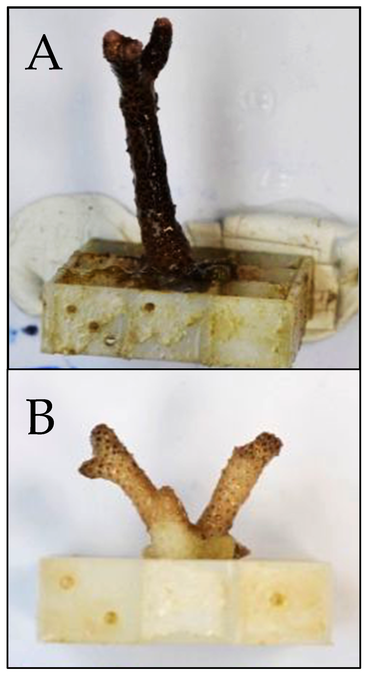
Figure 1. Representative images used in the estimation of relative photosymbiont abundance. Panel (A) depicts a healthy, unbleached coral from the control temperature, high pCO2 treatment (color score = 5.69). Panel (B) depicts a partially bleached coral from the high temperature, high pCO2 treatment (color score = 4.56).
Jmse 10 01106 g001
Figure 2. An example of a vertical pH profile generated during measurement of the coral calcifying fluid pH. This particular pH profile was generated for a specimen of S. hystrix cultured in the ‘1000 ppm pCO2, 28 °C’ treatment.
Jmse 10 01106 g002
Figure 3. The relationship between pCO2 and coral calcification rates at ambient and high temperature ((A) = S. pistillata, (B) = S. hystrix, (C) = P. damicornis, and (D) = L. pertusa). Shaded boundaries represent 95% confidence intervals. Calcification rate was significantly impacted by an interaction between pCO2 and temperature for all coral species.
Jmse 10 01106 g003
Figure 4. The effect of seawater pCO2 on calcifying fluid pH (n = 3 individuals per treatment; panels (AD)) and Δ[H+] (panels (EH)) for three species of tropical corals ((A,E) = S. pistillata; (B,F) = S. hystrix, and (C,G) = P. damicornis) and one species of cold-water coral (L. pertusa; (D,H)). The impact of elevated temperature on the response of S. pistillata to increasing pCO2 is also shown (panels (A) and (E)). Increasing pCO2 was significantly associated with declining calcifying fluid pH and increasing Δ[H+] for all four coral species. The calcifying fluid pH of S. pistillata decreased significantly in response to a 3 °C increase in temperature, and Δ[H+] significantly responded to an interaction between increased pCO2 and increased temperature. Shaded boundaries represent 95% confidence intervals. Solid black lines represent seawater pH under control temperature; dashed black line represents seawater pH under high temperature.
Jmse 10 01106 g004
Figure 5. Slopes of regressions calculated from linear mixed effects models investigating the impacts of pCO2 and temperature on calcification rate, calcifying fluid pH, and Δ[H+]. Significant differences were found amongst the slopes of the different coral species’ calcification responses to ocean acidification and warming. No significant difference was observed amongst the slopes of the different coral species’ calcifying fluid pH response to ocean acidification. The slopes of the different coral species’ proton regulation response (i.e., Δ[H+]) to ocean acidification were not significantly different from each other. Vertical bars represent 95% confidence intervals.
Jmse 10 01106 g005
Figure 6. The effect of seawater pCO2 and warming on the color score of the three species of tropical corals ((A): S. pistillata; (B): S. hystrix; (C): P. damicornis)). Color score is a proxy for photosymbiont abundance (‘bleaching’), where 0 = bleached and 6 = healthy. Trendlines indicate significant correlations between seawater pCO2 and color score at 28 (orange) and 31 (red) °C. The color score of both S. pistillata and P. damicornis increased in response to increasing pCO2 (lmers, S. pistillata, p < 0.001; P. damicornis, p = 0.001) and decreased in response to a 3 °C temperature increase (lmers, S. pistillata, p < 0.001, P. damicornis, p = 0.001). Color score of S. hystrix was significantly impacted (indicating bleaching) by an interaction between pCO2 and temperature (lmer, p < 0.001), whereby color score increased significantly with increasing pCO2 at 28 °C (lmer, p < 0.001) but showed no significant change with increasing pCO2 at 31 °C (lmer, p = 0.056). Shaded boundaries represent 95% confidence intervals.
Jmse 10 01106 g006
Figure 7. The relationship between coral color score and calcification rate of three species of tropical corals ((A): S. pistillata; (B): S. hystrix; (C): P. damicornis). Color score is a proxy for photosymbiont abundance (‘bleaching’), where ‘0’ = bleached and ‘6’ = healthy. Trendlines indicate significant correlations between color score and calcification rate at 28 (orange) and 31 (red) °C. Temperature significantly impacted the relationship between color score and calcification rate of S. pistillata, and caused a significant decline in calcification rate of S. hystrix. No significant relationship existed between calcification rate and color score of P. damicornis, and temperature had no significant impact on this relationship. Shaded boundaries represent 95% confidence intervals.
Jmse 10 01106 g007
Table 1. Average calculated parameters for the tropical corals and all treatments: pCO2 of the mixed gases in equilibrium with seawaters (pCO2 (gas-e)), pH on seawater scale (pHSW), carbonate ion concentration ([CO32−]), bicarbonate ion concentration ([HCO3]), dissolved carbon dioxide ([CO2]SW), and aragonite saturation state (ΩA). Average measured parameters for all treatments: salinity (Sal), temperature (Temp), pH on NBS scale (pHNBS), total alkalinity (TA), and dissolved inorganic carbon (DIC). ‘SE’ represents standard error and ‘n’ is the sample size.
400 ppm (9 °C)400 ppm (12 °C)1000 ppm (9 °C)1000 ppm
(12 °C)
2800 ppm (9 °C)2800 ppm
(12 °C)
CALCULATED PARAMETERS
pCO2 (gas-e)(ppm-v)46649992588528073194
SE891512119135
Range362–540425–607808–1144772–10501728–43022298–4945
n323231323232
pHSW 8.118.067.857.877.427.42
SE0.020.020.010.010.010.02
Range8.03–8.307.97–8.277.73–7.987.80–7.997.28–7.517.20–7.57
n323131323232
[CO32−](µM)33432021726590113
SE9116434
Range235–395211–442159–274226–30964–11376–149
n323231323232
[HCO3](µM)213320172424246826973030
SE333635357754
Range1846–24331772–23892138–27482193–28422104–32892476–3475
n323131323232
[CO2] (SW)(µM)12.312.324.622.074.379.5
SE0.20.30.40.43.23.4
Range9–1410–1721–3019–2745–11256–121
n323231323232
ΩA 5.45.23.54.31.41.8
SE0.10.20.10.10.00.1
Range3.8–6.43.5–7.22.6–4.43.7–5.01.0–1.81.2–2.4
n323231323232
MEASURED PARAMETERS
Sal(psu)34.8735.6335.4435.9935.7535.68
SE0.060.080.050.060.050.06
Range33.75–36.2534.45–37.5534.55–36.6534.85–37.2534.55–36.7534.35–37.05
n104104104104104104
Temp(°C)28.2931.7227.8830.8328.1730.93
SE0.010.060.020.020.020.02
Range28.1–28.530.8–32.327.6–28.230.5–31.028.0–28.430.6–31.2
n104104104104104104
pHNBS 8.278.248.048.127.627.69
SE0.010.010.010.010.010.01
Range8.02–8.478.12–8.487.88–8.337.92–8.347.47–7.987.48–7.97
n104104104104104104
TA(µM)291527742939308329073290
SE495446408156
Range2420–33042309–32232524–33062735–34622260–34932705–3794
n323232323232
DIC(µM)248023502645275528613223
SE404444378257
Range2097–28242009–27262013–30032445–31342231–34902634–3700
n323232323232
Table 2. Average calculated parameters for Lophelia pertusa and all treatments: pCO2 of the mixed gases in equilibrium with seawaters (pCO2 (gas-e)), pH on seawater scale (pHSW), carbonate ion concentration ([CO32−]), bicarbonate ion concentration ([HCO3]), dissolved carbon dioxide ([CO2]SW), and aragonite saturation state (ΩA). Average measured parameters for all treatments: salinity (Sal), temperature (Temp), pH on NBS scale (pHNBS), total alkalinity (TA), and dissolved inorganic carbon (DIC). ‘SE’ represents standard error and ‘n’ is the sample size.
400 ppm (9 °C)400 ppm (12 °C)1000 ppm
(9 °C)
1000 ppm
(12 °C)
2800 ppm
(9 °C)
2800 ppm
(12 °C)
CALCULATED PARAMETERS
pCO2 (gas-e)(ppm-v)4514941096107928643167
SE24169960222202
Range238–580399–595672–1815834–14961600–41621821–4669
n161616161616
pHSW 8.208.217.877.867.427.42
SE0.040.040.040.030.040.03
Range8.01–8.547.96–8.407.62–8.127.70–8.067.17–7.677.19–7.63
n202020202020
[CO32−](µM)2442891211405260
SE23259865
Range128–486149–45887–201102–21125–9935–110
n202020202020
[HCO3](µM)267427752794283530343058
SE10711189956981
Range2028–33562215–33482299–33662350–33492655–34202604–3486
n202020202020
[CO2] (SW)(µM)20194541127118
SE114288
Range11–2614–2428–8330–5972–19072–187
n202020202020
ΩA 3.74.41.82.10.80.9
SE0.30.40.10.10.10.1
Range1.9–7.32.3–6.91.3–3.01.6–3.20.4–1.50.5–1.7
n202020202020
MEASURED PARAMETERS
Sal(psu)34.9835.1334.9635.2335.0135.40
SE0.020.030.030.060.020.05
Range34.55–35.2534.65–35.6534.55–35.6534.20–36.1534.45–35.2534.35–36.05
n606060606060
Temp(°C)8.8612.179.0712.528.8312.67
SE0.030.040.030.040.020.03
Range8.5–9.411.6–12.68.7–9.412.0–12.98.5–9.112.0–13.1
n606060606060
pHNBS 8.078.137.807.827.397.40
SE0.010.020.020.020.020.02
Range7.82–8.397.85–8.437.48–8.157.52–8.017.13–7.757.12–7.63
n606060606060
TA(µM)324534423080316331573198
SE148161931068087
Range2355–41062601–43492598–35702609–37832752–35702759–3655
n202020202020
DIC(µM)293830832960301632143235
SE125134931016985
Range2180–36592395–37932449–35362494–35562805–35992741–3676
n202020202020
Publisher’s Note: MDPI stays neutral with regard to jurisdictional claims in published maps and institutional affiliations.