Article

Synergistic Antibiofilm Effects of Exopolymers Produced by the Marine, Thermotolerant Bacillus licheniformis B3-15 and Their Potential Medical Applications

by
Vincenzo Zammuto
1,2,3,*,
Eleonora Agostino
1,
Angela Macrì
1,
Antonio Spanò
1,2,
Emanuela Grillo
4,
Marco Sebastiano Nicolò
1,2 and
Concetta Gugliandolo
1,2
1
Department of Chemical, Biological, Pharmaceutical and Environmental Sciences, University of Messina, Viale Ferdinando Stagno D’Alcontres 31, 98166 Messina, Italy
2
Research Centre for Extreme Environments and Extremophiles, Department of Chemical, Biological, Pharmaceutical and Environmental Sciences, University of Messina, Viale Ferdinando Stagno D’Alcontres 31, 98166 Messina, Italy
3
ATHENA Green Solutions S.r.l., Viale Ferdinando Stagno d’Alcontres 31, 98166 Messina, Italy
4
Department of Health Sciences, University Magna Græcia of Catanzaro, 88100 Catanzaro, Italy
*
Author to whom correspondence should be addressed.
J. Mar. Sci. Eng. 2023, 11(9), 1660; https://doi.org/10.3390/jmse11091660
Submission received: 26 July 2023 / Revised: 19 August 2023 / Accepted: 22 August 2023 / Published: 24 August 2023
(This article belongs to the Special Issue Marine Microbes and Their Application in Biotechnology)

Abstract

:
The exopolysaccharide (EPS B3-15) and biosurfactant (BS B3-15), produced by the marine Bacillus licheniformis B3-15, were recently reported to possess different antibiofilm activities, with the EPS being more active in preventing the adhesion of Pseudomonas aeruginosa and Staphylococcus aureus and the BS in destroying their preformed biofilms on different surfaces. In this study, the synergistic effects of the two exopolymers on the bacterial adhesion and biofilm disruption of P. aeruginosa and S. aureus were evaluated on polystyrene, a medical polyvinyl chloride (PVC) device, and contact lenses (CLs) in order to address their potential use in biomedical applications. To this purpose, EPS B3-15 and BS B3-15 were equally combined (1:1 w/w), and the mixture (BPS B3-15) was added at different concentrations (from 50 to 300 µg mL−1) and at different times of bacterial development. Compared to each polymer, the BPS B3-15 (300 µg mL−1) more efficiently reduced the adhesion of P. aeruginosa and S. aureus on polystyrene (65 and 58%, respectively), PVC devices (62 and 42%, respectively), and CLs (39 and 35%, respectively), also in combination with a CLs care solution (88 and 39%, respectively). Furthermore, BPS B3-15 was able to disrupt mature biofilms, acting more effectively against S. aureus (72%) than P. aeruginosa (6%). The combination of exopolymers at low concentrations exhibited synergistic effects to prevent and eradicate biofilms.

1. Introduction

Biofilms are organized communities of bacterial cells enclosed in a self-synthesized matrix mainly composed of exopolysaccharides, proteins, extracellular DNA, and lipids [1]. Unlike free-living bacteria, bacterial cells encased in the biofilm are less susceptible to biocides (disinfectants and antimicrobial agents) and to the host’s immune responses, and therefore they are difficult to eradicate, with significant impacts in several areas, including food spoilage, aquaculture, and human health [1,2,3,4]. Bacterial biofilms are involved in different tissue infections, including lung infections of cystic fibrosis, urethritis, otitis, periodontitis, and endocarditis, and device-related infections, such as ventricular derivations, endotracheal tubes, urinary catheters, and contact lenses [5,6].
Biofilm formation is a complex process consisting of five phases: adhesion, reversible attachment, irreversible attachment, maturation of the biofilm, and dispersion [7,8]. The initial adhesion on abiotic or biotic surfaces, which is considered the most crucial phase in biofilm formation, depends on both the surface properties, including roughness, chemical composition, hydrophobicity, and the presence of charges, and the cell-surface characteristics (i.e., hydrophobicity and surface charges) [9,10,11,12]. After the initial attachment, the bacterial cells start to replicate into microcolonies and begin to express genes involved in the synthesis of the extracellular matrix as well as those linked to antibiotic resistance [1,13,14]. In the mature biofilm, the bacterial cells grow into a more complex multicellular form, characterized by the presence of differentiated mushroom- or pillar-like structures [15]. The extracellular matrix, mainly composed of exopolysaccharides (EPSs) (40–95%), confers the structure of biofilms, retains water and nutrients, and offers a barrier against environmental stresses, pathogens, and predator injuries. Several bacterial EPSs are also reported to be able to prevent the adhesion of a wide spectrum of bacteria and, therefore, their biofilm formation by acting as surface-active molecules [16,17]. Biosurfactants (BSs), such as lipopeptides, glycolipids, and phospholipids, with lower molecular weights and higher emulsifying activity than EPSs, efficiently reduce the surface tension [18,19,20,21,22].
We previously reported that the non-biocidal EPS B3-15 and BS B3-15, produced by the marine thermotolerant Bacillus licheniformis B3-15, isolated from a shallow hydrothermal vent of Vulcano Island (Italy), acted differently on the biofilm formation of Gram-negative and Gram-positive bacteria [18,23,24]. The EPS B3-15, composed of mannose and poly-gamma-glutamic acid, at a very low concentration (300 µg mL−1), was able to prevent the adhesion of Pseudomonas aeruginosa (51%), and Staphylococcus aureus (52%), on different abiotic (i.e., polystyrene, medical device) and biotic surfaces (human nasal epithelial cells), but it did not affect their preformed biofilm [23,25]. Independently from quorum sensing, EPS B3-15 modes of action were hypothetically referred to: (i) the modification of surface properties, cell-surface charges, and hydrophobicity; and (ii) the inhibition of cell-to-cell aggregation, as confirmed by the downregulation of the gene expression of P. aeruginosa and S. aureus encoding for lectins or adhesins (lecA and clfA, respectively) [23]. Similarly, to the EPS B3-15, the lichenysin-like lipopeptide BS B3-15 was able to reduce the adhesion of P. aeruginosa (47%) and S. aureus (36%), but in contrast, the BS B3-15 disrupted their preformed biofilm (26% and 47%, respectively) on polystyrene surfaces [18,24]. These two exopolymers have been proposed as candidates for applications in different fields because of their versatility, biocompatibility, absorbability, and absence of cytotoxicity. The search for new strategies capable of counteracting the formation or removing preformed biofilms on different surfaces and devices is important in medical and non-medical fields, such as nasal spray, coating agents in functionalized devices (i.e., orthopedic and endotracheal devices, vascular and urinary catheters), and detergents and antiadhesive agents in industry (e.g., food, agriculture, cosmeceuticals) [23,24].
In this study, we evaluated the effects of the two exopolymers, equally combined (1:1 w/w) (BPS B3-15), on the adhesion and biofilm formation of Pseudomonas aeruginosa ATCC 27853 and Staphylococcus aureus ATCC 29213, used as biofilm models of clinically significant bacteria, on a polyvinyl chloride medical device and contact lenses. Biofilm inhibition of exopolymers was also investigated in combination with a commercial contact lens care solution.

2. Materials and Methods

2.1. Bacillus licheniformis Strain B3-15 and Exopolymer Production

Bacillus licheniformis strain B3-15, isolated from a shallow hydrothermal vent origin (Porto di Levante of Vulcano Island, Messina, Italy), was previously reported to produce exopolymers, EPS B3-15 and BS B3-15 [23,25,26]. Briefly, optimal growth conditions of strain B3-15 were 45 °C, a pH value equal to 7, and NaCl 2% (w/v). It is routinely maintained on plates of Tryptic Soy Agar (TSA, Sigma Aldrich, Burlington, MA, USA), modified with the addition of 1% NaCl (Oxoid, Cambridge, UK). Strain cultures are kept in 20% (v/v) glycerol (Panreac Química, Barcelona, Spain) at −80 °C for long-term storage.
To produce the EPS B3-15, the B3-15 strain was cultivated in a 1 L flask containing 250 mL of the medium SG17 under optimized conditions (containing 5% glucose) and incubated at its optimal growth conditions for 48 h under agitation at 250 rpm [25]. To obtain the EPS B3-15, the culture was centrifuged 9318× g rpm for 10 min, and the cell-free supernatant (CFS) was treated with an equal volume of absolute ethanol. The precipitated EPS B3-15 was dialyzed and lyophilized. The final yield of the obtained EPS B3-15 was 240 mg L−1 [25]. The EPS B3-15 was constituted by a disaccharide repeating unit having a manno-pyranosidic configuration and a component attributed to poly-gamma-glutamic acid [25].
To obtain BS B3-15, the strain was grown under optimized conditions (sucrose 5% and phosphate deprivation) (MGV-op) as previously reported [24]. The CFS, obtained by centrifugation, was acidified by adding HCl (2N) drop by drop up to pH 2.0 and then incubated at 4 °C overnight. To extract the BS, an equal volume of acidified CFS was mixed with a solvent solution (chloroform methanol, 2:1 v/v), and after removing the aqueous solution, the organic layer was dried using a rotary evaporator (Rotavapor® R-300, BUCHI Italia S.r.l., Cornaredo, Italy). The yield of BS B3-15 was 1.5 g L−1 after 48 h of incubation at 45 °C, and its structure was attributed to a lichenysin-like lipopeptide [18,24].
EPS B3-15 and BS B3-15 were dissolved in phosphate buffer saline (PBS, Thermo Fisher Scientific, Milan, Italy) in equal weight (1:1 w/w) (BPS B3-15) and sterilized by filtration through a nitrocellulose membrane (0.2 µm pore size) (Biogenerica, Catania, Italy).

2.2. Bacterial Pathogens

Pseudomonas aeruginosa ATCC 27853 and Staphylococcus aureus ATCC 29213 strains were purchased from the American Type Culture Collection (LGC Promochem, Teddington, UK). P. aeruginosa is maintained in Luria Bertani broth (LB, Sigma Aldrich, Milan, Italy) or solidified with 2% Bacto agar (Difco, Baltimore, MD, USA), whereas S. aureus is maintained in Tryptic Soy Broth (TSB, Sigma Aldrich, Milan, Italy) or solidified with 2% Bacto agar, both at 37 °C. The strains are kept at −80 °C in the presence of 20% (v/v) glycerol for long-term storage.

2.3. Antibacterial Activity of BPS B3-15 and Antiseptic Solution for Contact Lens Care

To determine the minimum inhibitory concentration (MIC) values of BPS B3-15, the serial dilution assay was used [27]. Serial dilutions of the BPS (4, 2, 1, 0.5, and 0.25 mg mL−1) were prepared in tubes of Mueller–Hinton broth (MHB, Sigma Aldrich, Milan, Italy) and then inoculated with suitable aliquots (10 mL) of an overnight culture (comparable to 0.5 McFarland standard) of each strain in MHB. The tubes were incubated at 37 °C overnight, and then the growth was estimated spectrophotometrically (OD600 nm).
The effects of BPS B3-15 on bacterial growth were spectrophotometrically determined. Aliquots (180 µL) (OD600 nm = 0.1) from each overnight strain culture (in MHB) were distributed in 96-well polystyrene microtiter plates (six replicates), and 20 μL of BPS B3-15 (300 μg mL−1 final concentration, in PBS) or PBS used as a control were added to each well. The microplates were incubated at 37 °C for 24 h without shaking, and OD600 nm values were registered every 2 h.
A CL-care solution, commercially available in the Italian market, is a sterile isotonic solution containing EDTA 0.01%, polyhexamethylenebiguanide 0.0002%, hydroxyethylcellulose, poloxamer 407 0.18%, and polyquaternium 0.004%. The antibacterial activity of the CL-care solution on the model strains was evaluated at different final concentrations (0, 10, 20, 30, 50%, and 100% v/v).
Additionally, 100 µL of each culture were spread on plates of Mueller–Hinton agar (MHA) in triplicate; the plates were incubated at 37 °C for 24 h, and the CFU were counted. The results were expressed as an average of CFU mL−1.

2.4. Antibiofilm Activity of BPS B3-15

2.4.1. BPS B3-15 Addition to Polystyrene at Different Concentrations

To evaluate the effects of BPS B3-15 on biofilm formation against P. aeruginosa and S. aureus, 96-well polystyrene microplates (Falcon®, Fisher Scientific, Milan, Italy) were used, as previously reported by O’Toole et al. [28]. Aliquots (180 μL) of a culture of P. aeruginosa or S. aureus grown overnight in LB or TSB, respectively, were poured into microwells (six replicates, OD600 nm = 0.1), and 20 μL of the solution of BPS B3-15, dissolved in PBS, were added in each well to ensure a final concentration of 50, 100, 200, or 300 µg mL−1, or aliquots of PBS (20 μL) as a negative control. After the incubation at 37 °C for 48 h (for P. aeruginosa) or 24 h (for S. aureus) without shaking to allow mature biofilm formation [23,24], the nonadherent bacteria were removed from each well of the microplates by washing five times with distilled water. To highlight the formed biofilm, a crystal violet (0.1% w/v) solution was poured into each microwell, and the microplates were incubated at room temperature for 20 min. To remove the excess stain, the microplates were washed 5 times and air-dried for 45 min. The crystal violet bonded by the biofilm was solubilized with absolute ethanol to allow the determination of the biofilm mass spectrophotometrically (OD585 nm) using a microtiter plate reader (Multiskan GO, Thermo Scientific, Waltham, MA, USA).
The reduction of biofilm formation by each strain was expressed as antibiofilm activity (%) by applying the following formula:
Reduction   of   biofilm   formation   ( % ) = ( OD   585   nm   control   OD 585   nm   sample OD 585   nm   control ) × 100
The average and standard deviation of six replicates were calculated, and statistically significant differences among the groups were calculated using a two-way ANOVA followed by Tukey’s multiple comparison tests (** p ≤ 0.01 or * p ≤ 0.05) using GraphPad Prism (version 9.0; GraphPad Software, San Diego, CA, USA).

2.4.2. BPS B3-15 Addition to Polystyrene at Different Times

Aliquots of 20 μL of BPS B3-15 solution (final concentration of 300 μg mL−1) or 20 μL of PBS (as control) were added to each well of 96-well polystyrene microplates at different times, corresponding to initial adhesion (0 h), reversible adhesion (2 h), irreversible adhesion (4–8 h), and mature biofilm (48 h for P. aeruginosa or 24 h for S. aureus). The biofilm formation was determined as reported above.

2.4.3. BPS B3-15 Addition to a PVC Medical Device

The effects of BPS B3-15 at different concentrations (50, 100, 200, or 300 µg mL−1) on the biofilm formation on PVC medical devices were evaluated as described previously [23]. Briefly, the PVC tube of a medical device (external and internal diameters 4.1 and 3 mm, respectively) (BENIFIS SRL, Genova, Italy) was sliced into sterile segments (1 cm × 0.5 cm × 0.1 cm), and each segment was placed in each well of a 96-well polystyrene microplate containing 180 µL of P. aeruginosa or S. aureus culture (OD600 nm = 0.1) and 20 µL of PBS, as control, or BPS B3-15, were added. The microplates were incubated at 37 °C for 48 h or 24 h for P. aeruginosa or S. aureus, respectively. To remove the non-adherent bacterial cells, each PVC segment was washed with PBS and stained with 0.1% (v/v) crystal violet solution. The segments were washed 5 times with deionized water, and after drying, stained biofilms were solubilized with absolute ethanol for 30 min at room temperature. The destain solution was spectrophotometrically measured (OD585 nm), and the reduction of biofilm formation was calculated as reported above.

2.4.4. BPS B3-15 Addition on Contact Lenses (CLs)

To evaluate the antibiofilm activity on CLs of bacterial exopolymers (EPS B3-15, BS B3-15, and BPS B3-15), 900 µL of each bacterial culture of P. aeruginosa and S. aureus, grown overnight in LB and TSB, respectively (OD600 nm = 0.1), were added to each well of 24-well polystyrene microplates (Falcon ®, Fisher Scientific, Milan, Italy) containing a daily contact lens (Zeiss, Jena, Germany), composed by Dayfilcon A 42% and water 58%, previously washed with sterile PBS, as reported by Mordmuang et al. [29]. An aliquot (100 µL) of each solution of EPS B3-15, BS B3-15, or BPS B3-15 (at final concentrations from 50 to 300 µg mL−1) dissolved in PBS or PBS (as control) was added in each well. The microplates were incubated at 37 °C for 48 h (for P. aeruginosa) or 24 h (for S. aureus), as reported above, without shaking. After the incubation, the CLs were transferred into new wells, and non-adherent bacteria were removed by washing 5 times with distilled water. The biofilms were stained with an optimized solution of crystal violet (0.001% w/v) for 20 min to avoid overstaining of the lenses. The excess stain was removed by aspiration, and the CLs were washed 10 times with distilled water and air dried for 15 min. The stained biofilms were solubilized with acetic acid at 33% (v/v) [29], and the biofilm mass was determined spectrophotometrically (OD585 nm) as reported above.
Contact lens care solution (CS) at different concentrations or the exopolymers (EPS B3-15, BS B3-15, or BPS B3-15) dissolved in CS at their best concentration were added to each overnight culture of P. aeruginosa and S. aureus in LB or TSB (OD600 nm = 0.1) into the wells of 24-well polystyrene microplates containing CLs. The microplates were incubated at 37 °C as above, and the antibiofilm assay was carried out as previously described. The antibiofilm activity of the CS was calculated by normalizing the biofilm mass (OD585 nm) with respect to the bacterial growth (OD600 nm) as follows: ODnormalized = OD585 nm/OD600 nm [29].

2.5. BPS B3-15 Surface-Active Properties and Effects on Polystyrene Surface Adhesion

2.5.1. Surface-Active Properties

The emulsifying activity of BPS B3-15 (300 µg mL−1) was evaluated as reported by Cooper and Goldenberg [30]. BPS B3-15 solution (2 mL), dissolved in distilled water, was mixed with an equal amount of kerosene in a tube and shaken at high speed for 2 min. Triton X-100 (Sigma-Aldrich, Milan, Italy) and distilled water were used as positive and negative controls, respectively. The height of the emulsion was used to calculate the emulsifying index (E24) following the equation:
E 24 % = height   of   emulsion height   of   total   × 100 .
To assess the ability of the BPS B3-15 (300 µg mL−1) to modify the hydrophobic surfaces, the contact angle (θ) in the water solution was measured using the sessile-drop technique. Briefly, 5 µL of the BPS B3-15 solution was spotted onto the lid of a 96-well polystyrene microplate, incubated at room temperature for 15 min, and photographed with a high-resolution camera. To measure the angle (θ) on a sessile drop, the images were analyzed (in triplicate) using the software ImageJ Drop Snake plugin, and the average value and standard deviation were calculated [31,32].

2.5.2. Surface Coating Assay

The surface coating assay on the polystyrene surface was performed as described previously [23]. Briefly, each well of a 24-well polystyrene microtiter plate was coated with an aliquot (20 μL) of BPS B3-15 (300 μg mL−1 final concentration) or 20 μL of PBS used as a control [33], and the plates were dried at 37 °C for 30 min. A suitable diluted overnight bacterial culture (1 mL, containing 105 CFU mL−1) in LB for P. aeruginosa or TSB for S. aureus was poured into each well of the microplates. The plates were incubated at 37 °C for 18 h, and each well was washed with distilled water and stained with 1 mL of crystal violet solution (0.1%, w/v). Excess dye was removed by washing the wells with distilled water and air drying. The inhibitory activity of BPS B3-15 was indicated by the halo in the center of the wells.

2.5.3. Cell-Surface Charges and Hydrophobicity Properties

To investigate the charges and hydrophobicity of the cellular surfaces of P. aeruginosa and S. aureus, the microbial affinity to hydrocarbons (MATH) assay was carried out according to Bellon-Fontaine et al. [34]. Aliquots (10 mL) of overnight bacterial cultures, grown in LB or TSB at 37 °C, were centrifuged at 5242× g rpm for 10 min, and the pellets were washed two times and suspended in 10 mL of sterile PBS. Each bacterial suspension (10 mL) was treated for 30 min with BPS B3-15 (300 μg mL−1 in PBS) or with PBS as a control. The suspensions of treated and untreated bacteria were centrifuged 5242× g rpm for 10 min, and PBS was added to reach OD400 nm values ranging from 0.5 to 0.7 (A0). Bacterial suspension aliquots (3 mL) were mixed in a glass tube containing 0.4 mL of the following solvents (Sigma Aldrich, Milan, Italy): ethyl acetate, chloroform, decane, and hexadecane. After mixing for 1 min, each tube was placed on the bench at room temperature for 10 min to allow phase separation. The absorbance (OD400 nm) of the aqueous phase of each tube was measured (A1), and the percentage of affinity for hydrocarbons was calculated as follows:
%   Affinity = A 0 A 1 A 0 × 100 .
All assays are representative of three independent experiments.

3. Results

3.1. Antibacterial Activity of BPS B3-15

BPS B3-15 did not affect the growth of P. aeruginosa or S. aureus up to 1000 µg mL−1, indicating that it did not exert any antibacterial activity. The growth curves of P. aeruginosa and S. aureus were evaluated in the absence or presence of BPS B3-15 (300 µg mL−1) (Figure 1), since each component of BPS B3-15 (EPS B3-15 and BS B3-15) has been previously reported to possess low toxicity towards human cells up to 300 µg mL−1 [23,24].

3.2. Antibiofilm Activity of BPS B3-15

3.2.1. BPS B3-15 Addition on Polystyrene Surfaces at Increasing Concentrations and at Different Times of Bacterial Growth

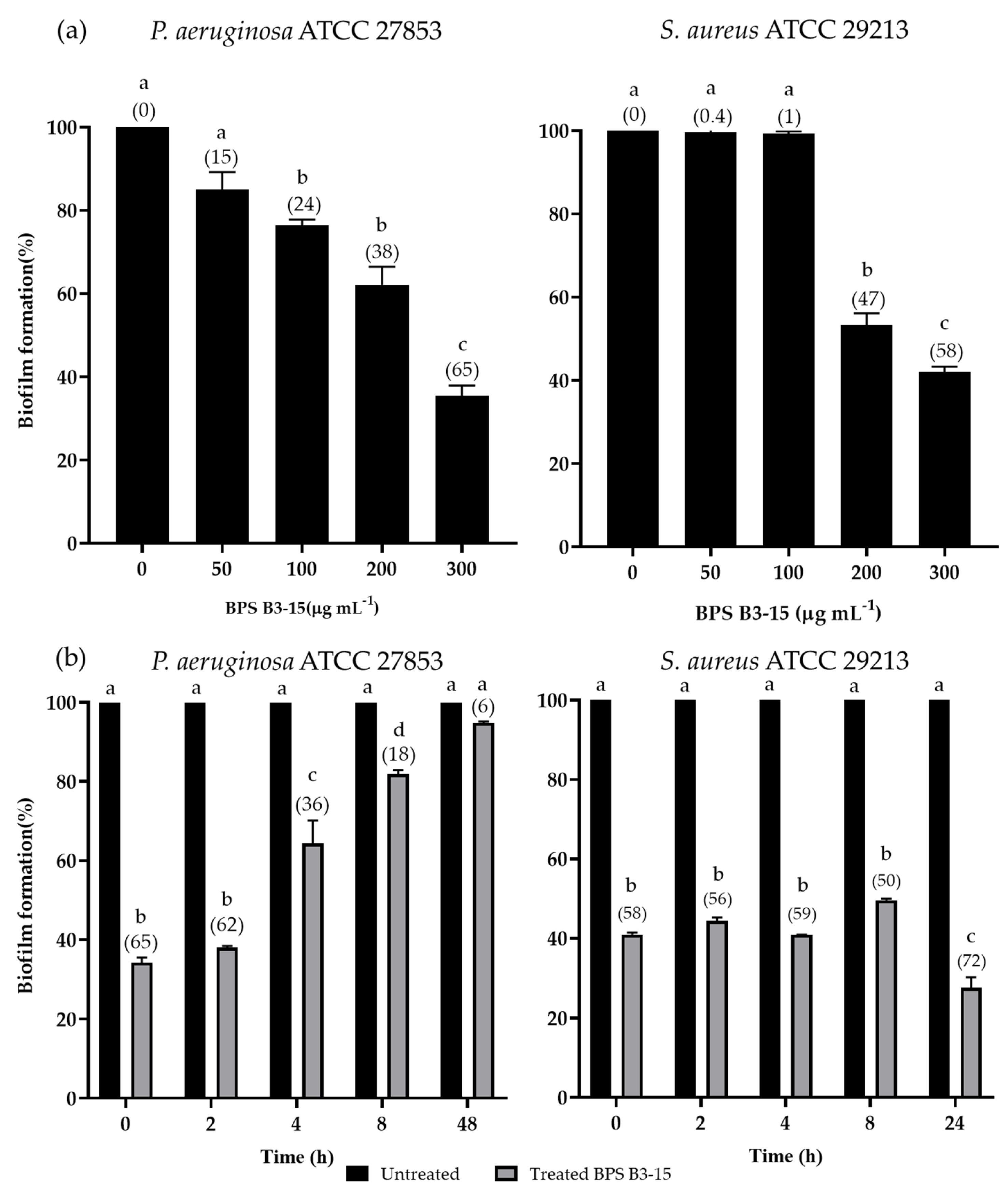
The inhibition effects of BPS B3-15 at increased doses (from 50 to 300 μg mL−1) on biofilm formation by P. aeruginosa and S. aureus on polystyrene microplates are reported in Figure 2a.
BPS B3-15 exhibited dose-dependent inhibitory effects, with 300 μg mL−1 the most active concentration in reducing the biofilm of P. aeruginosa (65%) and S. aureus (58%) (Figure 2a). To assess the ability to interfere with the different phases of biofilm formation, BPS B3-15 (300 µg mL−1) was added at different times of bacterial growth (0, 2, 4, and 8 h) and at 48 h for P. aeruginosa or 24 h for S. aureus, when their biofilms were completely established (Figure 2b). When added at T0 and T2, BPS B3-15 strongly inhibited the initial and reversible adhesion of P. aeruginosa (65% and 62% inhibition, respectively) and S. aureus (58% and 56% inhibition, respectively). When BPS B3-15 was added at T4 and T8, P. aeruginosa biofilm formation was moderately inhibited (36% and 18%, respectively), whereas that of S. aureus was greatly inhibited (59% and 50%). The mature biofilm of P. aeruginosa (T48) was moderately inhibited (6%) by the addition of BPS B3-15, whereas that of S. aureus (T24) was almost completely removed (72%; Figure 2b).

3.2.2. BPS B3-15 Addition to a Polyvinyl Chloride Medical Device

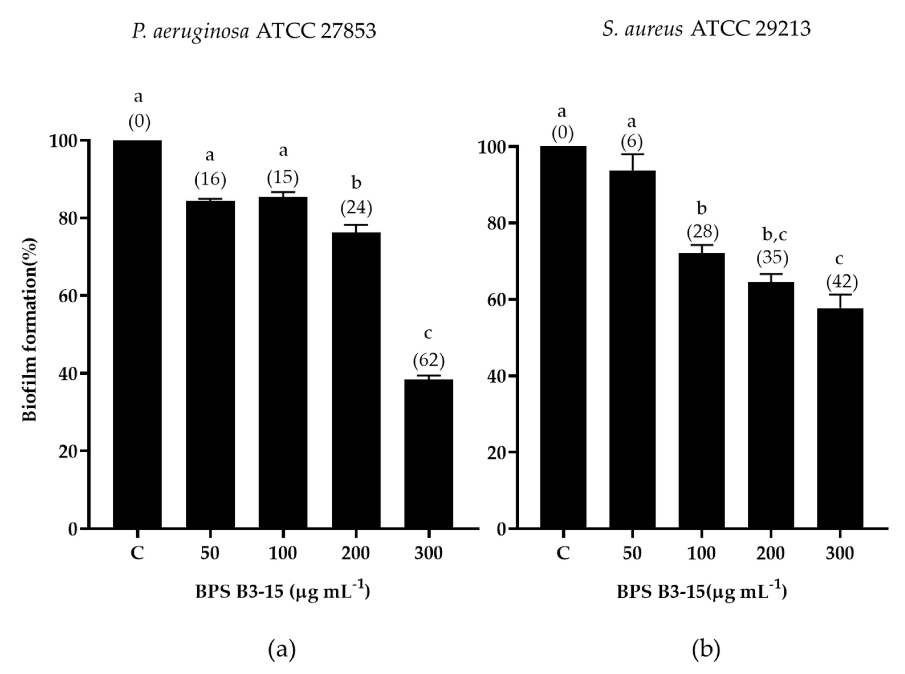
The effect of BPS B3-15 addition (from 50 to 300 µg mL−1) on the biofilm formation of P. aeruginosa and S. aureus on a polyvinyl chloride (PVC) section of a medical device is shown in Figure 3.
BPS B3-15 addition at the highest concentration (300 µg mL−1) reduced the biofilm formation of P. aeruginosa (62%) on PVC more efficiently than that of S. aureus (42%) (Figure 3).

3.3. BPS B3-15 Addition on Contact Lenses (CL) and CL-Care Solution (CS)

3.3.1. Antibacterial Activity of CL-Care Solution (CS)

CL-care solution (CS) effects on bacterial biomass (OD600 nm) and viable cell counts (CFU mL−1), grown in MHB and MHA, respectively, after incubation for 24 h at 37 °C, are shown in Table 1.
The antibacterial activity of CS was dose-dependent, with a significant reduction of bacterial growth at concentrations above 30%. Without affecting the bacterial biomass (OD600 nm = 0.07), CS at concentrations of 50 and 100% (v/v) strongly reduced viable cells of P. aeruginosa (4 and 6 log scale, respectively) and S. aureus (5 and 6 log scale), indicating bactericidal activity.

3.3.2. CL-Care Solution Antibiofilm Activity

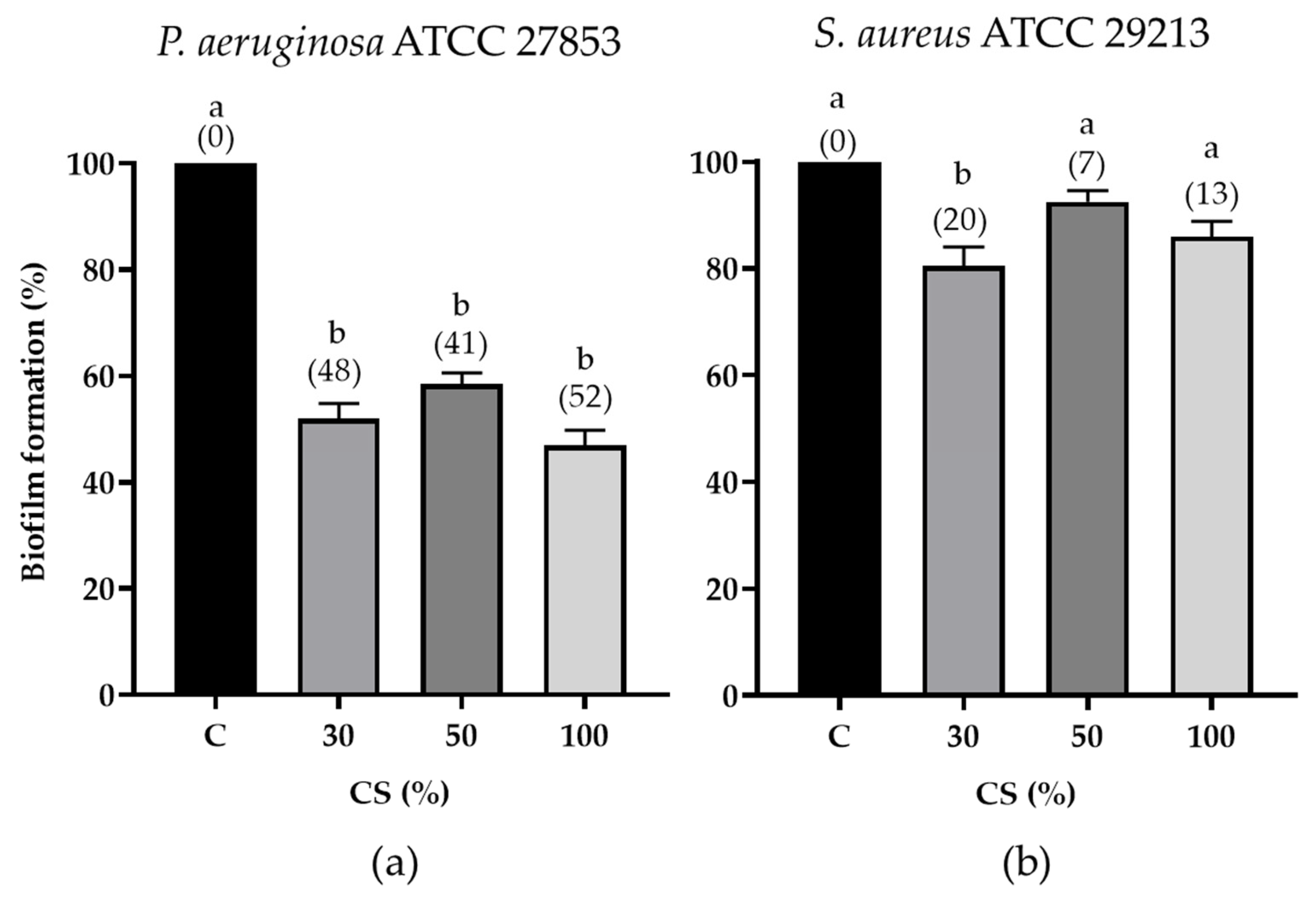
The effects of the CS at different concentrations (30, 50, and 100%) on the biofilm formation by P. aeruginosa or S. aureus on CLs are shown in Figure 4.
The CS at a concentration of 30% showed, after normalization by the bacterial biomass, the highest antibiofilm activity against S. aureus (20%) and similar activity to CS undiluted (100%) against P. aeruginosa (48%); therefore, this concentration was used for further experiments.

3.3.3. Antibiofilm Effects of EPS B3-15, BS B3-15, and BPS B3-15 in Combination with CS on Contact Lenses

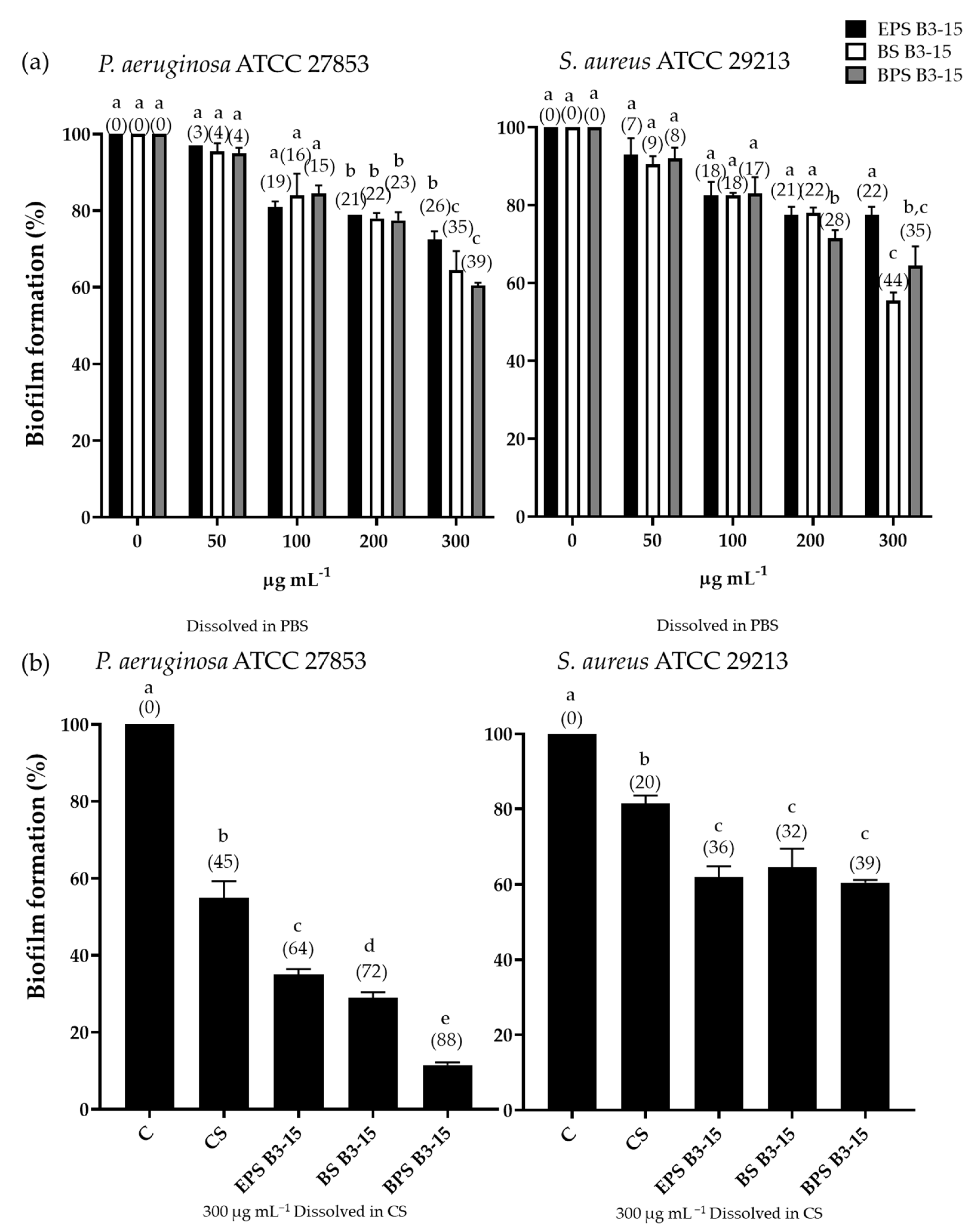
The effects of EPS B3-15, BS B3-15, and BPS B3-15 (300 µg mL−1) alone or in combination with CS (30%) on the biofilm formation of P. aeruginosa or S. aureus on contact lenses are shown in Figure 5.
The presence of the EPS B3-15, BS B3-15, and BPS B3-15 on CLs moderately reduced, in a dose-dependent manner, the biofilm of P. aeruginosa and S. aureus, with the highest activity at 300 µg mL−1 (26, 35, and 39%, respectively for P. aeruginosa and 22, 44, and 35%, respectively for S. aureus) (Figure 5a). The combination of EPS B3-15, BS B3-15, and BPS B3-15 with CS reduced the biofilm of P. aeruginosa on CLs (64, 72, and 88%, respectively) more efficiently than the CS (55%) or each exopolymer alone (Figure 5b). Furthermore, each exopolymer in combination with CS moderately inhibited the biofilm formation of S. aureus (<40%), with BPS B3-15 the most active (39%) and similar to BS.

3.4. BPS B3-15 Surface-Active Properties and Effects on Polystyrene Surface Adhesion

3.4.1. Surface-Active Properties

The effects of BPS B3-15 (300 µg mL−1) on the surface properties of polystyrene by the emulsifying activity and the contact angle measure are reported in Figure 6.
BPS B3-15 emulsifies the kerosene (1:1 v/v) with an E24 value of 82% higher than Triton X-100 (74%) (Figure 6a), used as a positive control. The surface activity of BPS B3-15 was also confirmed by its ability to modify the wettability of polystyrene surfaces, as indicated by the contact angle that was greatly reduced from 79° to 36° (Figure 6b).

3.4.2. Coating Assay

The adhesion of P. aeruginosa or S. aureus on a pre-coated crude BPS B3-15 (300 µg mL−1) polystyrene surface is reported in Figure 7.
BPS B3-15 hindered the adhesion of the two strains to polystyrene surfaces and was more effective against the adhesion of S. aureus than P. aeruginosa (Figure 7).

3.4.3. Cell-Surface Charges and Hydrophobicity Properties

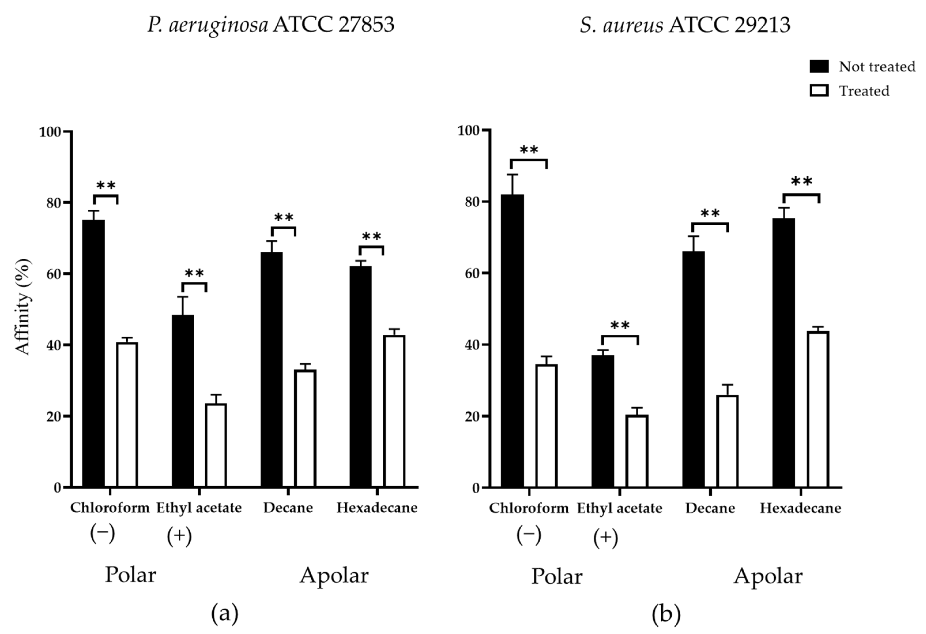
The P. aeruginosa and S. aureus affinity to polar (ethyl acetate and chloroform) and non-polar solvents (decane and hexadecane) treated and untreated with BPS B3-15 are shown in Figure 8.
The cells of P. aeruginosa not treated exhibited high affinity to chloroform (75%), decane (66%), and hexadecane (62%), and low affinity (<50%) to ethyl acetate, suggesting the presence of negative charges on cellular surfaces and a high hydrophobic level (Figure 8a).
S. aureus possessed high affinity (82%) to chloroform and low affinity (39%) to ethyl-acetate, whereas the affinity to decane and hexadecane was high (66% and 74%, respectively), indicating that cellular surfaces were negatively charged and hydrophobic (Figure 8b).
BPS B3-15 significantly modified the bacterial cells affinity for the hydrocarbons, with the highest differences occurring for the cells of S. aureus towards chloroform and decane (43% and 37% reduction, respectively).

4. Discussion

Bacterial biofilms are considered a great concern for human and environmental health due to their resistance to various biocidal treatments [2,3,4,5]. In the present study, the potential synergistic effects of two different exopolymers, i.e., the mannose-rich EPS B3-15 and the lichenysin-like biosurfactant BS B3-15, produced by the same marine thermotolerant strain B. licheniformis B3-15, were evaluated against the biofilm formation of P. aeruginosa and S. aureus on a polyvinyl chloride medical device and contact lenses commercially available. To this purpose, the two exopolymers were combined (1:1 w/w) (BPS B3-15), and the mixture was also investigated in combination with a contact lens care solution.
As confirmed by the coating assay on polystyrene, BPS B3-15 possessed antiadhesive effects that were related to the ability to modify both the hydrophobic properties and surface charges of the substratum more efficiently than those observed for EPS B3-15 and BS B3-15 alone. Moreover, the contact angle assay and emulsifying activity confirmed that BPS acted as a surfactant. The negative charges exhibited by the poly-gamma-glutamic acid (γ-PGA) component of EPS B3-15, as well as those of the aminoacidic residues of the lipopeptide BS B3-15, could contribute to altering the surface charges of polystyrene. These properties may justify the antiadhesive activity of the mixture BPS B3-15 towards both P. aeruginosa (65%) and S. aureus (58%) on polystyrene that resulted in being more effective than that observed for EPS B3-15 or BS B3-15 alone (Table 2) [23,24]. When compared with EPS B3-15 or BS B3-15, the activity of BPS B3-15 was also higher, contrasting the irreversible adhesion and the disruption of mature biofilms. This could be related to the stronger BPS B3-15 emulsifying activity (E24 = 82%) than EPS B3-15 (37%) [23] and BS B3-15 (67%) [24]. EPS B3-15 as a surfactant could act by reducing the interfacial tension between substratum surfaces and established biofilm and therefore inducing the detachment and destabilization of the bacterial biofilms, as reported similarly for other polymers [19,35,36,37,38].
BPS B3-15 reduced the bacterial adhesion to PVC, a negatively charged hydrophobic plastic polymer, more effectively (61.6% for P. aeruginosa and 42.3% for S. aureus) than EPS B3-15 (52.7% and 32.3%, respectively) [23]. As revealed by the MATH assay, the antiadhesive activity of BPS B3-15 on PVC could be explained by the modification of both the cell-surface charges and hydrophobicity levels. Specifically, BPS B3-15 decreased the affinity to chloroform and decane of P. aeruginosa and decreased the affinity to ethyl acetate of S. aureus, suggesting a reduction of the cell surface positive charges. BPS B3-15 could be proposed as a coating agent to prevent or eradicate biofilms in medical devices [23,39,40]. In addition, the exopolymers EPS B3-15, BS B3-15, and their combination BPS B3-15 were evaluated against the biofilm formation of P. aeruginosa and S. aureus on contact lenses composed by the polymer Dayfilcon A. All the exopolymers were less efficient in reducing biofilm formation on CLs than on the other surfaces (polystyrene and PVC) (Table 2). These differences could be related to the high hydrophilicity of the CLs polymer, which consists of approximately 50% water. Few studies have reported antibiofilm activity on CLs of natural products. Allium sativum fermented inhibited the biofilm formation of P. aeruginosa (35%) [41], similar to BS B3-15 (35%), but less than BPS B3-15 (39%), whereas the cannabinol oil extract was more active (50%) [42] than BPS B3-15. However, no activity of these natural products has been reported on S. aureus.
Commercially available CSs may not be effective against biofilms even if they comply with international standards ISO 14729 and FDA 510(k), which determine adequate antimicrobial activity [41]. This is because they have only been tested against a limited number of reference strains of planktonic bacteria and fungi [42]. The CS inhibited the P. aeruginosa biofilm on CLs (48%) more than S. aureus (20%); these data are according to those reported by Di Onofrio et al. [41]. When the exopolymers were added to commercial CS (30%), the antibiofilm effects increased, with BPS B3-15 being the most active to inhibit the biofilm formation of P. aeruginosa (88% of biofilm reduction) and of S. aureus (39%). Interestingly, the combination of BPS B3-15 with CS (30%) was twofold more active than CS alone. In a future perspective, BPS B3-15 could be proposed in combination with disinfectants to prevent and remove biofilms and, therefore, reduce risks related to eye infections.

5. Conclusions

The exopolysaccharide (EPS) and the lichenysin-like lipopeptide (BS) produced by the marine thermotolerant B. licheniformis B3-15, with distinctive properties, have been reported to possess bioactive properties for both environmental and human health. Without exerting any antibacterial activity, the two mixed exopolymers (BPS) reduced more efficiently the adhesion of P. aeruginosa (65%) and S. aureus (58%) to polystyrene and PVC medical devices (62 and 42%, respectively) than did each compound separately. Interestingly, the synergic action of BPS also disrupted the preformed biofilm of S. aureus (72%). Due to its ability to modify the abiotic surface properties and alter the hydrophobicity of Gram-negative and Gram-positive bacterial cell surfaces, BPS at low concentrations could have potential use as a surface coating agent for medical devices (i.e., orthopedic and endotracheal devices, vascular and urinary catheters) to prevent adhesion and eradicate bacterial biofilms, and it could also be exploited to develop more effective care solutions.

Author Contributions

Conceptualization, V.Z. and C.G.; methodology, V.Z., E.A. and A.M.; software, V.Z.; validation C.G. and V.Z.; formal analysis, E.A. and A.M.; investigation, V.Z., E.A., A.M. and E.G.; resources, C.G.; data curation, C.G., V.Z. and A.S.; writing—original draft preparation, V.Z.; writing—review and editing, C.G., V.Z., M.S.N. and A.S.; visualization, V.Z.; supervision, C.G.; project administration, C.G.; funding acquisition, C.G. All authors have read and agreed to the published version of the manuscript.

Funding

This research was supported by the project “PON R&I 2014–2020—“BONE++” ARS01_00693–CUP J66C18000340005”.

Institutional Review Board Statement

Not applicable.

Informed Consent Statement

Not applicable.

Data Availability Statement

Not applicable.

Conflicts of Interest

The authors declare no conflict of interest.

References

  1. Abdallah, M.; Benoliel, C.; Drider, D.; Dhulster, P.; Chihib, N.-E. Biofilm formation and persistence on abiotic surfaces in the context of food and medical environments. Arch. Microbiol. 2014, 196, 453–472. [Google Scholar] [CrossRef]
  2. Laganà, P.; Melcarne, L.; Delia, S. Acinetobacter baumannii and endocarditis, rare complication but important clinical relevance. Int. J. Cardiol. 2015, 187, 678–679. [Google Scholar] [CrossRef]
  3. Zammuto, V.; Rizzo, M.G.; Spanò, A.; Genovese, G.; Morabito, M.; Spagnuolo, D.; Capparucci, F.; Gervasi, C.; Smeriglio, A.; Trombetta, D.; et al. In vitro evaluation of antibiofilm activity of crude extracts from macroalgae against pathogens relevant in aquaculture. Aquaculture 2022, 549, 737729. [Google Scholar] [CrossRef]
  4. Zammuto, V.; Rizzo, M.G.; Spanò, A.; Spagnuolo, D.; Di Martino, A.; Morabito, M.; Gugliandolo, C. Effects of crude polysaccharides from marine macroalgae on the adhesion and biofilm formation of Pseudomonas aeruginosa and Staphylococcus aureus. Algal Res. 2022, 63, 102646. [Google Scholar] [CrossRef]
  5. Schulze, A.; Mitterer, F.; Pombo, J.P.; Schild, S. Biofilms by Bacterial Human Pathogens: Clinical Relevance—Development, Composition and Regulation—Therapeutical Strategies. Microb. Cell 2021, 8, 28–56. [Google Scholar] [CrossRef] [PubMed]
  6. Lebeaux, D.; Chauhan, A.; Rendueles, O.; Beloin, C. From in vitro to in vivo Models of Bacterial Biofilm-Related Infections. Pathogens 2013, 2, 288–356. [Google Scholar] [CrossRef] [PubMed]
  7. Mirghani, R.; Saba, T.; Khaliq, H.; Mitchell, J.; Do, L.; Chambi, L.; Diaz, K.; Kennedy, T.; Alkassab, K.; Huynh, T.; et al. Biofilms: Formation, drug resistance and alternatives to conventional approaches. AIMS Microbiol. 2022, 8, 239–277. [Google Scholar] [CrossRef]
  8. Rizzo, C.; Zammuto, V.; Lo Giudice, A.; Rizzo, M.G.; Spanò, A.; Laganà, P.; Martinez, M.; Guglielmino, S.; Gugliandolo, C. Antibiofilm activity of Antarctic sponge-associated bacteria against Pseudomonas aeruginosa and Staphylococcus aureus. J. Mar. Sci. Eng. 2021, 9, 243. [Google Scholar] [CrossRef]
  9. Flemming, H.C.; van Hullebusch, E.D.; Neu, T.R.; Nielsen, P.H.; Seviour, T.; Stoodley, P.; Wingender, J.; Wuertz, S. The biofilm matrix: Multitasking in a shared space. Nat. Rev. Microbiol. 2023, 21, 70–86. [Google Scholar] [CrossRef]
  10. Karygianni, L.; Ren, Z.; Koo, H.; Thurnheer, T. Biofilm Matrixome: Extracellular Components in Structured Microbial Communities. Trends Microbiol. 2020, 28, 668–681. [Google Scholar] [CrossRef]
  11. Balebona, M.C.; Moriñigo, M.A.; Borrego, J.J. Hydrophobicity and adhesion to fish cells and mucus of Vibrio strains isolated from infected fish. Int. Microbiol. 2001, 4, 21–26. [Google Scholar] [CrossRef] [PubMed]
  12. Tuson, H.H.; Weibel, D.B. Bacteria–surface interactions. Soft Matter. 2013, 9, 4368–4380. [Google Scholar] [CrossRef] [PubMed]
  13. Verderosa, A.D.; Totsika, M.; Fairfull-Smith, K.E. Bacterial Biofilm Eradication Agents: A Current Review. Front. Chem. 2019, 7, 824. [Google Scholar] [CrossRef] [PubMed]
  14. Bagge, N.; Schuster, M.; Hentzer, M.; Ciofu, O.; Givskov, M.; Greenberg, E.P.; Høiby, N. Pseudomonas aeruginosa Biofilms Exposed to Imipenem Exhibit Changes in Global Gene Expression and β-Lactamase and Alginate Production. Antimicrob. Agents Chemother. 2004, 48, 1175–1187. [Google Scholar] [CrossRef] [PubMed]
  15. Davey, M.E.; O’Toole, G.A. Microbial biofilms: From ecology to molecular genetics. Microbiol. Mol. Biol. Rev. 2000, 64, 847–867. [Google Scholar] [CrossRef] [PubMed]
  16. Rendueles, O.; Kaplan, J.B.; Ghigo, J.M. Antibiofilm polysaccharides. Environ. Microbiol. 2013, 15, 334–346. [Google Scholar] [CrossRef] [PubMed]
  17. Uneputty, A.; Dávila-Lezama, A.; Garibo, D.; Oknianska, A.; Bogdanchikova, N.; Hernández-Sánchez, J.F.; Susarrey-Arce, A. Strategies applied to modify structured and smooth surfaces: A step closer to reduce bacterial adhesion and biofilm formation. Colloid Interface Sci. Commun. 2022, 46, 100560. [Google Scholar] [CrossRef]
  18. Zammuto, V.; Spanò, A.; Nicolò, M.S.; Grillo, E.; Caccamo, M.T.; Magazù, S.; Cappello, S.; Gugliandolo, C. Thermophilic hydrocarbon-utilizing bacilli from marine shallow hydrothermal vents as producers of biosurfactants. J. Mar. Sci. Eng. 2022, 10, 1077. [Google Scholar] [CrossRef]
  19. Cruz Mendoza, I.; Villavicencio-Vasquez, M.; Aguayo, P.; Coello Montoya, D.; Plaza, L.; Romero-Peña, M.; Marqués, A.M.; Coronel-León, J. Biosurfactant from Bacillus subtilis DS03: Properties and Application in Cleaning Out Place System in a Pilot Sausages Processing. Microorganisms 2022, 10, 1518. [Google Scholar] [CrossRef]
  20. Joshi, S.J.; Geetha, S.J.; Desai, A.J. Characterization and application of biosurfactant produced by Bacillus licheniformis R2. Appl. Biochem. Biotechnol. 2015, 177, 346–361. [Google Scholar] [CrossRef]
  21. Janek, T.; Lukaszewicz, M.; Krasowska, A. Identification and characterization of biosurfactants produced by the Arctic bacterium Pseudomonas putida BD2. Colloids Surf. B Biointerfaces 2013, 110, 379–386. [Google Scholar] [CrossRef] [PubMed]
  22. De França, Í.W.L.; Lima, A.P.; Lemos, J.A.M.; Lemos, C.G.F.; Melo, V.M.M.; de Sant’ana, H.B.; Gonçalves, L.R.B. Production of a biosurfactant by Bacillus subtilis ICA56 aiming bioremediation of impacted soils. Catal. Today 2015, 255, 10–15. [Google Scholar] [CrossRef]
  23. Zammuto, V.; Spanò, A.; Agostino, E.; Macrì, A.; De Pasquale, C.; Ferlazzo, G.; Rizzo, M.G.; Nicolò, M.S.; Guglielmino, S.; Gugliandolo, C. Anti-bacterial adhesion on abiotic and biotic surfaces of the exopolysaccharide from the marine Bacillus licheniformis B3-15. Mar. Drugs 2023, 21, 313. [Google Scholar] [CrossRef] [PubMed]
  24. Zammuto, V.; Rizzo, M.G.; De Pasquale, C.; Ferlazzo, G.; Caccamo, M.T.; Magazù, S.; Guglielmino, S.P.P.; Gugliandolo, C. Lichenysin-like polypeptide production by Bacillus licheniformis B3-15 and Its antiadhesive and antibiofilm properties. Microorganisms 2023, 11, 1842. [Google Scholar] [CrossRef]
  25. Spanò, A.; Zammuto, V.; Macrì, A.; Agostino, E.; Nicolò, M.S.; Scala, A.; Trombetta, D.; Smeriglio, A.; Ingegneri, M.; Caccamo, M.T.; et al. Arsenic adsorption and toxicity reduction of an exopolysaccharide produced by Bacillus licheniformis B3-15 of shallow hydrothermal vent origin. J. Mar. Sci. Eng. 2023, 11, 325. [Google Scholar] [CrossRef]
  26. Maugeri, T.L.; Gugliandolo, C.; Caccamo, D.; Panico, A.; Lama, L.; Gambacorta, A.; Nicolaus, B. A halophilic thermotolerant Bacillus isolated from a marine hot spring able to produce a new exopolysaccharide. Biotechnol. Lett. 2002, 24, 515–519. [Google Scholar] [CrossRef]
  27. European Committee for Antimicrobial Susceptibility Testing (EUCAST) of the European Society of Clinical Microbiology and Infectious Diseases (ESCMID). Determination of Minimum Inhibitory Concentrations (MICs) of antibacterial agents by broth dilution. Clin. Microbiol. Infect. 2003, 9, 509–515. [Google Scholar]
  28. O’Toole, G.A. Microtiter dish biofilm formation assay. J. Vis. Exp. 2011, 47, e2437. [Google Scholar]
  29. Mordmuang, A.; Udomwech, L.; Karnjana, K. Influence of contact lens materials and cleaning procedures on bacterial adhesion and biofilm formation. Clin. Ophthalmol. 2021, 15, 2391. [Google Scholar] [CrossRef]
  30. Cooper, D.G.; Goldenberg, B.G. Surface-active agents from two Bacilllus species. Appl. Environ. Microbiol. 1987, 53, 224–229. [Google Scholar] [CrossRef]
  31. Daerr, A.; Mogne, A. Pendent_Drop: An ImageJ Plugin to Measure the Surface Tension. J. Open Res. Softw. 2016, 4, 2–6. [Google Scholar] [CrossRef]
  32. Caccamo, M.T.; Zammuto, V.; Spanò, A.; Gugliandolo, C.; Magazù, S. Hydrating capabilities of the biopolymers produced by the marine thermophilic Bacillus horneckiae SBP3 as evaluated by ATR-FTIR spectroscopy. Materials 2022, 15, 5988. [Google Scholar] [CrossRef] [PubMed]
  33. Karwacki, M.T.; Kadouri, D.E.; Bendaoud, M.; Izano, E.A.; Sampathkumar, V.; Inzana, T.J.; Kaplan, J. Antibiofilm activity of Actinobacillus pleuropneumoniae serotype 5 capsular polysaccharides. PLoS ONE 2013, 8, e63844. [Google Scholar]
  34. Bellon-Fontaine, M.N.; Rault, J.; Van Oss, C.J. Microbial adhesion to solvents: A novel method to determine the electrondonor/ electron-acceptor or Lewis acid-base properties of microbial cells. Colloids Surf. B Biointerfaces 1996, 7, 47–53. [Google Scholar] [CrossRef]
  35. Kavita, K.; Singh, V.K.; Mishra, A.; Jha, B. Characterization and anti-biofilm activity of extracellular polymeric substances from Oceanobacillus iheyensis. Carbohydr. Polym. 2014, 101, 29–35. [Google Scholar] [CrossRef]
  36. Coronel-León, J.; de Grau, G.; Grau-Campistany, A.; Farfan, M.; Rabanal, F.; Manresa, A.; Marqués, A.M. Biosurfactant production by AL 1.1, a Bacillus licheniformis strain isolated from Antarctica: Production, chemical characterization and properties. Ann. Microbiol. 2015, 65, 2065–2078. [Google Scholar] [CrossRef]
  37. Paraszkiewicz, K.; Moryl, M.; Płaza, G.; Bhagat, D.; Satpute, S.K.; Bernat, P. Surfactants of microbial origin as antibiofilm agents. Int. J. Environ. Health Res. 2019, 31, 401–420. [Google Scholar] [CrossRef]
  38. Quadriya, H.; Ali, S.A.M.; Parameshwar, J.; Manasa, M.; Khan, M.Y.; Hameeda, B. Microbes Living Together: Exploiting the Art for Making Biosurfactants and Biofilms. In Implication of Quorum Sensing System in Biofilm Formation and Virulence; Springer: Berlin/Heidelberg, Germany, 2018; pp. 161–177. [Google Scholar]
  39. Katsikogianni, M.; Missirlis, Y. Concise review of mechanisms of bacterial adhesion to biomaterials and of techniques used in estimating bacteria-material interactions. Eur. Cell Mater. 2004, 8, 37–57. [Google Scholar] [CrossRef]
  40. Li, X.; Sun, L.; Zhang, P.; Wang, Y. Novel Approaches to combat medical device-associated biofilms. Coatings 2021, 11, 294. [Google Scholar] [CrossRef]
  41. Di Onofrio, V.; Gesuele, R.; Maione, A.; Liguori, G.; Liguori, R.; Guida, M.; Nigro, R.; Galdiero, E. Prevention of Pseudomonas aeruginosa biofilm formation on soft contact lenses by Allium sativum fermented extract (BGE) and Cannabinol oil extract (CBD). Antibiotics 2019, 8, 258. [Google Scholar] [CrossRef]
  42. Kilvington, S.; Huang, L.; Kao, E.; Powell, C.H. Development of a new contact lens multipurpose solution: Comparative analysis of microbiological, biological, and clinical performance. J. Optom. 2010, 13, 134–142. [Google Scholar] [CrossRef]
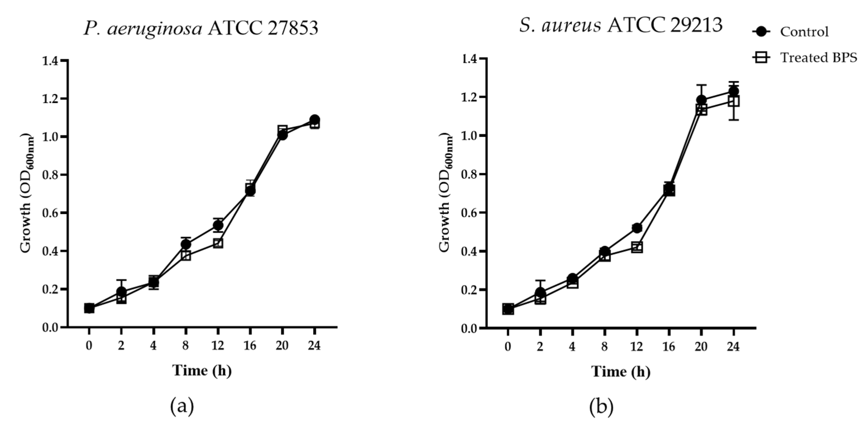
Figure 1. Growth curves of (a) Pseudomonas aeruginosa ATCC 27853 and (b) Staphylococcus aureus ATCC 29213 in the absence (control) and in the presence of BPS B3-15 (300 µg mL−1). The points are the averages of three replicates (n = 3) and standard deviations.
Jmse 11 01660 g001
Figure 2. Biofilm formation (%) on polystyrene microplates by Pseudomonas aeruginosa ATCC 27853 and Staphylococcus aureus ATCC 29213 (a) in the presence of BPS B3-15 at increasing concentrations (from 50 to 300 µg mL−1), and (b) after addition of BPS B3-15 (300 µg mL−1) at different times of bacterial growth (0, 2, 4, 8, 24, or 48 h). Statistical differences were evaluated using a two-way ANOVA with Tukey’s multiple comparisons test. Different lowercase letters above the bar graph indicate significant statistical differences (p ≤ 0.05). Values of biofilm inhibition (%) are reported in brackets.
Jmse 11 01660 g002
Figure 3. Biofilm formation (%) on a PVC medical device by (a) Pseudomonas aeruginosa ATCC 27853 and (b) Staphylococcus aureus ATCC 29213 in the absence (C) and after the addition of BPS B3-15 (from 50 to 300 µg mL−1, after 48 h or 24 h incubation, respectively. Statistical differences were evaluated using a two-way ANOVA with Tukey’s multiple comparisons test. Different lowercase letters above the bar graph indicate significant statistical differences (p ≤ 0.05). Values of biofilm inhibition (%) are reported in brackets.
Jmse 11 01660 g003
Figure 4. Biofilm formation (%, normalized by each biomass) on contact lenses by (a) Pseudomonas aeruginosa ATCC 27853 and (b) Staphylococcus aureus ATCC 29213, in the absence (C, control) and after the addition of contact lens care solution (CS) at different concentrations (30, 50, and 100% v/v). Statistical differences were evaluated using a two-way ANOVA with Tukey’s multiple comparisons test. Different lowercase letters above the bar graph indicate significant statistical differences (p ≤ 0.05). Values of biofilm inhibition (%) are reported in brackets.
Jmse 11 01660 g004
Figure 5. Effects of EPS B3-15, BS B3-15, and BPS B3-15 (a) alone (at different concentrations from 50 to 300 µg mL−1) or (b) in combination (at 300 µg mL−1 with contact lens care solution (CS) (30% v/v) on the biofilm formation (normalized by each biomass, %) of Pseudomonas aeruginosa ATCC 27853 and Staphylococcus aureus ATCC 29213 on contact lenses. Statistical differences were evaluated using a two-way ANOVA with Tukey’s multiple comparisons test. Different lowercase letters above the bar graph indicate significant statistical differences (p ≤ 0.05). In brackets are values of biofilm inhibition (%).
Jmse 11 01660 g005
Figure 6. Surface active properties. (a) Ability of BPS B3-15 to emulsify (E24) kerosene (1:1, v/v). (b) Contact angle of BPS B3-15 aqueous solution (300 µg mL−1) on polystyrene surfaces. The data are expressed as averages and standard deviations (n = 3). ** Significantly different p ≤ 0.01 compared with the untreated condition.
Jmse 11 01660 g006
Figure 7. Pseudomonas aeruginosa ATCC 27853 and Staphylococcus aureus ATCC 29213 adhesions to polystyrene surfaces in the absence (control) or pre-coated with BPS B3-15 (300 µg mL−1) after 18 h treatment.
Jmse 11 01660 g007
Figure 8. Affinity (%) to polar (chloroform and ethyl acetate) and non−polar (decane and hexadecane) solvents of (a) Pseudomonas aeruginosa ATCC 27853 and (b) Staphylococcus aureus ATCC 29213 cells not treated or treated with BPS B3-15 (300 µg mL−1). Bars represent the means of three independent experiments (n = 3). ** Significantly different p ≤ 0.01 compared with the untreated condition.
Jmse 11 01660 g008
Table 1. Pseudomonas aeruginosa ATCC 27853 and S. aureus ATCC 29213 biomass (OD600 nm) and viable cells (CFU mL−1) in the presence of contact lens care solution (CS) diluted (10, 20, 30, and 50%) or not diluted (100%) in PBS.
CL-Care Solution (%)StrainBacterial Biomass (OD600 nm)Viable Cells (CFU mL−1)
0P. aeruginosa1.235.05 × 108
S. aureus1.279.60 × 108
10P. aeruginosa0.974.13 × 108
S. aureus1.178.79 × 108
20P. aeruginosa0.773.24 × 108
S. aureus0.543.95 × 108
30P. aeruginosa0.602.50 × 108
S. aureus0.403.00 × 108
50P. aeruginosa0.071.00 × 104
S. aureus0.063.00 × 103
100P. aeruginosa0.073.50 × 102
S. aureus0.062.00 × 102
Table 2. Antibiofilm activity of EPS B3-15, BS B3-15, and BPS B3-15 (300 µg mL−1) against P. aeruginosa and S. aureus on different surfaces.
Antibiofilm Activity (%)
P. aeruginosaS. aureus
PolystyreneEPS B3-155152
BS B3-154736
BPS B3-156558
PVCEPS B3-155332
BS B3-154830
BPS B3-156242
Contact lensesEPS B3-152622
BS B3-153544
BPS B3-153935
CS4820
EPS B3-15 + CS7236
BS B3-15 + CS6432
BPS B3-15 + CS8839
Disclaimer/Publisher’s Note: The statements, opinions and data contained in all publications are solely those of the individual author(s) and contributor(s) and not of MDPI and/or the editor(s). MDPI and/or the editor(s) disclaim responsibility for any injury to people or property resulting from any ideas, methods, instructions or products referred to in the content.