Article

Extracts from Microalgae and Archaea from the Andalusian Coast: A Potential Source of Antiproliferative, Antioxidant, and Preventive Compounds

by
Cristina Luque
1,†,
Gloria Perazzoli
1,2,3,†,
Patricia Gómez-Villegas
4,
Javier Vigara
4,
Rosario Martínez
5,
Alejandro García-Beltrán
5,
Jesús M. Porres
5,
Jose Prados
1,2,3,*,
Rosa León
4 and
Consolación Melguizo
1,2,3
1
Institute of Biopathology and Regenerative Medicine (IBIMER), Biomedical Research Center (CIBM), 18016 Granada, Spain
2
Biosanitary Research Institute of Granada (ibs.GRANADA), 18012 Granada, Spain
3
Department of Anatomy and Embryology, University of Granada, 18016 Granada, Spain
4
Laboratory of Biochemistry, Center for Natural Resources, Health, and Environment, University of Huelva, 21004 Huelva, Spain
5
Department of Physiology, Institute of Nutrition and Food Technology (INyTA), Biomedical Research Center (CIBM), Sport and Health University Research Institute (IMUDS), Campus of International Excellence of the Sea (CEIMAR), University of Granada, 18016 Granada, Spain
*
Author to whom correspondence should be addressed.
These authors contributed equally to this work.
J. Mar. Sci. Eng. 2024, 12(6), 996; https://doi.org/10.3390/jmse12060996
Submission received: 20 May 2024 / Revised: 8 June 2024 / Accepted: 13 June 2024 / Published: 14 June 2024

Abstract

:
Marine and extreme environments harbor a huge diversity of microorganisms able to produce new bioactive metabolites with beneficial health effects. In this study, ethanol, aqueous, methanol, and acetone extracts and protein hydrolysates were obtained from five different microalgae species and two haloarchaea. An in vitro study of cytotoxicity, migration, angiogenic effect, antioxidant capacity, and modulation of detoxifying enzyme expression was carried out using resistant (HCT-15) and non-resistant (T84) colon cancer tumor lines. Our results showed that the aqueous extract of the microalga Chlorella sorokiniana induced the greatest cytotoxic effect in both cell lines, while the ethanolic extracts of the archaea Haloarcula hispanica and Halobacterium salinarum caused the greatest inhibition on the migratory capacity. Meanwhile, the protein hydrolyzate and the aqueous extract of the microalga Chlorella sorokiniana significantly protected cells against hydrogen peroxide damage. Moreover, the aqueous extracts of Haloarcula hispanica and Halobacterium salinarum resulted in inducing the greatest increase in the activity of the detoxifying enzymes enzyme quinone oxidoreductase and glutathione S-transferase. These preliminary results suggest that aqueous extracts of some microalgae and haloarchaea may be promising candidates for an adjuvant therapy against colorectal cancer. However, additional research is required to identify the active principles and elucidate the mechanisms of action involved.

Graphical Abstract

1. Introduction

The need for new therapeutic molecules has encouraged researchers to screen less explored environments, where a large variety of marine and freshwater microorganisms, many of which are still undiscovered, can be a source of new and unexplored bioactive compounds [1,2]. Concretely, screening of marine extreme environments can allow the isolation of new strains and the identification of novel metabolites with bioactive properties. Microalgae are a huge, ubiquitous, heterogeneous group of microorganisms which have been proposed as a sustainable source of functional ingredients [3,4,5]. In hypersaline environments, Haloarchaea are extremely halophilic microorganisms that face challenges, such as high temperatures, UV radiation, and high salinity. This adaptation to extreme conditions has led to the development of unique survival strategies, allowing them to produce compounds such as C50 carotenoids, ether lipids, and extremozymes. These distinctive characteristics make extracts from various Haloarchaea species display a variety of biological activities, such as antimicrobial, antihemolytic, neuroprotective, antiviral, and anti-inflammatory [6,7,8,9]. However, not many studies analyze the antitumor properties of these extracts and their possible application in patients affected by cancer.
In this context, haloarchaea species produce carotenoid pigments, many of which have been reported to exert antitumor activity by inducing apoptosis and/or suppressing the cell cycle. Some of these carotenoids showed a greater cytotoxic effect than 5-fluorouracil in cell lines of different types of tumors, including colorectal cancer (CRC) or breast cancer, decreasing damage in nontumor cells [7,10]. Likewise, microalgae species have been used to obtain numerous functional extracts and bioactive compounds with interesting antitumor capacities [11]. Specifically, it has been reported that crude extract of Chlorella protothecoides at 25, 50, and 100 μg/mL showed an antiproliferative effect against CRC HCT-116 cells. Furthermore, the methanolic extract of the microalga Amphidinium portfolioe at 10, 30, and 100 μg/mL showed high cytotoxicity against tumor cells from CRC (HCT-116), breast cancer (MDA-MB-231), lung cancer (A549), and pancreatic adenocarcinoma (PSN-1) [12,13].
The use of biomolecules and/or extracts from different origins, including marine environments, that can be used as adjuvants in the treatment of CRC is of great interest because this tumor, especially in advanced stages, shows a poor prognosis and no effective treatment. CRC, the third most common cancer and the second in terms of mortality, represents a public health problem [14]. The current treatments for CRC are primarily based on the stage and location of the tumor. While early-stage of the disease can often be cured through surgical resection, advanced-stage CRC poses a significant challenge and includes surgery, chemotherapy, and radiation therapy. The most used chemotherapeutic agents include fluoropyrimidines (such as 5-fluorouracil and capecitabine), oxaliplatin, and irinotecan. These drugs are often used in combination regimens, such as FOLFOX (5-fluorouracil, leucovorin, and oxaliplatin) or FOLFIRI (5-fluorouracil, leucovorin, and irinotecan), to maximize their effectiveness [15,16]. However, while chemotherapy has significantly improved the outcomes of CRC patients, it also has several limitations. One of the main challenges is the development of resistance to chemotherapy acquired in tumor cells because it can lead to treatment failure and disease progression. Another limitation is the toxicity associated with the chemotherapeutic agents that, by acting against cell division, also damage normal healthy cells, leading to side effects such as nausea, vomiting, hair loss, or fatigue. These side effects can significantly impact the quality of life of patients and may require dose reductions or treatment interruptions, potentially compromising the effectiveness of chemotherapeutic management [17,18]. These limitations in current treatments highlight the need for innovative therapeutic approaches in CRC.
In this context, there is an intense search for combined therapy systems that improve results against CRC. In fact, a polyphenol fraction from a marine plant (Thalassia testudinum) has been recently proposed as an adjuvant treatment in CRC therapy [19]. Therefore, there is evidence that isolated compounds or extracts from many marine organisms can improve the chemotherapeutic efficacy of commercial anticancer drugs and may represent a promising source for new anticancer strategies [20]. Specifically, the antitumor effect of extracts of the microalgae Chlorella vulgaris has been recently studied. A methanolic extract of this microalga was tested on hepatocellular carcinoma (HEPG-2), human prostate cancer (PC-3) and colorectal cancer (HCT-116) cell lines. The data obtained show that for the colorectal cancer line there was an inhibition of cell growth of around 70% at a dose of 100 µg/mL [21]. The ethanolic extract of Chlorella vulgaris has also been studied in vitro in lung (A549), breast (MCF7), cervical (Hela), and hepatocellular (Huh7) cancer cell lines, showing an inhibition of cell growth in all lines tested [22].
The main goal of this study was to screen a collection of microalgae isolated from continental and marine waters of the Andalusian coast and a series of extremophilic microorganisms, including microalgae and archaea, which thrive at extremely high salinity in the crystallization ponds of the saline works located in the Odiel Marshlands in the southwest of Spain and to develop extracts by diverse methods. Some selected extracts were analyzed to determine their antitumor activity against cell lines derived from human colon adenocarcinoma. Our studies demonstrate that some microalgae and haloarchaea contain bioactive molecules that should be analyzed in more depth for their possible use in adjuvant therapies against CRC.

2. Materials and Methods

2.1. Isolation and Identification of Halophilic Microalgae

Halophilic microalgae were isolated from water samples collected from a crystallizer pond in Odiel Saltworks (37.255025, −6.972945) at 33% salinity, located in the Odiel River Marshlands in Huelva (SW, Spain). Water samples (50 µL) were plated on Johnson’s medium [23] agar plates and incubated at 22 °C with light radiation (100 µM photon·m−2s−1) for 20 days. Several green colonies were selected, and pure cultures were established by successive streaking rounds on agar plates. The selected microorganisms were identified by 18S ribosomal RNA gene sequencing. Gene JET Genomic Purification kit (Thermo Fisher Scientific, Waltham, MA, USA) was used to extract genome DNA, following the manufacturer’s instructions. The 18S rRNA coding gene was amplified with specific primers for microalgae NS1-X (5′-CCAGTAGTCATATGCTTGTC-3′) and 18L-X (5′-ACCTTGTTACGACTTCTCC-3′) [24,25]. Polymerase chain reaction (PCR) was performed in a total volume of 25 µL, using an Eppendorf thermocycler. The reaction mixture included 1 µL of genomic DNA, 2.5 µL of a 10× specific buffer (10 pM of each primer, 0.2 mM dNTPs and 2.5 mM MgCl2), and 0.2 U of RED Taq® DNA polymerase (Sigma-Aldrich, St. Louis, MO, USA). The thermal profile consisted of 30 cycles of 0.5 min at 96 °C, 0.5 min at 55 °C, and 1 min at 72 °C, followed by a final extension of 10 min. To verify the PCR products, an electrophoresis in a 1% agarose gel was carried out. This was subsequently sent to Stabvida (Lisbon, Portugal) for sequencing by the Sanger method. The genetic sequences obtained were compared with those available in the GenBank and European Molecular Biology Laboratory (EMBL) databases using the BLASTn tool of the National Center for Biotechnology Information (NCBI). For phylogenetic analysis, multiple alignments were generated by Multiple Sequence Comparison by Log-Expectation (MUSCLE), and a molecular phylogenetic tree was constructed using the Molecular Evolutionary Genetics Analysis (MEGA X, Version 10, USA), including the new microalgal isolates with related reference species. The bootstrap was set to 1000 replicates, and Chlamydomonas reinhardtii was used as an outgroup for halophilic microalga.

2.2. Microbial Biomass and Culture Conditions

Microbial biomass was obtained from seven different species, including two freshwater microalgae of the genera, Chlorella and Chlamydomonas, and five marine and halophilic microorganisms. The freshwater microalga species Chlorella sorokiniana (211-32), obtained from the culture collection of the Institute of Plant Biochemistry and Photosynthesis (IBVF, Seville, Spain), and Chlamydomonas reinhardtii (21gr), kindly provided by Dr. Emilio Fernández from the University of Córdoba, were cultured in TAP (Tris–Acetate–Phosphate) medium at constant temperature (22 ± 1 °C) under continuous irradiance (100 µM photon·m−2s−1) in rotatory shakers at 150 rpm. The halophyllic microalgal strains Dunaliella HM13 and HM5 were isolated from the Odiel Marshlands Saltworks located on the Atlantic Coast (SW, Spain) during the course of this study, as described in Section 2.1, and cultured in in Johnson’s media [23] with 2 M NaCL. The halotolerant microalgae Picochlorum sp. HM1 (CCAP 6079/1) was formerly isolated from brackish waters of the Odiel Marshlands [24] and deposited in the CCAP, the Culture Collection of Algae and Protozoa (Scotland, UK), with deposit number CCAP 6079/1 and cultured in in F/2 medium [26]. All marine microalgal cultures were maintained at a thermostatic chamber at constant temperature (22 ± 1 °C) under continuous irradiance (100 µM photon·m−2s−1) and cultured in with agitation in rotatory shakers (150 rpm) and bubbled with 3% CO2-enriched air. Finally, the two species of haloarchaea, Haloarcula hispanica HM1 and Halobacterium salinarum HM2, were previously isolated from Odiel Marshlands crystallization ponds [27] and cultured in the medium described by Fang and coworkers [28], at 37 °C and 120 rpm. Growth of every culture was monitored at 580 nm for archaea and 660 nm for microalgae and collected by centrifugation at 19,800× g for 15 min at 4 °C, when they reached the end of the exponential phase of growth. The obtained cellular pellets were frozen at 80 °C and lyophilized for further uses.

2.3. Extract Preparation

First, microalgae and haloarchaea were freeze-dried and stored at −20 °C to obtain functional extracts rich in bioactive compounds. Then, five different treatments were developed to obtain various extracts: in Treatment 1 (T1), a cold extraction method with ethanol based on Martinez et al. [3] was followed; Treatment 2 (T2) consisted of the T1 procedure combined with a 40 s mechanical disruption using lysis beads (Precellys Tissue Homogenizer from Bertin Instruments) and a sonication process to obtain a forced cold ethanolic extraction; Treatment 3 (T3) consisted of a forced aqueous extraction, also with a mechanical disruption of 40 s and sonication treatment. On the other hand, Treatment 4 (T4) was carried out according to Kapravelou et al. [29] with some modifications, combining mechanical disruption for 40 s with sonication to achieve a forced aqueous extraction, and protein hydrolysis using recombinant proteases. Finally, an extraction was carried out using methanol and acetone (T5). These steps allowed different types of extracts to be obtained from haloarchaea and microalgae, each with a unique processing method to maximize the presence of bioactive compounds.

2.3.1. Ethanolic Extracts

To obtain the ethanolic extracts (EEs), 1 g of dehydrated biomass of haloarchaea or microalgae was added to 15 mL of hydroalcoholic solution (ethanol/water type I/12N HCl; 50:50:0.25) at pH 2 and 4 °C in a magnetic stirrer during 30 min in a reducing atmosphere with N2 (T1). After the first extraction, a centrifugation for 5 min at 3000 rpm was carried out. The pellet was recovered for a second extraction with 10 mL of hydroalcoholic solution and the supernatant was stored. After the second extraction, the centrifugation process was repeated, and the supernatant recovered. The supernatants from both extractions were combined and stored at −20 °C. To determine the concentration and yield of the ethanol extract, ethanol was evaporated from aliquots (1 mL) using a vacuum evaporator (Savant DNA120 SpeedVac Concentrator, ThermoSci, Waltham, MA, USA). The evaporated extracts were frozen in liquid nitrogen and lyophilized for 24 h (Cryodos-50 lyophilizer, TELSTAR, Madrid, Spain). To calculate the dry weight of the extract, it was necessary to subtract the weight of the empty container and referring it to the total volume of the extract obtained and the grams of biomass used for extraction. Forced ethanolic extraction (T2) was performed similarly, but with an initial mechanical milling step in the ethanolic extraction solution using lysis beads in a Precellys Tissue Homogenizer (Bertin Technologies, Paris, France). The extraction solution (4 mL) and the sample (0.75 g) were placed in the lysis tube, subjecting the mixture of two rounds of lysis at 6500 rpm for 20 s, followed by 15 min of ultrasonication in an ultrasonic water bath with a 60–70% intensity and a 37 kHz frequency (Fisherbrand, Fisher Scientific). Then, a cold extraction solution (10 mL) was added, adjusting the pH to 2, and the extraction was carried out under a N2 atmosphere, generating ethanolic extracts with prior mechanical disruption (EEMs).

2.3.2. Protein Extraction (T3)

The aqueous extract (AE) obtention was carried out using microalgae or haloarchaea (0.75 g), which were placed in a lysis tube with ice-cold type I water (4 mL). The mixture was subjected to a lysis at 6500 rpm for 20 s using a Precellys Tissue Homogenizer (Bertin Technologies, Paris, France), before freezing overnight at −80 °C. The next day, the frozen mixture was thawed at 23 °C for 30 min, accompanied by ultrasound treatment in an ultrasonic water bath with a 60–70% intensity and a 37 kHz frequency (fisherbrand, Fisher Scientific). After thawing, type I water (20 mL) was added to the mixture while adjusting the pH to 9.0 using 3 M KOH. Protein extraction was carried out by stirring with a magnetic stirrer for 45 min at room temperature. After the first extraction, the lysate was centrifuged at 3000 rpm for 10 min and the supernatant was stored on ice. The resulting pellet was resuspended in type I water (30 mL), adjusting the pH back to 9.0 with 3 M KOH if necessary. This pellet was subjected to a second extraction process, without mechanical treatment with lysis beads, under equal conditions as the first extraction, followed by another centrifugation. The supernatants from both extractions were combined and the total volume was measured. To evaluate the concentration of the aqueous extract, three aliquots of 500 µL were lyophilized during 24 h with a Cryodos-50 lyophilizer (TELSTAR, Madrid, Spain).

2.3.3. Protein Hydrolysis (T4)

For the hydrolysis of proteins from the previous extraction process and the obtaining of protein hydrolysates (PHs), the following procedure was implemented: Initially, CaCl2 and MgSO4 (1 mM) were added to the solution before the pH was set to 8.5 with KOH and the temperature of the solution was adjusted to 45 °C. Bacillus licheniformis protease (Sigma-Aldrich, Madrid, Spain) at a rate of 0.3 AU/g was added, and the mixture was stirred for 30 min at 45 °C. After completing the first hydrolysis stage, the mixture was subjected to an additional period of 30 min with Aspergillus oryzae protease (Sigma-Aldrich, Madrid, Spain) at a rate of 100 LAPU/g protein, maintaining the same pH and temperature conditions. Once the hydrolysis process was completed, the resulting total volume was measured. To evaluate the concentration of the aqueous extract, three aliquots of 500 µL were lyophilized during 24 h with a Cryodos-50 lyophilizer (TELSTAR, Madrid, Spain). The remainder of the protein hydrolysate was also lyophilized and stored at −80 °C for future use in subsequent studies.

2.3.4. Methanolic and Acetonic Fractions (T5)

For the preparation of lipidic methanolic (ME) and acetonic (AceE) pigment-containing extracts, algal and archaeal biomass was treated as previously indicated [27,30]. Briefly, to obtain methanolic extracts, 50 mg of lyophilized microalgal biomass sample was mixed with 2 mL of methanol shacked in a Vortex Genie 2 (Scientific Industries, Bohemia, NY, USA) with glass beads (0.5 mm) for 3 cycles of 2 min at room temperature in the dark. Similarly, to obtain acetonic extracts, 0.1 g of lyophilized haloarchaeal cells was treated with 2 mL of cold acetone (−20 °C) in the dark, and manually vortexed several times. In both cases, samples were centrifuged, and the process was repeated until obtaining color-less pellets. The supernatants were evaporated in a rotary evaporator at 30 °C, combined, lyophilized using a freeze-dryer, and then stored at −20 °C under a N2 atmosphere for further studies.

2.4. Cell Lines and In Vitro Culture

The cell lines T84, HCT-15, and HT29, derived from human CRC, as well as L929 mouse fibroblast cells, were obtained from the American Type Culture Collection (ATCC, Manassas, VA, USA). Cells were maintained in Dulbecco’s Modified Eagle’s Medium (DMEM) (Sigma-Aldrich, Madrid, Spain) supplemented with 10% fetal bovine serum (FBS) and 1% penicillin-streptomycin (Sigma-Aldrich, Madrid, Spain). The culture was incubated at 37 °C with a 5% CO2 atmosphere.

2.5. Cytotoxicity Assay

T84 and HCT-15 cells were plated in 48-well plates at densities of 5 × 10³ and 6 × 10³ cells per well, respectively. After 24 h, the cells were exposed to an increasing range of concentrations (100–1000 µg/mL) of extracts for a total of 72 h. As a control for ME and AceE, a plate with DMSO was used in the same range of doses tested; the rest of the extracts were resuspended in aqueous solvents nontoxic to cell cultures. Cell proliferation percentages were determined using a modified colorimetric assay involving sulforhodamine B (SRB) (Sigma-Aldrich, Madrid, Spain). To fix the cells, a 10% solution of trichloroacetic acid (TCA) (Sigma-Aldrich, Madrid, Spain) was applied as a cell fixative at 4 °C for 20 min. After that, the cells were stained with a solution containing 0.4% SRB diluted in 1% acetic acid, followed by a 20 min incubation at room temperature with agitation. Trizma® (Sigma-Aldrich, Madrid, Spain) at a concentration of 10 mM and pH 10.5 was used to solubilize the SRB, and the optical density (OD) was measured at 492 nm using a spectrophotometer, specifically the EX-Thermo Multiskan (Waltham, MA, USA). Cell viability (%) was calculated as follows: %Proliferation = ((DO Sample − blank)/(DO negative control − blank)) × 100.

2.6. Wound-Healing Assay

T84, HCT-15, and L929 cell lines were seeded in 6-well plates at a density of 4 × 10⁵ cells per well for HCT-15 and 3 × 10⁵ cells/well for T84 and L929, allowing them to form a complete monolayer in 1 mL of complemented DMEM. The next day, a wound was created in the central region of each well following the methodology described by Grada et al. [31]. After rinsing with PBS, 1 mL of DMEM without FBS was added, along with treatment using non-toxic doses of each extract (0.1 µg/mL). Over the next 72 h, the progress of cell migration was monitored at 24 h intervals using a DM IL LED microscope (Leica, Wetzlar, Germany) to capture images. The images were analyzed using a specific ImageJ plugin (NIH, Bethesda, MD, USA) to calculate the percentage of cell migration by measuring the cell-free area, as described by Suarez-Arnedo et al. [32].

2.7. Chicken Chorioallantoic Membrane (CAM) Assay

A CAM assay was employed to evaluate angiogenesis. Fertilized eggs were sourced from a certified poultry farm and subjected to a rigorous cleaning procedure with 70% ethanol, marking the initiation of the experimental timeline on day 0. The eggs were then placed in a controlled environment with a temperature of 37.0 °C and appropriate humidity, undergoing 180° turns thrice daily for the next three days. On day 3, 2 mL of albumen was meticulously extracted from the apex of each egg. A window measuring 1.5 cm2 was strategically created on the side of eggshell, and a polypropylene ring was positioned over the CAM. The window was safeguarded with tape before returning the eggs to the incubator, ensuring horizontal orientation to facilitate continued incubation. On day 7, viable eggs were randomly assigned to five groups (n = 10 for treatment eggs and n = 9 for control eggs). The CAM within the ring of each egg was treated with 50 μg/mL and 100 μg/mL of AE and PH of Chlorella sorokiniana in a 40 μL volume, while the negative control received an equivalent volume of PBS. Following a 72 h incubation period, the CAM regions of viable eggs were photographed both inside and outside the ring against a white background using a Motic SMZ-171 stereo microscope (Motic, Barcelona, Spain). Image analysis was performed using the “Vessel Analysis” plugin from FIJI 2.9.0.

2.8. In Vitro Antioxidant Analysis

T84 cells were seeded at a density of 2.5 × 10⁴ cells per well in 96-well plates, with supplemented DMEM (150 µL). The next day, the culture medium was replaced with serum-free DMEM. After 24 h, treatments of each extract were administered at two non-toxic doses (between 0.05 and 0.1 µg/mL), lasting for 24 h more. Then, the medium containing the treatments was discarded, and a fresh serum-free medium was added. Some wells were also treated with varying concentrations of H2O2 (ranging from 1 to 1.5 mM). After 6 h, the medium was once again replaced with a fresh serum-free medium and incubated for an additional 12 h. The doses of H2O2 and the extracts alone were used as a control of the experiment. Cell viability was assessed using the MTT (3-(4,5-Dimethylthiazol-2-yl)-2,5-Diphenyltetrazolium Bromide) protocol. In brief, MTT at 10% of well volume was added to each well and incubated under culture conditions for 2.5 h. Then, the medium was discarded, and a solution was added to dissolve the formazan crystals, composed of 200 μL of dimethyl sulfoxide (DMSO) and 25 μL of a Sorensen’s glycine buffer (0.1 M glycine, 0.1 M NaCl, and pH 10.5 adjusted with 0.1 M NaOH). The absorbance of the wells was measured using the EX-Thermo Multiskan spectrophotometer (Waltham, MA, USA) at a wavelength of 570 nm, with a reference at 690 nm.

2.9. Analysis of the Potential to Induce Detoxifying Enzymes

2.9.1. Obtention of Cytosolic Fractions

Cellular cytosolic content was extracted using a previously developed methodology by our research group [33]. The HT29 cell line was seeded in duplicate in 6-well plates at a density of 5 × 10⁵ cells per well, with 1.5 mL of culture medium, and incubated for 24 h. Subsequently, the cells were exposed to non-cytotoxic concentrations of each extract (0.05 µg/mL for AE from all extracts; 0.01 µg/mL for PH from Haloarcula hispanica and 0.1 µg/mL from Chlorella sorokiniana and Halobacterium salinarum; and 0.1 µg/mL for AceE from Haloarcula hispanica and Halobacterium salinarum and ME from Chlorella sorokiniana.) DL-Sulforaphane (SFN) (Sigma-Aldrich, Madrid, Spain) at a concentration of 10 µM served as a positive control. After 48 h of treatment, the cells were washed, trypsinized, and centrifuged. Following two washes with PBS, the cell pellet was resuspended in 500 µL of Tris-HCl buffer (25 mM and pH 7.4) and sonicated on ice for 20 s. Finally, the cell suspension was centrifuged at 10,000× g and 4 °C for 5 min, and the supernatant containing the cytosolic fraction was preserved at −80 °C. The protein concentration was determined using Bradford Reagent (Bio-Rad, Hercules, CA, USA).

2.9.2. Glutathione S-Transferase Measuring

The activity of the glutathione S-transferase (GST) enzyme was evaluated by measuring the colorimetric change resulting from a GST-catalyzed reaction between reduced glutathione (GSH) (Sigma-Aldrich, Madrid, Spain) and a GST substrate, 1-chloro-2,4-dinitrobenzene (CDNB) (Sigma-Aldrich, Madrid, Spain), with a molar extinction coefficient of 0.0096 μmol−1/cm−1. The reaction mixture comprised 870 μL of 100 mM phosphate buffer (pH 6.5), 20 μL of 50 mM CDNB, and 10 μL of 100 mM GSH. This mixture was incubated at 30 °C for 5 min using a UV–Vis Spectrophotometer UV-1900i. Next, 100 μL of the cytosolic fraction sample was added to a quartz cuvette containing 900 μL of the reaction mixture, and the absorbance at 340 nm was measured using a UV–Vis Spectrophotometer UV-1900i every minute for 5 min. The GST activity was determined by calculating the increase in absorbance per minute per milligram of total protein and comparing it with the untreated cells.

2.9.3. NAD(P)H: Quinone Oxidoreductase Determination

The activity of NAD(P)H: quinone oxidoreductase (QR) was measured colorimetrically by monitoring the reduction in 2,6-dichloroindophenol (DCIP) (Sigma-Aldrich, Madrid, Spain) with a molar extinction coefficient of 0.0205 μmol−1/cm−1, resulting in a decrease in optical density. For the assay, a reaction mixture was prepared by combining 881.5 μL of 25 mM Tris-HCl solution (pH 7.4), 60 μL of 1 mg/mL Bovine Serum Albumin (BSA) (Sigma-Aldrich, Madrid, Spain), 2.5 μL of 20% Tween, 5 μL of 1 mM Flavin adenine dinucleotide disodium (FAD) (Sigma-Aldrich, Madrid, Spain), 10 μL of 20 mM β-nicotinamide adenine dinucleotide (NADH) (Sigma-Aldrich, Madrid, Spain), and 16 μL of 5 mM DCIP. Next, 25 μL of the cytosolic fraction sample was added to a plastic cuvette containing 975 μL of the reaction mixture, and the absorbance at 600 nm was recorded every minute for a duration of 5 min using a UV–Vis Spectrophotometer UV-1900i (Shimadzu, Duisburg, Germany). The QR activity was determined by calculating the decrease in absorbance per minute per milligram of total protein and comparing it with the untreated cells.

2.10. Statistical Evaluation

The data were expressed as mean ± standard deviation (SD). Statistical analysis was conducted using the Statistical Package for the Social Sciences (SPSS) software, version 26. Student’s t-tests were employed with a significance level set at α = 5% using unique controls for each treatment.

3. Results

3.1. Selection and Identification of Halophilic Microalgae

Several halophilic microalgal strains were isolated from a crystallizer pond in Odiel Saltworks with 33% salinity, as detailed in the Materials and Methods Section. Among all the isolates, two colonies were selected for further studies for their better growth rate. The identification of these isolates was performed according to the amplification and sequencing of the 18S rRNA encoding gene. The obtained DNA sequences were compared to the corresponding ribosomal sequences available in the NCBI database for other extremophilic microalga, using the BLASTn tool. Results confirmed that both isolated microalgae belong, as expected, to the genus Dunaliella sp. and were named Dunaliella HM5 and Dunaliella HM13. Dunaliella sp. is a ubiquitous genus, which comprises some species able to tolerate extraordinarily high saline concentrations, near salt saturation, and able to accumulate high concentrations of β-carotene when subjected to stressing conditions. The phylogenetic analysis performed with the new isolated strains demonstrated that they are closely related to the most halophilic species of the genus, clustering largely with D. bardawil (Figure 1).

3.2. Antitumor Effects of Functional Extracts

3.2.1. Antiproliferative Effect against Cancer Cells

Both archaea and microalgae extracts were analyzed for their antitumor activity using the T84 and HCT-15 (drug-resistant) CRC cell lines. Neither the aqueous solvent nor the DMSO were toxic at the doses tested. All the AEs tested, as well as the PHs from Chlorella sorokiniana, Haloarcula hispanica, and Halobacterium salinarum, showed half-maximal inhibitory concentration (IC50) values lower than 200 µg/mL in T84 cancer cells (Table 1). Interestingly, the highest antitumor activity in this CRC cell line was observed with the use of Chlorella sorokiniana AE and PH (IC50 54.05 and 66.73 µg/mL, respectively). Regarding the HCT-15 cell line, functional extracts with an IC50 lower than 200 µg/mL were the PHs obtained from Chlorella sorokiniana and Halobacterium salinarum and all AEs (except Chlamydomonas reinhardtii) (Table 2). In this resistant cell line, Chlorella sorokiniana AE showed the lowest IC50 value (59.21 µg/mL). The ME from Chlorella sorokiniana and AceE from Haloarcula hispanica and Halobacterium salinarum and the EEs or EEMs from all the species studied were less effective than the AE, with an IC50 higher than 200 µg/mL (Table S1). Thus, our results suggest that AE and PH extracts showed the best antiproliferative activity on these two CRC cell lines and MEs and AceEs did not show significant antitumor activity (Supplementary Materials).

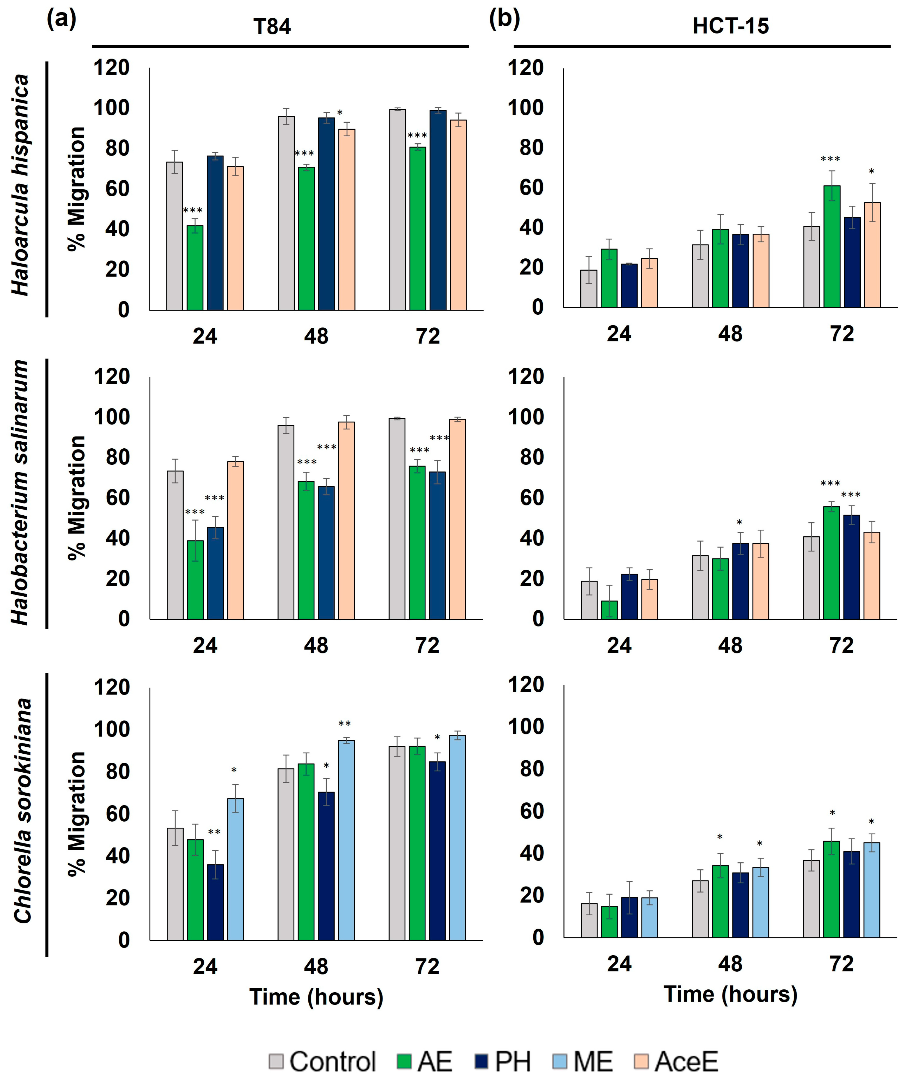
3.2.2. Alteration of Cell Migration Capacity Study

Based on the antiproliferative effect of previously tested archaea and microalgae extracts, we selected Haloarcula hispanica, Halobacterium salinarum, and Chlorella sorokiniana to analyze further antitumor properties. Thus, both T84 and HCT-15 colon cancer cells were exposed to non-toxic concentrations of the different extracts to analyze the modulation of cell migratory capacity (Figure 2). Our results showed that Haloarcula hispanica AE, Halobacterium salinarum AE and PH, and Chlorella sorokiniana PH significantly reduced the migratory ability of T84 tumor cells (p < 0.05) (Figure 2a). In fact, the AE of Haloarcula hispanica and Halobacterium salinarum induced the greatest inhibition of migratory capacity at 24 h (31.6% and 34.4%, respectively) (p < 0.001). It is necessary to mention that HCT-15 cells not only did not decrease their migratory capacity, but even showed a slight increase in their migratory capacity with some extracts (Figure 2b).

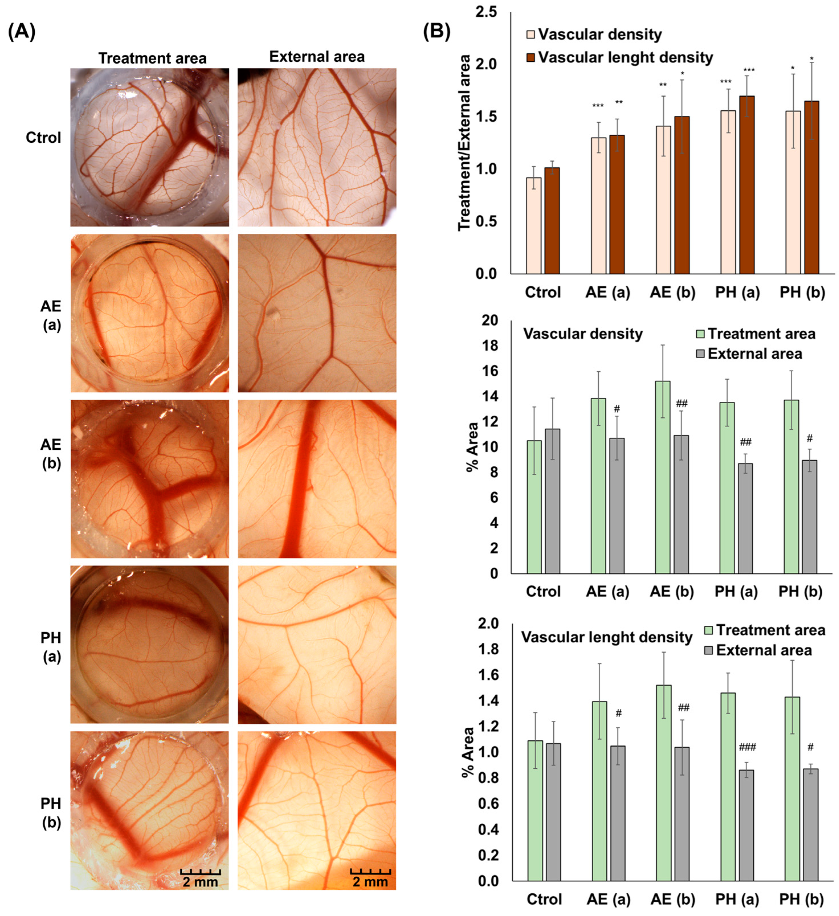
3.2.3. Angiogenesis Study

Since the most promising antiproliferative results were observed in Chlorella sorokiniana AE and PH, these extracts were selected to study their effect on angiogenesis in the CAM assay. Our results revealed that both tested doses of Chlorella sorokiniana AE and PH, 25 and 100 µg/mL, significantly increased the formation of blood vessels and their size in the CAM compared to the control eggs, increasing the percentage of area occupied by blood vessels in the treated area versus the external area of the CAM (Figure 3). Specifically, both doses of HP increased vascular density and vascular length density by 1.6 and 1.7-fold respectively compared to the control, while the AE improved both parameters between 1.3- and 1.5-fold.

3.3. Antioxidant and Preventive Effect of Functional Extracts

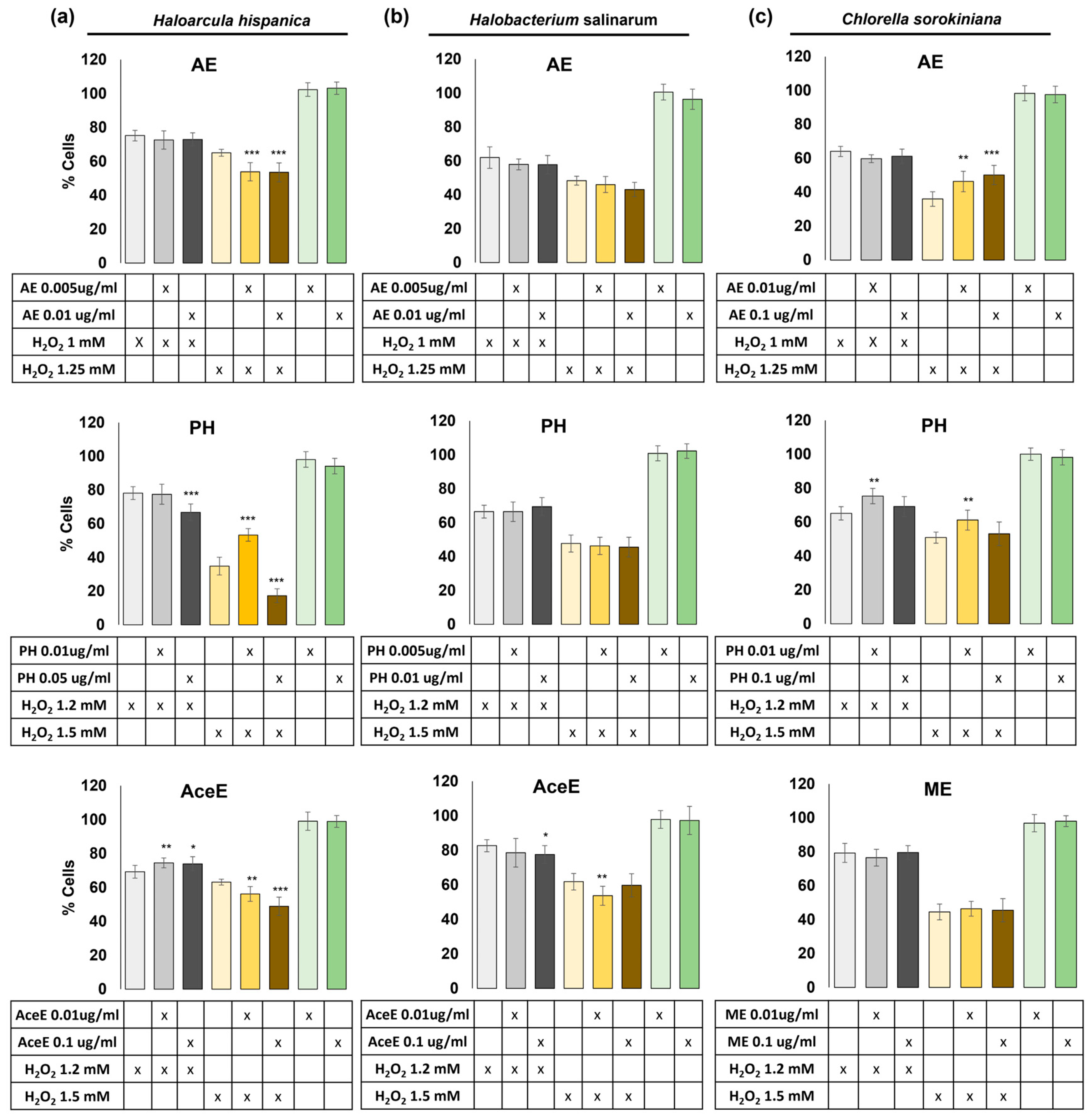
3.3.1. Protection Study against Reactive Oxygen Species

Analysis of the protective capacity of the extracts against oxidative stress showed that only the PH and AceE from Haloarcula hispanica and the AE and PH from Chlorella sorokiniana significantly protected cells against hydrogen peroxide damage. As shown in Figure 4, the AceE from Haloarcula hispanica provided the highest protection against reactive oxygen species when used at 0.01 µg/mL in combination with 1.2 mM of hydrogen peroxide (5.2% protection) (p < 0.01), while 0.1 µg/mL of the extract with the same concentration of hydrogen peroxide only protected 4.7% (p < 0.05). Similarly, Haloarcula hispanica PH and Chlorella sorokiniana AE and PH showed the highest degree of protection when used at 0.01 µg/mL, 0.1 µg/mL, and 0.01 µg/mL, respectively, both in combination with 1.5 mM of hydrogen peroxide (18.4, 14.1 and 10.3% of protection) (p < 0.01).

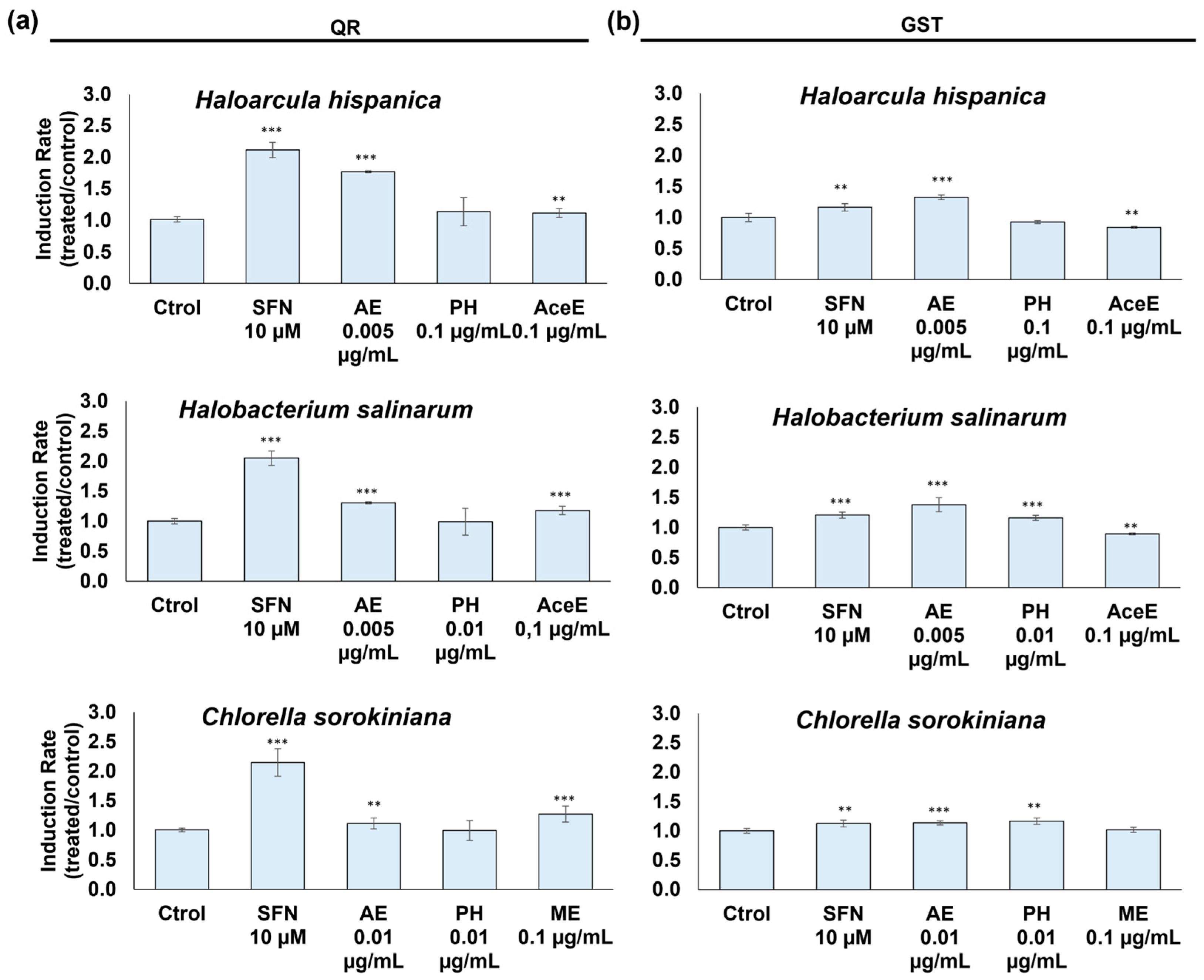
3.3.2. Detoxifying Enzyme Activity Enhancement

Analysis of the effect of functional extracts on detoxifying enzymes showed that Haloarcula hispanica AE induced the greatest increase in QR enzyme activity. As shown in Figure 5a, this enzyme increased its activity 1.77-fold (p < 0.001) after exposure to the AE for 48 h. The other extracts modulated the enzymatic activity to a lesser extent except the PH of Haloarcula hispanica, Chlorella sorokiniana, and Halobacterium salinarum, which did not exert any effect. On the other hand, the AE from Halobacterium salinarum induced the largest increase in GST enzyme activity (up to 1.38-fold) (p < 0.001), as can be seen in Figure 5b. Conversely, the HP from Haloarcula hispanica and ME from Chlorella sorokiniana did not induce any effect in this enzyme.

4. Discussion

Cancer represents a global health challenge which requires innovative therapeutic approaches. This paper explores the potential therapeutic contributions of archaeal and microalgal species, as understanding the molecular mechanisms underlying their effects could reveal innovative approaches to complement existing treatments, promoting advances in the search for more effective and targeted cancer therapies.
The antitumor activity of the extracts obtained was evaluated by cytotoxicity and tumor cell migration assays. Our results suggest that AE or PH extracts from the microalgae Dunaliella sp., Picochlorum sp., and Chlamydomonas reinhardtii showed a weak cytotoxic activity against CRC cells, with the lowest IC50 value observed for the AE of Picochlorum sp. against the HCT-15 cell line (114.33 ± 8.91 µg/mL). Similar results were previously obtained in other studies in which the IC50 values observed for different Dunaliella sp. and Picochlorum sp. extracts (ethanol, aqueous, dichloromethane, methanol, ethyl acetate, etc.) were higher than 100 µg/mL in breast, lung, and liver cancer cell lines [34,35,36]. By contrast, the AE and PH from the microalgae Chlorella sorokiniana showed high antitumor activity against the CRC cell lines tested, reaching IC50 values between 54.05 and 66.73 µg/mL for the T84 CRC line. The AE of Chlorella sorokiniana also showed high cytotoxicity (IC50 = 59.21 ± 10.3 µg/mL) against the drug-resistant HCT-15 CRC line, although in this case, cytotoxic activity decreased (IC50 = 165.41 ± 7.34 µg/mL) when the AE was obtained after a treatment with proteases (HP), indicating the possible contribution of a protein in the antiproliferative activity of this drug-resistant cell line. Our results are even more promising than those reported in the literature obtained with various extraction methods from different species of Chlorella [21,22,37]. Regarding the two archaea species analyzed, it should be noted that of the different extraction methods used, only the AE and HP showed antiproliferative activity against CRC cells. The most interesting results, however, were obtained by the HP from Halobacterium salinarum (IC50 value 110.56 ± 17.12 and 111.20 ± 22.20 µg/mL in HCT-15 and T84 cells, respectively). This is in agreement with previous results for both archaea species [38,39]. Since the best antitumor effects were observed in Chlorella sorokiniana, Haloarcula hispanica, and Halobacterium salinarum, these extracts were selected to examine their possible effect on inhibiting the migratory capacity of T84 and HCT-15 tumor cells. These results are summarized in Table 3.
Moreover, our results suggest that the AE resulted in the greatest inhibition of T84 migratory activity at 24 h of treatment, with Haloarcula hispanica and Halobacterium salinarum extracts reducing it by 31.6% and 34.4%, respectively. Similar results were obtained by Alateyah et al. [40], who observed that the methanolic extract of the microalgae Haematococcus pluvialis significantly reduced the migratory capacity of the breast cancer cell line MDA-MB-231 by 68% at 48 h of treatment.
This antitumor activity that has been seen in the extracts tested may be having its effect mainly due to different cellular mechanisms, including inhibition of migration, modulation of the cell cycle, and induction of autophagy or apoptosis [41]. Migration is highly related to tumors since it facilitates tumor growth and the spread of tumor cells to other organs, giving rise to metastasis [42]. Carotenoids such as fucoxanthin have resulted in the inhibition of cell migration in the HT-29 and HCT116 CCR cell lines in a dose-dependent manner [43], although it has also been seen in other tumor types such as sarcomas with extracts from the brown seaweed Cladosiphon okamuranus Tokida [44]. Alterations in the cell cycle are directly related to cell growth and apoptosis. Crude ethanol extracts from the microalga Chaetoceros calcitrans have been shown to have a modulating effect on the cell cycle in MCF7 breast cancer tumor cells, increasing cells in the subG0/G1 phase and thus promoting apoptosis [45]. Similar results have been obtained in ethanolic extracts of the microalga Micractinium sp., a study in the colorectal cancer cell line HCT116 shown an arrest of the cell cycle in the G1 phase through the regulation of genes such as CDKN1A and CDK6, characteristic of the G1/S phase transition [46]. Finally, autophagy and apoptosis involve the genetically programmed elimination of cells and is a fundamental process associated with development, physiology, and homeostasis, whose alteration can lead to various pathological conditions, including cancer [47]. There are studies that relate the ability of microalgae such as Chlorella vulgaris or Dunaliella salina to produce compounds such as phytosterols that can produce alterations in the apoptosis signaling pathway [48]. Analyses such as that of Jiang et al. show that these phytosterols, in addition to promoting cell apoptosis, are capable of reducing cell progression, invasion, and migration [49]. The presence of carotenoids has also been related to an induction of early apoptosis, as is the case for Chlorella ellipsoidea and Chlorella vulgaris, which are capable of inhibiting cell proliferation in the CCR cell line HCT15, producing IC50 values of around 40 μg/mL [50].
In the results obtained in the in vitro CAM assay, both EA and HP from Chlorella sorokiniana microalgae showed interesting angiogenic potential, although there are very little data on the effect of extracts or biomolecules derived from microalgae on vessel development. Some authors such as Jarquín-Cordero et al. have demonstrated the ability of the microalga Chlamydomonas reinhardtii to express proangiogenic growth factors [51,52,53,54]. Therefore, in the context of the activity of the extracts on tumors, these results should be evaluated with new assays.
Finally, the preventive activity of the different functional extracts of Chlorella sorokiniana, Haloarcula hispanica, and Halobacterium salinarum was assessed by testing their ability to protect cells against oxidative damage and their effect on the detoxifying enzymes QR and GST. Interestingly, in our results, the HP from Haloarcula hispanica and EA and HP from Chlorella sorokiniana showed the best protective effect for oxidative damage caused by H2O2 (18.4, 14.1, and 10.3% protection, respectively), while the EA from both archaea Haloarcula hispanica and Halobacterium salinarum resulted in the most interesting increase in QR and GST enzymes, respectively. In this context, our results support those of other authors who have already demonstrated certain antioxidant activity of different microalgae. In particular, Chlorella vulgaris has been found to contain significant amounts of pigments such as carotenes, lutein, chlorophylls, and vitamins such as vitamin C, all of which have antioxidant capacity [37,55,56]. The antioxidant effect of various haloarchaea has been studied and associated with the presence of carotenes [57,58]. While our results suggest that Halobacterium salinarum does not exhibit antioxidant activity, several studies have reported that the AE of Halobacterium salinarum showed significant antioxidant capacity [27,38]. On the other hand, although the effect of archaea on the activity of the detoxifying enzyme GST is understudied, Selim et al. demonstrated that species of the archaea genus Haloferax were able to promote the enzymatic activity of the detoxifying enzyme GST [59]. Furthermore, the ability of diverse microalgae to promote GST enzyme activity has been previously described by some authors [60,61,62].

5. Conclusions

The present study highlights the importance of exploring marine microorganisms and microalgae in the search for new therapies against colorectal cancer. Through the selection and analysis of functional extracts of microalgae and halophilic archaea, their potential as sources of bioactive compounds with antitumor properties has been demonstrated. The results revealed that Chlorella sorokiniana aqueous extracts exhibited significant antitumor activity against colorectal cancer cell lines (T84 and HCT-15), with IC50 values of 54.05 and 59.21 µg/mL, respectively, suggesting their promising potential as therapeutic agents against colorectal cancer. On the other hand, it was found that protein hydrolysate from Haloarcula hispanica and Chlorella sorokiniana showed antioxidant capacity, protecting cells against oxidative stress, and the aqueous extracts of Haloarcula hispanica and Halobacterium salinarum increased the activity of detoxifying enzymes such as QR and GST. These results support the idea that extremophilic marine microorganisms and microalgae can be valuable sources of compounds with antioxidant and protective properties against cellular damage.
Taken together, these findings suggest that functional extracts of microalgae and halophilic archaea have great potential as adjuvants in the treatment of colorectal cancer, both due to their direct antitumor activity and their ability to modulate key cellular processes related to tumor progression and antioxidant response. These results underscore the importance of continuing to explore marine biodiversity to identify new cancer therapies and improve existing treatments. However, further studies are needed to identify the active compounds in these extracts and for a better understanding of the underlying mechanisms of action.

Supplementary Materials

The following supporting information can be downloaded at: https://www.mdpi.com/article/10.3390/jmse12060996/s1, Table S1: IC50 values of methanol extract (ME) and acetonic extract (AceE) extracts in the CRC cell line T84 and HCT-15.

Author Contributions

Conceptualization, R.L. and C.M.; methodology, C.L., G.P. and P.G.-V.; validation, J.V. and R.M.; formal analysis, J.V., R.M., G.P., P.G.-V. and A.G.-B.; investigation, C.L., G.P. and P.G.-V.; data curation, J.M.P. and J.P.; writing—original draft preparation, C.L., G.P. and P.G.-V.; writing—review and editing, J.M.P. and R.L.; supervision, R.L. and C.M.; funding acquisition, G.P., P.G.-V. and R.L. All authors have read and agreed to the published version of the manuscript.

Funding

This research was funded by the Campus de Excelencia Internacional Global del Mar (CEI·Mar 2022) (Project CEI-JD-19.1 and CEI-JD-19.2) and Agencia Estatal de Investigación (grant PID2022-140995OB-C21 by MICIU/AEI/10.13039/501100011033 and ERDF/EU).

Institutional Review Board Statement

Not applicable.

Informed Consent Statement

Not applicable.

Data Availability Statement

Data are contained within the article.

Acknowledgments

The authors want to thank to Scientific Instrumentation Centre (CIC, UGR) for her excellent technical assistance.

Conflicts of Interest

The authors declare no conflicts of interest.

References

  1. Cragg, G.M.; Grothaus, P.G.; Newman, D.J. Impact of Natural Products on Developing New Anti-Cancer Agents. Chem. Rev. 2009, 109, 3012–3043. [Google Scholar] [CrossRef]
  2. Shukla, Y.; George, J. Combinatorial Strategies Employing Nutraceuticals for Cancer Development. Ann. N. Y Acad. Sci. 2011, 1229, 162–175. [Google Scholar] [CrossRef]
  3. Martínez, C.; Mairet, F.; Bernard, O. Theory of Turbid Microalgae Cultures. J. Theor. Biol. 2018, 456, 190–200. [Google Scholar] [CrossRef] [PubMed]
  4. Ferdous, U.T.; Yusof, Z.N.B. Medicinal Prospects of Antioxidants From Algal Sources in Cancer Therapy. Front. Pharmacol. 2021, 12, 593116. [Google Scholar] [CrossRef] [PubMed]
  5. Wu, J.; Gu, X.; Yang, D.; Xu, S.; Wang, S.; Chen, X.; Wang, Z. Bioactive Substances and Potentiality of Marine Microalgae. Food Sci. Nutr. 2021, 9, 5279–5292. [Google Scholar] [CrossRef] [PubMed]
  6. Hou, J.; Cui, H.-L. In Vitro Antioxidant, Antihemolytic, and Anticancer Activity of the Carotenoids from Halophilic Archaea. Curr. Microbiol. 2018, 75, 266–271. [Google Scholar] [CrossRef]
  7. Hegazy, G.E.; Abu-Serie, M.M.; Abo-Elela, G.M.; Ghozlan, H.; Sabry, S.A.; Soliman, N.A.; Abdel-Fattah, Y.R. In Vitro Dual (Anticancer and Antiviral) Activity of the Carotenoids Produced by Haloalkaliphilic Archaeon Natrialba sp. M6. Sci. Rep. 2020, 10, 5986. [Google Scholar] [CrossRef]
  8. Lizama, C.; Romero-Parra, J.; Andrade, D.; Riveros, F.; Bórquez, J.; Ahmed, S.; Venegas-Salas, L.; Cabalín, C.; Simirgiotis, M.J. Analysis of Carotenoids in Haloarchaea Species from Atacama Saline Lakes by High Resolution UHPLC-Q-Orbitrap-Mass Spectrometry: Antioxidant Potential and Biological Effect on Cell Viability. Antioxidants 2021, 10, 1230. [Google Scholar] [CrossRef]
  9. Sahli, K.; Gomri, M.A.; Esclapez, J.; Gómez-Villegas, P.; Bonete, M.-J.; León, R.; Kharroub, K. Characterization and Biological Activities of Carotenoids Produced by Three Haloarchaeal Strains Isolated from Algerian Salt Lakes. Arch. Microbiol. 2021, 204, 6. [Google Scholar] [CrossRef]
  10. Moghimipour, E.; Abedishirehjin, S.; Baghbadorani, M.A.; Handali, S. Bacteria and Archaea: A New Era of Cancer Therapy. J. Control. Release 2021, 338, 1–7. [Google Scholar] [CrossRef]
  11. Esposito, R.; Federico, S.; Glaviano, F.; Somma, E.; Zupo, V.; Costantini, M. Bioactive Compounds from Marine Sponges and Algae: Effects on Cancer Cell Metabolome and Chemical Structures. Int. J. Mol. Sci. 2022, 23, 10680. [Google Scholar] [CrossRef] [PubMed]
  12. Karakaş, C.Y.; Tekarslan Şahin, H.; İnan, B.; Özçimen, D.; Erginer, Y.Ö. In Vitro Cytotoxic Activity of Microalgal Extracts Loaded Nano–Micro Particles Produced via Electrospraying and Microemulsion Methods. Biotechnol. Prog. 2019, 35, e2876. [Google Scholar] [CrossRef] [PubMed]
  13. Abreu, A.C.; Molina-Miras, A.; Aguilera-Sáez, L.M.; López-Rosales, L.; Cerón-García, M.d.C.; Sánchez-Mirón, A.; Olmo-García, L.; Carrasco-Pancorbo, A.; García-Camacho, F.; Molina-Grima, E.; et al. Production of Amphidinols and Other Bioproducts of Interest by the Marine Microalga Amphidinium Carterae Unraveled by Nuclear Magnetic Resonance Metabolomics Approach Coupled to Multivariate Data Analysis. J. Agric. Food Chem. 2019, 67, 9667–9682. [Google Scholar] [CrossRef] [PubMed]
  14. Sung, H.; Ferlay, J.; Siegel, R.L.; Laversanne, M.; Soerjomataram, I.; Jemal, A.; Bray, F. Global Cancer Statistics 2020: GLOBOCAN Estimates of Incidence and Mortality Worldwide for 36 Cancers in 185 Countries. CA Cancer J. Clin. 2021, 71, 209–249. [Google Scholar] [CrossRef] [PubMed]
  15. Argilés, G.; Tabernero, J.; Labianca, R.; Hochhauser, D.; Salazar, R.; Iveson, T.; Laurent-Puig, P.; Quirke, P.; Yoshino, T.; Taieb, J.; et al. Localised Colon Cancer: ESMO Clinical Practice Guidelines for Diagnosis, Treatment and Follow-Up. Ann. Oncol. 2020, 31, 1291–1305. [Google Scholar] [CrossRef] [PubMed]
  16. Dekker, E.; Tanis, P.J.; Vleugels, J.L.A.; Kasi, P.M.; Wallace, M.B. Colorectal Cancer. Lancet 2019, 394, 1467–1480. [Google Scholar] [CrossRef] [PubMed]
  17. Barin-Le Guellec, C.; Lafay-Chebassier, C.; Ingrand, I.; Tournamille, J.-F.; Boudet, A.; Lanoue, M.-C.; Defossez, G.; Ingrand, P.; Perault-Pochat, M.-C.; Etienne-Grimaldi, M.-C. Toxicities Associated with Chemotherapy Regimens Containing a Fluoropyrimidine: A Real-Life Evaluation in France. Eur. J. Cancer 2020, 124, 37–46. [Google Scholar] [CrossRef] [PubMed]
  18. Hu, S.; Huang, K.M.; Adams, E.J.; Loprinzi, C.L.; Lustberg, M.B. Recent Developments of Novel Pharmacologic Therapeutics for Prevention of Chemotherapy-Induced Peripheral Neuropathy. Clin. Cancer Res. 2019, 25, 6295–6301. [Google Scholar] [CrossRef] [PubMed]
  19. Delgado-Roche, L.; González, K.; Mesta, F.; Couder, B.; Tavarez, Z.; Zavala, R.; Hernandez, I.; Garrido, G.; Rodeiro, I.; Vanden Berghe, W. Polyphenolic Fraction Obtained From Thalassia Testudinum Marine Plant and Thalassiolin B Exert Cytotoxic Effects in Colorectal Cancer Cells and Arrest Tumor Progression in a Xenograft Mouse Model. Front. Pharmacol. 2020, 11, 592985. [Google Scholar] [CrossRef]
  20. Ercolano, G.; De Cicco, P.; Ianaro, A. New Drugs from the Sea: Pro-Apoptotic Activity of Sponges and Algae Derived Compounds. Mar. Drugs 2019, 17, 31. [Google Scholar] [CrossRef]
  21. Hamouda, R.A.; Abd El Latif, A.; Elkaw, E.M.; Alotaibi, A.S.; Alenzi, A.M.; Hamza, H.A. Assessment of Antioxidant and Anticancer Activities of Microgreen Alga Chlorella Vulgaris and Its Blend with Different Vitamins. Molecules 2022, 27, 1602. [Google Scholar] [CrossRef] [PubMed]
  22. Sawasdee, N.; Jantakee, K.; Wathikthinnakon, M.; Panwong, S.; Pekkoh, J.; Duangjan, K.; Yenchitsomanus, P.; Panya, A. Microalga Chlorella sp. Extract Induced Apoptotic Cell Death of Cholangiocarcinoma via AKT/mTOR Signaling Pathway. Biomed. Pharmacother. 2023, 160, 114306. [Google Scholar] [CrossRef] [PubMed]
  23. Johnson, M.K.; Johnson, E.J.; MacElroy, R.D.; Speer, H.L.; Bruff, B.S. Effects of Salts on the Halophilic Alga Dunaliella Viridis. J. Bacteriol. 1968, 95, 1461–1468. [Google Scholar] [CrossRef] [PubMed]
  24. de la Vega, M.; Sayago, A.; Ariza, J.; Barneto, A.G.; León, R. Characterization of a Bacterioruberin-Producing Haloarchaea Isolated from the Marshlands of the Odiel River in the Southwest of Spain. Biotechnol. Prog. 2016, 32, 592–600. [Google Scholar] [CrossRef] [PubMed]
  25. Fawley, K.P.; Fawley, M.W. Observations on the Diversity and Ecology of Freshwater Nannochloropsis (Eustigmatophyceae), with Descriptions of New Taxa. Protist 2007, 158, 325–336. [Google Scholar] [CrossRef] [PubMed]
  26. Guillard, R.R.L. Culture of Phytoplankton for Feeding Marine Invertebrates. In Culture of Marine Invertebrate Animals: Proceedings—1st Conference on Culture of Marine Invertebrate Animals Greenport; Smith, W.L., Chanley, M.H., Eds.; Springer: Boston, MA, USA, 1975; pp. 29–60. ISBN 978-1-4615-8714-9. [Google Scholar]
  27. Gómez-Villegas, P.; Vigara, J.; Vila, M.; Varela, J.; Barreira, L.; Léon, R. Antioxidant, Antimicrobial, and Bioactive Potential of Two New Haloarchaeal Strains Isolated from Odiel Salterns (Southwest Spain). Biology 2020, 9, 298. [Google Scholar] [CrossRef] [PubMed]
  28. Fang, C.-J.; Ku, K.-L.; Lee, M.-H.; Su, N.-W. Influence of Nutritive Factors on C50 Carotenoids Production by Haloferax Mediterranei ATCC 33500 with Two-Stage Cultivation. Bioresour. Technol. 2010, 101, 6487–6493. [Google Scholar] [CrossRef] [PubMed]
  29. Kapravelou, G.; Martínez, R.; Andrade, A.M.; Sánchez, C.; Chaves, C.L.; López-Jurado, M.; Aranda, P.; Cantarero, S.; Arrebola, F.; Fernández-Segura, E.; et al. Health Promoting Effects of Lupin (Lupinus albus Var. Multolupa) Protein Hydrolyzate and Insoluble Fiber in a Diet-Induced Animal Experimental Model of Hypercholesterolemia. Food Res. Int. 2013, 54, 1471–1481. [Google Scholar] [CrossRef]
  30. Lichtenthaler, H.K. [34] Chlorophylls and Carotenoids: Pigments of Photosynthetic Biomembranes. In Methods in Enzymology; Plant Cell Membranes; Academic Press: Cambridge, MA, USA, 1987; Volume 148, pp. 350–382. [Google Scholar]
  31. Grada, A.; Otero-Vinas, M.; Prieto-Castrillo, F.; Obagi, Z.; Falanga, V. Research Techniques Made Simple: Analysis of Collective Cell Migration Using the Wound Healing Assay. J. Investig. Dermatol. 2017, 137, e11–e16. [Google Scholar] [CrossRef]
  32. Suarez-Arnedo, A.; Torres Figueroa, F.; Clavijo, C.; Arbeláez, P.; Cruz, J.C.; Muñoz-Camargo, C. An Image J Plugin for the High Throughput Image Analysis of in Vitro Scratch Wound Healing Assays. PLoS ONE 2020, 15, e0232565. [Google Scholar] [CrossRef]
  33. Fuel, M.; Mesas, C.; Martínez, R.; Ortiz, R.; Quiñonero, F.; Prados, J.; Porres, J.M.; Melguizo, C. Antioxidant and Antiproliferative Potential of Ethanolic Extracts from Moringa Oleifera, Tropaeolum Tuberosum and Annona Cherimola in Colorrectal Cancer Cells. Biomed. Pharmacother. 2021, 143, 112248. [Google Scholar] [CrossRef] [PubMed]
  34. Abolhasani, M.H.; Safavi, M.; Goodarzi, M.T.; Kassaee, S.M.; Azin, M. Identification and Anti-Cancer Activity in 2D and 3D Cell Culture Evaluation of an Iranian Isolated Marine Microalgae Picochlorum sp. RCC486. DARU J. Pharm. Sci. 2018, 26, 105–116. [Google Scholar] [CrossRef] [PubMed]
  35. Elleuch, F.; Baril, P.; Barkallah, M.; Perche, F.; Abdelkafi, S.; Fendri, I.; Pichon, C. Deciphering the Biological Activities of Dunaliella sp. Aqueous Extract from Stressed Conditions on Breast Cancer: From in Vitro to in Vivo Investigations. Int. J. Mol. Sci. 2020, 21, 1719. [Google Scholar] [CrossRef] [PubMed]
  36. Pasquet, V.; Morisset, P.; Ihammouine, S.; Chepied, A.; Aumailley, L.; Berard, J.-B.; Serive, B.; Kaas, R.; Lanneluc, I.; Thiery, V.; et al. Antiproliferative Activity of Violaxanthin Isolated from Bioguided Fractionation of Dunaliella Tertiolecta Extracts. Mar. Drugs 2011, 9, 819–831. [Google Scholar] [CrossRef] [PubMed]
  37. El-fayoumy, E.A.; Shanab, S.M.M.; Gaballa, H.S.; Tantawy, M.A.; Shalaby, E.A. Evaluation of Antioxidant and Anticancer Activity of Crude Extract and Different Fractions of Chlorella Vulgaris Axenic Culture Grown under Various Concentrations of Copper Ions. BMC Complement. Med. Ther. 2021, 21, 51. [Google Scholar] [CrossRef] [PubMed]
  38. Abbes, M.; Baati, H.; Guermazi, S.; Messina, C.; Santulli, A.; Gharsallah, N.; Ammar, E. Biological Properties of Carotenoids Extracted from Halobacterium Halobium Isolated from a Tunisian Solar Saltern. BMC Complement. Altern. Med. 2013, 13, 255. [Google Scholar] [CrossRef] [PubMed]
  39. Shahbazi, S.; Zargar, M.; Zolfaghari, M.R.; Amoozegar, M.A. Carotenoid Pigment of Halophilic Archaeon Haloarcula sp. A15 Induces Apoptosis of Breast Cancer Cells. Cell Biochem. Funct. 2023, 41, 344–354. [Google Scholar] [CrossRef] [PubMed]
  40. Alateyah, N.; Ahmad, S.M.S.; Gupta, I.; Fouzat, A.; Thaher, M.I.; Das, P.; Al Moustafa, A.-E.; Ouhtit, A. Haematococcus Pluvialis Microalgae Extract Inhibits Proliferation, Invasion, and Induces Apoptosis in Breast Cancer Cells. Front. Nutr. 2022, 9, 882956. [Google Scholar] [CrossRef] [PubMed]
  41. Ruiz-Torres, V.; Encinar, J.A.; Herranz-López, M.; Pérez-Sánchez, A.; Galiano, V.; Barrajón-Catalán, E.; Micol, V. An Updated Review on Marine Anticancer Compounds: The Use of Virtual Screening for the Discovery of Small-Molecule Cancer Drugs. Molecules 2017, 22, 1037. [Google Scholar] [CrossRef]
  42. Coultas, L.; Chawengsaksophak, K.; Rossant, J. Endothelial Cells and VEGF in Vascular Development. Nature 2005, 438, 937–945. [Google Scholar] [CrossRef]
  43. Méresse, S.; Fodil, M.; Fleury, F.; Chénais, B. Fucoxanthin, a Marine-Derived Carotenoid from Brown Seaweeds and Microalgae: A Promising Bioactive Compound for Cancer Therapy. Int. J. Mol. Sci. 2020, 21, 9273. [Google Scholar] [CrossRef]
  44. Rokkaku, T.; Kimura, R.; Ishikawa, C.; Yasumoto, T.; Senba, M.; Kanaya, F.; Mori, N. Anticancer Effects of Marine Carotenoids, Fucoxanthin and Its Deacetylated Product, Fucoxanthinol, on Osteosarcoma. Int. J. Oncol. 2013, 43, 1176–1186. [Google Scholar] [CrossRef]
  45. Ebrahimi Nigjeh, S.; Yusoff, F.M.; Mohamed Alitheen, N.B.; Rasoli, M.; Keong, Y.S.; bin Omar, A.R. Cytotoxic Effect of Ethanol Extract of Microalga, Chaetoceros Calcitrans, and Its Mechanisms in Inducing Apoptosis in Human Breast Cancer Cell Line. Biomed. Res. Int. 2013, 2013, 783690. [Google Scholar] [CrossRef]
  46. Suh, S.-S.; Hong, J.-M.; Kim, E.J.; Jung, S.W.; Kim, S.-M.; Kim, J.E.; Kim, I.-C.; Kim, S. Anti-Inflammation and Anti-Cancer Activity of Ethanol Extract of Antarctic Freshwater Microalga, Micractinium sp. Int. J. Med. Sci. 2018, 15, 929–936. [Google Scholar] [CrossRef]
  47. Abaza, A.; Vasavada, A.M.; Sadhu, A.; Valencia, C.; Fatima, H.; Nwankwo, I.; Anam, M.; Maharjan, S.; Amjad, Z.; Khan, S. A Systematic Review of Apoptosis in Correlation with Cancer: Should Apoptosis Be the Ultimate Target for Cancer Treatment? Cureus 2022, 14, e28496. [Google Scholar] [CrossRef]
  48. Luo, X.; Su, P.; Zhang, W. Advances in Microalgae-Derived Phytosterols for Functional Food and Pharmaceutical Applications. Mar. Drugs 2015, 13, 4231–4254. [Google Scholar] [CrossRef]
  49. Jiang, L.; Zhao, X.; Xu, J.; Li, C.; Yu, Y.; Wang, W.; Zhu, L. The Protective Effect of Dietary Phytosterols on Cancer Risk: A Systematic Meta-Analysis. J. Oncol. 2019, 2019, e7479518. [Google Scholar] [CrossRef]
  50. Cha, K.H.; Koo, S.Y.; Lee, D.-U. Antiproliferative Effects of Carotenoids Extracted from Chlorella ellipsoidea and Chlorella vulgaris on Human Colon Cancer Cells. J. Agric. Food Chem. 2008, 56, 10521–10526. [Google Scholar] [CrossRef]
  51. Adamczyk-Grochala, J.; Wnuk, M.; Duda, M.; Zuczek, J.; Lewinska, A. Treatment with Modified Extracts of the Microalga Planktochlorella Nurekis Attenuates the Development of Stress-Induced Senescence in Human Skin Cells. Nutrients 2020, 12, 1005. [Google Scholar] [CrossRef]
  52. Bari, E.; Arciola, C.R.; Vigani, B.; Crivelli, B.; Moro, P.; Marrubini, G.; Sorrenti, M.; Catenacci, L.; Bruni, G.; Chlapanidas, T.; et al. In Vitro Effectiveness of Microspheres Based on Silk Sericin and Chlorella vulgaris or Arthrospira platensis for Wound Healing Applications. Materials 2017, 10, 983. [Google Scholar] [CrossRef]
  53. Jarquín-Cordero, M.; Chávez, M.N.; Centeno-Cerdas, C.; Bohne, A.-V.; Hopfner, U.; Machens, H.-G.; Egaña, J.T.; Nickelsen, J. Towards a Biotechnological Platform for the Production of Human Pro-Angiogenic Growth Factors in the Green Alga Chlamydomonas Reinhardtii. Appl. Microbiol. Biotechnol. 2020, 104, 725–739. [Google Scholar] [CrossRef]
  54. Syarina, P.N.A.; Karthivashan, G.; Abas, F.; Arulselvan, P.; Fakurazi, S. Wound Healing Potential of Spirulina Platensis Extracts on Human Dermal Fibroblast Cells. EXCLI J. 2015, 14, 385–393. [Google Scholar] [CrossRef]
  55. Safafar, H.; Van Wagenen, J.; Møller, P.; Jacobsen, C. Carotenoids, Phenolic Compounds and Tocopherols Contribute to the Antioxidative Properties of Some Microalgae Species Grown on Industrial Wastewater. Mar. Drugs 2015, 13, 7339–7356. [Google Scholar] [CrossRef]
  56. Safi, C.; Zebib, B.; Merah, O.; Pontalier, P.-Y.; Vaca-Garcia, C. Morphology, Composition, Production, Processing and Applications of Chlorella Vulgaris: A Review. Renew. Sustain. Energy Rev. 2014, 35, 265–278. [Google Scholar] [CrossRef]
  57. Baati, H.; Siala, M.; Azri, C.; Ammar, E.; Trigui, M. Hydrolytic Enzyme Screening and Carotenoid Production Evaluation of Halophilic Archaea Isolated from Highly Heavy Metal-Enriched Solar Saltern Sediments. Braz. J. Microbiol. 2022, 53, 1893–1906. [Google Scholar] [CrossRef]
  58. Kesbiç, F.I.; Gültepe, N. Carotenoid Characterization, Fatty Acid Profiles, and Antioxidant Activities of Haloarchaeal Extracts. J. Basic. Microbiol. 2024, 64, 2300330. [Google Scholar] [CrossRef]
  59. Selim, S.; Akhtar, N.; Hagagy, N.; Alanazi, A.; Warrad, M.; El Azab, E.; Elamir, M.Y.M.; Al-Sanea, M.M.; Jaouni, S.K.A.; Abdel-Mawgoud, M.; et al. Selection of Newly Identified Growth-Promoting Archaea Haloferax Species with a Potential Action on Cobalt Resistance in Maize Plants. Front. Plant Sci. 2022, 13, 872654. [Google Scholar] [CrossRef]
  60. Castro, M.S.; Silva, J.C.; Machado, B.R.; Guimarães, P.S.; Lombardi, A.T.; Martins, C.D.M.G.; Zanette, J. Differential Effects of Atrazine on Chlorophyceae Species and Association with Morphology, Photosynthesis, Chlorophyll Content, and Glutathione-S-Transferase Activity. Environ. Toxicol. Chem. 2022, 41, 1675–1685. [Google Scholar] [CrossRef]
  61. Hamed, S.M.; Okla, M.K.; Al-Saadi, L.S.; Hozzein, W.N.; Mohamed, H.S.; Selim, S.; AbdElgawad, H. Evaluation of the Phycoremediation Potential of Microalgae for Captan Removal: Comprehensive Analysis on Toxicity, Detoxification and Antioxidants Modulation. J. Hazard. Mater. 2022, 427, 128177. [Google Scholar] [CrossRef]
  62. Zhang, Y.; He, D.; Chang, F.; Dang, C.; Fu, J. Combined Effects of Sulfamethoxazole and Erythromycin on a Freshwater Microalga, Raphidocelis Subcapitata: Toxicity and Oxidative Stress. Antibiotics 2021, 10, 576. [Google Scholar] [CrossRef]
Figure 1. Molecular phylogenetic analysis by Maximum Likelihood method. The tree represents the relationship among the 18S rRNA sequences from the two strains isolated (HM5 and HM13) from Odiel Saltworks Marshlands and reference sequences of Dunaliella species. Multiple alignments were generated by MUSCLE and the tree was constructed with MEGA X, using 1000 bootstrap replicates. The name and the NCBI access number are indicated for all the reference sequences.
Jmse 12 00996 g001
Figure 2. Study of a possible alteration of cell migratory capacity after exposing cells to non-toxic doses of different extracts. Results obtained on the cell lines of CCR T84 (a) and HCT-15 (b). Values are expressed as the mean ± standard deviation of at least three cultures. Student’s t-tests were employed with a significance level set at α = 5%; * p < 0.05; ** p < 0.01; and *** p < 0.001 compared with untreated cells. AE: aqueous extract; PH: protein hydrolysate; ME: methanol extract; and AceE: acetone extract.
Jmse 12 00996 g002
Figure 3. Effect on angiogenesis on the CAM of AE and PH Chlorella sorokiniana extracts. Doses of 25 (a) and 100 µg/mL (b) were applied for 72 h of treatment. Negative control was stablished as PBS-treated eggs. (A) Images of treatment and external area using a Motic SMZ-171 stereo microscope at 4× zoom. (B) Graphical representation of the Vascular density and Vascular length density measurements given by the “Vessel Analysis” plugin from FIJI 2.9.0 software. Results are shown as mean ± SD of at least four available eggs per experimental group. Student’s t-tests were employed with a significance level set at α = 5%, *: p < 0.05, **: p < 0.01, and ***: p < 0.001 compared to control eggs; #: p < 0.05, ##: p < 0.01, and ###: p < 0.01 compared to external area of the CAM.
Jmse 12 00996 g003
Figure 4. Assay of in vitro antioxidant activity of functional extracts. HT29 cells were pre-treated with functional extracts of Haloarcula hispanica (a), Halobacterium salinarum (b), and Chlorella sorokiniana (c) and subsequently exposed to hydrogen peroxide for 6 h of treatment. Data are represented as the mean ± SD of up to octuplicate. Student’s t-tests were employed with a significance level set at α = 5%; *: p < 0.05; **: p < 0.01; and ***: p < 0.001 compared to its treatment with H2O2. AE: aqueous extract; PH: protein hydrolysate; ME: methanol extract; and AceE: acetone extract.
Jmse 12 00996 g004
Figure 5. Effect of functional extracts on detoxifying enzyme activity. NAD(P)H: quinone oxidoreductase (QR) activity (a) and glutathione S-transferase (GST) analysis (b). Data are shown as the mean ± standard deviation of three independent cultures. Student’s t-tests were employed with a significance level set at α = 5%; **: p < 0.01; and ***: p < 0.001 compared to untreated cells. AE: aqueous extract; PH: protein hydrolysate; ME: methanol extract; and AceE: acetone extract.
Jmse 12 00996 g005
Table 1. IC50 values of the different extracts in the CRC cell line T84. Results are represented as the mean ± standard deviation of three replicates.
Microbial SpeciesEE IC50 (µg/mL)EEM IC50 (µg/mL)AE IC50 (µg/mL)PH IC50 (µg/mL)
Dunaliella HM13>1000>1000184.67 ± 15.71477.98 ± 29.16
Dunaliella HM5>1000>1000137.56 ± 7.58356.36 ± 11.22
Picochlorum sp.>600>600194.47 ± 57.52229.89 ± 14.84
Chlorella sorokiniana523.20 ± 47.71569.33 ± 88.5954.05 ± 27.7966.73 ± 7.64
Chlamydomonas reinhardtii>800399.29 ± 9.42>150>400
Haloarcula hispanica>1000>1000118.83 ± 57.52153.56 ± 3.26
Halobacterium salinarum>1000>1000138.47 ± 33.94111.20 ± 22.20
EE: ethanol extract; AE: aqueous extract; PH: protein hydrolysate; and EEM: ethanolic extract with previous mechanical disruption.
Table 2. IC50 values of the different extracts in the CRC cell line HCT-15. Results are represented as the mean ± standard deviation of three replicates.
Microbial SpeciesEE IC50 (µg/mL)EEM IC50 (µg/mL)AE IC50 (µg/mL)PH IC50 (µg/mL)
Dunaliella HM13345.02 ± 33.10198.75 ± 6.20959.23 ± 24.53718.24 ± 30.64
Dunaliella HM5346.46 ± 21.52137.39 ± 8.91593.04 ± 33.91553.11 ± 57.24
Picochlorum sp.575.85 ± 42.11114.33 ± 8.91>600749.90 ± 27.87
Chlorella sorokiniana301.27 ± 22.6159.21 ± 10.30165.41 ± 7.34242.99 ± 15.55
Chlamydomonas reinhardtii>800>300>400316.25 ± 6.84
Haloarcula hispanica>1000181.05 ± 7.69160.44 ± 4.67>1000
Halobacterium salinarum>1000171.29 ± 10.89110.56 ± 17.12>1000
EE: ethanol extract; AE: aqueous extract; PH: protein hydrolysate; and EEM: ethanolic extract with previous mechanical disruption.
Table 3. Comparison of the results discussed for the antitumor effect.
Ref.Microbial Species Human Cancer Cell LineExtractIC50 Value (μg/mL)Main Results
[21]Chlorella vulgarisPC-3 prostate
Hep-G2 liver
HCT-116 colorectal
Hela cervical
Methanol <100Supplementing the microalgae C. vulgaris with several vitamins showed an increase not only in its antioxidant and antitumor capacity but also an increase in its total proteins, biomass, and pigment content.
Methanol supplemented with Thiamine (vitamin B1)<100
[22]Chlorella sp.A-549 lung
Hela cervical
MCF7 breast
Huh7 hepatocellular
CCA and KKU213A cholangiocarcinoma
Polysaccharide >2000The antitumor effect of the ethanolic extract of Chlorella sp. was demonstrated by promoting cell death through the AKT/mTOR pathway.
Ethanol>300
[34]Dunaliella sp.MDA-MB-231 and MCF-7 breast
Hep-G2 liver
A-549 lung
Methanol>150Methanol and ethyl acetate extracts have a high content of phenolic compounds and carotenoids that are associated with an increase in apoptosis through the activation of caspase-3.
Ethyl acetate>200
Chloroform>500
Hexane>500
[36]Dunaliella tertiolectaMCF-7 breast
A-549 lung
LNCaP prostate
Ethanol >100The compound violaxanthin was identified as the molecule with the greatest antiproliferative potential present in the extract obtained with dichloromethane.
Dichloromethane>100
[37]Chlorella vulgarisHela cervical Methanol >125The methanolic extraction of C. vulgaris under copper-mediated stress conditions results in an antitumor effect on the Hela cell line.
[38]Halobacterium halobiumHep-G2 liver Carotenoid extraction>500Carotenoids obtained from Halobacterium halobium present an antitumor and antioxidant effect in the HepG2 line.
[39]Haloarcula sp.MCF-7 breast Carotenoid extraction>600The pigments obtained from the Haloarcula sp. archaea induce the expression of genes involved in apoptosis, thus having an antitumor effect in the breast cancer line MCF-7.
Disclaimer/Publisher’s Note: The statements, opinions and data contained in all publications are solely those of the individual author(s) and contributor(s) and not of MDPI and/or the editor(s). MDPI and/or the editor(s) disclaim responsibility for any injury to people or property resulting from any ideas, methods, instructions or products referred to in the content.

