Article

Responses of Different Morphological Cells of Phaeocystis globosa to UV-B Radiation

1
School of Resources, Environment and Materials, Guangxi University, Nanning 530004, China
2
Guangxi Key Laboratory of Marine Environmental Science, Guangxi Academy of Marine Sciences, Guangxi Academy of Sciences, Nanning 530007, China
3
Beibu Gulf Marine Industry Research Institute, Fangchenggang 538000, China
*
Authors to whom correspondence should be addressed.
J. Mar. Sci. Eng. 2024, 12(9), 1619; https://doi.org/10.3390/jmse12091619
Submission received: 31 July 2024 / Revised: 30 August 2024 / Accepted: 6 September 2024 / Published: 11 September 2024
(This article belongs to the Section Marine Ecology)

Abstract

:
Phaeocystis globosa is an important member of the plankton community and was considered to be a typical bloom-forming algae. Its life cycle is variable, comprising both solitary and colony cells. The growth process of P. globosa is vulnerable to UV-B radiation. However, the influence of UV-B on photosynthetic activity and the resulting programmed cell death (PCD) process is not entirely understood. Our findings demonstrated that Fv/Fm, rETRmax, Y(II) and α of solitary and colony cells were significantly decreased after UV-B treatment (p < 0.05). The colony cells showed a lower damage rate and higher repair rate than solitary cells (p < 0.05), suggesting that colony cells have better UV-B radiation resistance. After UV-B radiation, we found the characteristic markers of PCD-phosphatidylserine (PS) externalization and DNA fragmentation were discovered in the two cell morphologies, with increased caspase-3-like activity, proving the onset of PCD. In addition, the reactive oxygen species (ROS) content and antioxidant enzyme activities were examined. The results showed that, the ROS content went up, the solitary cells were significantly greater than colony cells under UV-B radiation (p < 0.001). In addition, the superoxide dismutase (SOD) and catalase (CAT) activities increased, and solitary cells always had significantly higher activity than colony cells (p < 0.05), but the changing trend in ROS content did not match the changes in CAT and SOD activities. This may have been due to the necrosis of solitary cells. The findings show that, besides PCD, solitary cells also developed necrosis under UV-B radiation. This study provides evidence that different morphological cells of marine microalgae present different reactions to UV-B radiation. It helps to further improve the knowledge of the environmental adaptation mechanism of P. globosa.

1. Introduction

Phaeocystis globosa causes harmful algae blooms in tropical, subtropical, and temperate coastal waters worldwide and has a strong influence on the cycling of carbon and sulfur in nearshore waters [1,2]. P. globosa blooms in the form of colonies floating on the surface of seawater, which can lead to the discoloration of seawater when many colonies are formed, resulting in marine ecological disasters [3,4]. Blooms can cause severe environmental and human safety problems, including the degradation of water quality, death of aquatic animals, and destruction of aquatic ecosystems [5,6,7]. P. globosa has a complex heterogonic life cycle. It consists of solitary cells, and colonies are made up of numerous cells enclosed in a polysaccharide film [8,9]. This polysaccharide outer membrane can help colony cells resist predators, bacteria, and viruses [10].
As an autotroph, P. globosa relies on the photosynthetic oxidative system for energy conversion to accomplish cell reproduction, polysaccharide secretion, and bloom formation [8,11]. When photosynthetic organs absorb sunlight for photosynthesis, they also inevitably absorb UV-B. UV-B radiation is one significant environmental factor affecting the growth and reproduction of P. globosa [12]. With rapid urbanization and industrialization, the ozone layer is being destroyed. This phenomenon has resulted in the increase in the amount of UV-B radiation reaching the Earth’s surface [13]. The increased UV exposure’s effects on marine algae have attracted considerable attention [14]. In particular, the effect of UV-B on photosynthetic oxidative systems can profoundly affect cellular growth, reproduction, and even cell life history [15]. Scholz et al. reported that the growth rate of intertidal diatoms (Achnanthes exigua, Cocconeis peltoides, Diploneis littoralis, Navicula digitoradiata, and Amphora exigua) was significantly inhibited under higher-intensity UV-B exposure, and biomass and pigment content were decreased with increasing doses of UV-B exposure [16]. Additionally, UV-B radiation causes photo-inhibition in the marine diatom Skeletonema costatum [17]. Similarly, a decrease in the maximum electron transfer rate was observed after 4 d of UV-B treatment in Isochrysis galbana [18]. However, the impacts of UV-B radiation on the evolution of P. globosa are complicated due to the specialized heterotypic life cycle of this organism. At present, it is unclear how P. globosa responds to high doses of UV-B exposure. The polysaccharide outer membrane of colony cells may provide protection against UV-B radiation.
Reports of the effects of UV radiation on marine algae are numerous [19,20]. The majority of UV radiation stress in algae is related to the generation of reactive oxygen species (ROS) [21]. UV-B treatment can have a direct effect on chloroplasts, leading to the inhibition of photosynthesis, which results in high levels of ROS production and accumulation [22]. In addition, UV-B treatment can affect the differentiation, photosynthesis capacity, and pigmentation in algae, leading to many types of cell damage, such as DNA damage and membrane damage, which eventually result in cell death [23,24,25]. Programmed cell death (PCD) in algae can be induced by UV radiation exposure [26]. PCD is a gene-regulated suicide mechanism and it occurs when the cell is stimulated by environmental factors [27,28,29]. A number of changes occur, involving phosphatidylserine (PS) externalization, DNA fragmentation, and the initiation of caspase-3-like activity [30,31]. When cells are subjected to stress from the external environment, microalgae may eliminate damaged cells by triggering PCD to protect community cells [32]. PCD detection methods have been widely applied to explore algae responses to UV radiation [33,34,35]. Nevertheless, the PCD process of P. globosa solitary and colony cells under UV-B-induced stress is poorly understood.
In addition to PCD, necrosis occurs in algae under UV radiation [36]. Necrosis is a passive death mode that occurs simultaneously with apoptosis or independently [37]. UV-B radiation strongly inhibits the activities of algae. El-Sheekh et al. reported that after 7 h of exposure to UV-B radiation, cyanobacteria showed necrosis [38]. Moreover, necrosis and apoptosis were observed in the UV-induced death of Chlamydomonas reinhardtii [39]. In fact, the extent of cellular damage is correlated with the level of environmental stress [40]. Necrosis occurs when cells are stressed beyond their ability to cope with environmental stress [41].
Few reports have focused on UV-B radiation-induced PCD in P. globosa or on differences between phenotypes (solitary and colony) in response to UV-B radiation. Exploring the process of cell death in different types of P. globosa cells under heavy environmental stress provides a valuable way to understand how cells change to adapt and survive in a dynamic environment. In this study, P. globosa colony and solitary cells were selected to determine their photosynthetic activity, antioxidant enzyme activities, caspase-3-like activity, PS externalization, and DNA fragmentation after UV-B radiation. The responses of P. globosa solitary and colony cells to UV-B radiation stress are discussed from a PCD point of view. Furthermore, by comparing the differences between PCD in colony and solitary cells, new insights into the adaptation of P. globosa to UV-B radiation can be obtained. These results highlight the importance of switching between different cell types in determining the fate of P. globosa under UV irradiation stress. The study also provides insight into the potential survival strategies and different death mechanisms of microalgal cells in high-pressure environments.

2. Materials and Methods

2.1. Algal Culture and Treatment with UV-B Radiation

P. globosa cells were separated from the nearshore waters of Beibu Gulf and preserved in the Microalgae Resource Centre of Guangxi Academy of Science in 2016. This strain was cultivated using f/2 medium at a temperature of 20 °C [42]. The light intensity for the culture was 50 µmol photons·m−2·s−1 and the light–dark cycle was 12 h:12 h. P. globosa cells were collected in the period of exponential growth and were filtered by a 10 μm mesh to separate the solitary cells from the colony cells. UV-B radiation was obtained from a UV lamp (Philips, Amsterdam, The Netherlands) and radiation intensity was 485 μw·cm−2. P. globosa cells were radiated by UV-B for 2, 4 h. Cultures without UV-B exposure were used as the control group (0 h). Before the experiment, the UV lamp was left on for 8 h to ensure that the radiation level was stable during the experiments. Both the control group and experimental group had three replicates.

2.2. Photosynthetic Activity

P. globosa solitary and colony cells were measured for their photosynthetic activity via a Phyto-PAM fluorometer (Walz, Effeltrich, Germany). The measurement was performed based on a method reported by Zhuang et al. [43]. The maximum photosynthetic efficiency (Fv/Fm), actual photosynthetic efficiency (Y(II)), light energy utilization (α), and relative maximum electron transport rate (rETRmax) of the samples were recorded.

2.3. Damage Rate (k, min−1) and Repair Rate (r, min−1) of PSII

The Fv/Fm values of P. globosa solitary and colony cells were measured at 0, 60, 120, 180, and 240 min during UV-B radiation and at 300, 360, 420, and 480 min during the recovery period (5 µmol photons·m−2·s−1). The damage rate and repair rate of P. globosa were calculated according to the Kok model [44]. The variation in the maximum photosynthetic efficiency of cells with radiation time was calculated according to Formula (1):
P / P i n i t i a l = a + b e x p c t
a = r / ( r + k )
b = k / ( r + k )
c = r + k  
where P indicates the Fv/Fm at the corresponding time, and Pinitial indicates the initial Fv/Fm. The values of a, b, and c can be calculated by fitting an exponential equation, and the values of r and k can be calculated by linking (2), (3), and (4).

2.4. ROS Content and Antioxidant Enzyme Activity

The ROS contents in P. globosa solitary and colony cells were measured by 2′,7′-dichlorodihydrofluorescein diacetate (Nanjing Jiancheng, Nanjing, China). P. globosa cells were collected via centrifugation and incubated with 10 μM DCFH-DA in darkness for 20 min at 37 °C. The fluorescence of the cells was measured with a multifunctional enzyme labeler system (Tecan, Männedorf, Switzerland).
The superoxide dismutase (SOD) and catalase (CAT) activities in P. globosa solitary and colony cells were measured via a SOD assay kit (Nanjing Jiancheng, Nanjing, China) and a CAT assay kit (Nanjing Jiancheng, Nanjing, China), respectively. The cells were collected by centrifugation. The collected cells were transferred to a new tube and then subjected to ultrasonic processing at low temperature by an ultrasonic pulverizer (Xinzhi, Ningbo, China) at 200 W for 5 min (ultrasonication 2 s; rest 3 s). The supernatants were obtained by centrifugation and used to analyze the activities of SOD and CAT following the instructions provided by the test kit manufacturers.

2.5. Caspase-3-like Activity

The caspase-3-like activities of P. globosa solitary and colony cells were measured via a caspase-3-like activity assay kit (Solarbio, Beijing, China). The cells were collected by centrifugation and were subsequently lysed at low temperature for 15 min with 100 μL of lysis buffer. The supernatants were obtained via centrifugation and measured following the kit manufacturer’s instructions. The caspase-3-like activity of P. globosa solitary and colony cells was determined by measuring the absorbance at 405 nm with an enzyme labeler instrument (Thermo, Waltham, MA, USA).

2.6. PS Detected by Annexin V-FITC

PS externalization of P. globosa cells was detected via an Annexin V-FITC detection kit (Nanjing Jiancheng, Nanjing, China). Cells were collected by centrifugation. The supernatant was removed, and the residue was retained. The residue was incubated with 500 μL of binding solution and 5 μL of Annexin V-FITC in the dark for 10 min at 20 °C. The samples were subsequently washed with PBS three times. Cells were collected in low light and were analyzed via fluorescence microscopy (Nikon, Tokyo, Japan).

2.7. TUNEL Detected by Fluorescence Microscopy

The DNA fragmentation in P. globosa cells was detected with a TUNEL assay kit (Beyotime, Shanghai, China). The cells were collected by centrifugation. The supernatant was removed, and the residue was retained. The residue was treated for 30 min with paraformaldehyde (Beyotime, Shanghai, China) and kept for 5 min at 20 °C in cell lysis buffer supplemented with 0.3% Triton X-100 (Biosharp, Hefei, China). After permeabilization, the cells were added to 50 μL of TUNEL assay solution, then kept at 37 °C in low light for 1 h. Subsequently, the cells were washed with PBS three times. The sample cells were collected in the dark and were analyzed via fluorescence microscopy.

2.8. Statistical Analysis

Data on the photosynthetic activity, PSII damage rate and repair rate, caspase-3-like activity, SOD and CAT activities, and ROS content of P. globosa solitary and colony cells are presented as the means ± standard deviations (SDs). All data analyses were performed via the software SPSS 20.0. The differences between solitary and colony cells were analyzed via a t-test, with a significance level of 5%.

3. Result

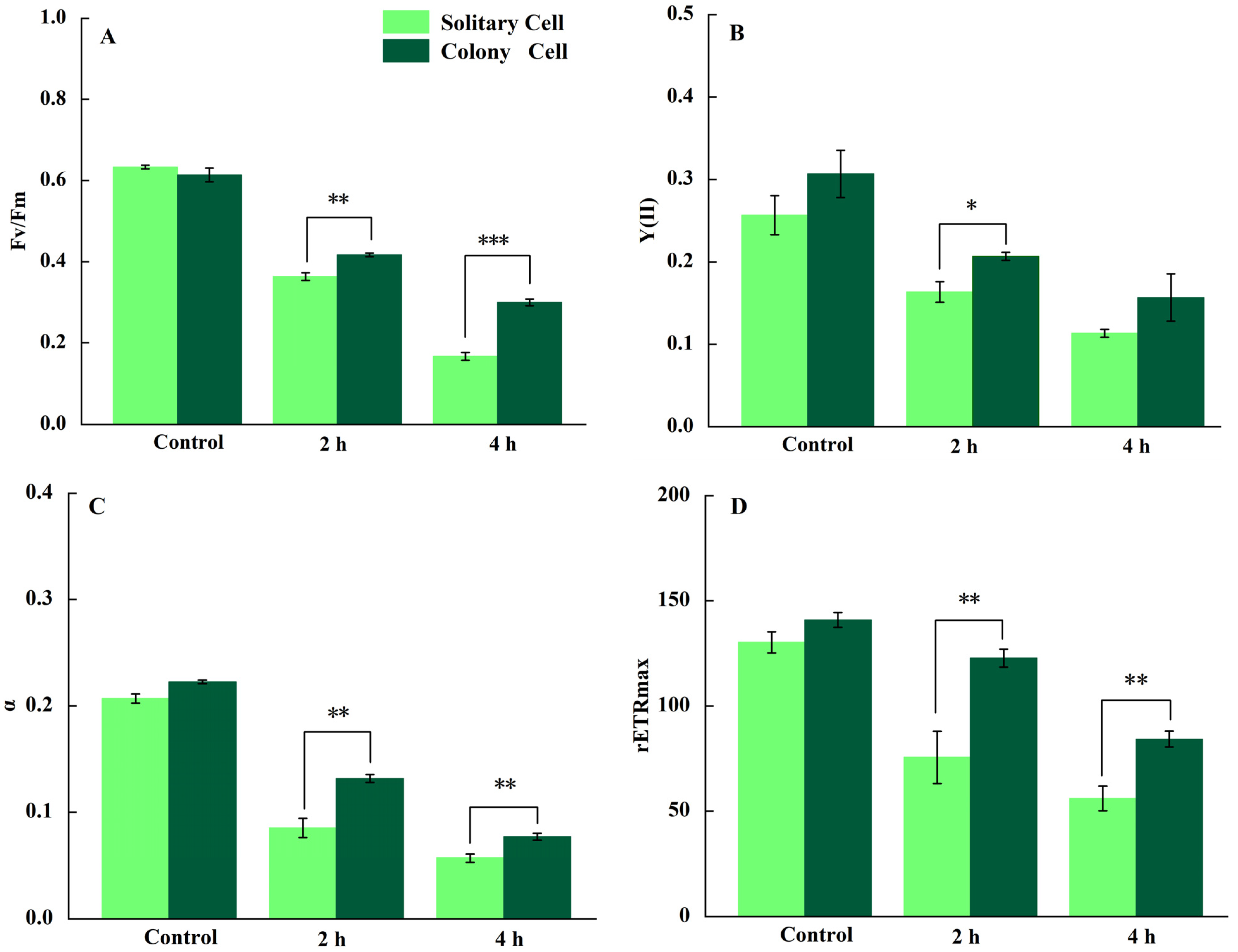
3.1. Photosynthetic Activity

Compared to 0 h, the Fv/Fm in P. globosa solitary and colony cells decreased gradually within 2, 4 h of UV-B radiation (p < 0.01). Initial Fv/Fm values of P. globosa solitary and colony cells were not significantly different (p > 0.05, Figure 1A). Under 2, 4 h of UV-B radiation, the Fv/Fm of solitary cells decreased by 42.6% and 73.7%, respectively; the Fv/Fm of colony cells decreased by 32.0% and 49.5%, respectively. These findings indicated that the Fv/Fm in the solitary cells was inhibited to a greater extent than that in the colony cells. The Y(II) of P. globosa solitary and colony cells significantly decreased with longer exposure time to UV-B (p < 0.05). The initial value of Y(II) did not differ between the solitary and colony cells. After 2 h of UV-B radiation, the Y(II) of solitary cells was significantly lower than that of colony cells (p < 0.05; Figure 1B). Initially, the α and rETRmax of solitary P. globosa cells did not differ from those of colony cells. After 2, 4 h of UV-B radiation, the α, rETRmax values in P. globosa solitary and colony cells decreased significantly (p < 0.01) and continued to decrease with time. During 2 h, 4 h of UV-B radiation, the α and rETRmax values in P. globosa solitary were significantly lower than those of colony cells (p < 0.01; Figure 1C,D).

3.2. Damage Rate and Repair Rate of PSII

Table 1 shows the damage rates and repair rates of P. globosa solitary and colony cells under UV-B radiation. The damage rates of P. globosa solitary and colony cells were 10.87 ± 0.252 min−1 and 7.74 ± 0.157 min−1, respectively, and the damage rate of solitary cells was significantly greater than that of colony cells (p < 0.05). Following 4 h of recovery in poor light, P. globosa solitary cells presented a lower repair rate (5.62 ± 0.42 min−1) than did colony cells (16.10 ± 1.00 min−1). Compared with solitary cells, colony cells had a significantly greater repair rate (p < 0.001).

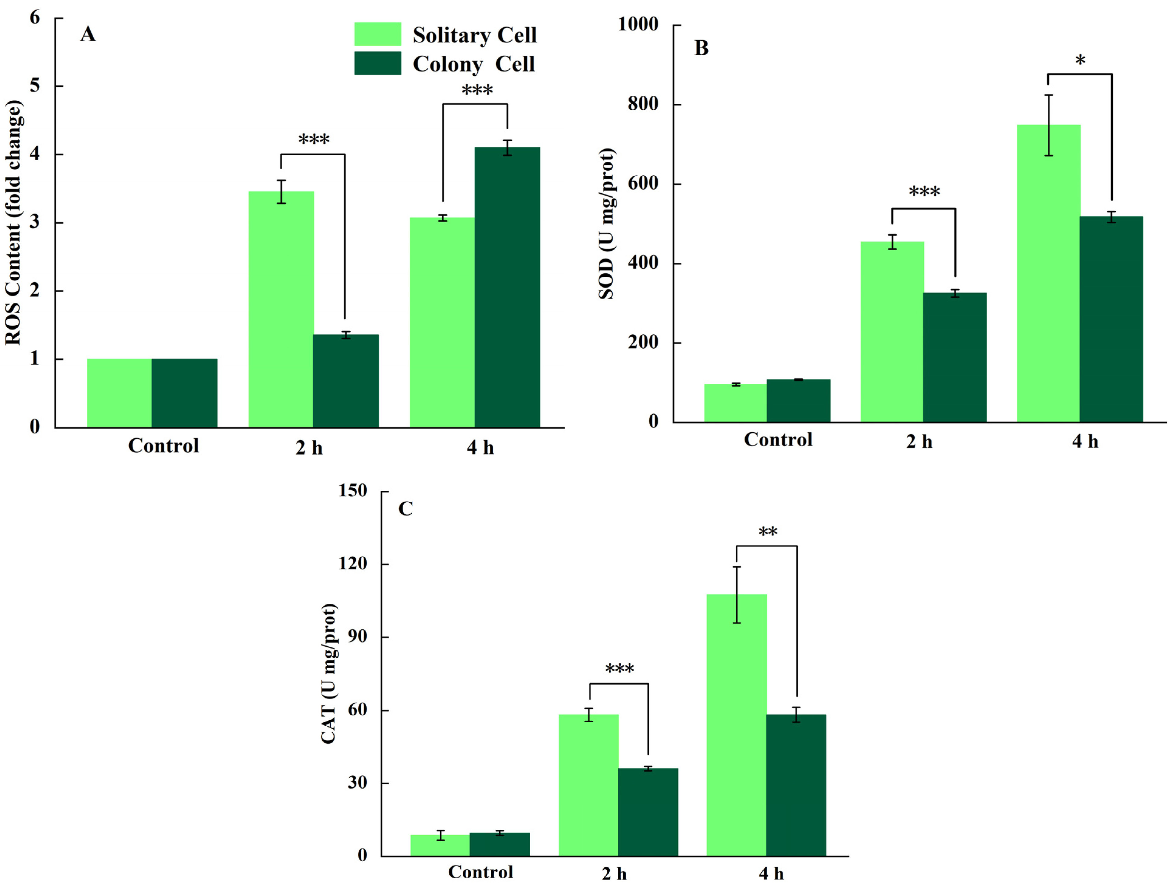
3.3. Determination of ROS and Antioxidant Enzyme Activities

After 2, 4 h of UV-B radiation, the increase in ROS content in P. globosa solitary cells was significant compared with that at 0 h (p < 0.01). In P. globosa solitary cells, the ROS contents after 2, 4 h of UV-B radiation were 3.5 and 3.1 times greater than those at 0 h, respectively. In P. globosa colony cells, the ROS contents after 2, 4 h of UV-B radiation were 1.4 and 4.1 times greater than those in the control group, respectively. The ROS of solitary cells were significantly greater than those of colony cells after being exposed to UV-B radiation for 2 h (p < 0.001), whereas the ROS of colony cells were significantly greater than those of solitary cells after being exposed to UV-B radiation for 4 h (p < 0.001; Figure 2A).
When P. globosa solitary cells were radiated with UV-B for 2 h, the SOD activity rapidly increased by 3.7 times and then increased by 6.9 times at 4 h compared with 0 h. A rapid increase in the SOD activity was also detected in P. globosa colony cells with UV-B radiation, but the level was significantly (p < 0.05) lower than that in solitary cells. Compared with 0 h, the SOD activity of P. globosa colony cells increased 2.0 and 3.8 times with 2, 4 h of UV-B radiation, respectively (Figure 2B). Compared with 0 h, the CAT activity in P. globosa solitary and colony cells significantly increased by 6.1 and 2.8 times, respectively (p < 0.01), after radiation with UV-B for 2 h; it significantly (p < 0.001) increased by 11.9 and 5.1 times, respectively, after radiation with UV-B for 4 h (Figure 2B). The CAT activity of P. globosa solitary cells was significantly higher than that of colony cells under UV-B radiation at 2, 4 h (p < 0.01; Figure 2C).

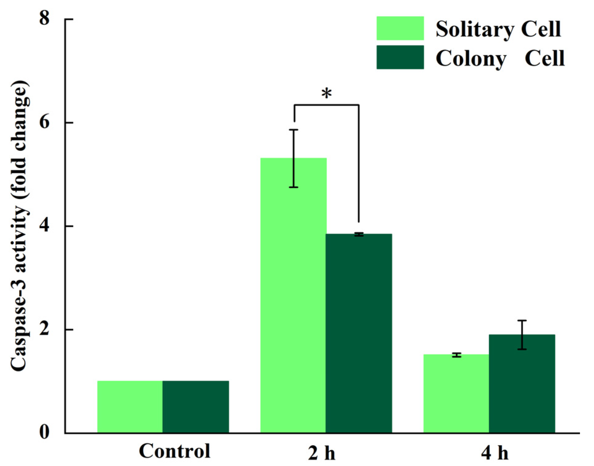
3.4. Measurement of Caspase-3-like Activity

After UV-B radiation for 2 h, the caspase-3-like activities of P. globosa solitary and colony cells significantly increased, with increases of 5.3 and 3.8 times, respectively (p < 0.01). The caspase-3-like activity of P. globosa solitary cells was significantly higher than that of colony cells after being exposed to UV-B radiation for 2 h (p < 0.05). After 4 h of UV-B radiation, caspase-3-like activity in P. globosa solitary and colony cells decreased, but both groups had higher values than at 0 h (Figure 3).

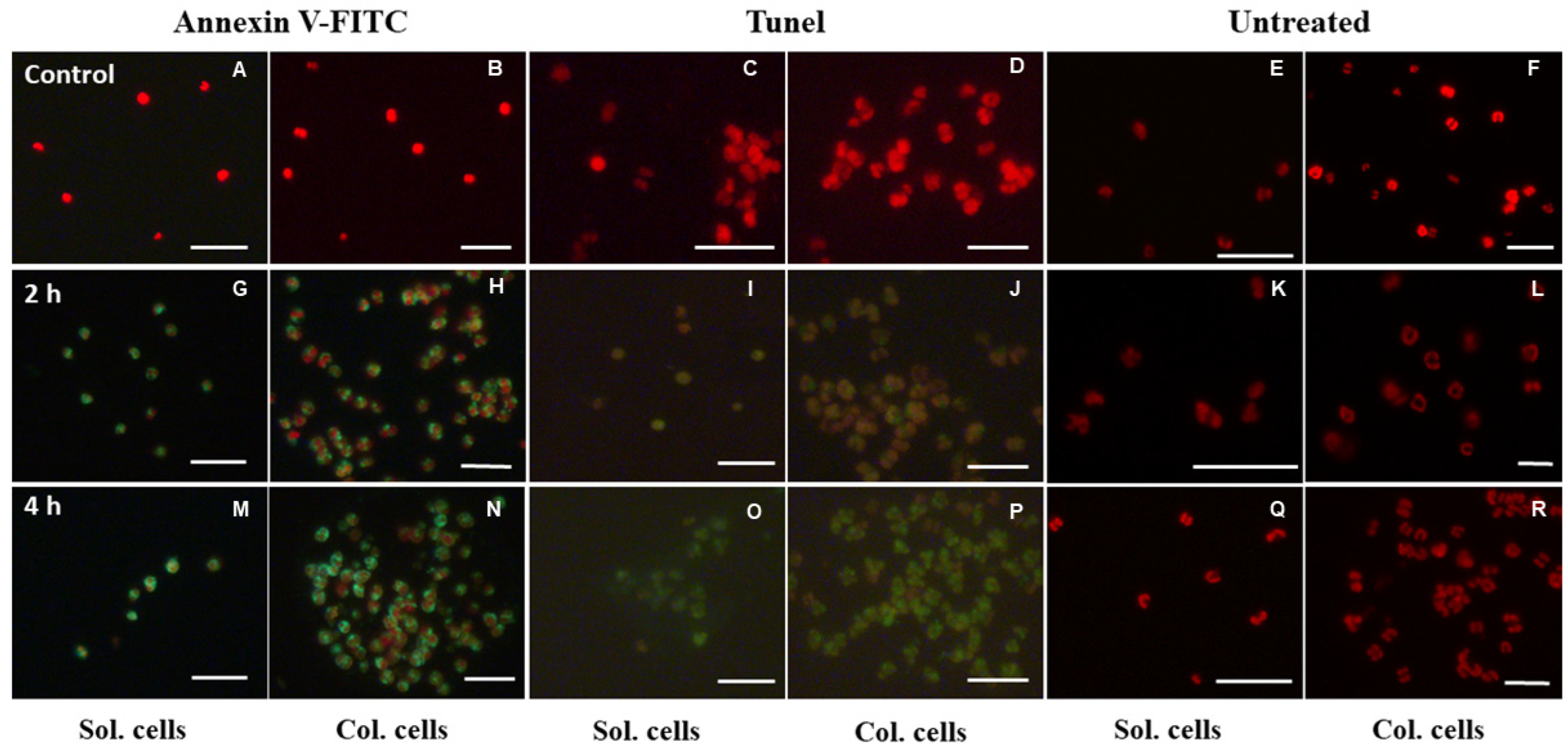
3.5. Determination of PS Externalization and DNA Fragmentation

PS externalization, as revealed by Annexin V-FITC staining, was monitored in P. globosa solitary and colony cells. P. globosa solitary and colony cells radiated by UV-B presented bright green fluorescence, which indicated positive PS externalization at 2, 4 h (Figure 4G,H,M,N). In contrast, the control group exhibited bright red fluorescence (Figure 4A,B). Detection of DNA fragmentation by TUNEL assay. Unlike the red fluorescence found in the control group (Figure 4C,D), the experimental group presented bright green fluorescence, indicating that high levels of DNA fragmentation occurred in P. globosa solitary and colony cells (Figure 4I,J,O,P). In addition, P. globosa solitary and colony cells were observed under a microscope. After 4 h of exposure to UV-B, both solitary and colony cells were complete (Figure 4K,L). However, the solitary cells lysed after UV-B radiation for 4 h (Figure 4Q). This phenomenon was not detected in the colony cells (Figure 4R). These findings suggested that under the same UV-B dose, solitary cells suffered more damage than colony cells.

4. Discussion

4.1. Effect of UV-B on the Photosynthesis of P. globosa

One of the most basic and vital life activities in marine algae is photosynthesis [45,46]. PSII is a key component of photosynthesis and is responsible for light energy absorption and electron transfer [47,48]. UV-B radiation attacks the D1 and D2 proteins, which in turn causes PSII damage and hinders electron transfer [49,50]. Energy utilization in PSII can be quickly and effectively monitored via chlorophyll fluorescence analysis to characterize the photosynthetic activity of cells.
The Fv/Fm, Y(II) of P. globosa solitary and colony cells significantly declined under UV-B exposure (p < 0.05; Figure 1A,B), indicating that UV-B radiation reduced the Fv/Fm, Y(II) of P. globosa solitary and colony cells. Similarly, α and rETRmax decreased, indicating that the light capture capability and the efficiency of relative electron transfer in P. globosa solitary and colony cells were impaired under UV-B radiation (Figure 1C,D). According to earlier research, UV-B radiation disrupts photosynthesis in marine algae [51]. For example, the Fv/Fm and Y(II) of Tussaudia salina decreased dramatically when algae were radiated by UV-B [52]. Similar results were reported in Trichodesmium erythraeum, in which the Y(II) of the cells significantly decreased under brief exposure to UV-B [24]. The decrease in the efficiency of energy absorption, transfer, and conversion in PSII of P. globosa solitary and colony cells under exposure to UV-B resulted in a decrease in the potential and actual photosynthetic capacity, inhibiting photosynthetic activity.
Notably, the decrease in the Fv/Fm of P. globosa solitary cells was significantly greater than that in colony cells (p < 0.01), indicating better UV-B radiation tolerance in the colony cells. In addition, according to the damage and repair rate of PSII in P. globosa solitary and colony cells, colony cells presented greater UV-B radiation tolerance and greater recovery than solitary cells (Table 1). A prior study found a similar result: Microcystis aeruginosa solitary and colony cells radiated with UV-B radiation were allowed to recover under low light for 240 min, and under these conditions, the Fv/Fm of M. aeruginosa colony cells was higher compared to that of solitary cells [53]. UV-B radiation can trigger the synthesis of substances like MAAs and Car (carotenoids) that have the ability to absorb UV-B [50,54]. The polysaccharide outer membrane of the colony likely plays a protective role against UV-B radiation. A study revealed that Phaeocystis spp. colonies contain a variety of MAAs that can absorb UV radiation [55]. In addition, previous studies confirmed that Phaeocystis pouchetii solitary cells are significantly deficient in UV-absorbing substances compared with P. pouchetii colony cells [56]. The greater tolerance of P. globosa colony cells to UV-B exposure may be attributed to the UV-absorbing substances contained in the polysaccharide outer membrane.
In short, P. globosa solitary and colony cells’ photosynthetic activity was not hampered to the same extent by UV-B radiation. Compared with solitary cells, colony cells suffered less damage to photosynthetic activity, which may have been caused by protection from the polysaccharide outer membrane.

4.2. Response of P. globosa Solitary and Colony Cells to UV-B Radiation

Our study clearly shows that PCD occurred in P. globosa solitary and colony cells under UV-B radiation. The typical characteristics of PCD, notably DNA fragmentation, PS externalization and caspase-3-like activity, were observed. PS is on the inner side of the cell membrane under normal conditions, which is altered during the process of PCD, when PS is dislocated to outside of the cell membrane [57,58]. During the early stages of PCD, PS externalization can be detected by staining with Annexin V-FITC. DNA fragmentation is a hallmark of late-stage PCD [59] and occurs in P. globosa solitary and colony cells after 2 or 4 h of UV-B radiation (Figure 4). Another important indicator of PCD is the increase of caspase-3-like activity (Figure 3). Caspase-3-like proteins were activated in P. globosa in reaction to UV-B radiation. This means that an intrinsic pathway was involved in the PCD process.
UV-B exposure can raise cellular levels of ROS, such as hydroxyl radicals and hydrogen peroxide radicals [60]. ROS attack different targets inside cells [61]. Generally, the overproduction of ROS in cells leads to internal damage, such as overoxidation of proteins and lipids, alterations in membrane permeability, ultimately leading to PCD [62]. In fact, ROS can function as signaling molecules during the early stages of PCD, which has been confirmed in other microalgae, such as Thalassiosira pseudonana and Peridinium gatunense [63,64]. In our study, excess ROS in P. globosa cells was induced by UV-B radiation (Figure 2). Excess ROS can trigger caspase-3-like activity to initiate a series of intracellular cascade reactions and subsequent degradation of intracellular proteins [65,66]. These findings suggest that UV-B exposure causes the accumulation of ROS in P. globosa, which subsequently stimulates an increase in capase-3-like activity and ultimately leads to PCD.
Aerobic microorganisms can regulate the level of ROS via CAT and SOD to ensure that the intracellular oxidative system is in balance [61,67]. Thus, the intracellular ROS content is likely to show the same trend as that of CAT and SOD under extrinsic environmental stress conditions. However, activities of CAT and SOD in reaction to UV-B radiation for 2, 4 h did not match the ROS content in the antioxidant system of P. globosa cells. After 4 h of UV-B exposure, the CAT and SOD activities of P. globosa colony cells were significantly lower than solitary cells (p < 0.05, Figure 2). Higher antioxidant enzyme activities are related to higher ROS contents within a normal antioxidant system [68]. As mentioned above, solitary cells suffer more damage from UV-B exposure due to a lack of protection from the polysaccharide outer membrane, which is thought to lead to more ROS accumulation in solitary cells than in colony cells. Our results in this study showed the opposite trend. A reasonable explanation for this finding is that, unlike colony cells, some solitary cells might become necrotic after 4 h of UV-B radiation. Cell lysis due to necrosis allows for the loss of ROS in solitary cells, leading to a lower ROS content in solitary cells than in colony cells (Figure 2A). Moreover, this finding is supported by the results shown in Figure 4. After 4 h of UV-B radiation, lysis was observed via fluorescence microscopy in solitary cells, whereas colony cells were intact. A previous study reported that Dunaliella viridis also experienced necrosis after UV-B exposure [36].
The main difference between colony and solitary cells is that the former are protected by a polysaccharide outer membrane, which attenuates UV-B radiation and ensures cellular integrity. In brief, PCD in colony cells is induced by ROS, with the protection of the polysaccharide outer membrane. In contrast, solitary cells exhibited PCD induced by both ROS and necrosis.

5. Conclusions

In this study, we characterized the photosynthetic capacity and PCD process of P. globosa colony and solitary cells exposed to UV-B radiation, and found that both colony and solitary cells had PCD characteristics. UV-B radiation causes the overproduction of ROS and activation of caspase-3-like activity. We can conservatively suggest that UV-B radiation could assist in accelerating the death of P. globosa. Moreover, the two cell types still showed a difference in responses to UV-B radiation. This study highlights the fact that solitary cells experienced a passive cell death - necrosis compared to colony cells. The colony cells of P. globosa are considered to be an evolutionary way to actively accommodate changes in the natural environment. Photosynthetic activity confirms that colony cells have a better recovery ability and are more adaptable to UV-B radiation than solitary cells. It can be speculated that when P. globosa is floating on the sea surface, the colony cells defend against UV-B radiation due to the protection of the polysaccharide outer membrane. Colony cells have better growth advantages. In contrast, solitary cells are subjected to more stress from UV-B radiation. This study reveals the lethal mechanism of UV-B radiation on P. globosa, which provides a basis for further understanding of the interaction on algae by UV radiation.

Author Contributions

Conceptualization, W.W., J.L. (Jie Li); methodology, W.W., J.L. (Jie Li); C.L.; writing—original draft preparation, W.W., J.L. (Jie Li); writing—review and editing, W.W., J.L. (Jie Li); project administration, J.L. (Junxiang Lai); funding acquisition, J.L. (Junxiang Lai). All authors have read and agreed to the published version of the manuscript.

Funding

This work was supported by the National Natural Science Foundation of China (No: 42266007); National Natural Science Foundation of China (No: U23A2038); Science and Technology Major Project of Guangxi (No: AA17202020); The Guangxi Natural Science Foundation (No. 2023GXNSFBA026210).

Institutional Review Board Statement

Not applicable.

Informed Consent Statement

Not applicable.

Data Availability Statement

The data are available on request.

Conflicts of Interest

The authors declare no conflict of interest.

References

  1. Rousseau, V.; Chrétiennot Dinet, M.J.; Jacobsen, A.; Verity, P.; Whipple, S. The life cycle of Phaeocystis: State of knowledge and presumptive role in ecology. Biogeochemistry 2007, 83, 29–47. [Google Scholar] [CrossRef]
  2. Wang, X.; Wang, Y.; Ou, L. The roles of light–dark cycles in the growth of Phaeocystis globosa from the South China Sea: The cost of colony enlargement. J. Sea Res. 2014, 85, 518–523. [Google Scholar] [CrossRef]
  3. Rousseau, V.; Vaulot, D.; Casotti, R.; Cariou, V.; Lenz, J.; Gunkel, J.; Baumann, M. The life cycle of Phaeocystis (Prymnesiophycaea): Evidence and hypotheses. J. Mar. Syst. 1994, 5, 23–39. [Google Scholar] [CrossRef]
  4. Chen, Y.Q.; Wang, N.; Zhang, P.; Zhou, H.; Qu, L. Molecular evidence identifies bloom-forming Phaeocystis (Prymnesiophyta) from coastal waters of southeast China as Phaeocystis globosa. Biochem. Syst. Ecol. 2002, 30, 15–22. [Google Scholar] [CrossRef]
  5. Schoemann, V.; Becquevort, S.; Stefels, J.; Rousseau, V.; Lancelot, C. Phaeocystis blooms in the global ocean and their controlling mechanisms: A review. J. Sea Res. 2005, 53, 43–66. [Google Scholar] [CrossRef]
  6. Peperzak, L.; van Wezel, R. Human fatalities related to a Phaeocystis harmful algal bloom in the North Sea. Harmful Algae 2023, 130, 102545. [Google Scholar] [CrossRef]
  7. Smith, W.O., Jr.; Trimborn, S. Phaeocystis: A Global Enigma. Ann. Rev. Mar. Sci. 2024, 16, 417–441. [Google Scholar] [CrossRef]
  8. Riegman, R.; Van Boekel, W. The ecophysiology of Phaeocystis globosa: A review. J. Sea Res. 1996, 35, 235–242. [Google Scholar] [CrossRef]
  9. Zhang, S.F.; Zhang, K.; Cheng, H.M.; Lin, L.; Wang, D.Z. Comparative transcriptomics reveals colony formation mechanism of a harmful algal bloom species Phaeocystis globosa. Sci. Total Environ. 2020, 719, 137454. [Google Scholar] [CrossRef]
  10. Wang, X.; Song, H.; Wang, Y.; Chen, N. Research on the biology and ecology of the harmful algal bloom species Phaeocystis globosa in China: Progresses in the last 20 years. Harmful Algae 2021, 107, 102057. [Google Scholar] [CrossRef]
  11. Wu, K.; Tang, S.; Wu, X.; Zhu, J.; Song, J.; Zhong, Y.; Zhou, J.; Cai, Z. Colony formation of Phaeocystis globosa: A case study of evolutionary strategy for competitive adaptation. Mar. Pollut. Bull. 2023, 186, 114453. [Google Scholar] [CrossRef] [PubMed]
  12. Chen, S.; Gao, K. Solar ultraviolet radiation and CO2-induced ocean acidification interacts to influence the photosynthetic performance of the red tide alga Phaeocystis globosa (Prymnesiophyceae). Hydrobiologia 2011, 675, 105–117. [Google Scholar] [CrossRef]
  13. Neale, P.J.; Williamson, C.E.; Banaszak, A.T.; Häder, D.P.; Hylander, S.; Ossola, R.; Rose, K.C.; Wängberg, S.Å.; Zepp, R. The response of aquatic ecosystems to the interactive effects of stratospheric ozone depletion, UV radiation, and climate change. Photochem. Photobiol. Sci. 2023, 22, 1093–1127. [Google Scholar] [CrossRef] [PubMed]
  14. Häder, D.-P.; Williamson, C.E.; Wängberg, S.-Å.; Rautio, M.; Rose, K.C.; Gao, K.; Helbling, E.W.; Sinha, R.P.; Worrest, R. Effects of UV radiation on aquatic ecosystems and interactions with other environmental factors. Photochem. Photobiol. Sci. 2015, 14, 108–126. [Google Scholar] [CrossRef]
  15. Holzinger, A.; Lütz, C. Algae and UV irradiation: Effects on ultrastructure and related metabolic functions. Micron 2006, 37, 190–207. [Google Scholar] [CrossRef] [PubMed]
  16. Scholz, B.; Rúa, A.; Liebezeit, G. Effects of UV radiation on five marine microphytobenthic Wadden sea diatoms, isolated from the Solthrn tidal flat (Lower Saxony, southern North Sea)—Part II: Changes in carbohydrate, amino acid and fatty acid composition. Eur. J. Phycol. 2014, 49, 97–114. [Google Scholar] [CrossRef]
  17. Pessoa, M. Algae and aquatic macrophytes responses to cope to ultraviolet radiation—A Review. Emir. J. Food Agric. 2012, 24, 527–545. [Google Scholar] [CrossRef]
  18. Montero, O.; Klisch, M.; Häder, D.P.; Lubian, L.M. Comparative Sensitivity of Seven Marine Microalgae to Cumulative Exposure to Ultraviolet-B Radiation with Daily Increasing Doses. Bot. Mar. 2002, 45, 305–315. [Google Scholar] [CrossRef]
  19. Saber, H.; El Sheekh, M.M.; Ibrahim, A.; Alwaleed, E.A. Effect of UV-B radiation on amino acids profile, antioxidant enzymes and lipid peroxidation of some cyanobacteria and green algae. Int. J. Radiat. Biol. 2020, 96, 1192–1206. [Google Scholar] [CrossRef]
  20. Xue, S.; Zang, Y.; Chen, J.; Shang, S.; Gao, L.; Tang, X. Ultraviolet-B radiation stress triggers reactive oxygen species and regulates the antioxidant defense and photosynthesis systems of intertidal red algae Neoporphyra haitanensis. Front. Mar. Sci. 2022, 9, 1043462. [Google Scholar] [CrossRef]
  21. Lesser, M. Acclimation of phytoplankton to UV-B radiation: Oxidative stress and photoinhibition of photosynthesis are not prevented by UV-absorbing compounds in the dinoflagellate Prorocentrum micans. Mar. Ecol.-Prog. Ser. 1996, 132, 287–297. [Google Scholar] [CrossRef]
  22. Fryer, M.J.; Oxborough, K.; Mullineaux, P.M.; Baker, N.R. Imaging of photo-oxidative stress responses in leaves. J. Exp. Bot. 2002, 53, 1249–1254. [Google Scholar]
  23. Zhang, P.Y.; Yu, J.; Tang, X.X. UV-B Radiation Suppresses the Growth and Antioxidant Systems of Two Marine Microalgae, Platymonas subcordiformis (Wille) Hazen and Nitzschia closterium (Ehrenb.) W. Sm. J. Integr. Plant Biol. 2005, 47, 683–691. [Google Scholar] [CrossRef]
  24. Cai, X.; Hutchins, D.A.; Fu, F.; Gao, K. Effects of ultraviolet radiation on photosynthetic performance and N2 fixation in IMS 101. Biogeosciences 2017, 14, 4455–4466. [Google Scholar] [CrossRef]
  25. Gao, K.; Beardall, J.; Häder, D.P.; Hall Spencer, J.M.; Gao, G.; Hutchins, D.A. Effects of Ocean Acidification on Marine Photosynthetic Organisms Under the Concurrent Influences of Warming, UV Radiation, and Deoxygenation. Front. Mar. Sci. 2019, 6, 322. [Google Scholar] [CrossRef]
  26. Edreva, A.M.; Pouneva, I.D.; Gesheva, E.Z. UV-B radiation induces biphasic burst of hydrogen peroxide in mesophyll Chlorella vulgaris. Russ. J. Plant Physiol. 2015, 62, 219–223. [Google Scholar] [CrossRef]
  27. Tanouchi, Y.; Lee, A.J.; Meredith, H.; You, L. Programmed cell death in bacteria and implications for antibiotic therapy. Trends Microbiol. 2013, 21, 265–270. [Google Scholar] [CrossRef]
  28. Bidle, K.D. Programmed Cell Death in Unicellular Phytoplankton. Curr. Biol. 2016, 26, R594–R607. [Google Scholar] [CrossRef]
  29. Valandro, F.; Menguer, P.K.; Cabreira-Cagliari, C.; Margis-Pinheiro, M.; Cagliari, A. Programmed cell death (PCD) control in plants: New insights from the Arabidopsis thaliana deathosome. Plant Sci. 2020, 299, 110603. [Google Scholar] [CrossRef]
  30. Franklin, D.J.; Brussaard, C.P.D.; Berges, J.A. What is the role and nature of programmed cell death in phytoplankton ecology? Eur. J. Phycol. 2006, 41, 1–14. [Google Scholar] [CrossRef]
  31. Rzymski, P.; Klimaszyk, P.; Jurczak, T.; Poniedziałek, B. Oxidative Stress, Programmed Cell Death and Microcystin Release in Microcystis aeruginosa in Response to Daphnia Grazers. Front. Microbiol. 2020, 11, 1201. [Google Scholar] [CrossRef] [PubMed]
  32. Orellana, M.V.; Pang, W.L.; Durand, P.M.; Whitehead, K.; Baliga, N.S. A Role for Programmed Cell Death in the Microbial Loop. PLoS ONE 2013, 8, e62595. [Google Scholar] [CrossRef] [PubMed]
  33. Rotari, V.; Gordon, A.; He, R.; Gallois, P. Caspase-like activities and UV-induced programmed cell death in Arabidopsis. BMC Plant Biol. 2005, 5, S18. [Google Scholar] [CrossRef]
  34. Gao, C.; Zhang, L.; Wen, F.; Xing, D. Sorting out the role of reactive oxygen species during plant programmed cell death induced by ultraviolet-C overexposure. Plant Signal. Behav. 2008, 3, 197–198. [Google Scholar] [CrossRef]
  35. Li, S.; Tao, Y.; Dao, G.H.; Hu, H.Y. Synergetic suppression effects upon the combination of UV-C irradiation and berberine on Microcystis aeruginosa and Scenedesmus obliquus in reclaimed water: Effectiveness and mechanisms. Sci. Total Environ. 2020, 744, 140937. [Google Scholar] [CrossRef] [PubMed]
  36. Jiménez, C.; Capasso, J.M.; Edelstein, C.L.; Rivard, C.J.; Lucia, S.; Breusegem, S.; Berl, T.; Segovia, M. Different ways to die: Cell death modes of the unicellular chlorophyte Dunaliella viridis exposed to various environmental stresses are mediated by the caspase-like activity DEVDase. J. Exp. Bot. 2009, 60, 815–828. [Google Scholar] [CrossRef]
  37. Zheng, N.; Lin, X.; Huang, P.; Liu, Y.; Bartlam, M.; Wang, Y. Tea polyphenols inhibit blooms caused by eukaryotic and prokaryotic algae. Ecotoxicol. Environ. Saf. 2023, 265, 115531. [Google Scholar] [CrossRef]
  38. El-Sheekh, M.M.; Alwaleed, E.A.; Ibrahim, A.; Saber, H. Detrimental effect of UV-B radiation on growth, photosynthetic pigments, metabolites and ultrastructure of some cyanobacteria and freshwater chlorophyta. Int. J. Radiat. Biol. 2021, 97, 265–275. [Google Scholar] [CrossRef]
  39. Moharikar, S.; D’Souza, J.; Kulkarni, A.; Rao, B. Apoptotic-like cell death pathway is induced in unicellular chlorophyte Chlamydomonas reinhardtii (Chlorophyceae) cells following UV irradiation: Detection and functional analyses. J. Phycol. 2006, 42, 423–433. [Google Scholar] [CrossRef]
  40. Dunn, S.R.; Bythell, J.C.; Le Tissier, M.D.A.; Burnett, W.J.; Thomason, J.C. Programmed cell death and cell necrosis activity during hyperthermic stress-induced bleaching of the symbiotic sea anemone Aiptasia sp. J. Exp. Mar. Biol. Ecol. 2002, 272, 29–53. [Google Scholar] [CrossRef]
  41. Zhou, T.; Cao, H.; Zheng, J.; Teng, F.; Wang, X.; Lou, K.; Zhang, X.; Tao, Y. Suppression of water-bloom cyanobacterium Microcystis aeruginosa by algaecide hydrogen peroxide maximized through programmed cell death. J. Hazard. Mater. 2020, 393, 122394. [Google Scholar] [CrossRef] [PubMed]
  42. Guillard, R.R.L. Culture of Phytoplankton for Feeding Marine Invertebrates. In Culture of Marine Invertebrate Animals: Proceedings—1st Conference on Culture of Marine Invertebrate Animals Greenport; Smith, W.L., Chanley, M.H., Eds.; Springer: Boston, MA, USA, 1975; pp. 29–60. [Google Scholar]
  43. Zhuang, J.; Lu, J.; Cao, K.-F.; Li, J. Haploid Helps Phaeocystis globosa distribute to deeper dim water, as evidenced by growth and photosynthetic physiology. Front. Mar. Sci. 2022, 9, 902330. [Google Scholar] [CrossRef]
  44. Heraud, P.; Beardall, J. Changes in chlorophyll fluorescence during exposure of Dunaliella tertiolecta to UV radiation indicate a dynamic interaction between damage and repair processes. Photosynth. Res. 2000, 63, 123–134. [Google Scholar] [CrossRef] [PubMed]
  45. Moejes, F.W.; Matuszyńska, A.; Adhikari, K.; Bassi, R.; Cariti, F.; Cogne, G.; Dikaios, I.; Falciatore, A.; Finazzi, G.; Flori, S.; et al. A systems-wide understanding of photosynthetic acclimation in algae and higher plants. J. Exp. Bot. 2017, 68, 2667–2681. [Google Scholar] [CrossRef]
  46. Singh, S.P.; Singh, P. Effect of temperature and light on the growth of algae species: A review. Renew. Sustain. Energy Rev. 2015, 50, 431–444. [Google Scholar] [CrossRef]
  47. Melis, A. Photosystem-II damage and repair cycle in chloroplasts: What modulates the rate of photodamage in vivo? Trends Plant Sci. 1999, 4, 130–135. [Google Scholar] [CrossRef]
  48. Wang, B.; Jia, J. Photoprotection mechanisms of Nannochloropsis oceanica in response to light stress. Algal Res. 2020, 46, 101784. [Google Scholar] [CrossRef]
  49. Campbell, D.; Eriksson, M.J.; Öquist, G.; Gustafsson, P.; Clarke, A.K. The cyanobacterium Synechococcus resists UV-B by exchanging photosystem II reaction-center D1 proteins. Proc. Natl. Acad. Sci. 1998, 95, 364–369. [Google Scholar] [CrossRef] [PubMed]
  50. Rastogi, R.P.; Madamwar, D.; Nakamoto, H.; Incharoensakdi, A. Resilience and self-regulation processes of microalgae under UV radiation stress. J. Photochem. Photobiol. C 2020, 43, 100322. [Google Scholar] [CrossRef]
  51. Abo-Shady, A.M.; El-Sheekh, M.M.; El-Naggar, A.H.; Abomohra, A.E.-F. Effect of UV-B radiation on growth, photosynthetic activity and metabolic activities of Chlorococcum sp. Ann. Microbiol. 2008, 58, 21–27. [Google Scholar] [CrossRef]
  52. Zhang, X.; Tang, X.; Wang, M.; Zhang, W.; Zhou, B.; Wang, Y. ROS and calcium signaling mediated pathways involved in stress responses of the marine microalgae Dunaliella salina to enhanced UV-B radiation. J. Photochem. Photobiol. B Biol. 2017, 173, 360–367. [Google Scholar] [CrossRef] [PubMed]
  53. Qin, H.; Li, S.; Li, D. Differential responses of different phenotypes of Microcystis (Cyanophyceae) to UV-B radiation. Phycologia 2015, 54, 118–129. [Google Scholar] [CrossRef]
  54. Taira, H.; Taguchi, S. Cellular Mycosporine-like amino acids protect photosystem II of the Dinoflagellate Scrippsiella sweeneyae from ultraviolet radiation damage. J. Photochem. Photobiol. B Biol. 2017, 174, 27–34. [Google Scholar] [CrossRef] [PubMed]
  55. Ha, S.Y.; Lee, D.B.; Kang, S.H.; Shin, K.H. Strategy of photo-protection in phytoplankton assemblages in the Kongsfjorden, Svalbard, Arctic. Chin. J. Oceanol. Limnol. 2016, 34, 1–12. [Google Scholar] [CrossRef]
  56. Marchant, H.J.; Davidson, A.T.; Kelly, G.J. UV-B protecting compounds in the marine alga Phaeocystis pouchetii from Antarctica. Mar. Biol. 1991, 109, 391–395. [Google Scholar] [CrossRef]
  57. Sperandio, S.; de Belle, I.; Bredesen, D.E. An alternative, nonapoptotic form of programmed cell death. Proc. Natl. Acad. Sci. USA 2000, 97, 14376–14381. [Google Scholar] [CrossRef]
  58. Xu, C.; Yu, S.; Hu, J.; Effiong, K.; Ge, Z.; Tang, T.; Xiao, X. Programmed cell death process in freshwater Microcystis aeruginosa and marine Phaeocystis globosa induced by a plant derived allelochemical. Sci. Total Environ. 2022, 838, 156055. [Google Scholar] [CrossRef]
  59. Zuppini, A.; Andreoli, C.; Baldan, B. Heat stress: An inducer of programmed cell death in Chlorella saccharophila. Plant Cell Physiol. 2007, 48, 1000–1009. [Google Scholar] [CrossRef]
  60. Malanga, G.; Susana, P. Oxidative stress and antioxidant content in Chlorella vulgaris after exposure to ultraviolet—B radiation. Physiol. Plant. 1995, 94, 672–679. [Google Scholar] [CrossRef]
  61. Sun, X.M.; Geng, L.J.; Ren, L.J.; Ji, X.J.; Hao, N.; Chen, K.Q.; Huang, H. Influence of oxygen on the biosynthesis of polyunsaturated fatty acids in microalgae. Bioresour. Technol. 2018, 250, 868–876. [Google Scholar] [CrossRef]
  62. Gechev, T.S.; Hille, J. Hydrogen peroxide as a signal controlling plant programmed cell death. J. Cell Biol. 2005, 168, 17–20. [Google Scholar] [CrossRef] [PubMed]
  63. Luo, C.S.; Liang, J.R.; Lin, Q.; Li, C.; Bowler, C.; Anderson, D.M.; Wang, P.; Wang, X.W.; Gao, Y.H. Cellular Responses Associated with ROS Production and Cell Fate Decision in Early Stress Response to Iron Limitation in the Diatom Thalassiosira pseudonana. J. Proteome Res. 2014, 13, 5510–5523. [Google Scholar] [CrossRef] [PubMed]
  64. Vardi, A.; Berman Frank, I.; Rozenberg, T.; Hadas, O.; Kaplan, A.; Levine, A. Programmed cell death of the dinoflagellate Peridinium gatunense is mediated by CO2 limitation and oxidative stress. Curr. Biol. 1999, 9, 1061–1064. [Google Scholar] [CrossRef] [PubMed]
  65. Spungin, D.; Bidle, K.D.; Berman Frank, I. Metacaspase involvement in programmed cell death of the marine cyanobacterium Trichodesmium. Environ. Microbiol. 2019, 21, 667–681. [Google Scholar] [CrossRef]
  66. Ye, C.; Zheng, S.; Jiang, D.; Lu, J.; Huang, Z.; Liu, Z.; Zhou, H.; Zhuang, C.; Li, J. Initiation and Execution of Programmed Cell Death and Regulation of Reactive Oxygen Species in Plants. Int. J. Mol. Sci. 2021, 22, 12942. [Google Scholar] [CrossRef]
  67. Coulombier, N.; Jauffrais, T.; Lebouvier, N. Antioxidant Compounds from Microalgae: A Review. Mar. Drugs 2021, 19, 549. [Google Scholar] [CrossRef]
  68. Lee, T.M.; Shiu, C.T. Implications of mycosporine-like amino acid and antioxidant defenses in UV-B radiation tolerance for the algae species Ptercladiella capillacea and Gelidium amansii. Mar. Environ. Res. 2009, 67, 8–16. [Google Scholar] [CrossRef]
Figure 1. Effect of UV-B on the photosynthetic activity of solitary and colony cells of P. globosa. Note: (A) maximum photosynthetic reaction rate; (B) actual photosynthetic reaction rate; (C) utilization of light energy by photosynthetic organs; (D) relative maximum electron transfer rate. * indicates statistically significant differences in the data. * p < 0.05; ** p < 0.01; *** p < 0.001.
Jmse 12 01619 g001
Figure 2. Effect of UV-B on the ROS content and antioxidant enzyme activity of solitary and colony cells of P. globosa. Note: (A) ROS content; (B) SOD activity; (C) CAT activity. * indicates statistically significant differences in the data. * p < 0.05; ** p < 0.01; *** p < 0.001.
Jmse 12 01619 g002
Figure 3. Effect of UV-B on the caspase-3-like activity of P. globosa’s solitary and colony cells. Note: * indicates statistically significant differences in the data. * p < 0.05.
Jmse 12 01619 g003
Figure 4. Effect of UV-B on the TUNEL assay of P. globosa’s solitary and colony cells. Note: control group (AF); UV-B radiation at 2 h (GL); UV-B radiation at 4 h (MR). Red fluorescence indicates chlorophyll and green fluorescence indicates positive cells. The scale in the picture is 10 µm.
Jmse 12 01619 g004
Table 1. The damage rate (k) repair rate (r) in PSⅡ for solitary and colony cells of P. globosa.
Cell Morphologyr (×10−3 min−1)k (×10−3 min−1)
Solitary cell5.62 ± 0.4210.87 ± 0.252
Colony cell16.10 ± 1.00 ***7.74 ± 0.157 *
Note: * indicates statistically significant differences in the data. * p < 0.05; *** p < 0.001.
Disclaimer/Publisher’s Note: The statements, opinions and data contained in all publications are solely those of the individual author(s) and contributor(s) and not of MDPI and/or the editor(s). MDPI and/or the editor(s) disclaim responsibility for any injury to people or property resulting from any ideas, methods, instructions or products referred to in the content.

