Review

PEEK Biomaterial in Long-Term Provisional Implant Restorations: A Review

by
Suphachai Suphangul
1,
Dinesh Rokaya
2,
Chatruethai Kanchanasobhana
1,
Pimduen Rungsiyakull
1 and
Pisaisit Chaijareenont
1,*
1
Department of Prosthodontics, Faculty of Dentistry, Chiang Mai University, Chiang Mai 50200, Thailand
2
Department of Clinical Dentistry, Walailak University International College of Dentistry, Walailak University, Bangkok 10400, Thailand
*
Author to whom correspondence should be addressed.
J. Funct. Biomater. 2022, 13(2), 33; https://doi.org/10.3390/jfb13020033
Submission received: 27 February 2022 / Revised: 17 March 2022 / Accepted: 21 March 2022 / Published: 22 March 2022
(This article belongs to the Special Issue Recent Advances in Dental Implants and Biomaterials)

Abstract

:
Polyetheretherketone (PEEK) has become a useful polymeric biomaterial due to its superior properties and has been increasingly used in dentistry, especially in prosthetic dentistry and dental implantology. Promising applications of PEEK in dentistry are dental implants, temporary abutment, implant-supported provisional crowns, fixed prosthesis, removable denture framework, and finger prosthesis. PEEK as a long-term provisional implant restoration has not been studied much. Hence, this review article aims to review PEEK as a long-term provisional implant restoration for applications focusing on implant dentistry. Articles published in English on PEEK biomaterial for long-term provisional implant restoration were searched in Google Scholar, ScienceDirect, PubMed/MEDLINE, and Scopus. Then, relevant articles were selected and included in this literature review. PEEK presents suitable properties for various implant components in implant dentistry, including temporary and long-term provisional restorations. The modifications of PEEK result in wider applications in clinical dentistry. The PEEK reinforced by 30–50% carbon fibers can be a suitable material for the various implant components in dentistry.

Graphical Abstract

1. Introduction

Currently, digital technologies have been integrated into implant dentistry for prosthetic restoration rehabilitation of missing teeth [1,2,3,4]. For implant components, titanium (Ti) and its alloys are commonly used but they have disadvantages of causing black coloration in the anterior zone and causing allergy [5,6]. Recently there have been tried thermoplastic materials in implant dentistry. Polymers are one of the most important materials in dentistry. They have present good physical and mechanical properties and biocompatibility. Denture base, removable appliances, and restorations are fabricated mainly from polymers [7]. In addition to these, various polymeric thin films are also available for various dental applications [8].
The polyaryletherketone (PAEK) family are superior performance polymers among all thermoplastic composites, as shown in Figure 1 [9,10]. Polyetheretherketone (PEEK) and polyetherketoneketone (PEKK) are the two most common sub-members of the PAEK family launched in the engineering field in the 1980s [11]. PAEK family has a linear aromatic polyether ketone. PEEK presents ultrahigh performance with superior properties and chemical resistance [12,13]. These remarkable outcomes of PEEK caused it to receive much attention in the field of medicine and dentistry [14,15,16,17,18,19].
Recently, although PEEK has been increasingly used in implant dentistry, PEEK as a long-term provisional implant restoration has not been studied extensively. Hence, this review article aims to review PEEK as a long-term provisional implant restoration for applications in dentistry. Articles published in English on PEEK biomaterial for long-term provisional implant restoration were searched in Google Scholar, ScienceDirect, PubMed/MEDLINE, and Scopus. Then, relevant articles were selected and included in this literature review.

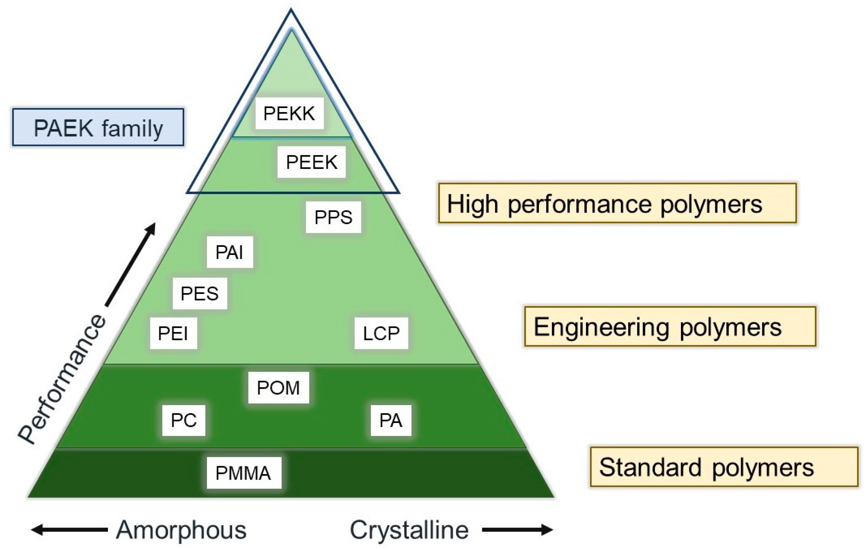
2. PEEK: Structure, Properties, and Prosthetic Applications

PEEK was developed in the late 1990s as a biomaterial that has superior physical, mechanical, and biological properties for biomedical applications [18]. It is a thermoplastic, monochromatic, semicrystalline polymer and is considered the most important member of the PEAK family [20]. The structure of PEEK comprises repeated aromatic rings of the ether and ketone groups (Figure 2) [21].
The high-performance PEEK polymer is being used in the field of orthopedics, spinal, and dental prostheses [18,22]. Recently, PEEK has been increasingly used in implant dentistry. Promising applications of PEEK in dentistry include dental implants [12,23], temporary abutments [22], implant-supported provisional crowns [24,25], fixed prostheses [21], removable denture frameworks [17], and finger prostheses [26]. Little information is available in the dental literature on PEEK as a long-term provisional implant restoration [14,15]. Hence, this review article aims to review PEEK as a long-term provisional implant restoration for applications in dentistry.
A three-dimensional (3D) finite element analysis of implants, abutments, screws, crowns, and peri-implant bones showed that using PEEK materials such as PEEK abutments and crowns reduces the stresses on abutment [27]. In addition, a PEEK framework veneered with composite resin has an elastic modulus of around 4 GPa, which can dampen the occlusal forces on tooth structures, thus protecting structures better than ceramic materials and preventing debonding rates [16,25,28]. Hence, they can be used as alternative materials in endocrown for severely damaged molar and implant restorations.
PEEK has some advantages in dentistry compared with metallic restorations such as Ti. Ti is an allergen, whereas PEEK causes fewer hypersensitive and allergic reactions [5,6]. In addition, PEEK has a more aesthetic appearance than Ti, and PEEK has less/no effect on magnetic resonance imaging artifacts, as it is radiolucent [29,30]. Hence, PEEK abutments can be used as alternatives to Ti abutments.
Recently, studies have focused on improving the surface of PEEK, the bioactivity of PEEK implants at the nanoscale, and reducing the biofilm formation in PEEK materials in implant dentistry [12,23,31,32]. Surface modification of PEEK enhances the cell adhesion, proliferation, biocompatibility, and osteogenic properties of PEEK implant materials. PEEK also influences the biofilm structure and reduces the chances of peri-implant inflammations [31].
A scan body is being manufactured by various companies and varies according to the material, size, shape, connection type, reusability, scanner compatibility, computer design software, and cost [33,34]. Various factors of the scan body (material, design, light reflectance, manufacturing tolerance, and screwing) affect digital transfer in implants [35,36,37,38,39,40]. PEEK scan bodies have been used in prosthetic dentistry using digital technologies. Digital technologies have increased clinical success [41,42], and digital impressions are more accurate and time-efficient than conventional impressions [33,42,43,44]. Still, there can be PEEK displacements in PEEK scan bodies, especially with hand tightening [35]. Figure 3 shows the vertical displacements of PEEK scan bodies (>100 µm) (Myfit and Dentium). Hence, it is suggested to be tightening torque (5 Ncm) but not hand tightening [35].

3. Provisional Restorations

PEEK is a suitable material for provisional restorations, including fixed dental prostheses during dental implant treatment [14,16,25]. Hexagonal-shaped healing abutments can help to provide adequate retention and resistance for implant prostheses [14]. PEEK biomaterials are becoming popular as healing abutments and in PEEK with titanium base temporary abutments. Figure 4 shows the various PEEK abutments. PEEK healing abutment (Figure 4B) is used as an alternative for classic titanium healing abutment, and PEEK with titanium base temporary abutment (Figure 4C) is used for long-term interim restoration, especially in areas with esthetic consideration.

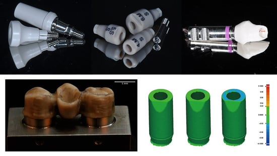
4. A Case of PEEK Temporary Abutment Fabrication

The use of a PEKK temporary abutment (RC Temporary Abutment, Straumann, Basel, Switzerland) as a provisional restoration is shown in Figure 5. Following implant placement, a PEKK temporary abutment try-in is performed, and the temporary abutment is adjusted until desired shape and size were achieved. Then, a temporary crown (Protemp 4, 3M ESPE, Seefeld, Germany) is attached to the customized PEEK abutment, after which it can be inserted into the patient. The patient can use it for a few weeks to months. Then, after some months, a definitive implant prosthesis can be fabricated and placed in the patient.
In a study by Santing et al. [24], the authors measured the failure types and fracture strength of implant-supported PEEK abutments with crowns made from composite crowns and solid Ti temporary abutments and found no difference between different abutment types. In addition, the fracture strength of temporary crowns on PEEK abutments was similar to the Ti temporary abutments except for the central incisors. In addition, Stawarczyk et al. [45] studied the effect of pretreatments of PEEK and adhesive systems on the fracture load of PEEK frameworks for fixed dental prostheses (FDPs). They found that all studied FDPs showed some cracks in the veneering composite crowns in the pontic region irrespective of the pretreatment or adhesive used. In addition, the PEEK frameworks did not fracture after loading, but chipping of the composite was found (Figure 6).
In a study by Rodríguez et al. [46], the authors studied the fracture pattern and fracture load of frameworks for FPDs made with three different subtractive computers aided design and computer-aided manufacturing (CAD/CAM) materials (milled metal made from cobalt–chromium, PEEK, and Lava zirconia), and they found that PEEK had a higher fracture load than zirconia but a lower one than milled metal, as shown in Figure 7. All groups showed clinically acceptable fracture load (>1000 N). Hence, PEEK might be considered a favorable alternative for FPDs.

5. PEEK Modifications

In screw-retained implant prostheses, a PEEK abutment screw has an advantage over a metal screw due to similar elastic properties [47]. In addition, the stress distribution in implant and abutment can be improved by composing the PEEK and its fiber-reinforced composites [47,48,49,50,51]. Figure 8 shows the surfaces of a 3D-printed PEEK and PEEK matrix composite [52]. The unfilled PEEK has low elastic modulus and strength. It has been found that addition of 30–50% carbon fibers does not affect the stress compared with unfilled PEEK [48], but >50% carbon fibers show higher elastic modulus and strength than those of the PEEK composites with lower percentage of carbon fibers [47]. Increasing short carbon fibers (CFs) up to 60% results in higher stress distribution through abutment and implants toward the bone [48]. Hence, the strength and stiffness of PEEK composites can be enhanced by adding more carbon fiber networks. Furthermore, PEEK veneered onto Ti structures can also enhance the stress distribution, especially at the interface between the implant and bone [48].
Similarly, Neumann et al. [49] compared the fracture resistance of Ti, PEEK, and PEEK reinforced with 30% carbon fiber abutment screws, and they found that the Ti screws had higher fracture resistance than screws with PEEK and PEEK reinforced with 30% carbon fiber but no difference between screws with PEEK and PEEK reinforced with 30% carbon fiber. In addition, Schwitalla et al. [47] conducted a study on PEEK screw-in screw-retained implant prostheses, in which they used four PEEK-modified abutments: two screw types with 15% short carbon fibers and 40% short carbon, and another two screw types with 20% TiO2 powder and >50% continuous parallel carbon fibers. Controls included Ti6Al4V abutments screws. They found that the maximum tensile strength was 76.08 MPa for 20% TiO2 powder, 152.67 MPa for 15% short carbon fibers, 157.29 MPa for 40% short carbon fibers, and 191.69 MPa for 15% continuous carbon fibers. The maximum tensile strength of the Ti screws amounted to 1196.29 MPa. The PEEK screw could be removed more easily in case of abutment screw fracture. Hence, the PEEK reinforced >50% continuous carbon fibers could be a suitable material to use in implant dentistry.
In another study, Schwitalla et al. [50] studied the insertion torque at the failure of PEEK using implants made from unfilled PEEK, PEEK-reinforced carbon fiber (>50 vol% continuous axially parallel fibers), and Ti. The insertion torque at the failure of the unfilled PEEK implants was 22.6 Ncm, which was higher than that of the PEEK-reinforced carbon fiber (20.2 Ncm) but significantly less than that of the Ti specimens (92.6 ± 2.3 Ncm). Hence, the PEEK and PEEK-reinforced carbon fiber implants are not able to withstand the insertion force necessary to obtain primary stability for immediate loading.
Although the PEEK has good aesthetics and mechanical properties similar to the human bone, PEEK materials are at risk of fracture and abrasion [52]. Further studies are needed on PEEK implants so that they can replace titanium in the future [31,53,54]. In addition, it is necessary to improve the fracture properties of PEEK implants. Considering this, the development of a PEEK composite by adding continuous multidirectional carbon fibers may help to improve the properties, but more studies are needed [51].

6. Conclusions

PEEK presents suitable properties for various implant components in implant dentistry, including temporary and long-term provisional restorations. Although PEEK has good aesthetics and mechanical properties, similar to the human bone, PEEK materials are at risk of fracture and abrasion. The modification of PEEK is necessary for its wider applications in dentistry. PEEK reinforced by 30–50% carbon fibers would be the material of choice for various dental implant components. Further studies are needed on PEEK implants so that they can replace Ti in the future and be used in long-term provisional restorations.

Author Contributions

Conceptualization, S.S. and P.C.; methodology, S.S. and P.C.; software, S.S. and D.R.; validation, C.K., P.R. and P.C.; formal analysis, S.S. and D.R.; investigation, S.S. and D.R.; resources, S.S. and D.R.; data curation, S.S. and D.R.; writing—original draft preparation, S.S.; writing—review and editing, S.S. and D.R.; visualization, C.K., P.R. and P.C.; supervision, P.C.; project administration, P.C.; funding acquisition, S.S. All authors have read and agreed to the published version of the manuscript.

Funding

This research received no external funding.

Institutional Review Board Statement

The study was conducted in accordance with the Declaration of Helsinki.

Informed Consent Statement

Not applicable.

Data Availability Statement

Not applicable.

Acknowledgments

The authors would like to thank the Faculty of Dentistry, Chiang Mai University, Chiang Mai 50200, Thailand, for the support for this study.

Conflicts of Interest

The authors declare no conflict of interest.

Sample Availability

Not applicable.

References

  1. Amornvit, P.; Rokaya, D.; Bajracharya, S.; Keawcharoen, K.; Supavanich, W. Management of obstructive sleep apnea with implant retained mandibular advancement device. World J. Dent. 2014, 5, 184–189. [Google Scholar] [CrossRef]
  2. Rungsiyakull, P.; Kujarearntaworn, K.; Khongkhunthian, P.; Swain, M.; Rungsiyakull, C. Effect of the location of dental mini-implants on strain distribution under mandibular kennedy class 1 implant-retained removable partial dentures. Int. J. Dent. 2021, 2021, 6688521. [Google Scholar] [CrossRef] [PubMed]
  3. Rattanapanich, P.; Aunmeungtong, W.; Chaijareenont, P.; Khongkhunthian, P. Comparative study between an immediate loading protocol using the digital workflow and a conventional protocol for dental implant treatment: A randomized clinical trial. J. Clin. Med. 2019, 8, 622. [Google Scholar] [CrossRef] [PubMed]
  4. Paepoemsin, T.; Reichart, P.A.; Chaijareenont, P.; Strietzel, F.P.; Khongkhunthian, P. Removal torque evaluation of three different abutment screws for single implant restorations after mechanical cyclic loading. Oral Implantol. 2016, 9, 213–221. [Google Scholar]
  5. Sicilia, A.; Cuesta, S.; Coma, G.; Arregui, I.; Guisasola, C.; Ruiz, E.; Maestro, A. Titanium allergy in dental implant patients: A clinical study on 1500 consecutive patients. Clin. Oral Implant. Res. 2008, 19, 823–835. [Google Scholar] [CrossRef] [PubMed]
  6. Rokaya, D.; Srimaneepong, V.; Qin, J. Modification of titanium alloys for dental applications. In Metal, Metal Oxides and Metal Sulphides for Biomedical Applications; Rajendran, S., Naushad, M., Durgalakshmi, D., Lichtfouse, E., Eds.; Springer: Cham, Switzerland, 2021; Volume 58, pp. 51–85. [Google Scholar]
  7. Xu, X.; He, L.; Zhu, B.; Li, J.; Li, J. Advances in polymeric materials for dental applications. Polym. Chem. 2017, 8, 807–823. [Google Scholar] [CrossRef]
  8. Rokaya, D.; Srimaneepong, V.; Sapkota, J.; Qin, J.; Siraleartmukul, K.; Siriwongrungson, V. Polymeric materials and films in dentistry: An overview. J. Adv. Res. 2018, 14, 25–34. [Google Scholar] [CrossRef] [PubMed]
  9. Choupin, T. Mechanical Performances of Pekk Thermoplastic Composites Linked to Their Processing Parameters; Paris Institue of Technology, ENSAM: Paris, France, 2017. [Google Scholar]
  10. Alqurashi, H.; Khurshid, Z.; Syed, A.U.Y.; Rashid Habib, S.; Rokaya, D.; Zafar, M.S. Polyetherketoneketone (pekk): An emerging biomaterial for oral implants and dental prostheses. J. Adv. Res. 2021, 28, 87–95. [Google Scholar] [CrossRef] [PubMed]
  11. Huang, B.; Qian, J.; Wang, G.; Cai, M. Synthesis and properties of novel copolymers of poly(ether ketone diphenyl ketone ether ketone ketone) and poly(ether amide ether amide ether ketone ketone). Polym. Eng. Sci. 2014, 54, 1757–1764. [Google Scholar] [CrossRef]
  12. Najeeb, S.; Zafar, M.S.; Khurshid, Z.; Siddiqui, F. Applications of polyetheretherketone (peek) in oral implantology and prosthodontics. J. Prosthodont. Res. 2016, 60, 12–19. [Google Scholar] [CrossRef]
  13. Abhay, S.S.; Ganapathy, D.; Veeraiyan, D.N.; Ariga, P.; Heboyan, A.; Amornvit, P.; Rokaya, D.; Srimaneepong, V. Wear resistance, color stability and displacement resistance of milled peek crowns compared to zirconia crowns under stimulated chewing and high-performance aging. Polymers 2021, 13, 3761. [Google Scholar] [CrossRef] [PubMed]
  14. Kwan, J.C.; Kwan, N. Clinical application of peek as a provisional fixed dental prosthesis retained by reciprocated guide surfaces of healing abutments during dental implant treatment. Int. J. Oral Maxillofac. Implant. 2021, 36, 581–586. [Google Scholar] [CrossRef] [PubMed]
  15. Papathanasiou, I.; Kamposiora, P.; Papavasiliou, G.; Ferrari, M. The use of peek in digital prosthodontics: A narrative review. BMC Oral Health 2020, 20, 217. [Google Scholar] [CrossRef] [PubMed]
  16. Zoidis, P.; Bakiri, E.; Polyzois, G. Using modified polyetheretherketone (peek) as an alternative material for endocrown restorations: A short-term clinical report. J. Prosthet. Dent. 2017, 117, 335–339. [Google Scholar] [CrossRef] [PubMed]
  17. Zoidis, P.; Papathanasiou, I.; Polyzois, G. The use of a modified poly-ether-ether-ketone (peek) as an alternative framework material for removable dental prostheses. A clinical report. J. Prosthodont. 2016, 25, 580–584. [Google Scholar] [CrossRef] [PubMed]
  18. Kurtz, S.M.; Devine, J.N. Peek biomaterials in trauma, orthopedic, and spinal implants. Biomaterials 2007, 28, 4845–4869. [Google Scholar] [CrossRef]
  19. Kang, J.; Zhang, J.; Zheng, J.; Wang, L.; Li, D.; Liu, S. 3d-printed peek implant for mandibular defects repair—A new method. J. Mech. Behav. Biomed. Mater. 2021, 116, 104335. [Google Scholar] [CrossRef]
  20. Sorte, N.; Bhat, V.; Hegde, C. Poly-ether-ether-ketone (peek): A review. Int. J. Recent. Sci. Res. 2017, 8, 19208–19211. [Google Scholar]
  21. Stawarczyk, B.; Eichberger, M.; Uhrenbacher, J.; Wimmer, T.; Edelhoff, D.; Schmidlin, P.R. Three-unit reinforced polyetheretherketone composite fdps: Influence of fabrication method on load-bearing capacity and failure types. Dent. Mater. J. 2015, 34, 7–12. [Google Scholar] [CrossRef] [PubMed]
  22. Bathala, L.; Majeti, V.; Rachuri, N.; Singh, N.; Gedela, S. The role of polyether ether ketone (peek) in dentistry—A review. J. Med. Life. 2019, 12, 5–9. [Google Scholar] [CrossRef]
  23. Najeeb, S.; Khurshid, Z.; Matinlinna, J.P.; Siddiqui, F.; Nassani, M.Z.; Baroudi, K. Nanomodified peek dental implants: Bioactive composites and surface modification—A review. Int. J. Dent. 2015, 2015, 381759. [Google Scholar] [CrossRef] [PubMed]
  24. Santing, H.J.; Meijer, H.J.; Raghoebar, G.M.; Özcan, M. Fracture strength and failure mode of maxillary implant-supported provisional single crowns: A comparison of composite resin crowns fabricated directly over peek abutments and solid titanium abutments. Clin. Implant Dent. Relat. Res. 2012, 14, 882–889. [Google Scholar] [CrossRef]
  25. Zoidis, P.; Bakiri, E.; Papathanasiou, I.; Zappi, A. Modified peek as an alternative crown framework material for weak abutment teeth: A case report. Gen. Dent. 2017, 65, 37–40. [Google Scholar] [PubMed]
  26. Amornvit, P.; Rokaya, D.; Sanohkan, S. Applications of peek in implant retained finger prosthesis. J. Int. Dent. Med. Res. 2019, 12, 1606–1609. [Google Scholar]
  27. Tekin, S.; Değer, Y.; Demirci, F. Evaluation of the use of peek material in implant-supported fixed restorations by finite element analysis. Niger. J. Clin. Pract. 2019, 22, 1252–1258. [Google Scholar] [PubMed]
  28. Zoidis, P.; Papathanasiou, I. Modified peek resin-bonded fixed dental prosthesis as an interim restoration after implant placement. J. Prosthet. Dent. 2016, 116, 637–641. [Google Scholar] [CrossRef] [PubMed]
  29. Lee, W.T.; Koak, J.Y.; Lim, Y.J.; Kim, S.K.; Kwon, H.B.; Kim, M.J. Stress shielding and fatigue limits of poly-ether-ether-ketone dental implants. J. Biomed. Mater. Res. B Appl. Biomater. 2012, 100, 1044–1052. [Google Scholar] [CrossRef]
  30. Wang, H.; Xu, M.; Zhang, W.; Kwok, D.T.; Jiang, J.; Wu, Z.; Chu, P.K. Mechanical and biological characteristics of diamond-like carbon coated poly aryl-ether-ether-ketone. Biomaterials 2010, 31, 8181–8187. [Google Scholar] [CrossRef]
  31. Mishra, S.; Chowdhary, R. Peek materials as an alternative to titanium in dental implants: A systematic review. Clin. Implant Dent. Relat. Res. 2019, 21, 208–222. [Google Scholar] [CrossRef]
  32. Brum, R.S.; Labes, L.G.; Volpato CÂ, M.; Benfatti, C.A.M.; Pimenta, A.L. Strategies to reduce biofilm formation in peek materials applied to implant dentistry-a comprehensive review. Antibiotics 2020, 9, 609. [Google Scholar] [CrossRef]
  33. Huang, R.; Liu, Y.; Huang, B.; Zhou, F.; Chen, Z.; Li, Z. Improved accuracy of digital implant impressions with newly designed scan bodies: An in vivo evaluation in beagle dogs. BMC Oral Health 2021, 21, 623. [Google Scholar] [CrossRef] [PubMed]
  34. Brandt, J.; Lauer, H.C.; Peter, T.; Brandt, S. Digital process for an implant-supported fixed dental prosthesis: A clinical report. J. Prosthet. Dent. 2015, 114, 469–473. [Google Scholar] [CrossRef] [PubMed]
  35. Kim, J.; Son, K.; Lee, K.B. Displacement of scan body during screw tightening: A comparative in vitro study. J. Adv. Prosthodont. 2020, 12, 307–315. [Google Scholar] [CrossRef] [PubMed]
  36. Albdour, E.A.; Shaheen, E.; Vranckx, M.; Mangano, F.G.; Politis, C.; Jacobs, R. A novel in vivo method to evaluate trueness of digital impressions. BMC Oral Health 2018, 18, 117. [Google Scholar] [CrossRef] [PubMed]
  37. Mizumoto, R.M.; Yilmaz, B. Intraoral scan bodies in implant dentistry: A systematic review. J. Prosthet. Dent. 2018, 120, 343–352. [Google Scholar] [CrossRef] [PubMed]
  38. Amornvit, P.; Rokaya, D.; Sanohkan, S. Comparison of accuracy of current ten intraoral scanners. Biomed. Res. Int. 2021, 2021, 2673040. [Google Scholar] [CrossRef] [PubMed]
  39. Rutkunas, V.; Larsson, C.; Vult von Steyern, P.; Mangano, F.; Gedrimiene, A. Clinical and laboratory passive fit assessment of implant-supported zirconia restorations fabricated using conventional and digital workflow. Clin. Implant Dent. Relat. Res. 2020, 22, 237–245. [Google Scholar] [CrossRef] [PubMed]
  40. Moreira, A.H.; Rodrigues, N.F.; Pinho, A.C.; Fonseca, J.C.; Vilaça, J.L. Accuracy comparison of implant impression techniques: A systematic review. Clin. Implant Dent. Relat. Res. 2015, 17 (Suppl. S2), e751–e764. [Google Scholar] [CrossRef] [PubMed]
  41. Humagain, M.; Rokaya, D. Integrating digital technologies in dentistry to enhance the clinical success. Kathmandu Univ. Med. J. (KUMJ) 2019, 17, 256–257. [Google Scholar]
  42. Huang, R.; Liu, Y.; Huang, B.; Zhang, C.; Chen, Z.; Li, Z. Improved scanning accuracy with newly designed scan bodies: An in vitro study comparing digital versus conventional impression techniques for complete-arch implant rehabilitation. Clin. Oral Implant. Res. 2020, 31, 625–633. [Google Scholar] [CrossRef] [PubMed]
  43. Yuzbasioglu, E.; Kurt, H.; Turunc, R.; Bilir, H. Comparison of digital and conventional impression techniques: Evaluation of patients’ perception, treatment comfort, effectiveness and clinical outcomes. BMC Oral Health 2014, 14, 10. [Google Scholar] [CrossRef] [PubMed]
  44. Alikhasi, M.; Siadat, H.; Nasirpour, A.; Hasanzade, M. Three-dimensional accuracy of digital impression versus conventional method: Effect of implant angulation and connection type. Int. J. Dent. 2018, 2018, 3761750. [Google Scholar] [CrossRef] [PubMed]
  45. Stawarczyk, B.; Thrun, H.; Eichberger, M.; Roos, M.; Edelhoff, D.; Schweiger, J.; Schmidlin, P.R. Effect of different surface pretreatments and adhesives on the load-bearing capacity of veneered 3-unit peek fdps. J. Prosthet. Dent. 2015, 114, 666–673. [Google Scholar] [CrossRef] [PubMed]
  46. Rodríguez, V.; Tobar, C.; López-Suárez, C.; Peláez, J.; Suárez, M.J. Fracture load of metal, zirconia and polyetheretherketone posterior cad-cam milled fixed partial denture frameworks. Materials 2021, 14, 959. [Google Scholar] [CrossRef] [PubMed]
  47. Schwitalla, A.D.; Abou-Emara, M.; Zimmermann, T.; Spintig, T.; Beuer, F.; Lackmann, J.; Müller, W.D. The applicability of peek-based abutment screws. J. Mech. Behav. Biomed. Mater. 2016, 63, 244–251. [Google Scholar] [CrossRef] [PubMed]
  48. Souza, J.C.M.; Pinho, S.S.; Braz, M.P.; Silva, F.S.; Henriques, B. Carbon fiber-reinforced peek in implant dentistry: A scoping review on the finite element method. Comput. Methods Biomech. Biomed. Eng. 2021, 24, 1355–1367. [Google Scholar] [CrossRef] [PubMed]
  49. Neumann, E.A.; Villar, C.C.; França, F.M. Fracture resistance of abutment screws made of titanium, polyetheretherketone, and carbon fiber-reinforced polyetheretherketone. Braz. Oral Res. 2014, 28, 1–5. [Google Scholar] [CrossRef] [PubMed]
  50. Schwitalla, A.D.; Zimmermann, T.; Spintig, T.; Abou-Emara, M.; Lackmann, J.; Müller, W.D.; Houshmand, A. Maximum insertion torque of a novel implant-abutment-interface design for peek dental implants. J. Mech. Behav. Biomed. Mater. 2018, 77, 85–89. [Google Scholar] [CrossRef] [PubMed]
  51. Guo, C.; Liu, X.; Liu, G. Surface finishing of fdm-fabricated amorphous polyetheretherketone and its carbon-fiber-reinforced composite by dry milling. Polymers 2021, 13, 2175. [Google Scholar] [CrossRef]
  52. Blanch-Martínez, N.; Arias-Herrera, S.; Martínez-González, A. Behavior of polyether-ether-ketone (peek) in prostheses on dental implants. A review. J. Clin. Exp. Dent. 2021, 13, e520–e526. [Google Scholar] [CrossRef] [PubMed]
  53. Paratelli, A.; Perrone, G.; Ortega, R.; Gómez-Polo, M. Polyetheretherketone in implant prosthodontics: A scoping review. Int. J. Prosthodont. 2020, 33, 671–679. [Google Scholar] [CrossRef] [PubMed]
  54. Wachtel, A.; Zimmermann, T.; Sütel, M.; Adali, U.; Abou-Emara, M.; Müller, W.D.; Mühlemann, S.; Schwitalla, A.D. Bacterial leakage and bending moments of screw-retained, composite-veneered peek implant crowns. J. Mech. Behav. Biomed. Mater. 2019, 91, 32–37. [Google Scholar] [CrossRef] [PubMed]
Figure 1. Various polymers and performance of PEKK: PMMA = polymethylmethacrylate, PC = polycarbonates, pA = polyamide, POM = polyoxymethylene, PPS = polyphenylene sulfide, PAI = polyamide-imide, PEI = polyethylenimine, PES = polyether sulfone, PEEK = polyetheretherketone, PEKK = polyetherketoneketone, PAEK = polyaryletherketone.
Jfb 13 00033 g001
Figure 2. Structure of polyetheretherketone (PEEK).
Jfb 13 00033 g002
Figure 3. Various scan bodies (left to right: 5 Ncm, 10 Ncm, and hand tightening). Myfit group (metal) (A), Myfit (PEEK) (B), Dentium (PEEK) (C), and Straumann (PEEK) (D) [35].
Jfb 13 00033 g003
Figure 4. PEEK abutments: (A) PEEK scannable healing abutment (Dentium Co., Seoul, Korea); (B) PEEK healing abutment (GM Customizable healing abutment, Neodent, Curitiba, Brazil); (C) PEEK with titanium base temporary abutment (GM PRO PEEK Abutment, Neodent, Curitiba, Brazil).
Jfb 13 00033 g004
Figure 5. Fabrication of the PEEK with Ti base temporary abutment: (A) adjustment of the prefabricated PEEK temporary abutment; (B) customized PEEK abutment; (C) customized PEEK abutment with temporary crown attached to the abutment; (D) temporary implant prosthesis with PEEK abutment and definitive implant prosthesis with Ti abutment.
Jfb 13 00033 g005
Figure 6. Failures of PEEK framework and composite crown: (A) cracks seen in veneering composite material following thermal cycling and (B) chipping of the veneering material seen after fracture testing [46].
Jfb 13 00033 g006
Figure 7. Fracture pattern (A) and fracture load (B) of metal, PEEK, and zirconia framework showed that PEEK had a higher fracture load than zirconia but a lower one than milled metal [46].
Jfb 13 00033 g007
Figure 8. Surfaces 3D morphology and scanning electron microscopy of printed PEEK and Carbon fiber/PEEK composite: (a) PEEK with 0.1 layer thickness; (b) carbon fiber/PEEK with 0.1 mm layer thickness; (c) PEEK part with 0.2 mm layer thickness; (d) carbon fiber/PEEK part with LT 0.2 mm layer thickness; (e) scanning electron microscopy image of the PEEK part with 0.1 mm layer thickness; (f) scanning electron microscopy image of the carbon fiber/PEEK part with 0.1 mm layer thickness [51].
Jfb 13 00033 g008
Publisher’s Note: MDPI stays neutral with regard to jurisdictional claims in published maps and institutional affiliations.

